Models and Methods for Computational Cardiology

A special issue of Journal of Cardiovascular Development and Disease (ISSN 2308-3425).

Deadline for manuscript submissions: closed (15 February 2023) | Viewed by 25290

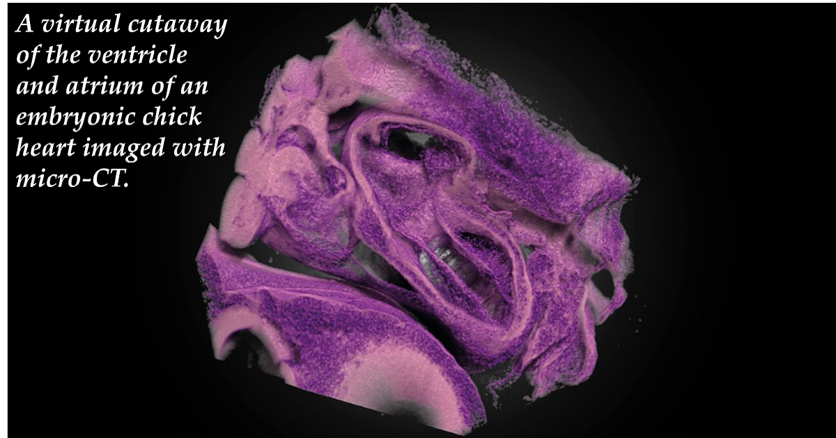
Image courtesy of Nina Kraus

Special Issue Editors


E-Mail Website
Guest Editor
Biomedical Engineering Department, College of Engineering and Computing, University of South Carolina, Greenville, SC 29605, USA
Interests: cardiovascular development; extracellular matrix development; the interplay between genomic programing and the mechanical environment during development and disease
Special Issues, Collections and Topics in MDPI journals

E-Mail Website
Guest Editor
Department of Biomedical Engineering, School of Medicine, Oregon Health Sciences University, Portland, OR 97239, USA
Interests: cardiovascular development; hemodynamics; heart function; computational fluid dynamics (CFD); mechanotransduction
Special Issues, Collections and Topics in MDPI journals

Special Issue Information

Dear Colleagues,

The Journal of Cardiovascular Development and Disease is planning to create a Special Issue that focuses on computational cardiology. In this Special Issue, we will highlight some of the recent developments that are advancing the understanding and diagnosis of cardiovascular disease. The enhancement of imaging modalities has allowed for the resolution of disease initiation and progression, which until recently was only available in humans post mortem. Advances in cardiovascular computational modeling and cardiovascular informatics together with these new imaging modalities create new opportunities for earlier interventions and better outcomes for cardiovascular disease, a global leader in human mortality. In this Special issue “Models and Methods for Computational Cardiology”, we welcome you to contribute a research paper or review article on any aspect of this topic including novel basic science or clinical approaches that better define the mechanisms of cardiovascular development and pathology. Novel models for pediatric and adult heart disease, including those that seek to improve outcomes from surgical interventions, are also relevant topics for this Special Issue. This is an excellent opportunity for clinical and basic sciences trainees in your group to contribute to the field.

Prof. Dr. Richard L. Goodwin
Prof. Dr. Sandra Rugonyi
Guest Editors

Manuscript Submission Information

Manuscripts should be submitted online at www.mdpi.com by registering and logging in to this website. Once you are registered, click here to go to the submission form. Manuscripts can be submitted until the deadline. All submissions that pass pre-check are peer-reviewed. Accepted papers will be published continuously in the journal (as soon as accepted) and will be listed together on the special issue website. Research articles, review articles as well as short communications are invited. For planned papers, a title and short abstract (about 100 words) can be sent to the Editorial Office for announcement on this website.

Submitted manuscripts should not have been published previously, nor be under consideration for publication elsewhere (except conference proceedings papers). All manuscripts are thoroughly refereed through a single-blind peer-review process. A guide for authors and other relevant information for submission of manuscripts is available on the Instructions for Authors page. Journal of Cardiovascular Development and Disease is an international peer-reviewed open access monthly journal published by MDPI.

Please visit the Instructions for Authors page before submitting a manuscript. The Article Processing Charge (APC) for publication in this open access journal is 2700 CHF (Swiss Francs). Submitted papers should be well formatted and use good English. Authors may use MDPI's English editing service prior to publication or during author revisions.

Keywords

  • cardiovascular development
  • congenital heart disease
  • cardiovascular imaging
  • patient-specific cardiovascular modeling
  • cardiovascular growth and remodeling
  • cardiovascular computational fluid dynamics
  • cardiovascular flow tissue interaction
  • hemodynamics
  • mechanotransduction

Related Special Issue

Published Papers (11 papers)

Order results
Result details
Select all
Export citation of selected articles as:

Research

Jump to: Review

19 pages, 6115 KiB  
Article
Energetics of Cardiac Blood Flow in Hypertrophic Cardiomyopathy through Individualized Computational Modeling
by Owen Baenen, Angie Carolina Carreño-Martínez, Theodore P. Abraham and Sandra Rugonyi
J. Cardiovasc. Dev. Dis. 2023, 10(10), 411; https://doi.org/10.3390/jcdd10100411 - 27 Sep 2023
Viewed by 1338
Abstract
Hypertrophic cardiomyopathy (HCM) is a congenital heart disease characterized by thickening of the heart’s left ventricle (LV) wall that can lead to cardiac dysfunction and heart failure. Ventricular wall thickening affects the motion of cardiac walls and blood flow within the heart. Because [...] Read more.
Hypertrophic cardiomyopathy (HCM) is a congenital heart disease characterized by thickening of the heart’s left ventricle (LV) wall that can lead to cardiac dysfunction and heart failure. Ventricular wall thickening affects the motion of cardiac walls and blood flow within the heart. Because abnormal cardiac blood flow in turn could lead to detrimental remodeling of heart walls, aberrant ventricular flow patterns could exacerbate HCM progression. How blood flow patterns are affected by hypertrophy and inter-patient variability is not known. To address this gap in knowledge, we present here strategies to generate personalized computational fluid dynamics (CFD) models of the heart LV from patient cardiac magnetic resonance (cMR) images. We performed simulations of CFD LV models from three cases (one normal, two HCM). CFD computations solved for blood flow velocities, from which flow patterns and the energetics of flow within the LV were quantified. We found that, compared to a normal heart, HCM hearts exhibit anomalous flow patterns and a mismatch in the timing of energy transfer from the LV wall to blood flow, as well as changes in kinetic energy flow patterns. While our results are preliminary, our presented methodology holds promise for in-depth analysis of HCM patient hemodynamics in clinical practice. Full article
(This article belongs to the Special Issue Models and Methods for Computational Cardiology)
Show Figures

Figure 1

18 pages, 5302 KiB  
Article
Uncertainty Quantification in the In Vivo Image-Based Estimation of Local Elastic Properties of Vascular Walls
by Benigno Marco Fanni, Maria Nicole Antonuccio, Alessandra Pizzuto, Sergio Berti, Giuseppe Santoro and Simona Celi
J. Cardiovasc. Dev. Dis. 2023, 10(3), 109; https://doi.org/10.3390/jcdd10030109 - 04 Mar 2023
Cited by 5 | Viewed by 1564
Abstract
Introduction: Patient-specific computational models are a powerful tool for planning cardiovascular interventions. However, the in vivo patient-specific mechanical properties of vessels represent a major source of uncertainty. In this study, we investigated the effect of uncertainty in the elastic module (E) [...] Read more.
Introduction: Patient-specific computational models are a powerful tool for planning cardiovascular interventions. However, the in vivo patient-specific mechanical properties of vessels represent a major source of uncertainty. In this study, we investigated the effect of uncertainty in the elastic module (E) on a Fluid–Structure Interaction (FSI) model of a patient-specific aorta. Methods: The image-based χ-method was used to compute the initial E value of the vascular wall. The uncertainty quantification was carried out using the generalized Polynomial Chaos (gPC) expansion technique. The stochastic analysis was based on four deterministic simulations considering four quadrature points. A deviation of about ±20% on the estimation of the E value was assumed. Results: The influence of the uncertain E parameter was evaluated along the cardiac cycle on area and flow variations extracted from five cross-sections of the aortic FSI model. Results of stochastic analysis showed the impact of E in the ascending aorta while an insignificant effect was observed in the descending tract. Conclusions: This study demonstrated the importance of the image-based methodology for inferring E, highlighting the feasibility of retrieving useful additional data and enhancing the reliability of in silico models in clinical practice. Full article
(This article belongs to the Special Issue Models and Methods for Computational Cardiology)
Show Figures

Figure 1

13 pages, 1384 KiB  
Article
Development of Prediction Models for Acute Myocardial Infarction at Prehospital Stage with Machine Learning Based on a Nationwide Database
by Arom Choi, Min Joung Kim, Ji Min Sung, Sunhee Kim, Jayoung Lee, Heejung Hyun, Hyeon Chang Kim, Ji Hoon Kim and Hyuk-Jae Chang
J. Cardiovasc. Dev. Dis. 2022, 9(12), 430; https://doi.org/10.3390/jcdd9120430 - 02 Dec 2022
Cited by 5 | Viewed by 2057
Abstract
Models for predicting acute myocardial infarction (AMI) at the prehospital stage were developed and their efficacy compared, based on variables identified from a nationwide systematic emergency medical service (EMS) registry using conventional statistical methods and machine learning algorithms. Patients in the EMS cardiovascular [...] Read more.
Models for predicting acute myocardial infarction (AMI) at the prehospital stage were developed and their efficacy compared, based on variables identified from a nationwide systematic emergency medical service (EMS) registry using conventional statistical methods and machine learning algorithms. Patients in the EMS cardiovascular registry aged >15 years who were transferred from the public EMS to emergency departments in Korea from January 2016 to December 2018 were enrolled. Two datasets were constructed according to the hierarchical structure of the registry. A total of 184,577 patients (Dataset 1) were included in the final analysis. Among them, 72,439 patients (Dataset 2) were suspected to have AMI at prehospital stage. Between the models derived using the conventional logistic regression method, the B-type model incorporated AMI-specific variables from the A-type model and exhibited a superior discriminative ability (p = 0.02). The models that used extreme gradient boosting and a multilayer perceptron yielded a higher predictive performance than the conventional logistic regression-based models for analyses that used both datasets. Each machine learning algorithm yielded different classification lists of the 10 most important features. Therefore, prediction models that use nationwide prehospital data and are developed with appropriate structures can improve the identification of patients who require timely AMI management. Full article
(This article belongs to the Special Issue Models and Methods for Computational Cardiology)
Show Figures

Figure 1

18 pages, 5523 KiB  
Article
Geometry Does Impact on the Plane Strain Directions of the Human Left Ventricle, Irrespective of Disease
by Paolo Piras, Ivan Colorado-Cervantes, Paola Nardinocchi, Stefano Gabriele, Valerio Varano, Giuseppe Esposito, Luciano Teresi, Concetta Torromeo and Paolo Emilio Puddu
J. Cardiovasc. Dev. Dis. 2022, 9(11), 393; https://doi.org/10.3390/jcdd9110393 - 15 Nov 2022
Cited by 1 | Viewed by 1401
Abstract
The directions of primary strain lines of local deformation in Epicardial and Endocardial layers have been the subject of debate in recent years. Different methods led to different conclusions and a complete assessment of strain direction patterns in large and variable (in terms [...] Read more.
The directions of primary strain lines of local deformation in Epicardial and Endocardial layers have been the subject of debate in recent years. Different methods led to different conclusions and a complete assessment of strain direction patterns in large and variable (in terms of pathology) cohorts of healthy and diseased patients is still lacking. Here, we use local deformation tensors in order to evaluate the angle of strain lines with respect to the horizontal circumferential direction in both Epi- and Endo-layers. We evaluated this on a large group of 193 subjects including 82 healthy control and 111 patients belonging to a great variety of pathological conditions. We found that Epicardial strain lines obliquely directed while those of Endocardium are almost circumferential. This result occurs irrespective of pathological condition. We propose that the geometric vinculum characterizing Endocardium and Epicardium in terms of different lever arm length and orientation of muscular fibers during contraction inescapably requires Endocardial strain lines to be circumferentially oriented and this is corroborated by experimental results. Further investigations on transmural structure of myocytes could couple results presented here in order to furnish additional experimental explanations. Full article
(This article belongs to the Special Issue Models and Methods for Computational Cardiology)
Show Figures

Figure 1

16 pages, 20381 KiB  
Article
OCT Meets micro-CT: A Subject-Specific Correlative Multimodal Imaging Workflow for Early Chick Heart Development Modeling
by Nina Kraus, Fabian Placzek and Brian Metscher
J. Cardiovasc. Dev. Dis. 2022, 9(11), 379; https://doi.org/10.3390/jcdd9110379 - 03 Nov 2022
Cited by 2 | Viewed by 1862
Abstract
Structural and Doppler velocity data collected from optical coherence tomography have already provided crucial insights into cardiac morphogenesis. X-ray microtomography and other ex vivo methods have elucidated structural details of developing hearts. However, by itself, no single imaging modality can provide comprehensive information [...] Read more.
Structural and Doppler velocity data collected from optical coherence tomography have already provided crucial insights into cardiac morphogenesis. X-ray microtomography and other ex vivo methods have elucidated structural details of developing hearts. However, by itself, no single imaging modality can provide comprehensive information allowing to fully decipher the inner workings of an entire developing organ. Hence, we introduce a specimen-specific correlative multimodal imaging workflow combining OCT and micro-CT imaging which is applicable for modeling of early chick heart development—a valuable model organism in cardiovascular development research. The image acquisition and processing employ common reagents, lab-based micro-CT imaging, and software that is free for academic use. Our goal is to provide a step-by-step guide on how to implement this workflow and to demonstrate why those two modalities together have the potential to provide new insight into normal cardiac development and heart malformations leading to congenital heart disease. Full article
(This article belongs to the Special Issue Models and Methods for Computational Cardiology)
Show Figures

Figure 1

13 pages, 3279 KiB  
Article
Single-Cell RNA Sequencing Reveals Distinct Cardiac-Derived Stromal Cell Subpopulations
by Jessica R. Hoffman, Arun R. Jayaraman, Sruti Bheri and Michael E. Davis
J. Cardiovasc. Dev. Dis. 2022, 9(11), 374; https://doi.org/10.3390/jcdd9110374 - 01 Nov 2022
Viewed by 2292
Abstract
Human cardiac-derived c-kit+ stromal cells (CSCs) have demonstrated efficacy in preclinical trials for the treatment of heart failure and myocardial dysfunction. Unfortunately, large variability in patient outcomes and cell populations remains a problem. Previous research has demonstrated that the reparative capacity of CSCs [...] Read more.
Human cardiac-derived c-kit+ stromal cells (CSCs) have demonstrated efficacy in preclinical trials for the treatment of heart failure and myocardial dysfunction. Unfortunately, large variability in patient outcomes and cell populations remains a problem. Previous research has demonstrated that the reparative capacity of CSCs may be linked to the age of the cells: CSCs derived from neonate patients increase cardiac function and reduce fibrosis. However, age-dependent differences between CSC populations have primarily been explored with bulk sequencing methods. In this work, we hypothesized that differences in CSC populations and subsequent cell therapy outcomes may arise from differing cell subtypes within donor CSC samples. We performed single-cell RNA sequencing on four neonatal CSC (nCSC) and five child CSC (cCSC) samples. Subcluster analysis revealed cCSC-enriched clusters upregulated in several fibrosis- and immune response-related genes. Module-based analysis identified upregulation of chemotaxis and ribosomal activity-related genes in nCSCs and upregulation of immune response and fiber synthesis genes in cCSCs. Further, we identified versican and integrin alpha 2 as potential markers for a fibrotic cell subtype. By investigating differences in patient-derived CSC populations at the single-cell level, this research aims to identify and characterize CSC subtypes to better optimize CSC-based therapy and improve patient outcomes. Full article
(This article belongs to the Special Issue Models and Methods for Computational Cardiology)
Show Figures

Figure 1

14 pages, 315 KiB  
Article
Stroke Severity in Ischemic Stroke Patients with a History of Diastolic Blood Pressure Treated in a Telestroke Network
by Christina Brown, Kameron Terrell, Richard Goodwin and Thomas Nathaniel
J. Cardiovasc. Dev. Dis. 2022, 9(10), 345; https://doi.org/10.3390/jcdd9100345 - 10 Oct 2022
Cited by 3 | Viewed by 1605
Abstract
Background: The relationship between diastolic blood pressure (DBP), risk factors, and stroke severity in acute ischemic stroke (AIS) patients treated in a telestroke network is not fully understood. The present study aims to determine the effect of risk factors on stroke severity in [...] Read more.
Background: The relationship between diastolic blood pressure (DBP), risk factors, and stroke severity in acute ischemic stroke (AIS) patients treated in a telestroke network is not fully understood. The present study aims to determine the effect of risk factors on stroke severity in AIS patients with a history of elevated DBP. Material and Methods: We retrospectively analyzed data on stroke severity for AIS patients treated between January 2014 and June 2016 treated in the PRISMA Health telestroke network. Data on the severity of stroke on admission were evaluated using NIHSS scores ≤7 for reduced, and >7 for increased, stroke severity. DBP was stratified as ≤80 mmHg for reduced DBP and >80 mmHg for elevated DBP. The study’s primary outcomes were risk factors associated with improving neurologic functions or reduced stroke severity and deteriorating neurologic functions or increased stroke severity. The associations between risk factors and stroke severity for AIS with elevated DBP were determined using multi-level logistic and regression models. Results: In the adjusted analysis, AIS patients with a DBP ≤ 80 mmHg, obesity (OR = 0.388, 95% Cl, 0.182–0.828, p = 0.014) was associated with reduced stroke severity, while an increased heart rate (OR = 1.025, 95% Cl, 1.001–1.050, p = 0.042) was associated with higher stroke severity. For AIS patients with a DBP > 80 mmHg, hypertension (OR = 3.453, 95% Cl, 1.137–10.491, p = 0.029), history of smoking (OR = 2.55, 95% Cl, 1.06–6.132, p = 0.037), and heart rate (OR = 1.036, 95% Cl, 1.009–1.064, p = 0.009) were associated with higher stroke severity. Caucasians (OR = 0.294, 95% Cl, 0.090–0.964, p = 0.002) and obesity (OR = 0.455, 95% Cl, 0.207–1.002, p = 0.05) were more likely to be associated with reduced stroke severity. Conclusions: Our findings reveal specific risk factors that can be managed to improve the care of AIS patients with elevated DBP treated in the telestroke network. Full article
(This article belongs to the Special Issue Models and Methods for Computational Cardiology)
14 pages, 796 KiB  
Article
20 Years of Real-World Data to Estimate the Prevalence of Heart Failure and Its Subtypes in an Unselected Population of Integrated Care Units
by Cristina Gavina, Daniel Seabra Carvalho, Filipa Valente, Filipa Bernardo, Ricardo Jorge Dinis-Oliveira, Carla Santos-Araújo and Tiago Taveira-Gomes
J. Cardiovasc. Dev. Dis. 2022, 9(5), 149; https://doi.org/10.3390/jcdd9050149 - 07 May 2022
Cited by 7 | Viewed by 4376
Abstract
Introduction: Heart failure (HF) is a clinical syndrome caused by structural and functional cardiac abnormalities resulting in the impairment of cardiac function, entailing significant mortality. The prevalence of HF has reached epidemic proportions in the last few decades, mainly in the elderly, but [...] Read more.
Introduction: Heart failure (HF) is a clinical syndrome caused by structural and functional cardiac abnormalities resulting in the impairment of cardiac function, entailing significant mortality. The prevalence of HF has reached epidemic proportions in the last few decades, mainly in the elderly, but recent evidence suggests that its epidemiology may be changing. Objective: Our objective was to estimate the prevalence of HF and its subtypes, and to characterize HF in a population of integrated care users. Material and Methods: A non-interventional cross-sectional study was performed in a healthcare center that provides primary, secondary and tertiary health cares. Echocardiographic parameters (left ventricle ejection fraction (LVEF) and evidence of structural heart disease) and elevated levels of natriuretic peptides were used to define two HF phenotypes: (i) HF with a reduced ejection fraction (HFrEF, LVEF ≤ 40% and either NT-proBNP ≥ 400 pg/mL (≥600 pg/mL if atrial fibrillation (AF)/flutter) or BNP ≥ 100 pg/mL (≥125 pg/mL if AF/flutter)) and (ii) HF with a non-reduced ejection fraction (HFnrEF), which encompasses both HFpEF (LVEF ≥ 50% and either NT-proBNP ≥ 200 pg/mL (≥600 pg/mL if AF/flutter) or BNP ≥ 100 pg/mL (≥125 pg/mL if AF/flutter) in the presence of at least one structural cardiac abnormality) and HF with a mildly reduced fraction (HFmrEF, LVEF within 40–50% and either NT-proBNP ≥ 200 pg/mL (≥600 pg/mL if AF/flutter) or BNP ≥ 100 pg/mL (≥125 pg/mL if AF/flutter) in the presence of at least one structural cardiac abnormality). The significance threshold was set at p ≤ 0.001. Results: We analyzed 126,636 patients with a mean age of 52.2 (SD = 18.3) years, with 57% (n = 72,290) being female. The prevalence of HF was 2.1% (n = 2700). The HF patients’ mean age was 74.0 (SD = 12.1) years, and 51.6% (n = 1394) were female. Regarding HF subtypes, HFpEF accounted for 65.4% (n = 1765); 16.1% (n = 434) had HFmrEF and 16.3% (n = 439) had HFrEF. The patients with HFrEF were younger (p < 0.001) and had a history of myocardial infarction more frequently (p < 0.001) compared to HFnrEF, with no other significant differences between the HF groups. The HFrEF patients were more frequently prescribed CV medications than HFnrEF patients. Type 2 Diabetes Mellitus (T2D) was present in 44.7% (n = 1207) of the HF patients. CKD was more frequently present in T2D vs. non-T2D HF patients at every stage (p < 0.001), as well as stroke, peripheral artery disease, and microvascular disease (p < 0.001). Conclusions: In this cohort, considering a contemporary definition, the prevalence of HF was 2.1%. HFrEF accounted for 16.3% of the cases, with a similar clinical–epidemiological profile having been previously reported in the literature. Our study revealed a high prevalence of patients with HFpEF (65.4%), raising awareness for the increasing prevalence of this entity in cardiology practice. These results may guide local and national health policies and strategies for HF diagnosis and management. Full article
(This article belongs to the Special Issue Models and Methods for Computational Cardiology)
Show Figures

Figure 1

11 pages, 1683 KiB  
Article
Three-Dimensional Adaptive Image Compression Concept for Medical Imaging: Application to Computed Tomography Angiography for Peripheral Arteries
by Guillaume Fahrni, David C. Rotzinger, Chiaki Nakajo, Jamshid Dehmeshki and Salah Dine Qanadli
J. Cardiovasc. Dev. Dis. 2022, 9(5), 137; https://doi.org/10.3390/jcdd9050137 - 27 Apr 2022
Cited by 1 | Viewed by 1778
Abstract
Advances in computed tomography (CT) have resulted in a substantial increase in the size of datasets. We built a new concept of medical image compression that provides the best compromise between compression rate and image quality. The method is based on multiple contexts [...] Read more.
Advances in computed tomography (CT) have resulted in a substantial increase in the size of datasets. We built a new concept of medical image compression that provides the best compromise between compression rate and image quality. The method is based on multiple contexts and regions-of-interest (ROI) defined according to the degree of clinical interest. High priority areas (primary ROIs) are assigned a lossless compression. Other areas (secondary ROIs and background) are compressed with moderate or heavy losses. The method is applied to a whole dataset of CT angiography (CTA) of the lower extremity vasculature. It is compared to standard lossy compression techniques in terms of quantitative and qualitative image quality. It is also compared to standard lossless compression techniques in terms of image size reduction and compression ratio. The proposed compression method met quantitative criteria for high-quality encoding. It obtained the highest qualitative image quality rating score, with a statistically significant difference compared to other methods. The average compressed image size was up to 61% lower compared to standard compression techniques, with a 9:1 compression ratio compared with original non-compressed images. Our new adaptive 3D compression method for CT images can save data storage space while preserving clinically relevant information. Full article
(This article belongs to the Special Issue Models and Methods for Computational Cardiology)
Show Figures

Figure 1

Review

Jump to: Research

31 pages, 6159 KiB  
Review
Recasting Current Knowledge of Human Fetal Circulation: The Importance of Computational Models
by Daibo Zhang and Stephanie E. Lindsey
J. Cardiovasc. Dev. Dis. 2023, 10(6), 240; https://doi.org/10.3390/jcdd10060240 - 30 May 2023
Viewed by 3300
Abstract
Computational hemodynamic simulations are becoming increasingly important for cardiovascular research and clinical practice, yet incorporating numerical simulations of human fetal circulation is relatively underutilized and underdeveloped. The fetus possesses unique vascular shunts to appropriately distribute oxygen and nutrients acquired from the placenta, adding [...] Read more.
Computational hemodynamic simulations are becoming increasingly important for cardiovascular research and clinical practice, yet incorporating numerical simulations of human fetal circulation is relatively underutilized and underdeveloped. The fetus possesses unique vascular shunts to appropriately distribute oxygen and nutrients acquired from the placenta, adding complexity and adaptability to blood flow patterns within the fetal vascular network. Perturbations to fetal circulation compromise fetal growth and trigger the abnormal cardiovascular remodeling that underlies congenital heart defects. Computational modeling can be used to elucidate complex blood flow patterns in the fetal circulatory system for normal versus abnormal development. We present an overview of fetal cardiovascular physiology and its evolution from being investigated with invasive experiments and primitive imaging techniques to advanced imaging (4D MRI and ultrasound) and computational modeling. We introduce the theoretical backgrounds of both lumped-parameter networks and three-dimensional computational fluid dynamic simulations of the cardiovascular system. We subsequently summarize existing modeling studies of human fetal circulation along with their limitations and challenges. Finally, we highlight opportunities for improved fetal circulation models. Full article
(This article belongs to the Special Issue Models and Methods for Computational Cardiology)
Show Figures

Figure 1

12 pages, 1760 KiB  
Review
Following the Beat: Imaging the Valveless Pumping Function in the Early Embryonic Heart
by Shang Wang and Irina V. Larina
J. Cardiovasc. Dev. Dis. 2022, 9(8), 267; https://doi.org/10.3390/jcdd9080267 - 15 Aug 2022
Cited by 4 | Viewed by 1806
Abstract
In vertebrates, the coordinated beat of the early heart tube drives cardiogenesis and supports embryonic growth. How the heart pumps at this valveless stage marks a fascinating problem that is of vital significance for understanding cardiac development and defects. The developing heart achieves [...] Read more.
In vertebrates, the coordinated beat of the early heart tube drives cardiogenesis and supports embryonic growth. How the heart pumps at this valveless stage marks a fascinating problem that is of vital significance for understanding cardiac development and defects. The developing heart achieves its function at the same time as continuous and dramatic morphological changes, which in turn modify its pumping dynamics. The beauty of this muti-time-scale process also highlights its complexity that requires interdisciplinary approaches to study. High-resolution optical imaging, particularly fast, four-dimensional (4D) imaging, plays a critical role in revealing the process of pumping, instructing numerical modeling, and enabling biomechanical analyses. In this review, we aim to connect the investigation of valveless pumping mechanisms with the recent advancements in embryonic cardiodynamic imaging, facilitating interactions between these two areas of study, in hopes of encouraging and motivating innovative work to further understand the early heartbeat. Full article
(This article belongs to the Special Issue Models and Methods for Computational Cardiology)
Show Figures

Figure 1

Back to TopTop