


Cells 2023, 12(4), 638; https://doi.org/10.3390/cells12040638 - 16 Feb 2023
Cited by 3 | Viewed by 1826
Abstract
►
Show Figures
Atrial fibrillation is the most prevalent tachyarrhythmia in clinical practice, with very high cardiovascular morbidity and mortality with a high-cost impact in health systems. Currently, it is one of the main causes of stroke and subsequent heart failure and sudden death. miRNAs mediate
[...] Read more.
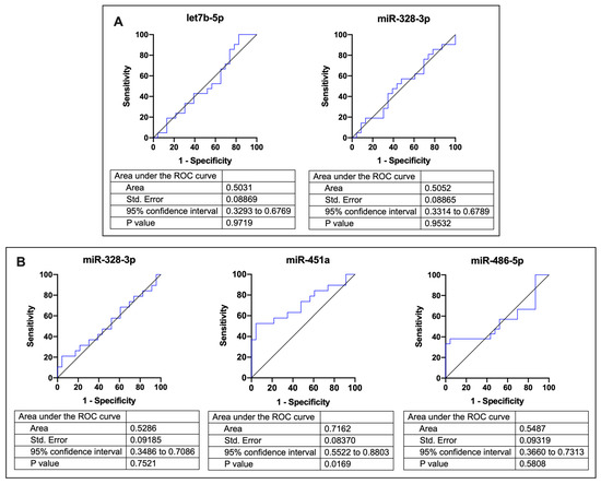
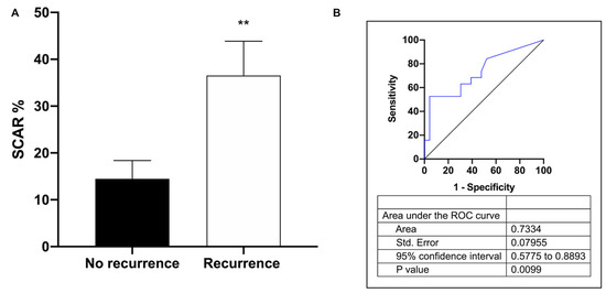
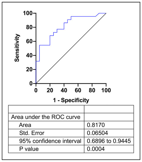
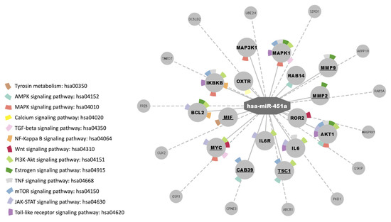
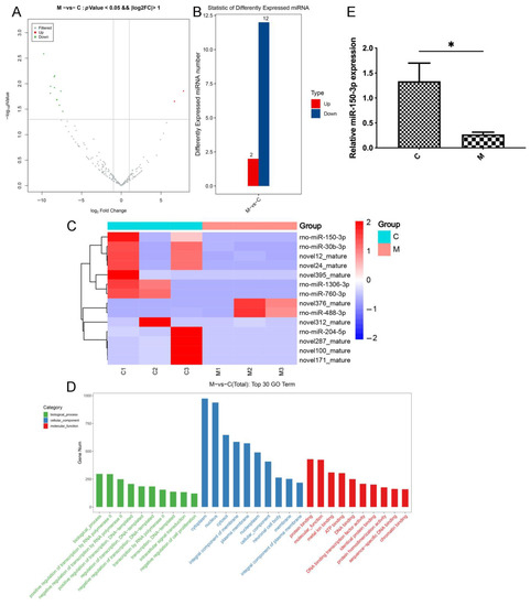
Atrial fibrillation is the most prevalent tachyarrhythmia in clinical practice, with very high cardiovascular morbidity and mortality with a high-cost impact in health systems. Currently, it is one of the main causes of stroke and subsequent heart failure and sudden death. miRNAs mediate in several processes involved in cardiovascular disease, including fibrosis and electrical and structural remodeling. Several studies suggest a key role of miRNAs in the course and maintenance of atrial fibrillation. In our study, we aimed to identify the differential expression of circulating miRNAs and their predictive value as biomarkers of recurrence in atrial fibrillation patients undergoing catheter pulmonary vein ablation. To this effect, 42 atrial fibrillation patients were recruited for catheter ablation. We measured the expression of 84 miRNAs in non-recurrent and recurrent groups (45.2%), both in plasma from peripheral and left atrium blood. Expression analysis showed that miRNA-451a is downregulated in recurrent patients. Receiver operating characteristic curve analysis showed that miR-451a in left atrium plasma could predict atrial fibrillation recurrence after pulmonary vein isolation. In addition, atrial fibrillation recurrence is positively associated with the increment of scar percentage. Our data suggest that miRNA-451a expression plays an important role in AF recurrence by controlling fibrosis and progression.
Full article
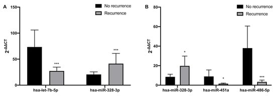
Figure 1
