


Cells 2024, 13(2), 107; https://doi.org/10.3390/cells13020107 - 05 Jan 2024
Viewed by 1153
Abstract
►
Show Figures
The world-wide COVID-19 pandemic has promoted a series of alternative vaccination strategies aiming to elicit neutralizing adaptive immunity in the human host. However, restricted efficacies of these vaccines targeting epitopes on the spike (S) protein that is involved in primary viral entry were
[...] Read more.
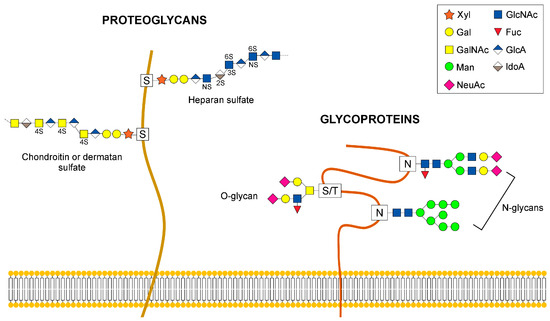
The world-wide COVID-19 pandemic has promoted a series of alternative vaccination strategies aiming to elicit neutralizing adaptive immunity in the human host. However, restricted efficacies of these vaccines targeting epitopes on the spike (S) protein that is involved in primary viral entry were observed and putatively assigned to viral glycosylation as an effective escape mechanism. Besides the well-recognized N-glycan shield covering SARS-CoV-2 spike (S) proteins, immunization strategies may be hampered by heavy O-glycosylation and variable O-glycosites fluctuating depending on the organ sites of primary infection and those involved in immunization. A further complication associated with viral glycosylation arises from the development of autoimmune antibodies to self-carbohydrates, including O-linked blood group antigens, as structural parts of viral proteins. This outline already emphasizes the importance of viral glycosylation in general and, in particular, highlights the impact of the site-specific O-glycosylation of virions, since this modification is independent of sequons and varies strongly in dependence on cell-specific repertoires of peptidyl-N-acetylgalactosaminyltransferases with their varying site preferences and of glycan core-specific glycosyltransferases. This review summarizes the current knowledge on the viral O-glycosylation of the SARS-CoV-2 spike protein and its impact on virulence and immune modulation in the host.
Full article

Figure 1