


Cells 2023, 12(8), 1190; https://doi.org/10.3390/cells12081190 - 19 Apr 2023
Cited by 2 | Viewed by 1566
Abstract
►
Show Figures
Schwann cells (SCs) are myelinating cells that promote peripheral nerve regeneration. When nerve lesions form, SCs are destroyed, ultimately hindering nerve repair. The difficulty in treating nerve repair is exacerbated due to SC’s limited and slow expansion capacity. Therapeutic use of adipose-derived stem
[...] Read more.
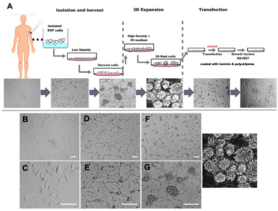
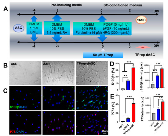
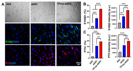
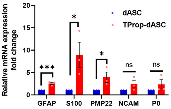
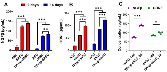
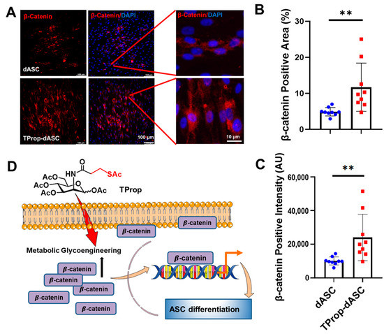
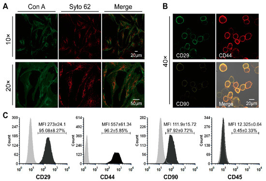
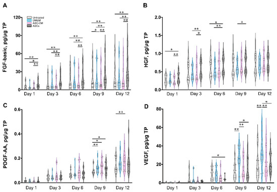
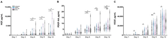
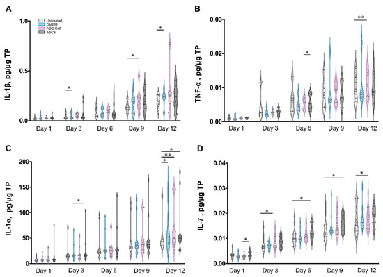
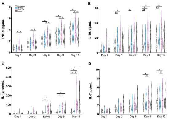
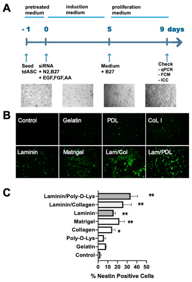
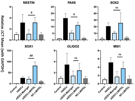
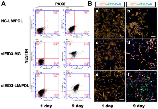
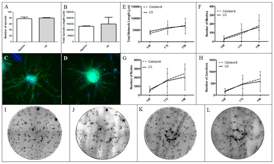
Schwann cells (SCs) are myelinating cells that promote peripheral nerve regeneration. When nerve lesions form, SCs are destroyed, ultimately hindering nerve repair. The difficulty in treating nerve repair is exacerbated due to SC’s limited and slow expansion capacity. Therapeutic use of adipose-derived stem cells (ASCs) is emerging in combating peripheral nerve injury due to these cells’ SC differentiation capability and can be harvested easily in large numbers. Despite ASC’s therapeutic potential, their transdifferentiation period typically takes more than two weeks. In this study, we demonstrate that metabolic glycoengineering (MGE) technology enhances ASC differentiation into SCs. Specifically, the sugar analog Ac5ManNTProp (TProp), which modulates cell surface sialylation, significantly improved ASC differentiation with upregulated SC protein S100β and p75NGFR expression and elevated the neurotrophic factors nerve growth factor beta (NGFβ) and glial cell-line-derived neurotrophic factor (GDNF). TProp treatment remarkably reduced the SC transdifferentiation period from about two weeks to two days in vitro, which has the potential to improve neuronal regeneration and facilitate future use of ASCs in regenerative medicine.
Full article
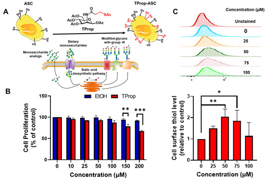
Figure 1


