Cyclophosphazene Intrinsically Derived Heteroatom (S, N, P, O)-Doped Carbon Nanoplates for Ultrasensitive Monitoring of Dopamine from Chicken Samples

 and
and 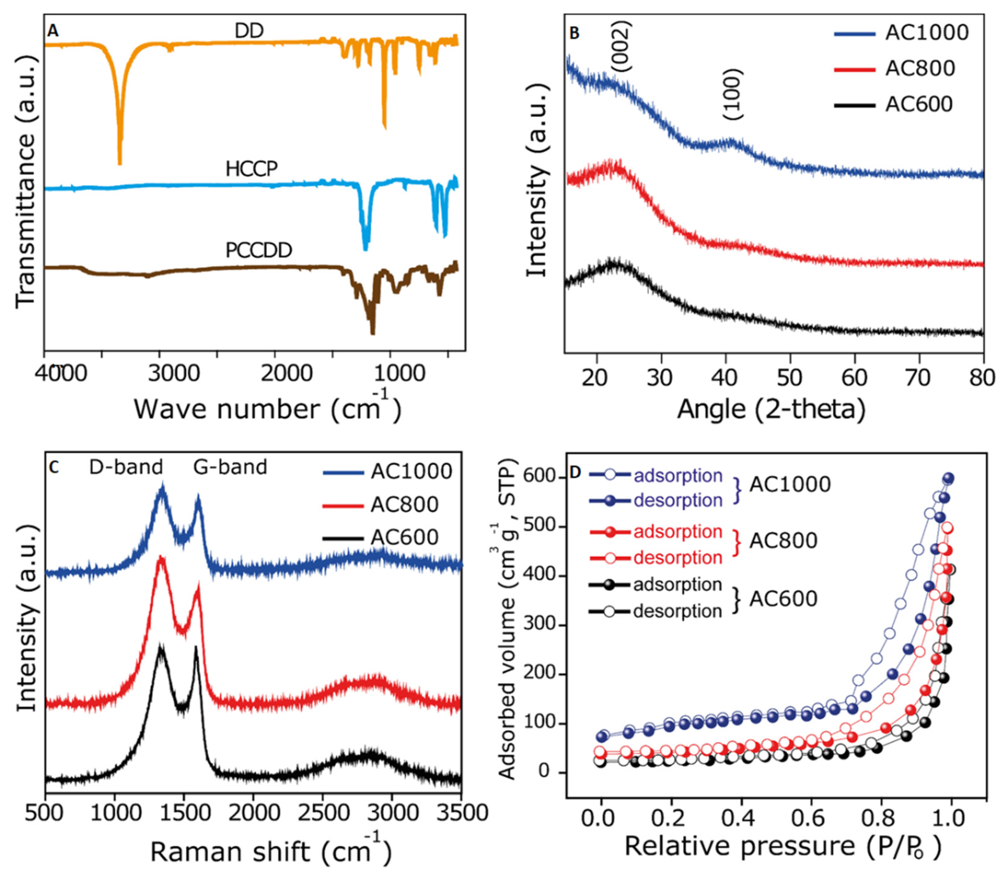
Abstract
:1. Introduction
2. Experimental Section
2.1. Reagents and Materials
2.2. Synthesis of Poly(cyclotriphosphazene)-1,4-dithiane-2,5-diol (PCD) Polymer
2.3. Synthesis of SNPO-CPL
2.4. Electrochemical Measurements
2.5. Real Sample Analysis
3. Results and Discussion
3.1. Structural and Morphological Assessment of PCD Polymer
3.2. Structural and Morphological Assessment of SNPO-CPL
3.3. Electrocatalytic Assessments of SNPO-CPL
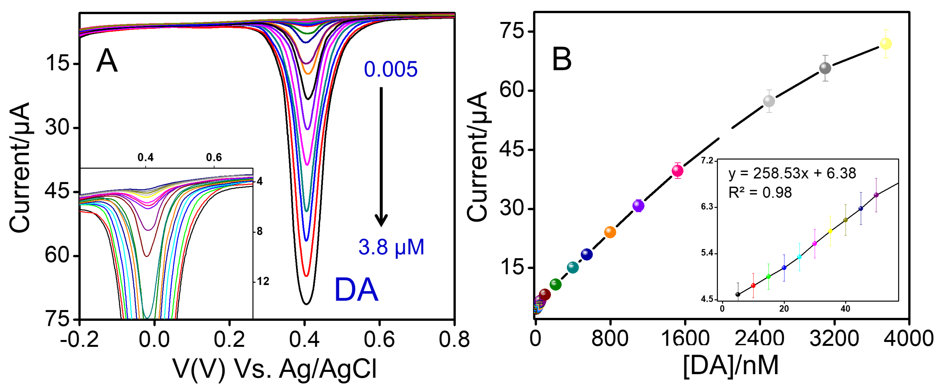
3.4. Sensing Efficacy of SNPO-CPL-800 toward DA Monitoring
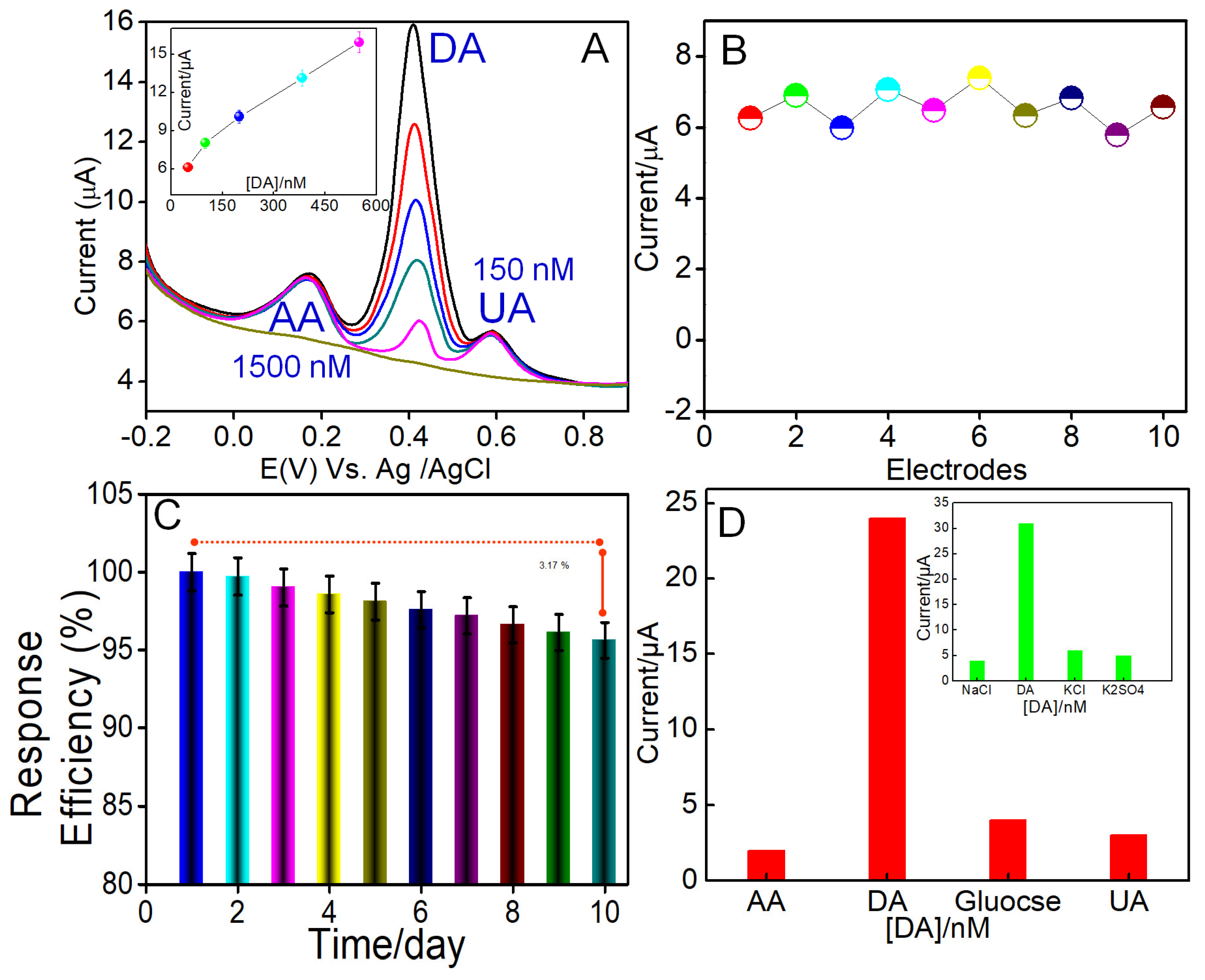
3.5. Selective Efficacy of SNPO-CPL-800 toward DA Monitoring
3.6. Reproducible Efficiency of the Designed SNPO-CPL-800 Electrode
3.7. DA Monitoring in Chicken Samples
4. Conclusions
Supplementary Materials
Author Contributions
Funding
Institutional Review Board Statement
Informed Consent Statement
Data Availability Statement
Conflicts of Interest
Abbreviations
References
- Yang, C.; Trikantzopoulos, E.; Nguyen, M.D.; Jacobs, C.B.; Wang, Y.; Mahjouri-Samani, M.; Ivanov, I.N.; Venton, B.J. Laser Treated Carbon Nanotube Yarn Microelectrodes for Rapid and Sensitive Detection of Dopamine in Vivo. ACS Sens. 2016, 1, 508–515. [Google Scholar] [CrossRef] [PubMed] [Green Version]
- Akhtar, N.; El-Safty, S.A.; Abdelsalam, M.E.; Shenashen, M.; Kawarada, H. Radially oriented nanostrand electrodes to boost glucose sensing in mammalian blood. Biosens. Bioelectron. 2016, 77, 656–665. [Google Scholar] [CrossRef] [PubMed]
- Ling, X.; Shi, R.; Zhang, J.; Liu, D.; Weng, M.; Zhang, C.; Lu, M.; Xie, X.; Huang, L.; Huang, W. Dual-Signal Luminescent Detection of Dopamine by a Single Type of Lanthanide-Doped Nanoparticles. ACS Sens. 2018, 3, 1683–1689. [Google Scholar] [CrossRef]
- Pradhan, T.; Jung, H.S.; Jang, J.H.; Kim, T.W.; Kang, C.; Kim, J.S. Chemical sensing of neurotransmitters. Chem. Soc. Rev. 2014, 43, 4684–4713. [Google Scholar] [CrossRef] [PubMed]
- Parchekani, J.; Hashemzadeh, H.; Allahverdi, A.; Siampour, H.; Abbasian, S.; Moshaii, A.; Naderi-Manesh, H. Zepto molar miRNA-21 detection in gold Nano-islands platform toward early cancer screening. Sens. Bio-Sens. Res. 2021, 34, 100449. [Google Scholar] [CrossRef]
- Demuru, S.; Nela, L.; Marchack, N.; Holmes, S.J.; Farmer, D.B.; Tulevski, G.S.; Lin, Q.; Deligianni, H. Scalable Nanostructured Carbon Electrode Arrays for Enhanced Dopamine Detection. ACS Sens. 2018, 3, 799–805. [Google Scholar] [CrossRef]
- Kumar, G.G.; Amala, G.; Gowtham, S.M. Recent advancements, key challenges and solutions in non-enzymatic electrochemical glucose sensors based on graphene platforms. RSC Adv. 2017, 7, 36949–36976. [Google Scholar] [CrossRef] [Green Version]
- Emran, M.Y.; Shenashen, M.A.; Mekawy, M.; Azzam, A.M.; Akhtar, N.; Gomaa, H.; Selim, M.M.; Faheem, A.; El-Safty, S.A. Ultrasensitive in-vitro monitoring of monoamine neurotransmitters from dopaminergic cells. Sens. Actuators B Chem. 2018, 259, 114–124. [Google Scholar] [CrossRef]
- Yi, X.; Wu, Y.; Tan, G.; Yu, P.; Zhou, L.; Zhou, Z.; Chen, J.; Wang, Z.; Pang, J.; Ning, C. Palladium nanoparticles entrapped in a self-supporting nanoporous gold wire as sensitive dopamine biosensor. Sci. Rep. 2017, 7, 7941. [Google Scholar] [CrossRef] [Green Version]
- Phung, V.-D.; Jung, W.-S.; Nguyen, T.-A.; Kim, J.-H.; Lee, S.-W. Reliable and quantitative SERS detection of dopamine levels in human blood plasma using a plasmonic Au/Ag nanocluster substrate. Nanoscale 2018, 10, 22493–22503. [Google Scholar] [CrossRef]
- Emran, M.Y.; Khalifa, H.; Gomaa, H.; Shenashen, M.A.; Akhtar, N.; Mekawy, M.; Faheem, A.; El-Safty, S.A. Hierarchical C-N doped NiO with dual-head echinop flowers for ultrasensitive monitoring of epinephrine in human blood serum. Microchim. Acta 2017, 184, 4553–4562. [Google Scholar] [CrossRef]
- Gao, G.; Zhang, Z.; Wang, K.; Yuan, Q.; Wang, X. One-pot synthesis of dendritic Pt3Ni nanoalloys as nonenzymatic electrochemical biosensors with high sensitivity and selectivity for dopamine detection. Nanoscale 2017, 9, 10998–11003. [Google Scholar] [CrossRef] [PubMed]
- Hsu, M.-S.; Chen, Y.-L.; Lee, C.-Y.; Chiu, H.-T. Gold Nanostructures on Flexible Substrates as Electrochemical Dopamine Sensors. ACS Appl. Mater. Interfaces 2012, 4, 5570–5575. [Google Scholar] [CrossRef] [PubMed]
- Josephine, D.S.R.; Babu, K.J.; Kumar, G.P.G.; Sethuraman, K. Titanium dioxide anchored graphene oxide nanosheets for highly selective voltammetric sensing of dopamine. Microchim. Acta 2017, 184, 781–790. [Google Scholar] [CrossRef]
- Shen, Y.; Sheng, Q.; Zheng, J. A high-performance electrochemical dopamine sensor based on a platinum–nickel bimetallic decorated poly(dopamine)-functionalized reduced graphene oxide nanocomposite. Anal. Methods 2017, 9, 4566–4573. [Google Scholar] [CrossRef]
- Choo, S.-S.; Kang, E.-S.; Song, I.; Lee, D.; Choi, J.-W.; Kim, T.-H. Electrochemical Detection of Dopamine Using 3D Porous Graphene Oxide/Gold Nanoparticle Composites. Sensors 2017, 17, 861. [Google Scholar] [CrossRef] [Green Version]
- He, Y.; Han, X.; Du, Y.; Zhang, B.; Xu, P. Heteroatom-Doped Carbon Nanostructures Derived from Conjugated Polymers for Energy Applications. Polymers 2016, 8, 366. [Google Scholar] [CrossRef] [Green Version]
- Ghanbari, K.; Moloudi, M. Flower-like ZnO decorated polyaniline/reduced graphene oxide nanocomposites for simultaneous determination of dopamine and uric acid. Anal. Biochem. 2016, 512, 91–102. [Google Scholar] [CrossRef] [Green Version]
- Feng, X.; Zhang, Y.; Zhou, J.; Li, Y.; Chen, S.; Zhang, L.; Ma, Y.; Wang, L.; Yan, X. Three-dimensional nitrogen-doped graphene as an ultrasensitive electrochemical sensor for the detection of dopamine. Nanoscale 2015, 7, 2427–2432. [Google Scholar] [CrossRef]
- Gao, W.; Feng, X.; Zhang, T.; Huang, H.; Li, J.; Song, W. One-Step Pyrolytic Synthesis of Nitrogen and Sulfur Dual-Doped Porous Carbon with High Catalytic Activity and Good Accessibility to Small Biomolecules. ACS Appl. Mater. Interfaces 2014, 6, 19109–19117. [Google Scholar] [CrossRef]
- Mastragostino, M.; Arbizzani, C.; Meneghello, L.; Paraventi, R. Electronically conducting polymers and activated carbon: Electrode materials in supercapacitor technology. Adv. Mater. 1996, 8, 331–334. [Google Scholar] [CrossRef]
- Zhang, J.; Zhao, Z.; Xia, Z.; Dai, L. A metal-free bifunctional electrocatalyst for oxygen reduction and oxygen evolution reactions. Nat. Nanotechnol. 2015, 10, 444–452. [Google Scholar] [CrossRef]
- Liu, W.; Zhang, S.; Dar, S.U.; Zhao, Y.; Akram, R.; Zhang, X.; Jin, S.; Wu, Z.; Wu, D. Polyphosphazene-derived heteroatoms-doped carbon materials for supercapacitor electrodes. Carbon 2018, 129, 420–427. [Google Scholar] [CrossRef]
- Yang, S.; Zhu, Y.; Cao, C.; Peng, L.; Li, S.; Zhai, D.; Song, W. A general route to coat poly(cyclotriphosphazene-co-4,4′-sulfonyldiphenol) on various substrates and the derived N, P, S-doped hollow carbon shells for catalysis. Nanoscale 2017, 9, 13538–13545. [Google Scholar] [CrossRef] [PubMed]
- Ali, S.; Zuhra, Z.; Butler, I.S.; Dar, S.U.; Hameed, M.U.; Wu, D.; Zhang, L.; Wu, Z. High-throughput synthesis of cross-linked poly(cyclotriphosphazene-co-bis(aminomethyl)ferrocene) microspheres and their performance as a superparamagnetic, electrochemical, fluorescent and adsorbent material. Chem. Eng. J. 2017, 315, 448–458. [Google Scholar] [CrossRef]
- Dar, S.U.; Ali, S.; Hameed, M.U.; Zuhra, Z.; Wu, Z. A facile synthesis, structural morphology and fluorescent properties of cross-linked poly(cyclotriphosphazene-co-1,3,5-tri(4-hydroxyphenyl)benzene) hybrid copolymer microspheres. New J. Chem. 2016, 40, 8418–8423. [Google Scholar] [CrossRef]
- Zhou, M.; Pu, F.; Wang, Z.; Guan, S. Nitrogen-doped porous carbons through KOH activation with superior performance in supercapacitors. Carbon 2014, 68, 185–194. [Google Scholar] [CrossRef]
- Li, Z.; Zhou, X.; Wang, K.; Zou, X.; Shi, J.; Huang, X.; Holmes, M. A novel sensor for determination of dopamine in meat based on ZnO-decorated reduced graphene oxide composites. Innov. Food Sci. Emerg. Technol. 2015, 31, 196–203. [Google Scholar]
- Wen, Z.; Wang, X.; Mao, S.; Bo, Z.; Kim, H.; Cui, S.; Lu, G.; Feng, X.; Chen, J. Crumpled Nitrogen-Doped Graphene Nanosheets with Ultrahigh Pore Volume for High-Performance Supercapacitor. Adv. Mater 2012, 24, 5610–5616. [Google Scholar] [CrossRef]
- Qie, L.; Chen, W.; Xiong, X.; Hu, C.; Zou, F.; Hu, P.; Huang, Y. Sulfur-doped carbon with enlarged interlayer distance as a high-performance anode material for sodium-ion batteries. Adv. Sci. 2015, 2, 1500195. [Google Scholar] [CrossRef] [Green Version]
- Deng, X.; Zhao, B.; Zhu, L.; Shao, Z. Molten salt synthesis of nitrogen-doped carbon with hierarchical pore structures for use as high-performance electrodes in supercapacitors. Carbon 2015, 93, 48–58. [Google Scholar] [CrossRef]
- Hu, L.; Hou, J.; Ma, Y.; Li, H.; Zhai, T. Multi-heteroatom self-doped porous carbon derived from swim bladders for large capacitance supercapacitors. J. Mater. Chem. A 2016, 4, 15006–15014. [Google Scholar] [CrossRef]
- Bock, C.; Paquet, C.; Couillard, M.; Botton, G.A.; MacDougall, B.R. Size-Selected Synthesis of PtRu Nano-Catalysts: Reaction and Size Control Mechanism. J. Am. Chem. Soc. 2004, 126, 8028–8037. [Google Scholar] [CrossRef] [PubMed] [Green Version]
- Liu, M.; Gan, L.; Xiong, W.; Zhao, F.; Fan, X.; Zhu, D.; Xu, Z.; Hao, Z.; Chen, L. Nickel-Doped Activated Mesoporous Carbon Microspheres with Partially Graphitic Structure for Supercapacitors. Energy Fuels 2013, 27, 1168–1173. [Google Scholar] [CrossRef]
- Mita, A.F.; Ekra, N.J.; Banik, B.K. Synthesis of Activated Carbon from Locally Available Rice Husk for Treating Saline Drinking Water. In Proceedings of the 6th ICERIE: 6th International Conference on Engineering Research, Innovation and Education, Sylhet, Bangladesh, 26–28 February 2021. [Google Scholar]
- Wang, Y.-Z.; Li, F.-M.; Kishimoto, K. Thermal effects on vibration properties of double-layered nanoplates at small scales. Compos. Part B Eng. 2011, 42, 1311–1317. [Google Scholar] [CrossRef]
- Emran, M.Y.; Shenashen, M.; Morita, H.; El-Safty, S.A. One-step selective screening of bioactive molecules in living cells using sulfur-doped microporous carbon. Biosens. Bioelectron. 2018, 109, 237–245. [Google Scholar] [CrossRef] [PubMed]
- Sheng, Z.-H.; Zheng, X.-Q.; Xu, J.-Y.; Bao, W.-J.; Wang, F.-B.; Xia, X.-H. Electrochemical sensor based on nitrogen doped graphene: Simultaneous determination of ascorbic acid, dopamine and uric acid. Biosens. Bioelectron. 2012, 34, 125–131. [Google Scholar] [CrossRef]
- Wiench, P.; González, Z.; Menéndez, R.; Grzyb, B.; Gryglewicz, G. Beneficial impact of oxygen on the electrochemical performance of dopamine sensors based on N-doped reduced graphene oxides. Sens. Actuators B Chem. 2018, 257, 143–153. [Google Scholar] [CrossRef]
- Taverniers, I.; De Loose, M.; Van Bockstaele, E. Trends in quality in the analytical laboratory. II. Analytical method validation and quality assurance. TrAC Trends Anal. Chem. 2004, 23, 535–552. [Google Scholar] [CrossRef]
- Asad, M.; Zulfiqar, A.; Raza, R.; Yang, M.; Hayat, A.; Akhtar, N. Orange Peel Derived C-dots Decorated CuO Nanorods for the Selective Monitoring of Dopamine from Deboned Chicken. Electroanalysis 2020, 32, 11–18. [Google Scholar] [CrossRef]
- Sun, C.-L.; Chang, C.-T.; Lee, H.-H.; Zhou, J.; Wang, J.; Sham, T.-K.; Pong, W.-F. Microwave-assisted synthesis of a core–shell MWCNT/GONR heterostructure for the electrochemical detection of ascorbic acid, dopamine, and uric acid. Acs Nano 2011, 5, 7788–7795. [Google Scholar] [CrossRef] [PubMed]
- Zhang, X.; Wang, K.-P.; Zhang, L.-N.; Zhang, Y.-C.; Shen, L. Phosphorus-doped graphene-based electrochemical sensor for sensitive detection of acetaminophen. Anal. Chim. Acta 2018, 1036, 26–32. [Google Scholar] [CrossRef] [PubMed]
- Zhao, L.; Cai, Z.; Yao, Q.; Zhao, T.; Lin, H.; Xiao, Y.; Chen, X. Electropolymerization fabrication of three-dimensional N, P-co-doped carbon network as a flexible electrochemical dopamine sensor. Sens. Actuators B Chem. 2017, 253, 1113–1119. [Google Scholar] [CrossRef]
- Li, S.M.; Yang, S.Y.; Wang, Y.S.; Lien, C.H.; Tien, H.W.; Hsiao, S.T.; Liao, W.-H.; Tsai, H.-P.; Chang, C.-L.; Ma, C.-C.; et al. Controllable synthesis of nitrogen-doped graphene and its effect on the simultaneous electrochemical determination of ascorbic acid, dopamine, and uric acid. Carbon 2013, 59, 418–429. [Google Scholar] [CrossRef]


Publisher’s Note: MDPI stays neutral with regard to jurisdictional claims in published maps and institutional affiliations. |
© 2022 by the authors. Licensee MDPI, Basel, Switzerland. This article is an open access article distributed under the terms and conditions of the Creative Commons Attribution (CC BY) license (https://creativecommons.org/licenses/by/4.0/).
Share and Cite
Abbas, Y.; Akhtar, N.; Ghaffar, S.; Al-Sulami, A.I.; Asad, M.; Mazhar, M.E.; Zafar, F.; Hayat, A.; Wu, Z. Cyclophosphazene Intrinsically Derived Heteroatom (S, N, P, O)-Doped Carbon Nanoplates for Ultrasensitive Monitoring of Dopamine from Chicken Samples. Biosensors 2022, 12, 1106. https://doi.org/10.3390/bios12121106
Abbas Y, Akhtar N, Ghaffar S, Al-Sulami AI, Asad M, Mazhar ME, Zafar F, Hayat A, Wu Z. Cyclophosphazene Intrinsically Derived Heteroatom (S, N, P, O)-Doped Carbon Nanoplates for Ultrasensitive Monitoring of Dopamine from Chicken Samples. Biosensors. 2022; 12(12):1106. https://doi.org/10.3390/bios12121106
Chicago/Turabian StyleAbbas, Yasir, Naeem Akhtar, Sania Ghaffar, Ahlam I. Al-Sulami, Muhammad Asad, Muhammad Ehsan Mazhar, Farhan Zafar, Akhtar Hayat, and Zhanpeng Wu. 2022. "Cyclophosphazene Intrinsically Derived Heteroatom (S, N, P, O)-Doped Carbon Nanoplates for Ultrasensitive Monitoring of Dopamine from Chicken Samples" Biosensors 12, no. 12: 1106. https://doi.org/10.3390/bios12121106