Next Issue
Volume 13, January
Previous Issue
Volume 12, November


Biosensors, Volume 12, Issue 12 (December 2022) – 124 articles

Cover Story (view full-size image): 
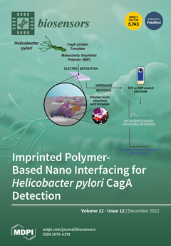
Helicobacter pylori is a microaerophilic, gastric, cancer-causing bacterium and develops colonization in gastric environments with the help of a major virulence factor, CagA (cytotoxin-associated gene A). In order to detect CagA, a nanomaterials-based molecularly imprinted sensing surface was fabricated by using CagA as a template. Pre-polymerization conditions were optimized through molecular dynamics simulations to obtain well-matched optimized molar ratios of monomers, cross-linkers, and templates. A simulation study revealed that a low binding energy was obtained upon template removal, which indicates the capability of MIP to recognize the CagA antigen through a strong binding affinity. Under the optimized electrochemical experimental conditions, the fabricated CagA-MIP/Au/rGO@SPE sensor exhibited high sensitivity and a low limit of detection. View this paper
  • Issues are regarded as officially published after their release is announced to the table of contents alert mailing list.
  • You may sign up for e-mail alerts to receive table of contents of newly released issues.
  • PDF is the official format for papers published in both, html and pdf forms. To view the papers in pdf format, click on the "PDF Full-text" link, and use the free Adobe Reader to open them.
Order results
Result details
Section
Select all
Export citation of selected articles as:
37 pages, 3405 KiB  
Review
The Roadmap of Graphene-Based Sensors: Electrochemical Methods for Bioanalytical Applications
by Ghazala Ashraf, Ayesha Aziz, Tayyaba Iftikhar, Zi-Tao Zhong, Muhammad Asif and Wei Chen
Biosensors 2022, 12(12), 1183; https://doi.org/10.3390/bios12121183 - 19 Dec 2022
Cited by 6 | Viewed by 2581
Abstract
Graphene (GR) has engrossed immense research attention as an emerging carbon material owing to its enthralling electrochemical (EC) and physical properties. Herein, we debate the role of GR-based nanomaterials (NMs) in refining EC sensing performance toward bioanalytes detection. Following the introduction, we briefly [...] Read more.
Graphene (GR) has engrossed immense research attention as an emerging carbon material owing to its enthralling electrochemical (EC) and physical properties. Herein, we debate the role of GR-based nanomaterials (NMs) in refining EC sensing performance toward bioanalytes detection. Following the introduction, we briefly discuss the GR fabrication, properties, application as electrode materials, the principle of EC sensing system, and the importance of bioanalytes detection in early disease diagnosis. Along with the brief description of GR-derivatives, simulation, and doping, classification of GR-based EC sensors such as cancer biomarkers, neurotransmitters, DNA sensors, immunosensors, and various other bioanalytes detection is provided. The working mechanism of topical GR-based EC sensors, advantages, and real-time analysis of these along with details of analytical merit of figures for EC sensors are discussed. Last, we have concluded the review by providing some suggestions to overcome the existing downsides of GR-based sensors and future outlook. The advancement of electrochemistry, nanotechnology, and point-of-care (POC) devices could offer the next generation of precise, sensitive, and reliable EC sensors. Full article
(This article belongs to the Special Issue Construction of Biosensors Using Nano- and Microtechnology)
Show Figures

Figure 1

34 pages, 13006 KiB  
Article
Feature-Based Information Retrieval of Multimodal Biosignals with a Self-Similarity Matrix: Focus on Automatic Segmentation
by João Rodrigues, Hui Liu, Duarte Folgado, David Belo, Tanja Schultz and Hugo Gamboa
Biosensors 2022, 12(12), 1182; https://doi.org/10.3390/bios12121182 - 19 Dec 2022
Cited by 26 | Viewed by 2730
Abstract
Biosignal-based technology has been increasingly available in our daily life, being a critical information source. Wearable biosensors have been widely applied in, among others, biometrics, sports, health care, rehabilitation assistance, and edutainment. Continuous data collection from biodevices provides a valuable volume of information, [...] Read more.
Biosignal-based technology has been increasingly available in our daily life, being a critical information source. Wearable biosensors have been widely applied in, among others, biometrics, sports, health care, rehabilitation assistance, and edutainment. Continuous data collection from biodevices provides a valuable volume of information, which needs to be curated and prepared before serving machine learning applications. One of the universal preparation steps is data segmentation and labelling/annotation. This work proposes a practical and manageable way to automatically segment and label single-channel or multimodal biosignal data using a self-similarity matrix (SSM) computed with signals’ feature-based representation. Applied to public biosignal datasets and a benchmark for change point detection, the proposed approach delivered lucid visual support in interpreting the biosignals with the SSM while performing accurate automatic segmentation of biosignals with the help of the novelty function and associating the segments grounded on their similarity measures with the similarity profiles. The proposed method performed superior to other algorithms in most cases of a series of automatic biosignal segmentation tasks; of equal appeal is that it provides an intuitive visualization for information retrieval of multimodal biosignals. Full article
(This article belongs to the Special Issue Advances in Biometrics and Biosensors Technologies and Applications)
Show Figures

Figure 1

24 pages, 2069 KiB  
Review
Recent Development of Fluorescent Nanodiamonds for Optical Biosensing and Disease Diagnosis
by Shahzad Ahmad Qureshi, Wesley Wei-Wen Hsiao, Lal Hussain, Haroon Aman, Trong-Nghia Le and Muhammad Rafique
Biosensors 2022, 12(12), 1181; https://doi.org/10.3390/bios12121181 - 19 Dec 2022
Cited by 18 | Viewed by 3471
Abstract
The ability to precisely monitor the intracellular temperature directly contributes to the essential understanding of biological metabolism, intracellular signaling, thermogenesis, and respiration. The intracellular heat generation and its measurement can also assist in the prediction of the pathogenesis of chronic diseases. However, intracellular [...] Read more.
The ability to precisely monitor the intracellular temperature directly contributes to the essential understanding of biological metabolism, intracellular signaling, thermogenesis, and respiration. The intracellular heat generation and its measurement can also assist in the prediction of the pathogenesis of chronic diseases. However, intracellular thermometry without altering the biochemical reactions and cellular membrane damage is challenging, requiring appropriately biocompatible, nontoxic, and efficient biosensors. Bright, photostable, and functionalized fluorescent nanodiamonds (FNDs) have emerged as excellent probes for intracellular thermometry and magnetometry with the spatial resolution on a nanometer scale. The temperature and magnetic field-dependent luminescence of naturally occurring defects in diamonds are key to high-sensitivity biosensing applications. Alterations in the surface chemistry of FNDs and conjugation with polymer, metallic, and magnetic nanoparticles have opened vast possibilities for drug delivery, diagnosis, nanomedicine, and magnetic hyperthermia. This study covers some recently reported research focusing on intracellular thermometry, magnetic sensing, and emerging applications of artificial intelligence (AI) in biomedical imaging. We extend the application of FNDs as biosensors toward disease diagnosis by using intracellular, stationary, and time-dependent information. Furthermore, the potential of machine learning (ML) and AI algorithms for developing biosensors can revolutionize any future outbreak. Full article
Show Figures

Figure 1

12 pages, 9174 KiB  
Article
A Simple Label-Free Aptamer-Based Electrochemical Biosensor for the Sensitive Detection of C-Reactive Proteins
by Huilin Gao, Yongchang Bai, Baixun He and Cherie S. Tan
Biosensors 2022, 12(12), 1180; https://doi.org/10.3390/bios12121180 - 18 Dec 2022
Cited by 2 | Viewed by 2325
Abstract
The level of C-reactive protein (CRP) in the human body is closely associated with cardiovascular diseases and inflammation. In this study, a label-free functionalized aptamer sensor was attached to an electrode trimmed with in-gold nanoparticles and carboxylated graphene oxide (AuNPs/GO-COOH) to achieve sensitive [...] Read more.
The level of C-reactive protein (CRP) in the human body is closely associated with cardiovascular diseases and inflammation. In this study, a label-free functionalized aptamer sensor was attached to an electrode trimmed with in-gold nanoparticles and carboxylated graphene oxide (AuNPs/GO-COOH) to achieve sensitive measurements relative to CRP. Gold nanoparticles were selected for this study due to super stability, remarkably high electrical conductivity, and biocompatibility. In addition, carboxylated graphene oxide was utilized to promote the anchorage of inducer molecules and to increase detection accuracies. The sensing signal was recorded using differential pulse voltammetry (DPV), and it produced a conspicuous peak current obtained at approximately −0.4 V. Furthermore, the adapted sensor manifested a broad linear span from 0.001 ng/mL to 100 ng/mL. The results also demonstrated that this aptamer sensor had superior stability, specificity, and reproducibility. This aptamer-based electrochemical sensor has enormous potential in complex application situations with interfering substances. Full article
(This article belongs to the Special Issue Advanced Label-Free Electrochemical Affinity Biosensors)
Show Figures

Figure 1

14 pages, 3323 KiB  
Article
A Novel Detachable, Reusable, and Versatile Acoustic Tweezer Manipulation Platform for Biochemical Analysis and Detection Systems
by Yukai Liu, Miaomiao Ji, Yichi Zhang, Xiaojun Qiao, Nanxin Yu, Chenxi Ding, Lingxiao Yang, Rui Feng, Xiujian Chou and Wenping Geng
Biosensors 2022, 12(12), 1179; https://doi.org/10.3390/bios12121179 - 18 Dec 2022
Cited by 1 | Viewed by 1647
Abstract
Multifunctional, integrated, and reusable operating platforms are highly sought after in biochemical analysis and detection systems. In this study, we demonstrated a novel detachable, reusable acoustic tweezer manipulation platform that is flexible and versatile. The free interchangeability of different detachable microchannel devices on [...] Read more.
Multifunctional, integrated, and reusable operating platforms are highly sought after in biochemical analysis and detection systems. In this study, we demonstrated a novel detachable, reusable acoustic tweezer manipulation platform that is flexible and versatile. The free interchangeability of different detachable microchannel devices on the acoustic tweezer platform was achieved by adding a waveguide layer (glass) and a coupling layer (polydimethylsiloxane (PDMS) polymer film). We designed and demonstrated the detachable multifunctional acoustic tweezer platform with three cell manipulation capabilities. In Demo I, the detachable acoustic tweezer platform is demonstrated to have the capability for parallel processing and enrichment of the sample. In Demo II, the detachable acoustic tweezer platform with capability for precise cell alignment is demonstrated. In Demo III, it was demonstrated that the detachable acoustic tweezer platform has the capability for the separation and purification of cells. Through experiments, our acoustic tweezer platform has good acoustic retention ability, reusability, and stability. More capabilities can be expanded in the future. It provides a simple, economical, and multifunctional reusable operating platform solution for biochemical analysis and detection systems. Full article
Show Figures

Figure 1

14 pages, 2683 KiB  
Article
Monitoring and Regulating Intracellular GPX4 mRNA Using Gold Nanoflare Probes and Enhancing Erastin-Induced Ferroptosis
by Xiaoyan Liu, Qiangqiang Yang, Yanan Sui, Qiaoli Yue, Shuqing Yan, Chuan Li and Min Hong
Biosensors 2022, 12(12), 1178; https://doi.org/10.3390/bios12121178 - 17 Dec 2022
Viewed by 1773
Abstract
Glutathione peroxidase 4 (GPX4) plays an important effect on ferroptosis. Down-regulating the expression of GPX4 mRNA can decrease the content of GPX4. In this work, a gold nanoflare (AuNF) probe loaded with anti-sense sequences targeting for GPX4 mRNA was designed to monitor and [...] Read more.
Glutathione peroxidase 4 (GPX4) plays an important effect on ferroptosis. Down-regulating the expression of GPX4 mRNA can decrease the content of GPX4. In this work, a gold nanoflare (AuNF) probe loaded with anti-sense sequences targeting for GPX4 mRNA was designed to monitor and down-regulate intracellular GPX4 mRNA using fluorescence imaging in situ and using anti-sense technology. The results revealed that there was a marked difference for the expression of GPX4 mRNA in different cell lines, and the survival rate of cancer cells was not significantly effected when the relative mRNA and protein expression levels of GPX4 was down-regulated by AuNF probes. However, when co-treated with AuNF probes, the low expression of GPX4 strengthened erastin-induced ferroptosis, and this synergy showed a better effect on inhibiting the proliferation of cancer cells. Full article
(This article belongs to the Special Issue Biosensing and Diagnosis)
Show Figures

Figure 1

11 pages, 2606 KiB  
Article
On-Chip Organoid Formation to Study CXCR4/CXCL-12 Chemokine Microenvironment Responses for Renal Cancer Drug Testing
by Adem Ozcelik, Burcin Irem Abas, Omer Erdogan, Evrim Cevik and Ozge Cevik
Biosensors 2022, 12(12), 1177; https://doi.org/10.3390/bios12121177 - 17 Dec 2022
Cited by 3 | Viewed by 1979
Abstract
Organoid models have gained importance in recent years in determining the toxic effects of drugs in cancer studies. Organoid designs with the same standardized size and cellular structures are desired for drug tests. The field of microfluidics offers numerous advantages to enable well-controlled [...] Read more.
Organoid models have gained importance in recent years in determining the toxic effects of drugs in cancer studies. Organoid designs with the same standardized size and cellular structures are desired for drug tests. The field of microfluidics offers numerous advantages to enable well-controlled and contamination-free biomedical research. In this study, simple and low-cost microfluidic devices were designed and fabricated to develop an organoid model for drug testing for renal cancers. Caki human renal cancer cells and mesenchymal stem cells isolated from human umbilical cord were placed into alginate hydrogels. The microfluidic system was implemented to form size-controllable organoids within alginate hydrogels. Alginate capsules of uniform sizes formed in the microfluidic system were kept in cell culture for 21 days, and their organoid development was studied with calcein staining. Cisplatin was used as a standard chemotherapeutic, and organoid sphere structures were examined as a function of time with an MTT assay. HIF-1α, CXCR4 and CXCL-12 chemokine protein, and CXCR4 and CXCL-12 gene levels were tested in organoids and cisplatin responses. In conclusion, it was found that the standard renal cancer organoids made on a lab-on-a-chip system can be used to measure drug effects and tumor microenvironment responses. Full article
(This article belongs to the Collection Recent Developments in Microfluidics)
Show Figures

Figure 1

44 pages, 9519 KiB  
Review
Emerging Materials, Wearables, and Diagnostic Advancements in Therapeutic Treatment of Brain Diseases
by Brindha Ramasubramanian, Vundrala Sumedha Reddy, Vijila Chellappan and Seeram Ramakrishna
Biosensors 2022, 12(12), 1176; https://doi.org/10.3390/bios12121176 - 16 Dec 2022
Cited by 1 | Viewed by 4141
Abstract
Among the most critical health issues, brain illnesses, such as neurodegenerative conditions and tumors, lower quality of life and have a significant economic impact. Implantable technology and nano-drug carriers have enormous promise for cerebral brain activity sensing and regulated therapeutic application in the [...] Read more.
Among the most critical health issues, brain illnesses, such as neurodegenerative conditions and tumors, lower quality of life and have a significant economic impact. Implantable technology and nano-drug carriers have enormous promise for cerebral brain activity sensing and regulated therapeutic application in the treatment and detection of brain illnesses. Flexible materials are chosen for implantable devices because they help reduce biomechanical mismatch between the implanted device and brain tissue. Additionally, implanted biodegradable devices might lessen any autoimmune negative effects. The onerous subsequent operation for removing the implanted device is further lessened with biodegradability. This review expands on current developments in diagnostic technologies such as magnetic resonance imaging, computed tomography, mass spectroscopy, infrared spectroscopy, angiography, and electroencephalogram while providing an overview of prevalent brain diseases. As far as we are aware, there hasn’t been a single review article that addresses all the prevalent brain illnesses. The reviewer also looks into the prospects for the future and offers suggestions for the direction of future developments in the treatment of brain diseases. Full article
Show Figures

Figure 1

17 pages, 3542 KiB  
Article
Graphene Oxide-Magnetic Nanoparticles Loaded Polystyrene-Polydopamine Electrospun Nanofibers Based Nanocomposites for Immunosensing Application of C-Reactive Protein
by Simge Ketmen, Simge Er Zeybekler, Sultan Sacide Gelen and Dilek Odaci
Biosensors 2022, 12(12), 1175; https://doi.org/10.3390/bios12121175 - 16 Dec 2022
Cited by 7 | Viewed by 2271
Abstract
The large surface area/volume ratio and controllable surface conformation of electrospun nanofibers (ENFs) make them highly attractive in applications where a large surface area is desired, such as sensors and affinity membranes. In this study, nanocomposite-based ENFs were produced and immobilization of Anti-CRP [...] Read more.
The large surface area/volume ratio and controllable surface conformation of electrospun nanofibers (ENFs) make them highly attractive in applications where a large surface area is desired, such as sensors and affinity membranes. In this study, nanocomposite-based ENFs were produced and immobilization of Anti-CRP was carried out for the non-invasive detection of C-reactive protein (CRP). Initially, the synthesis of graphene oxide (GO) was carried out and it was modified with magnetic nanoparticles (MNP, Fe3O4) and polydopamine (PDA). Catechol-containing and quinone-containing functional groups were created on the nanocomposite surface for the immobilization of Anti-CRP. Polystyrene (PS) solution was mixed with rGO-MNP-PDA nanocomposite and PS/rGO-MNP-PDA ENFs were produced with bead-free, smooth, and uniform. The surface of the screen-printed carbon electrode (SPCE) was covered with PS/rGO-MNP-PDA ENFs by using the electrospinning technique under the determined optimum conditions. Next, Anti-CRP immobilization was carried out and the biofunctional surface was created on the PS/rGO-MNP-PDA ENFs coated SPCE. Moreover, PS/rGO-PDA/Anti-CRP and PS/MNP-PDA/Anti-CRP immunosensors were also prepared and the effect of each component in the nanocomposite-based electrospun nanofiber (MNP, rGO) on the sensor response was investigated. The analytic performance of the developed PS/rGO-MNP-PDA/Anti-CRP, PS/rGO-PDA/Anti-CRP, and PS/MNP-PDA/Anti-CRP immunosensors were examined by performing electrochemical measurements in the presence of CRP. The linear detection range of PS/rGO-MNP-PDA/Anti-CRP immunosensor was found to be from 0.5 to 60 ng/mL and the limit of detection (LOD) was calculated as 0.33 ng/mL for CRP. The PS/rGO-MNP-PDA/Anti-CRP immunosensor also exhibited good repeatability with a low coefficient of variation. Full article
Show Figures

Figure 1

15 pages, 4730 KiB  
Article
Comparison of Biosensing Methods Based on Different Isothermal Amplification Strategies: A Case Study with Erwinia amylovora
by Aleksandr V. Ivanov, Irina V. Safenkova, Natalia V. Drenova, Anatoly V. Zherdev and Boris B. Dzantiev
Biosensors 2022, 12(12), 1174; https://doi.org/10.3390/bios12121174 - 15 Dec 2022
Cited by 6 | Viewed by 2073
Abstract
Isothermal amplifications allow for the highly sensitive detection of nucleic acids, bypassing the use of instrumental thermal cycling. This work aimed to carry out an experimental comparison of the four most promising techniques: recombinase polymerase amplification (RPA) and loop-mediated isothermal amplification (LAMP) coupled [...] Read more.
Isothermal amplifications allow for the highly sensitive detection of nucleic acids, bypassing the use of instrumental thermal cycling. This work aimed to carry out an experimental comparison of the four most promising techniques: recombinase polymerase amplification (RPA) and loop-mediated isothermal amplification (LAMP) coupled with lateral flow test or coupled with additional amplification based on CRISPR/Cas12a resulting from the fluorescence of the Cas12a-cleaved probe. To compare the four amplification techniques, we chose the bacterial phytopathogen Erwinia amylovora (causative agent of fire blight), which has a quarantine significance in many countries and possesses a serious threat to agriculture. Three genes were chosen as the targets and primers were selected for each one (two for RPA and six for LAMP). They were functionalized by labels (biotin, fluorescein) at the 5′ ends for amplicons recognition by LFT. As a result, we developed LAMP-LFT, LAMP-CRISPR/Cas, RPA-LFT, and RPA-CRISPR/Cas for E. amylovora detection. The detection limit was 104 CFU/mL for LAMP-LFT, 103 CFU/mL for LAMP-CRISPR/Cas, and 102 CFU/mL for RPA-LFT and RPA-CRISPR/Cas. The results of four developed test systems were verified by qPCR on a panel of real samples. The developed assays based on RPA, LAMP, CRISPR/Cas12a, and LFT are rapid (30–55 min), user-friendly, and highly sensitive for E. amylovora detection. All proposed detection methods can be applied to fire blight diagnosis and effective management of this disease. Full article
Show Figures

Graphical abstract

19 pages, 9767 KiB  
Review
Assessing the Food Quality Using Carbon Nanomaterial Based Electrodes by Voltammetric Techniques
by Shashanka Rajendrachari, Nagaraj Basavegowda, Vinayak M Adimule, Baris Avar, Prathap Somu, Saravana Kumar R. M. and Kwang-Hyun Baek
Biosensors 2022, 12(12), 1173; https://doi.org/10.3390/bios12121173 - 15 Dec 2022
Cited by 21 | Viewed by 3803
Abstract
The world is facing a global financial loss and health effects due to food quality adulteration and contamination, which are seriously affecting human health. Synthetic colors, flavors, and preservatives are added to make food more attractive to consumers. Therefore, food safety has become [...] Read more.
The world is facing a global financial loss and health effects due to food quality adulteration and contamination, which are seriously affecting human health. Synthetic colors, flavors, and preservatives are added to make food more attractive to consumers. Therefore, food safety has become one of the fundamental needs of mankind. Due to the importance of food safety, the world is in great need of developing desirable and accurate methods for determining the quality of food. In recent years, the electrochemical methods have become more popular, due to their simplicity, ease in handling, economics, and specificity in determining food safety. Common food contaminants, such as pesticides, additives, and animal drug residues, cause foods that are most vulnerable to contamination to undergo evaluation frequently. The present review article discusses the electrochemical detection of the above food contaminants using different carbon nanomaterials, such as carbon nanotubes (CNTs), graphene, ordered mesoporous carbon (OMC), carbon dots, boron doped diamond (BDD), and fullerenes. The voltammetric methods, such as cyclic voltammetry (CV) and differential pulse voltammetry (DPV), have been proven to be potential methods for determining food contaminants. The use of carbon-based electrodes has the added advantage of electrochemically sensing the food contaminants due to their excellent sensitivity, specificity, large surface area, high porosity, antifouling, and biocompatibility. Full article
(This article belongs to the Section Biosensor Materials)
Show Figures

Figure 1

22 pages, 3754 KiB  
Review
State-of-the-Art Fluorescent Probes: Duplex-Specific Nuclease-Based Strategies for Early Disease Diagnostics
by Ghazala Ashraf, Zi-Tao Zhong, Muhammad Asif, Ayesha Aziz, Tayyaba Iftikhar, Wei Chen and Yuan-Di Zhao
Biosensors 2022, 12(12), 1172; https://doi.org/10.3390/bios12121172 - 15 Dec 2022
Cited by 5 | Viewed by 2282
Abstract
Precision healthcare aims to improve patient health by integrating prevention measures with early disease detection for prompt treatments. For the delivery of preventive healthcare, cutting-edge diagnostics that enable early disease detection must be clinically adopted. Duplex-specific nuclease (DSN) is a useful tool for [...] Read more.
Precision healthcare aims to improve patient health by integrating prevention measures with early disease detection for prompt treatments. For the delivery of preventive healthcare, cutting-edge diagnostics that enable early disease detection must be clinically adopted. Duplex-specific nuclease (DSN) is a useful tool for bioanalysis since it can precisely digest DNA contained in duplexes. DSN is commonly used in biomedical and life science applications, including the construction of cDNA libraries, detection of microRNA, and single-nucleotide polymorphism (SNP) recognition. Herein, following the comprehensive introduction to the field, we highlight the clinical applicability, multi-analyte miRNA, and SNP clinical assays for disease diagnosis through large-cohort studies using DSN-based fluorescent methods. In fluorescent platforms, the signal is produced based on the probe (dyes, TaqMan, or molecular beacon) properties in proportion to the target concentration. We outline the reported fluorescent biosensors for SNP detection in the next section. This review aims to capture current knowledge of the overlapping miRNAs and SNPs’ detection that have been widely associated with the pathophysiology of cancer, cardiovascular, neural, and viral diseases. We further highlight the proficiency of DSN-based approaches in complex biological matrices or those constructed on novel nano-architectures. The outlooks on the progress in this field are discussed. Full article
(This article belongs to the Special Issue Advances in Fluorescent Probe Biosensing)
Show Figures

Figure 1

20 pages, 3076 KiB  
Review
Optical Methods for Label-Free Detection of Bacteria
by Pengcheng Wang, Hao Sun, Wei Yang and Yimin Fang
Biosensors 2022, 12(12), 1171; https://doi.org/10.3390/bios12121171 - 15 Dec 2022
Cited by 10 | Viewed by 3543
Abstract
Pathogenic bacteria are the leading causes of food-borne and water-borne infections, and one of the most serious public threats. Traditional bacterial detection techniques, including plate culture, polymerase chain reaction, and enzyme-linked immunosorbent assay are time-consuming, while hindering precise therapy initiation. Thus, rapid detection [...] Read more.
Pathogenic bacteria are the leading causes of food-borne and water-borne infections, and one of the most serious public threats. Traditional bacterial detection techniques, including plate culture, polymerase chain reaction, and enzyme-linked immunosorbent assay are time-consuming, while hindering precise therapy initiation. Thus, rapid detection of bacteria is of vital clinical importance in reducing the misuse of antibiotics. Among the most recently developed methods, the label-free optical approach is one of the most promising methods that is able to address this challenge due to its rapidity, simplicity, and relatively low-cost. This paper reviews optical methods such as surface-enhanced Raman scattering spectroscopy, surface plasmon resonance, and dark-field microscopic imaging techniques for the rapid detection of pathogenic bacteria in a label-free manner. The advantages and disadvantages of these label-free technologies for bacterial detection are summarized in order to promote their application for rapid bacterial detection in source-limited environments and for drug resistance assessments. Full article
(This article belongs to the Special Issue Label-Free Biosensor)
Show Figures

Figure 1

13 pages, 1735 KiB  
Article
Estimation of Respiratory Rate from Functional Near-Infrared Spectroscopy (fNIRS): A New Perspective on Respiratory Interference
by Naser Hakimi, Mohammad Shahbakhti, Sofia Sappia, Jörn M. Horschig, Mathijs Bronkhorst, Marianne Floor-Westerdijk, Gaetano Valenza, Jeroen Dudink and Willy N. J. M. Colier
Biosensors 2022, 12(12), 1170; https://doi.org/10.3390/bios12121170 - 14 Dec 2022
Cited by 5 | Viewed by 2222
Abstract
Objective: Respiration is recognized as a systematic physiological interference in functional near-infrared spectroscopy (fNIRS). However, it remains unanswered as to whether it is possible to estimate the respiratory rate (RR) from such interference. Undoubtedly, RR estimation from fNIRS can provide complementary information that [...] Read more.
Objective: Respiration is recognized as a systematic physiological interference in functional near-infrared spectroscopy (fNIRS). However, it remains unanswered as to whether it is possible to estimate the respiratory rate (RR) from such interference. Undoubtedly, RR estimation from fNIRS can provide complementary information that can be used alongside the cerebral activity analysis, e.g., sport studies. Thus, the objective of this paper is to propose a method for RR estimation from fNIRS. Our primary presumption is that changes in the baseline wander of oxygenated hemoglobin concentration (O2Hb) signal are related to RR. Methods: fNIRS and respiratory signals were concurrently collected from subjects during controlled breathing tasks at a constant rate from 0.1 Hz to 0.4 Hz. Firstly, the signal quality index algorithm is employed to select the best O2Hb signal, and then a band-pass filter with cut-off frequencies from 0.05 to 2 Hz is used to remove very low- and high-frequency artifacts. Secondly, troughs of the filtered O2Hb signal are localized for synthesizing the baseline wander (S1) using cubic spline interpolation. Finally, the fast Fourier transform of the S1 signal is computed, and its dominant frequency is considered as RR. In this paper, two different datasets were employed, where the first one was used for the parameter adjustment of the proposed method, and the second one was solely used for testing. Results: The low mean absolute error between the reference and estimated RRs for the first and second datasets (2.6 and 1.3 breaths per minute, respectively) indicates the feasibility of the proposed method for RR estimation from fNIRS. Significance: This paper provides a novel view on the respiration interference as a source of complementary information in fNIRS. Full article
(This article belongs to the Special Issue Optical Biosensing and Bioimaging)
Show Figures

Figure 1

12 pages, 2652 KiB  
Article
Gold Leaf-Based Microfluidic Platform for Detection of Essential Oils Using Impedance Spectroscopy
by Ankita Sinha, Adrian K. Stavrakis, Mitar Simić, Sanja Kojić and Goran M. Stojanović
Biosensors 2022, 12(12), 1169; https://doi.org/10.3390/bios12121169 - 14 Dec 2022
Viewed by 1586
Abstract
Drug delivery systems are engineered platforms for the controlled release of various therapeutic agents. This paper presents a conductive gold leaf-based microfluidic platform fabricated using xurography technique for its potential implication in controlled drug delivery operations. To demonstrate this, peppermint and eucalyptus essential [...] Read more.
Drug delivery systems are engineered platforms for the controlled release of various therapeutic agents. This paper presents a conductive gold leaf-based microfluidic platform fabricated using xurography technique for its potential implication in controlled drug delivery operations. To demonstrate this, peppermint and eucalyptus essential oils (EOs) were selected as target fluids, which are best known for their medicinal properties in the field of dentistry. The work takes advantage of the high conductivity of the gold leaf, and thus, the response characteristics of the microfluidic chip are studied using electrochemical impedance spectroscopy (EIS) upon injecting EOs into its micro-channels. The effect of the exposure time of the chip to different concentrations (1% and 5%) of EOs was analyzed, and change in electrical resistance was measured at different time intervals of 0 h (the time of injection), 22 h, and 46 h. It was observed that our fabricated device demonstrated higher values of electrical resistance when exposed to EOs for longer times. Moreover, eucalyptus oil had stronger degradable effects on the chip, which resulted in higher electrical resistance than that of peppermint. 1% and 5% of Eucalyptus oil showed an electrical resistance of 1.79 kΩ and 1.45 kΩ at 10 kHz, while 1% and 5% of peppermint oil showed 1.26 kΩ and 1.07 kΩ of electrical resistance at 10 kHz respectively. The findings obtained in this paper are beneficial for designing suitable microfluidic devices to expand their applications for various biomedical purposes. Full article
(This article belongs to the Special Issue Biosensor Nanoengineering: Design, Operation and Implementation)
Show Figures

Figure 1

15 pages, 1550 KiB  
Article
Noninvasive Monitoring of Simulated Hemorrhage and Whole Blood Resuscitation
by Jay F. Gupta, Saaid H. Arshad, Brian A. Telfer, Eric J. Snider and Victor A. Convertino
Biosensors 2022, 12(12), 1168; https://doi.org/10.3390/bios12121168 - 14 Dec 2022
Cited by 3 | Viewed by 1757
Abstract
Hemorrhage is the leading cause of preventable death from trauma. Accurate monitoring of hemorrhage and resuscitation can significantly reduce mortality and morbidity but remains a challenge due to the low sensitivity of traditional vital signs in detecting blood loss and possible hemorrhagic shock. [...] Read more.
Hemorrhage is the leading cause of preventable death from trauma. Accurate monitoring of hemorrhage and resuscitation can significantly reduce mortality and morbidity but remains a challenge due to the low sensitivity of traditional vital signs in detecting blood loss and possible hemorrhagic shock. Vital signs are not reliable early indicators because of physiological mechanisms that compensate for blood loss and thus do not provide an accurate assessment of volume status. As an alternative, machine learning (ML) algorithms that operate on an arterial blood pressure (ABP) waveform have been shown to provide an effective early indicator. However, these ML approaches lack physiological interpretability. In this paper, we evaluate and compare the performance of ML models trained on nine ABP-derived features that provide physiological insight, using a database of 13 human subjects from a lower-body negative pressure (LBNP) model of progressive central hypovolemia and subsequent progressive restoration to normovolemia (i.e., simulated hemorrhage and whole blood resuscitation). Data were acquired at multiple repressurization rates for each subject to simulate varying resuscitation rates, resulting in 52 total LBNP collections. This work is the first to use a single ABP-based algorithm to monitor both simulated hemorrhage and resuscitation. A gradient-boosted regression tree model trained on only the half-rise to dicrotic notch (HRDN) feature achieved a root-mean-square error (RMSE) of 13%, an R2 of 0.82, and area under the receiver operating characteristic curve of 0.97 for detecting decompensation. This single-feature model’s performance compares favorably to previously reported results from more-complex black box machine learning models. This model further provides physiological insight because HRDN represents an approximate measure of the delay between the ABP ejected and reflected wave and therefore is an indication of cardiac and peripheral vascular mechanisms that contribute to the compensatory response to blood loss and replacement. Full article
Show Figures

Figure 1

21 pages, 2678 KiB  
Review
A Review: Research Progress of Neural Probes for Brain Research and Brain–Computer Interface
by Jiahui Luo, Ning Xue and Jiamin Chen
Biosensors 2022, 12(12), 1167; https://doi.org/10.3390/bios12121167 - 14 Dec 2022
Cited by 6 | Viewed by 2701
Abstract
Neural probes, as an invasive physiological tool at the mesoscopic scale, can decipher the code of brain connections and communications from the cellular or even molecular level, and realize information fusion between the human body and external machines. In addition to traditional electrodes, [...] Read more.
Neural probes, as an invasive physiological tool at the mesoscopic scale, can decipher the code of brain connections and communications from the cellular or even molecular level, and realize information fusion between the human body and external machines. In addition to traditional electrodes, two new types of neural probes have been developed in recent years: optoprobes based on optogenetics and magnetrodes that record neural magnetic signals. In this review, we give a comprehensive overview of these three kinds of neural probes. We firstly discuss the development of microelectrodes and strategies for their flexibility, which is mainly represented by the selection of flexible substrates and new electrode materials. Subsequently, the concept of optogenetics is introduced, followed by the review of several novel structures of optoprobes, which are divided into multifunctional optoprobes integrated with microfluidic channels, artifact-free optoprobes, three-dimensional drivable optoprobes, and flexible optoprobes. At last, we introduce the fundamental perspectives of magnetoresistive (MR) sensors and then review the research progress of magnetrodes based on it. Full article
(This article belongs to the Section Biosensor and Bioelectronic Devices)
Show Figures

Figure 1

14 pages, 4714 KiB  
Article
Triple Enhancement for Sensitive Immunochromatographic Assay: A Case Study for Human Fatty Acid-Binding Protein Detection
by Nadezhda A. Taranova, Alisa A. Bulanaya, Anatoly V. Zherdev and Boris B. Dzantiev
Biosensors 2022, 12(12), 1166; https://doi.org/10.3390/bios12121166 - 14 Dec 2022
Viewed by 1394
Abstract
The work considers a combination of three enhancing approaches for immunochromatographic assay (ICA) and the integration of their impacts into changes of the limit of detection (LOD). Human fatty acid binding protein (FABP), an early biomarker of acute myocardial infarction, was the target [...] Read more.
The work considers a combination of three enhancing approaches for immunochromatographic assay (ICA) and the integration of their impacts into changes of the limit of detection (LOD). Human fatty acid binding protein (FABP), an early biomarker of acute myocardial infarction, was the target analyte. Starting from the common ICA protocol with an LOD equal to 11.2 ng/mL, three approaches were realized: (1) replacement of spherical gold nanoparticles with gold nanoflowers having a branched surface (20-fold lowering the LOD); (2) enhanced labeling of immune complexes via nanoparticle aggregates (15-fold lowering); (3) in-situ growth of bound nanoparticles by reduction of gold salts (3-fold lowering). Single and combined implementations of these approaches have been studied. It has been shown that the LOD decrease for combined approaches is close to the multiplied contribution of each of them. The final LOD for FABP was 0.05 ng/mL, which is 220 times lower than the LOD for the common ICA protocol. The efficiency of the enhanced ICA with three combined approaches was confirmed by testing human serum samples for FABP presence and content. The development presents a new efficient technique for rapid sensitive detection of FABP for medical diagnostics. Moreover, the demonstrated multiple enhancements could be applied for various demanded analytes. Full article
(This article belongs to the Special Issue Nanobiosensors and Immunoassay)
Show Figures

Figure 1

13 pages, 2248 KiB  
Article
Ultrasensitive Determination of Glial-Fibrillary-Acidic-Protein (GFAP) in Human Serum-Matrix with a Label-Free Impedimetric Immunosensor
by Goksu Ozcelikay, Fariba Mollarasouli, Mehmet Altay Unal, Kıvılcım Gucuyener and Sibel A. Ozkan
Biosensors 2022, 12(12), 1165; https://doi.org/10.3390/bios12121165 - 14 Dec 2022
Cited by 2 | Viewed by 2555
Abstract
In this work, immobilizing anti-GFAP antibodies via covalent attachment onto L-cysteine/gold nanoparticles that were modified with screen-printed carbon electrodes (Anti-GFAP/L-cys/AuNps/SPCE) resulted in the development of a sensitive label-free impedance immunosensor for the detection of Glial Fibrillary Acidic Protein (GFAP). The immunosensor’s stepwise construction [...] Read more.
In this work, immobilizing anti-GFAP antibodies via covalent attachment onto L-cysteine/gold nanoparticles that were modified with screen-printed carbon electrodes (Anti-GFAP/L-cys/AuNps/SPCE) resulted in the development of a sensitive label-free impedance immunosensor for the detection of Glial Fibrillary Acidic Protein (GFAP). The immunosensor’s stepwise construction was studied using cyclic voltammetry (CV) and electrochemical impedance spectroscopy (EIS). L-cysteine was chosen as the linker between GFAP antibodies and Au NPs/SPCE because it enables the guided and stable immobilization of GFAP antibodies, thus resulting in increased immunosensor sensitivity. As a redox probe, 5 mM of [Fe(CN)6]3−/4− was used to measure the electron–transfer resistance (Ret), which was raised by the binding of antigens to the immobilized anti-GFAP on the surface of the modified electrode. A linear correlation between Rct and GFAP concentration was achieved under optimum conditions in the range of 1.0–1000.0 pg/mL, with an extraordinarily low detection limit of 51.0 fg/mL. The suggested immunosensor was successfully used to detect the presence of GFAP in human blood serum samples, yielding good findings. As a result, the proposed platform may be utilized to monitor central nervous system injuries. Full article
(This article belongs to the Special Issue Biosensors for Earlier Diagnosis of Alzheimer’s Disease)
Show Figures

Figure 1

28 pages, 15028 KiB  
Review
Recent Advances in Wearable Biosensors for Non-Invasive Detection of Human Lactate
by Yutong Shen, Chengkun Liu, Haijun He, Mengdi Zhang, Hao Wang, Keyu Ji, Liang Wei, Xue Mao, Runjun Sun and Fenglei Zhou
Biosensors 2022, 12(12), 1164; https://doi.org/10.3390/bios12121164 - 13 Dec 2022
Cited by 8 | Viewed by 3551
Abstract
Lactate, a crucial product of the anaerobic metabolism of carbohydrates in the human body, is of enormous significance in the diagnosis and treatment of diseases and scientific exercise management. The level of lactate in the bio-fluid is a crucial health indicator because it [...] Read more.
Lactate, a crucial product of the anaerobic metabolism of carbohydrates in the human body, is of enormous significance in the diagnosis and treatment of diseases and scientific exercise management. The level of lactate in the bio-fluid is a crucial health indicator because it is related to diseases, such as hypoxia, metabolic disorders, renal failure, heart failure, and respiratory failure. For critically ill patients and those who need to regularly control lactate levels, it is vital to develop a non-invasive wearable sensor to detect lactate levels in matrices other than blood. Due to its high sensitivity, high selectivity, low detection limit, simplicity of use, and ability to identify target molecules in the presence of interfering chemicals, biosensing is a potential analytical approach for lactate detection that has received increasing attention. Various types of wearable lactate biosensors are reviewed in this paper, along with their preparation, key properties, and commonly used flexible substrate materials including polydimethylsiloxane (PDMS), polyethylene terephthalate (PET), paper, and textiles. Key performance indicators, including sensitivity, linear detection range, and detection limit, are also compared. The challenges for future development are also summarized, along with some recommendations for the future development of lactate biosensors. Full article
(This article belongs to the Special Issue Wearable Sensing for Health Monitoring)
Show Figures

Figure 1

12 pages, 1357 KiB  
Article
A Microfluidic Platform with an Embedded Miniaturized Electrochemical Sensor for On-Chip Plasma Extraction Followed by In Situ High-Sensitivity C-Reactive Protein (hs-CRP) Detection
by Zhi-Xuan Lai, Chia-Chien Wu and Nien-Tsu Huang
Biosensors 2022, 12(12), 1163; https://doi.org/10.3390/bios12121163 - 13 Dec 2022
Cited by 3 | Viewed by 2065
Abstract
Blood testing is a clinical diagnostic tool to evaluate physiological conditions, the immune system response, or the presence of infection from whole blood samples. Although conventional blood testing can provide rich biological information, it usually requires complicated and tedious whole blood processing steps [...] Read more.
Blood testing is a clinical diagnostic tool to evaluate physiological conditions, the immune system response, or the presence of infection from whole blood samples. Although conventional blood testing can provide rich biological information, it usually requires complicated and tedious whole blood processing steps operated by benchtop instruments and well-experienced technicians, limiting its usage in point-of-care (POC) settings. To address the above problems, we propose a microfluidic platform for on-chip plasma extraction directly from whole blood and in situ biomarker detection. Herein, we chose C-reactive protein (CRP) as the target biomarker, which can be used to predict fatal cardiovascular disease (CVD) events such as heart attacks and strokes. To achieve a rapid, undiluted, and high-purity on-chip plasma extraction, we combined two whole blood processing methods: (1) anti-D immunoglobulin-assisted sedimentation, and (2) membrane filtration. To perform in situ CRP detection, we fabricated a three-dimensional (3D) microchannel with an embedded electrochemical (EC) sensor, which has a modular design to attach the blood collector and buffer reservoir with standard Luer connectors. As a proof of concept, we first confirmed that the dual plasma extraction design achieved the same purity level as the standard centrifugation method with smaller sample (100 µL of plasma extracted from 400 µL of whole blood) and time (7 min) requirements. Next, we validated the functionalization protocol of the EC sensor, followed by evaluating the detection of CRP spiked in plasma and whole blood. Our microfluidic platform performed on-chip plasma extraction directly from whole blood and in situ CRP detection at a 0.1–10 μg/mL concentration range, covering the CVD risk evaluation level of the high-sensitivity CRP (hs-CRP) test. Based on the above features, we believe that this platform constitutes a flexible way to integrate the processing of complex samples with accurate biomarker detection in a sample-to-answer POC platform, which can be applied in CVD risk monitoring under critical clinical situations. Full article
(This article belongs to the Special Issue Biosensing for Point-of-Care Diagnostics)
Show Figures

Figure 1

22 pages, 5353 KiB  
Review
Recent Advances in Electrochemical Biosensors for Monitoring Animal Cell Function and Viability
by Kyeong-Mo Koo, Chang-Dae Kim, Fu Nan Ju, Huijung Kim, Cheol-Hwi Kim and Tae-Hyung Kim
Biosensors 2022, 12(12), 1162; https://doi.org/10.3390/bios12121162 - 13 Dec 2022
Cited by 9 | Viewed by 3447
Abstract
Redox reactions in live cells are generated by involving various redox biomolecules for maintaining cell viability and functions. These qualities have been exploited in the development of clinical monitoring, diagnostic approaches, and numerous types of biosensors. Particularly, electrochemical biosensor-based live-cell detection technologies, such [...] Read more.
Redox reactions in live cells are generated by involving various redox biomolecules for maintaining cell viability and functions. These qualities have been exploited in the development of clinical monitoring, diagnostic approaches, and numerous types of biosensors. Particularly, electrochemical biosensor-based live-cell detection technologies, such as electric cell–substrate impedance (ECIS), field-effect transistors (FETs), and potentiometric-based biosensors, are used for the electrochemical-based sensing of extracellular changes, genetic alterations, and redox reactions. In addition to the electrochemical biosensors for live-cell detection, cancer and stem cells may be immobilized on an electrode surface and evaluated electrochemically. Various nanomaterials and cell-friendly ligands are used to enhance the sensitivity of electrochemical biosensors. Here, we discuss recent advances in the use of electrochemical sensors for determining cell viability and function, which are essential for the practical application of these sensors as tools for pharmaceutical analysis and toxicity testing. We believe that this review will motivate researchers to enhance their efforts devoted to accelerating the development of electrochemical biosensors for future applications in the pharmaceutical industry and stem cell therapeutics. Full article
(This article belongs to the Special Issue Electrochemical (Bio-) Sensors in Biological Applications)
Show Figures

Figure 1

13 pages, 2354 KiB  
Article
An Electrochemical Immunosensor Based on Carboxylated Graphene/SPCE for IgG-SARS-CoV-2 Nucleocapsid Determination
by Luciana de Souza Freire, Camila Macena Ruzo, Bárbara Batista Salgado, Ariamna María Dip Gandarilla, Yonny Romaguera-Barcelay, Ana P. M. Tavares, Maria Goreti Ferreira Sales, Isabelle Cordeiro, Jaila Dias Borges Lalwani, Robert Matos, Henrique Fonseca Filho, Spartaco Astolfi-Filho, Ştefan Ţălu, Pritesh Lalwani and Walter Ricardo Brito
Biosensors 2022, 12(12), 1161; https://doi.org/10.3390/bios12121161 - 13 Dec 2022
Cited by 5 | Viewed by 2137
Abstract
The COVID-19 pandemic has emphasized the importance and urgent need for rapid and accurate diagnostic tests for detecting and screening this infection. Our proposal was to develop a biosensor based on an ELISA immunoassay for monitoring antibodies against SARS-CoV-2 in human serum samples. [...] Read more.
The COVID-19 pandemic has emphasized the importance and urgent need for rapid and accurate diagnostic tests for detecting and screening this infection. Our proposal was to develop a biosensor based on an ELISA immunoassay for monitoring antibodies against SARS-CoV-2 in human serum samples. The nucleocapsid protein (N protein) from SARS-CoV-2 was employed as a specific receptor for the detection of SARS-CoV-2 nucleocapsid immunoglobulin G. N protein was immobilized on the surface of a screen-printed carbon electrode (SPCE) modified with carboxylated graphene (CG). The percentage of IgG-SARS-CoV-2 nucleocapsid present was quantified using a secondary antibody labeled with horseradish peroxidase (HRP) (anti-IgG-HRP) catalyzed using 3,3′,5,5′-tetramethylbenzidine (TMB) mediator by chronoamperometry. A linear response was obtained in the range of 1:1000–1:200 v/v in phosphate buffer solution (PBS), and the detection limit calculated was 1:4947 v/v. The chronoamperometric method showed electrical signals directly proportional to antibody concentrations due to antigen-antibody (Ag-Ab) specific and stable binding reaction. Full article
Show Figures

Figure 1

14 pages, 4770 KiB  
Article
A Compact Control System to Enable Automated Operation of Microfluidic Bioanalytical Assays
by Alan M. Gonzalez-Suarez, Alexander Long, XuHai Huang and Alexander Revzin
Biosensors 2022, 12(12), 1160; https://doi.org/10.3390/bios12121160 - 13 Dec 2022
Cited by 2 | Viewed by 2151
Abstract
We describe a control system for operating valve-enabled microfluidic devices and leverage this control system to carry out a complex workflow of plasma separation from 8 μL of whole blood followed by on-chip mixing of plasma with assay reagents for biomarker detection. The [...] Read more.
We describe a control system for operating valve-enabled microfluidic devices and leverage this control system to carry out a complex workflow of plasma separation from 8 μL of whole blood followed by on-chip mixing of plasma with assay reagents for biomarker detection. The control system incorporates pumps, digital pressure sensors, a microcontroller, solenoid valves and off-the-shelf components to deliver high and low air pressure in the desired temporal sequence to meter fluid flow and actuate microvalves. Importantly, our control system is portable, which is suitable for operating the microvalve-enabled microfluidic devices in the point-of-care setting. Full article
(This article belongs to the Special Issue Advanced Microfluidic Chips and Their Applications)
Show Figures

Figure 1

18 pages, 3452 KiB  
Article
A Fiber-Optic Sensor-Embedded and Machine Learning Assisted Smart Helmet for Multi-Variable Blunt Force Impact Sensing in Real Time
by Yiyang Zhuang, Taihao Han, Qingbo Yang, Ryan O’Malley, Aditya Kumar, Rex E. Gerald II and Jie Huang
Biosensors 2022, 12(12), 1159; https://doi.org/10.3390/bios12121159 - 13 Dec 2022
Cited by 1 | Viewed by 1965
Abstract
Early on-site diagnosis of mild traumatic brain injury (mTBI) will provide the best guidance for clinical practice. However, existing methods and sensors cannot provide sufficiently detailed physical information related to the blunt force impact. In the present work, a smart helmet with a [...] Read more.
Early on-site diagnosis of mild traumatic brain injury (mTBI) will provide the best guidance for clinical practice. However, existing methods and sensors cannot provide sufficiently detailed physical information related to the blunt force impact. In the present work, a smart helmet with a single embedded fiber Bragg grating (FBG) sensor is developed, which can monitor complex blunt force impact events in real time under both wired and wireless modes. The transient oscillatory signal “fingerprint” can specifically reflect the impact-caused physical deformation of the local helmet structure. By combination with machine learning algorithms, the unknown transient impact can be recognized quickly and accurately in terms of impact magnitude, direction, and latitude. Optimization of the training dataset was also validated, and the boosted ML models, such as the S-SVM+ and S-IBK+, are able to predict accurately with complex databases. Thus, the ML-FBG smart helmet system developed by this work may become a crucial intervention alternative during a traumatic brain injury event. Full article
(This article belongs to the Special Issue Biosensors and Neuroscience)
Show Figures

Figure 1

16 pages, 4383 KiB  
Review
Research Progress in the Synthesis of Carbon Dots and Their Application in Food Analysis
by Yuan Yu, Lili Zhang, Xin Gao, Yuanmiao Feng, Hongyuan Wang, Caihong Lei, Yanhong Yan and Shuiping Liu
Biosensors 2022, 12(12), 1158; https://doi.org/10.3390/bios12121158 - 12 Dec 2022
Cited by 3 | Viewed by 2170
Abstract
Food safety is connected to public health, making it crucial to protecting people’s health. Food analysis and detection can assure food quality and effectively reduce the entry of harmful foods into the market. Carbon dots (CDs) are an excellent choice for food analysis [...] Read more.
Food safety is connected to public health, making it crucial to protecting people’s health. Food analysis and detection can assure food quality and effectively reduce the entry of harmful foods into the market. Carbon dots (CDs) are an excellent choice for food analysis and detection attributable to their advantages of good optical properties, water solubility, high chemical stability, easy functionalization, excellent bleaching resistance, low toxicity, and good biocompatibility. This paper focuses on the optical properties, synthesis methods, and applications of CDs in food analysis and detection, including the recent advances in food nutritional composition analysis and food quality detection, such as food additives, heavy metal ions, foodborne pathogens, harmful organic pollutants, and pH value. Moreover, this review also discusses the potentially toxic effects, current challenges, and prospects of CDs in basic research and applications. We hope that this review can provide valuable information to lay a foundation for subsequent research on CDs and promote the exploration of CDs-based sensing for future food detection. Full article
(This article belongs to the Special Issue Nanomaterial-Based Biosensors for Food Analysis)
Show Figures

Figure 1

12 pages, 2272 KiB  
Article
Molecularly Imprinted Polymer-Based Electrochemical Sensor for Rapid and Selective Detection of Hypoxanthine
by Diksha Garg, Neelam Verma and Monika
Biosensors 2022, 12(12), 1157; https://doi.org/10.3390/bios12121157 - 12 Dec 2022
Cited by 1 | Viewed by 1953
Abstract
In this paper, we report on the coupling of an electrochemical transducer with a specifically designed biomimetic and synthetic polymeric layer that serves as a recognition surface that demonstrates the molecular memory necessary to facilitate the stable and selective identification of the meat-freshness [...] Read more.
In this paper, we report on the coupling of an electrochemical transducer with a specifically designed biomimetic and synthetic polymeric layer that serves as a recognition surface that demonstrates the molecular memory necessary to facilitate the stable and selective identification of the meat-freshness indicator hypoxanthine. Consumer preferences and the food safety of meat products are largely influenced by their freshness, so it is crucial to monitor it so as to quickly identify when it deteriorates. The sensor consists of a glassy-carbon electrode, which can be regenerated in situ continuously, functionalized with molecularly imprinted polymers (MIPs) and a nanocomposite of curcumin-coated iron oxide magnetic nanospheres (C-IO-MNSs) and multiwalled carbon nanotubes (MWCNTs) that enhance the surface area as well as the electroactive characteristics. The electrochemical behavior of the fabricated sensor was analyzed by both cyclic voltammetry (CV) and electrochemical impedance spectroscopy (EIS). Differential pulse voltammetric studies revealed the rapid response of the proposed sol-gel-MIP/MWCNT/C-IO-MNS/GCE sensor to hypoxanthine in a concentration range of 2–50 µg/mL with a lower limit of detection at 0.165 μg/mL. Application of the newly fabricated sensor demonstrated acceptable recoveries and satisfactory accuracy when used to measure hypoxanthine in different meat samples. Full article
Show Figures

Figure 1

13 pages, 2285 KiB  
Article
3D Printed Voltammetric Sensor Modified with an Fe(III)-Cluster for the Enzyme-Free Determination of Glucose in Sweat
by Eleni Koukouviti, Alexios K. Plessas, Anastasios Economou, Nikolaos Thomaidis, Giannis S. Papaefstathiou and Christos Kokkinos
Biosensors 2022, 12(12), 1156; https://doi.org/10.3390/bios12121156 - 11 Dec 2022
Cited by 3 | Viewed by 1746
Abstract
In this work, a 3D printed sensor modified with a water-stable complex of Fe(III) basic benzoate is presented for the voltammetric detection of glucose (GLU) in acidic epidermal skin conditions. The GLU sensor was produced by the drop-casting of Fe(III)-cluster ethanolic mixture on [...] Read more.
In this work, a 3D printed sensor modified with a water-stable complex of Fe(III) basic benzoate is presented for the voltammetric detection of glucose (GLU) in acidic epidermal skin conditions. The GLU sensor was produced by the drop-casting of Fe(III)-cluster ethanolic mixture on the surface of a 3D printed electrode fabricated by a carbon black loaded polylactic acid filament. The oxidation of GLU was electrocatalyzed by Fe(III), which was electrochemically generated in-situ by the Fe(III)-cluster precursor. The GLU determination was carried out by differential pulse voltammetry without the interference from common electroactive metabolites presented in sweat (such as urea, uric acid, and lactic acid), offering a limit of detection of 4.3 μmol L−1. The exceptional electrochemical performance of [Fe3O(PhCO2)6(H2O)3]∙PhCO2 combined with 3D printing technology forms an innovative and low-cost enzyme-free sensor suitable for noninvasive applications, opening the way for integrated 3D printed wearable biodevices. Full article
(This article belongs to the Special Issue Printed Electrochemical Biosensors)
Show Figures

Figure 1

23 pages, 2064 KiB  
Review
Recent Advances in Nanotechnology for the Management of Klebsiella pneumoniae–Related Infections
by Mahmood Barani, Hadis Fathizadeh, Hassan Arkaban, Davood Kalantar-Neyestanaki, Majid Reza Akbarizadeh, Abduladheem Turki Jalil and Reza Akhavan-Sigari
Biosensors 2022, 12(12), 1155; https://doi.org/10.3390/bios12121155 - 10 Dec 2022
Cited by 12 | Viewed by 2591
Abstract
Klebsiella pneumoniae is an important human pathogen that causes diseases such as urinary tract infections, pneumonia, bloodstream infections, bacteremia, and sepsis. The rise of multidrug-resistant strains has severely limited the available treatments for K. pneumoniae infections. On the other hand, K. pneumoniae activity [...] Read more.
Klebsiella pneumoniae is an important human pathogen that causes diseases such as urinary tract infections, pneumonia, bloodstream infections, bacteremia, and sepsis. The rise of multidrug-resistant strains has severely limited the available treatments for K. pneumoniae infections. On the other hand, K. pneumoniae activity (and related infections) urgently requires improved management strategies. A growing number of medical applications are using nanotechnology, which uses materials with atomic or molecular dimensions, to diagnose, eliminate, or reduce the activity of different infections. In this review, we start with the traditional treatment and detection method for K. pneumoniae and then concentrate on selected studies (2015–2022) that investigated the application of nanoparticles separately and in combination with other techniques against K. pneumoniae. Full article
(This article belongs to the Special Issue Nanomaterials and Their Applications in Sensing and Biosensing)
Show Figures

Figure 1

28 pages, 9237 KiB  
Review
Optical Light Sources and Wavelengths within the Visible and Near-Infrared Range Using Photoacoustic Effects for Biomedical Applications
by Unsang Jung, Jaemyung Ryu and Hojong Choi
Biosensors 2022, 12(12), 1154; https://doi.org/10.3390/bios12121154 - 10 Dec 2022
Cited by 4 | Viewed by 1846
Abstract
The photoacoustic (PA) effect occurs when sound waves are generated by light according to the thermodynamic and optical properties of the materials; they are absorption spectroscopic techniques that can be applied to characterize materials that absorb pulse or continuous wave (CW)-modulated electromagnetic radiation. [...] Read more.
The photoacoustic (PA) effect occurs when sound waves are generated by light according to the thermodynamic and optical properties of the materials; they are absorption spectroscopic techniques that can be applied to characterize materials that absorb pulse or continuous wave (CW)-modulated electromagnetic radiation. In addition, the wavelengths and properties of the incident light significantly impact the signal-to-ratio and contrast with photoacoustic signals. In this paper, we reviewed how absorption spectroscopic research results have been used in applying actual photoacoustic effects, focusing on light sources of each wavelength. In addition, the characteristics and compositions of the light sources used for the applications were investigated and organized based on the absorption spectrum of the target materials. Therefore, we expect that this study will help researchers (who desire to study photoacoustic effects) to more efficiently approach the appropriate conditions or environments for selecting the target materials and light sources. Full article
(This article belongs to the Special Issue Advanced Optical Sensing Techniques for Applications in Biomedicine)
Show Figures

Figure 1

Previous Issue
Next Issue
Back to TopTop