Next Issue
Volume 11, April
Previous Issue
Volume 11, February


Pathogens, Volume 11, Issue 3 (March 2022) – 101 articles

Cover Story (view full-size image): 
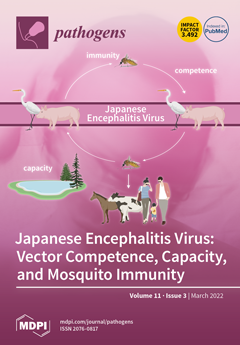
Japanese encephalitis virus (JEV) is a zoonotic mosquito-borne flavivirus that is maintained in a transmission cycle between mosquitoes and vertebrate hosts, mainly Ardeid birds and pigs. JEV is endemic in (South) East Asia and the Torres Strait region of Australia. Upon introduction into non-endemic areas, JEV could be transmitted and become established if competent vectors and suitable hosts are present. Here, an overview of the current knowledge on vector competence for JEV and JEV detection in field-caught mosquitoes is presented. Additionally, other parameters influencing vector capacity, e.g., temperature and abundance, are discussed. Furthermore, available knowledge on mosquito immunity in relation to JEV is summarized, covering physical and physiological barriers, molecular pathways, antimicrobial peptides, and the vector microbiome. View this paper.
  • Issues are regarded as officially published after their release is announced to the table of contents alert mailing list.
  • You may sign up for e-mail alerts to receive table of contents of newly released issues.
  • PDF is the official format for papers published in both, html and pdf forms. To view the papers in pdf format, click on the "PDF Full-text" link, and use the free Adobe Reader to open them.
Order results
Result details
Section
Select all
Export citation of selected articles as:
11 pages, 272 KiB  
Article
High Fecal Carriage of Multidrug Resistant Bacteria in the Community among Children in Northwestern Tanzania
by Delfina R. Msanga, Vitus Silago, Tulla Massoza, Benson R. Kidenya, Emmanuel Balandya, Mariam M. Mirambo, Bruno Sunguya, Blandina Theophil Mmbaga, Eligius Lyamuya, John Bartlet and Stephen E. Mshana
Pathogens 2022, 11(3), 379; https://doi.org/10.3390/pathogens11030379 - 21 Mar 2022
Cited by 4 | Viewed by 2186
Abstract
Colonization of multidrug resistant (MDR) bacteria is associated with subsequent invasive infections in children with comorbidities. This study aimed to determine the resistance profile and factors associated with MDR pathogen colonization among HIV−and HIV+ children below five years of age in Mwanza, Tanzania. [...] Read more.
Colonization of multidrug resistant (MDR) bacteria is associated with subsequent invasive infections in children with comorbidities. This study aimed to determine the resistance profile and factors associated with MDR pathogen colonization among HIV−and HIV+ children below five years of age in Mwanza, Tanzania. A total of 399 (HIV− 255 and HIV+ 144) children were enrolled and investigated for the presence of MDR bacteria. The median [IQR] age of children was 19 (10–36) months. Out of 27 Staphylococcus aureus colonizing the nasal cavity, 16 (59.5%) were methicillin resistant while 132/278 (47.2%) of Enterobacteriaceae from rectal swabs were resistant to third generation cephalosporins, with 69.7% (92/132) exhibiting extended spectrum beta lactamase (ESBL) phenotypes. The proportion of resistance to gentamicin, amoxicillin/clavulanic acid and meropenem were significantly higher among HIV+ than HIV− children. A history of antibiotic use in the last month OR 2.62 [1.1, 6.9] (p = 0.04) and history of a relative admitted from the same household in the past three months OR 3.73 [1.1, 13.2] (p = 0.03) independently predicted ESBL rectal colonization. HIV+ children had significantly more fecal carriage of isolates resistant to uncommonly used antibiotics. There is a need to strengthen antimicrobial stewardship and Infection Prevention and Control (IPC) programs to prevent the emergence and spread of MDR pathogens in children. Full article
16 pages, 1168 KiB  
Article
Pentoxifylline in the Treatment of Cutaneous Leishmaniasis: A Randomized Clinical Trial in Colombia
by Maria del Mar Castro, Alexandra Cossio, Adriana Navas, Olga Fernandez, Liliana Valderrama, Lyda Cuervo-Pardo, Ricardo Marquez-Oñate, María Adelaida Gómez and Nancy Gore Saravia
Pathogens 2022, 11(3), 378; https://doi.org/10.3390/pathogens11030378 - 21 Mar 2022
Cited by 1 | Viewed by 2112
Abstract
Addition of the immunomodulator pentoxifylline (PTX) to antimonial treatment of mucosal leishmaniasis has shown increased efficacy. This randomized, double-blind, placebo-controlled trial evaluated whether addition of pentoxifylline to meglumine antimoniate (MA) treatment improves therapeutic response in cutaneous leishmaniasis (CL) patients. Seventy-three patients aged 18–65 [...] Read more.
Addition of the immunomodulator pentoxifylline (PTX) to antimonial treatment of mucosal leishmaniasis has shown increased efficacy. This randomized, double-blind, placebo-controlled trial evaluated whether addition of pentoxifylline to meglumine antimoniate (MA) treatment improves therapeutic response in cutaneous leishmaniasis (CL) patients. Seventy-three patients aged 18–65 years, having multiple lesions or a single lesion ≥ 3 cm were randomized to receive: intramuscular MA (20 mg/kg/day × 20 days) plus oral PTX 400 mg thrice daily (intervention arm, n = 36) or MA plus placebo (control arm, n = 37), between 2012 and 2015. Inflammatory gene expression was evaluated by RT-qPCR in peripheral blood mononuclear cells from trial patients, before and after treatment. Intention-to-treat failure rate was 35% for intervention vs. 25% for control (OR: 0.61, 95% CI: 0.21–1.71). Per-protocol failure rate was 32% for PTX, and 24% for placebo (OR: 0.50, 95% CI: 0.13–1.97). No differences in frequency or severity of adverse events were found (PTX = 142 vs. placebo = 140). Expression of inflammatory mediators was unaltered by addition of PTX to MA. However, therapeutic failure was associated with significant overexpression of il1β and ptgs2 (p < 0.05), irrespective of study group. No clinical benefit of addition of PTX to standard treatment was detected in early mild to moderate CL caused by Leishmania (V.) panamensis. Full article
(This article belongs to the Special Issue Leishmania & Leishmaniasis)
Show Figures

Figure 1

18 pages, 700 KiB  
Review
Pathogenicity and Its Implications in Taxonomy: The Brucella and Ochrobactrum Case
by Edgardo Moreno, José María Blasco, Jean Jacques Letesson, Jean Pierre Gorvel and Ignacio Moriyón
Pathogens 2022, 11(3), 377; https://doi.org/10.3390/pathogens11030377 - 21 Mar 2022
Cited by 20 | Viewed by 4039
Abstract
The intracellular pathogens of the genus Brucella are phylogenetically close to Ochrobactrum, a diverse group of free-living bacteria with a few species occasionally infecting medically compromised patients. A group of taxonomists recently included all Ochrobactrum organisms in the genus Brucella based on [...] Read more.
The intracellular pathogens of the genus Brucella are phylogenetically close to Ochrobactrum, a diverse group of free-living bacteria with a few species occasionally infecting medically compromised patients. A group of taxonomists recently included all Ochrobactrum organisms in the genus Brucella based on global genome analyses and alleged equivalences with genera such as Mycobacterium. Here, we demonstrate that such equivalencies are incorrect because they overlook the complexities of pathogenicity. By summarizing Brucella and Ochrobactrum divergences in lifestyle, structure, physiology, population, closed versus open pangenomes, genomic traits, and pathogenicity, we show that when they are adequately understood, they are highly relevant in taxonomy and not unidimensional quantitative characters. Thus, the Ochrobactrum and Brucella differences are not limited to their assignments to different “risk-groups”, a biologically (and hence, taxonomically) oversimplified description that, moreover, does not support ignoring the nomen periculosum rule, as proposed. Since the epidemiology, prophylaxis, diagnosis, and treatment are thoroughly unrelated, merging free-living Ochrobactrum organisms with highly pathogenic Brucella organisms brings evident risks for veterinarians, medical doctors, and public health authorities who confront brucellosis, a significant zoonosis worldwide. Therefore, from taxonomical and practical standpoints, the Brucella and Ochrobactrum genera must be maintained apart. Consequently, we urge researchers, culture collections, and databases to keep their canonical nomenclature. Full article
(This article belongs to the Special Issue Brucella Species and Brucella melitensis)
Show Figures

Figure 1

15 pages, 8932 KiB  
Article
The Biological Traumatization of Crops Due to the Enzyme Stage of Enzyme-Mycotic Seed Depletion
by Sulukhan K. Temirbekova, Ivan M. Kulikov, Yuliya V. Afanasyeva, Mukhtar Z. Ashirbekov, Olga O. Beloshapkina, Elena A. Kalashnikova, Irina Sardarova, Marat Sh. Begeulov, Dmitry E. Kucher, Natalia E. Ionova and Nazih Y. Rebouh
Pathogens 2022, 11(3), 376; https://doi.org/10.3390/pathogens11030376 - 21 Mar 2022
Cited by 3 | Viewed by 1856
Abstract
In the light of Vavilov’s Law, grain traumatization in the standing crop of wheat and other crops due to the enzyme stage of enzyme-mycotic seed depletion (EMSD) was confirmed, the parameters of open and hidden harmfulness were detected, and a scale of plant [...] Read more.
In the light of Vavilov’s Law, grain traumatization in the standing crop of wheat and other crops due to the enzyme stage of enzyme-mycotic seed depletion (EMSD) was confirmed, the parameters of open and hidden harmfulness were detected, and a scale of plant resistance to such traumatization was developed. The current study demonstrates that pathogen contamination in grains occurs before harvesting and its degree is determined by favorable humidity and temperature conditions and by the open and hidden grain traumatization due to the enzyme stage of EMSD, i.e., the grain’s hydrolytic enzymes providing a growth substrate for a fungal spread that is later substituted by pathogen enzymes leading to grain spoiling and self-warming. The most common technique to preserve grain quality is to support a moisture level that prevents further spreading of the fungi. The grains that are contaminated with very low temperature and humidity levels facilitate the germinability and high quality of the grain. The new ways to withstand EMSD should, first of all, include a selection of activities. Using biological, biochemical and physical (X-ray) methods, genetic sources of resistance towards EMSD were found in the VIR world collection that is recommended for further selection. These sources have become a basis for the varieties, such as Moskovskaya 39, Ilot (winter wheat), Gremme and Gremme 2U (hulless spelt), Alcoran (winter spelt) and Kanysh (spring wheat). Full article
Show Figures

Figure 1

7 pages, 623 KiB  
Communication
European and American Strains of Porcine Parainfluenza Virus 1 (PPIV-1) Belong to Two Distinct Genetic Lineages
by Tomasz Stadejek, Piotr Cybulski, Phillip C. Gauger and Aleksandra Woźniak
Pathogens 2022, 11(3), 375; https://doi.org/10.3390/pathogens11030375 - 20 Mar 2022
Cited by 8 | Viewed by 1881
Abstract
Porcine parainfluenza virus 1 (PPIV-1) is a recently emerged respirovirus closely related to human parainfluenza virus 1 (HPIV-1) and Sendai virus (SenV). PPIV-1 has been detected in Asia, the Americas and Europe, but knowledge on its epidemiology and genetic diversity is very limited. [...] Read more.
Porcine parainfluenza virus 1 (PPIV-1) is a recently emerged respirovirus closely related to human parainfluenza virus 1 (HPIV-1) and Sendai virus (SenV). PPIV-1 has been detected in Asia, the Americas and Europe, but knowledge on its epidemiology and genetic diversity is very limited. In the present study, the complete nucleotide sequences of the fusion (F)-protein gene obtained from samples from 12 Polish and 11 US herds were analysed and compared to previously available genetic data from the Americas, Asia and Europe. The existence of two distinct clades was observed, grouping European sequences and one Hong Kong sequence (clade 1), or one American sequence and three Asian sequences (clade 2). The mean genetic distances measured with the p-distance were 0.04 (S.E., 0.000) within both clades, and 0.095 (S.E., 0.006) between the clades. Moreover, two distinct clusters of highly similar sequences were identified, which corresponded to the geographically distant nurseries and finishing units, from three pig flows within one Polish pig-production company. The obtained data indicate that the two PPIV-1 lineages may have evolved independently in Europe and America. More studies, particularly involving Asian viruses, are necessary to understand the virus’ emergence and epidemiology and the role of carriers in the spread of PPIV-1. Full article
(This article belongs to the Special Issue Swine Viral Diseases)
Show Figures

Figure 1

17 pages, 2077 KiB  
Review
Development and Structural Analysis of Antibody Therapeutics for Filoviruses
by Xiaoying Yu and Erica Ollmann Saphire
Pathogens 2022, 11(3), 374; https://doi.org/10.3390/pathogens11030374 - 18 Mar 2022
Cited by 5 | Viewed by 3674
Abstract
The filoviruses, including ebolaviruses and marburgviruses, are among the world’s deadliest pathogens. As the only surface-exposed protein on mature virions, their glycoprotein GP is the focus of current therapeutic monoclonal antibody discovery efforts. With recent technological developments, potent antibodies have been identified from [...] Read more.
The filoviruses, including ebolaviruses and marburgviruses, are among the world’s deadliest pathogens. As the only surface-exposed protein on mature virions, their glycoprotein GP is the focus of current therapeutic monoclonal antibody discovery efforts. With recent technological developments, potent antibodies have been identified from immunized animals and human survivors of virus infections and have been characterized functionally and structurally. Structural insight into how the most successful antibodies target GP further guides vaccine development. Here we review the recent developments in the identification and characterization of neutralizing antibodies and cocktail immunotherapies. Full article
(This article belongs to the Special Issue Characterization of Antibody Responses to Virus Infections in Humans)
Show Figures

Figure 1

15 pages, 1283 KiB  
Article
Trash to Treasure: How Insect Protein and Waste Containers Can Improve the Environmental Footprint of Mosquito Egg Releases
by Megan J. Allman, Aidan J. Slack, Nigel P. Abello, Ya-Hsun Lin, Scott L. O’Neill, Andrea J. Robinson, Heather A. Flores and D. Albert Joubert
Pathogens 2022, 11(3), 373; https://doi.org/10.3390/pathogens11030373 - 18 Mar 2022
Cited by 1 | Viewed by 2674
Abstract
Release and subsequent establishment of Wolbachia-infected Aedes aegypti in native mosquito populations has successfully reduced mosquito-borne disease incidence. While this is promising, further development is required to ensure that this method is scalable and sustainable. Egg release is a beneficial technique that [...] Read more.
Release and subsequent establishment of Wolbachia-infected Aedes aegypti in native mosquito populations has successfully reduced mosquito-borne disease incidence. While this is promising, further development is required to ensure that this method is scalable and sustainable. Egg release is a beneficial technique that requires reduced onsite resources and increases community acceptance; however, its incidental ecological impacts must be considered to ensure sustainability. In this study, we tested a more environmentally friendly mosquito rearing and release approach through the encapsulation of diet and egg mixtures and the subsequent utilization of waste containers to hatch and release mosquitoes. An ecologically friendly diet mix was specifically developed and tested for use in capsules, and we demonstrated that using either cricket or black soldier fly meal as a substitute for beef liver powder had no adverse effects on fitness or Wolbachia density. We further encapsulated both the egg and diet mixes and demonstrated no loss in viability. To address the potential of increased waste generation through disposable mosquito release containers, we tested reusing commonly found waste containers (aluminum and tin cans, PET, and glass bottles) as an alternative, conducting a case study in Kiribati to assess the concept’s cultural, political, and economic applicability. Our results showed that mosquito emergence and fitness was maintained with a variety of containers, including when tested in the field, compared to control containers, and that there are opportunities to implement this method in the Pacific Islands in a way that is culturally considerate and cost-effective. Full article
(This article belongs to the Special Issue Mosquito-Borne Diseases: Novel Control Strategies)
Show Figures

Figure 1

23 pages, 2812 KiB  
Article
Identification of Inflammatory and Regulatory Cytokines IL-1α-, IL-4-, IL-6-, IL-12-, IL-13-, IL-17A-, TNF-α-, and IFN-γ-Producing Cells in the Milk of Dairy Cows with Subclinical and Clinical Mastitis
by Zane Vitenberga-Verza, Māra Pilmane, Ksenija Šerstņova, Ivars Melderis, Łukasz Gontar, Maksymilian Kochański, Andżelika Drutowska, Gergely Maróti and Beatriz Prieto-Simón
Pathogens 2022, 11(3), 372; https://doi.org/10.3390/pathogens11030372 - 17 Mar 2022
Cited by 11 | Viewed by 3028
Abstract
In naturally occurring bovine mastitis, effects of infection depend on the host inflammatory response, including the effects of secreted cytokines. Knowledge about the inflammatory and regulatory cytokines in milk cells of free-stall barn dairy cows and in naturally occurring mastitis is lacking as [...] Read more.
In naturally occurring bovine mastitis, effects of infection depend on the host inflammatory response, including the effects of secreted cytokines. Knowledge about the inflammatory and regulatory cytokines in milk cells of free-stall barn dairy cows and in naturally occurring mastitis is lacking as most studies focus on induced mastitis. Hereby, the aim of the study was to determine inflammatory and regulatory cytokines in the milk of dairy cows with subclinical and clinical mastitis. The following examinations of milk samples were performed: differential counting of somatic cells (SCC), bacteriological examination, and immunocytochemical analysis. Mean SCC increased in subclinical and clinical mastitis cases. The number of pathogenic mastitis-causing bacteria on plates increased in subclinical mastitis cases but decreased in clinical mastitis. The inflammatory and regulatory markers in the milk cells of healthy cows showed the highest mean cell numbers (%). In mastitis cases, immunoreactivity was more pronounced for IL-4, IL-6, IL-12, IL-13, IL-17A, TNF-α, and IFN-γ. Data about subclinical and clinical mastitis demonstrate inflammatory responses to intramammary infection driven by IL-1α, IL-4, and IL-17A. Moreover, the host defense response in mastitis is characterized by continuation or resolution of initial inflammation. IL-12 and INF-γ immunoreactivity was recognized to differ mastitis cases from the relative health status. Full article
Show Figures

Figure 1

10 pages, 844 KiB  
Article
Unexpected TBEV Seropositivity in Serbian Patients Who Recovered from Viral Meningitis and Encephalitis
by Pavle Banović, Adrian Alberto Díaz-Sánchez, Selena Đurić, Siniša Sević, Vesna Turkulov, Dajana Lendak, Sandra Stefan Mikić, Verica Simin, Dragana Mijatović, Ivana Bogdan, Aleksandar Potkonjak, Sara Savić, Dasiel Obregón and Alejandro Cabezas-Cruz
Pathogens 2022, 11(3), 371; https://doi.org/10.3390/pathogens11030371 - 17 Mar 2022
Cited by 6 | Viewed by 2665
Abstract
The tick-borne encephalitis virus (TBEV) causes a life-threatening disease named Tick-borne encephalitis (TBE). The clinical symptoms associated with TBE range from non-specific to severe inflammation of the central nervous system and are very similar to the clinical presentation of other viral meningitis/encephalitis. In [...] Read more.
The tick-borne encephalitis virus (TBEV) causes a life-threatening disease named Tick-borne encephalitis (TBE). The clinical symptoms associated with TBE range from non-specific to severe inflammation of the central nervous system and are very similar to the clinical presentation of other viral meningitis/encephalitis. In consequence, TBE is often misclassified by clinical physicians, mainly in the non-identified high-risk areas where none or only a few TBE cases have been reported. Considering this situation, we hypothesized that among persons from northern Serbia who recovered from viral meningitis or encephalitis, there would be evidence of TBEV infection. To test this hypothesis, in this observational study, we evaluated the seroreactivity against TBEV antigens in patients from northern Serbia who were hospitalized due to viral meningitis and/or viral encephalitis of unknown etiology. Three cases of seroreactivity to TBEV antigens were discovered among convalescent patients who recovered from viral meningitis and/or encephalitis and accepted to participate in the study (n = 15). The clinical and laboratory findings of these patients overlap with that of seronegative convalescent patients. Although TBE has been a notifiable disease in Serbia since 2004, there is no active TBE surveillance program for the serologic or molecular screening of TBEV infection in humans in the country. This study highlights the necessity to increase the awareness of TBE among physicians and perform active and systematic screening of TBEV antibodies among patients with viral meningitis and/or encephalitis. Full article
(This article belongs to the Collection Regional Impact of Ticks and Tick-Borne Diseases)
Show Figures

Figure 1

7 pages, 1416 KiB  
Article
Repurposing Avasimibe to Inhibit Bacterial Glycosyltransferases
by Md Kamrul Hasan, Samir El Qaidi, Peter McDonald, Anuradha Roy and Philip R. Hardwidge
Pathogens 2022, 11(3), 370; https://doi.org/10.3390/pathogens11030370 - 17 Mar 2022
Viewed by 2021
Abstract
We are interested in identifying and characterizing small molecule inhibitors of bacterial virulence factors for their potential use as anti-virulence inhibitors. We identified from high-throughput screening assays a potential activity for avasimibe, a previously characterized acyl-coenzyme A: cholesterol acyltransferase inhibitor, in inhibiting the [...] Read more.
We are interested in identifying and characterizing small molecule inhibitors of bacterial virulence factors for their potential use as anti-virulence inhibitors. We identified from high-throughput screening assays a potential activity for avasimibe, a previously characterized acyl-coenzyme A: cholesterol acyltransferase inhibitor, in inhibiting the NleB and SseK arginine glycosyltransferases from Escherichia coli and Salmonella enterica, respectively. Avasimibe inhibited the activity of the Citrobacter rodentium NleB, E. coli NleB1, and S. enterica SseK1 enzymes, without affecting the activity of the human serine/threonine N-acetylglucosamine (O-GlcNAc) transferase. Avasimibe was not toxic to mammalian cells at up to 200 µM and was neither bacteriostatic nor bactericidal at concentrations of up to 125 µM. Doses of 10 µM avasimibe were sufficient to reduce S. enterica abundance in RAW264.7 macrophage-like cells, and intraperitoneal injection of avasimibe significantly reduced C. rodentium survival in mice, regardless of whether the avasimibe was administered pre- or post-infection. We propose that avasimibe or related derivates created using synthetic chemistry may have utility in preventing or treating bacterial infections by inhibiting arginine glycosyltransferases that are important to virulence. Full article
Show Figures

Figure 1

14 pages, 948 KiB  
Review
Calomys callosus: An Experimental Animal Model Applied to Parasitic Diseases Investigations of Public Health Concern
by Rafael Borges Rosa, Mylla Spirandelli da Costa, Samuel Cota Teixeira, Emilene Ferreira de Castro, Willyenne Marília Dantas, Eloisa Amália Vieira Ferro and Murilo Vieira da Silva
Pathogens 2022, 11(3), 369; https://doi.org/10.3390/pathogens11030369 - 17 Mar 2022
Cited by 3 | Viewed by 2634
Abstract
The appearance and spread of parasitic diseases around the world aroused the interest of the scientific community to discover new animal models for improving the quality and specificity of surveys. Calomys callosus is a rodent native to South America, an easy handling model, [...] Read more.
The appearance and spread of parasitic diseases around the world aroused the interest of the scientific community to discover new animal models for improving the quality and specificity of surveys. Calomys callosus is a rodent native to South America, an easy handling model, with satisfactory longevity and reproducibility. C. callosus is susceptible to toxoplasmosis and can be used as experimental model for the study the pathogenesis, treatment, vertical transmission, and ocular toxoplasmosis. C. callosus can also be used to study cutaneous and visceral leishmaniasis, as the animals present cutaneous lesions, as well as parasites in the organs. C. callosus has epidemiological importance in Chagas disease, and since it is a Trypanosoma cruzi natural host in which rodents show high parasitemia and lethality, they are also effective as a model of congenital transmission. In the study of schistosomiasis, Schistosoma mansoni was proven to be a C. callosus natural host; thus, this rodent is a great model for fibrosis, hepatic granulomatous reaction, and celloma associated with lymphomyeloid tissue (CALT) during S. mansoni infection. In this review, we summarize the leading studies of parasitic diseases that used C. callosus as a rodent experimental model, describing the main uses and characteristics that led them to be considered an effective model. Full article
(This article belongs to the Special Issue Advances in Parasitic Diseases)
Show Figures

Graphical abstract

14 pages, 297 KiB  
Review
A Review of SARS-CoV-2 Disease (COVID-19): Pandemic in Our Time
by Nasruddeen Al-Awwal, Ferris Dweik, Samira Mahdi, Majed El-Dweik and Stephen H. Anderson
Pathogens 2022, 11(3), 368; https://doi.org/10.3390/pathogens11030368 - 17 Mar 2022
Cited by 24 | Viewed by 3337
Abstract
Development and deployment of biosensors for the rapid detection of the 2019 novel severe acute respiratory syndrome—coronavirus 2 (SARS-CoV-2) are of utmost importance and urgency during this recent outbreak of coronavirus pneumonia (COVID-19) caused by SARS-CoV-2 infection, which spread rapidly around the world. [...] Read more.
Development and deployment of biosensors for the rapid detection of the 2019 novel severe acute respiratory syndrome—coronavirus 2 (SARS-CoV-2) are of utmost importance and urgency during this recent outbreak of coronavirus pneumonia (COVID-19) caused by SARS-CoV-2 infection, which spread rapidly around the world. Cases now confirmed in February 2022 indicate that more than 170 countries worldwide are affected. Recent evidence indicates over 430 million confirmed cases with over 5.92 million deaths scattered across the globe, with the United States having more than 78 million confirmed cases and over 920,000 deaths. The US now has many more cases than in China where coronavirus cases were first reported in late December 2019. During the initial outbreak in China, many leaders did not anticipate it could reach the whole world, spreading to many countries and posing severe threats to global health. The objective of this review is to summarize the origin of COVID-19, its biological nature, comparison with other coronaviruses, symptoms, prevention, treatment, potential, available methods for SARS-CoV-2 detection, and post-COVID-19 symptoms. Full article
(This article belongs to the Special Issue Viral Pathogenesis and Immunity)
8 pages, 1372 KiB  
Article
The Use of Pythium oligandrum in the Biological Control of Roundworm Infection in Dogs and Cats
by Iasmina Luca, Marius Stelian Ilie, Tiana Florea, Adrian Olariu-Jurca, Adrian Stancu and Gheorghe Dărăbuş
Pathogens 2022, 11(3), 367; https://doi.org/10.3390/pathogens11030367 - 17 Mar 2022
Cited by 2 | Viewed by 2475
Abstract
Pythium oligandrum is an oomycete used in veterinary medicine to treat dermatophytosis in animals. The ovicidal potential against various types of parasite eggs has not been investigated. Ascarids are very common in young animals and the resistance of eggs in the external environment [...] Read more.
Pythium oligandrum is an oomycete used in veterinary medicine to treat dermatophytosis in animals. The ovicidal potential against various types of parasite eggs has not been investigated. Ascarids are very common in young animals and the resistance of eggs in the external environment is very high. A commercial product containing P. oligandrum was used in the present study. Its ovicidal action against Toxocara spp. eggs was investigated. Thus, two categories of media (soil and sandstone) were used, on which the ascarid eggs were dispersed. The commercial product was prepared as a solution and was applied according to the manufacturer. The treatment scheme indicated in case of animals with dermatophytosis was used. Thus, the external natural conditions were recreated in the laboratory, in order to establish whether there is any applicability for this oomycete in the biological control of some parasitic diseases. The results indicated an ovicidal action of Pythium oligandrum, supporting the prospects of its use in the decontamination methods of various surfaces or environments where ascarid eggs from carnivores are found. Full article
(This article belongs to the Special Issue Advances in the Control of the Helminthosis in Domestic Animals)
Show Figures

Figure 1

16 pages, 2953 KiB  
Article
The Origin and Maintenance of Tuberculosis Is Explained by the Induction of Smear-Negative Disease in the Paleolithic
by Pere-Joan Cardona, Martí Català and Clara Prats
Pathogens 2022, 11(3), 366; https://doi.org/10.3390/pathogens11030366 - 17 Mar 2022
Cited by 4 | Viewed by 3892
Abstract
Is it possible that the origin of Mycobacterium tuberculosis (Mtb) infection was around 70,000 years before the common era? At that time Homo sapiens was just another primate species with discrete growth and a very low-density geographic occupation. Therefore, it is difficult to [...] Read more.
Is it possible that the origin of Mycobacterium tuberculosis (Mtb) infection was around 70,000 years before the common era? At that time Homo sapiens was just another primate species with discrete growth and a very low-density geographic occupation. Therefore, it is difficult to understand the origin of a highly virulent obligate human pathogen. We have designed a new SEIR model (TBSpectr) that allows the differentiation of smear-positive and -negative tuberculosis. The model reconciles currently accepted growth rates for the Middle Paleolithic (0.003%/year) and Neolithic (0.1%/year). The obtained data link the origin of Mtb infection in the Middle Paleolithic to the induction of smear-negative TB, and reveal that its persistence required interrelations among hunter–gatherer groups, while the risk of human extinction was negligible. It also highlights the number of people infected per case and the fast progression to disease for Mtb infection maintenance, as well as the link between poor health in the Neolithic with the increased incidence of more severe forms of TB (smear-positive). In conclusion, our data support the origin of TB as a well-tolerated, highly persistent disease, even in low-density populations, showing the difficulty of its eradication and highlighting the necessity for providing better health conditions to humans to reduce its severity. Full article
(This article belongs to the Special Issue Pathogenicity of Mycobacterium tuberculosis)
Show Figures

Figure 1

12 pages, 1878 KiB  
Article
Bronchoscopy and Lung Fine-Needle Aspiration for Antemortem Evaluation of Pulmonary Involvement in Dogs with Naturally Occurring Canine Leishmaniosis
by Ioannis Kavarnos, Dimitra Pardali, Georgia D. Brellou, Elias Papadopoulos, Maria Kritsepi-Konstantinou and Katerina K. Adamama-Moraitou
Pathogens 2022, 11(3), 365; https://doi.org/10.3390/pathogens11030365 - 17 Mar 2022
Cited by 2 | Viewed by 2500
Abstract
Clinical manifestations from the lower respiratory tract are rare in canine leishmaniosis (CanL), making bronchoscopy and lung fine-needle aspiration (FNA) seldomly justified. The aim of this prospective study was to investigate the involvement of Leishmania infantum in the lungs of dogs with naturally [...] Read more.
Clinical manifestations from the lower respiratory tract are rare in canine leishmaniosis (CanL), making bronchoscopy and lung fine-needle aspiration (FNA) seldomly justified. The aim of this prospective study was to investigate the involvement of Leishmania infantum in the lungs of dogs with naturally occurring CanL by bronchoscopy and examination of bronchoalveolar lavage fluid (BALF), bronchial mucosa biopsies, and FNA, using immunodiagnostics. Dogs with relevant concurrent diseases and azotemia were excluded. Cough was detected in 5/31 (16.1%) dogs. Lesions (hyperemia, edema, mucosal granularity, secretions) were identified upon bronchoscopy in 19/31 (61.3%) dogs. The cytology of BALF revealed histiocytic inflammation in 14/31 (45.2%) dogs; the parasite was identified in one dog (3.2%). The immunofluorescence antibody test in BALF was positive in 15/31 (48.4%) dogs. Histopathology of bronchial mucosa and/or adjacent alveoli revealed lesions (mononuclear cell infiltration, fibrosis, edema, thickening of the inter-alveolar septa) in 24/31 (77.4%) dogs, with no Leishmania amastigotes. Positive antigen staining was observed within the cytoplasm of mononuclear cells in immunocytochemistry and immunohistochemistry. Μononuclear cells showed antigenic positivity in bronchial mucosa (27/31; 87.1%), BALF (30/31; 96.8%), and lung FNA (27/31; 87.1%). In conclusion, lungs seem to be affected from CanL more commonly than previously believed, and bronchoscopy allows obtaining valuable samples for antemortem diagnosis. Full article
(This article belongs to the Special Issue New Insights on Canine Visceral Leishmaniasis)
Show Figures

Figure 1

6 pages, 224 KiB  
Editorial
Animal Chlamydiae: A Concern for Human and Veterinary Medicine
by Hanna Marti and Martina Jelocnik
Pathogens 2022, 11(3), 364; https://doi.org/10.3390/pathogens11030364 - 17 Mar 2022
Cited by 2 | Viewed by 1970
Abstract
The Chlamydiae are a phylum of obligate intracellular, Gram-negative bacteria with a biphasic lifecycle [...] Full article
(This article belongs to the Special Issue Animal Chlamydiae: A Concern for Human and Veterinary Medicine)
15 pages, 2812 KiB  
Article
Assay for Evaluating the Abundance of Vibrio cholerae and Its O1 Serogroup Subpopulation from Water without DNA Extraction
by Tania Nasreen, Nora A.S. Hussain, Jia Yee Ho, Vanessa Zhi Jie Aw, Munirul Alam, Stephanie K. Yanow and Yann F. Boucher
Pathogens 2022, 11(3), 363; https://doi.org/10.3390/pathogens11030363 - 16 Mar 2022
Cited by 4 | Viewed by 3226
Abstract
Cholera is a severe diarrheal disease caused by Vibrio cholerae, a natural inhabitant of brackish water. Effective control of cholera outbreaks depends on prompt detection of the pathogen from clinical specimens and tracking its source in the environment. Although the epidemiology of [...] Read more.
Cholera is a severe diarrheal disease caused by Vibrio cholerae, a natural inhabitant of brackish water. Effective control of cholera outbreaks depends on prompt detection of the pathogen from clinical specimens and tracking its source in the environment. Although the epidemiology of cholera is well studied, rapid detection of V. cholerae remains a challenge, and data on its abundance in environmental sources are limited. Here, we describe a sensitive molecular quantification assay by qPCR, which can be used on-site in low-resource settings on water without the need for DNA extraction. This newly optimized method exhibited 100% specificity for total V. cholerae as well as V. cholerae O1 and allowed detection of as few as three target CFU per reaction. The limit of detection is as low as 5 × 10³ CFU/L of water after concentrating biomass from the sample. The ability to perform qPCR on water samples without DNA extraction, portable features of the equipment, stability of the reagents at 4 °C and user-friendly online software facilitate fast quantitative analysis of V. cholerae. These characteristics make this assay extremely useful for field research in resource-poor settings and could support continuous monitoring in cholera-endemic areas. Full article
(This article belongs to the Special Issue Pathogens in Aquatic Environments)
Show Figures

Figure 1

4 pages, 198 KiB  
Editorial
Host–Pathogen Interactions: Organotypic Cultures to Unravel the Mysteries of the Primordial Hostility among Organisms
by Pasquale Marrazzo, Natalie Fischer, Claudia Nastasi, Monica Cricca and Daniela Fusco
Pathogens 2022, 11(3), 362; https://doi.org/10.3390/pathogens11030362 - 16 Mar 2022
Cited by 1 | Viewed by 2050
Abstract
The interaction of humans with microorganisms represents a subtle balance between harm and good [...] Full article
(This article belongs to the Special Issue New Tools in 3D Host-pathogen Interactions)
14 pages, 2725 KiB  
Article
Ex Vivo Infection of Human Placental Explants by Trypanosoma cruzi Reveals a microRNA Profile Similar to That Seen in Trophoblast Differentiation
by Lisvaneth Medina, Jesús Alejandro Guerrero-Muñoz, Ana Isabel Liempi, Christian Castillo, Yessica Ortega, Alfredo Sepúlveda, Fernando Salomó, Juan Diego Maya and Ulrike Kemmerling
Pathogens 2022, 11(3), 361; https://doi.org/10.3390/pathogens11030361 - 16 Mar 2022
Cited by 5 | Viewed by 2093
Abstract
Congenital Chagas disease, caused by the protozoan parasite Trypanosoma cruzi, is responsible for 22.5% of new cases each year. However, placental transmission occurs in only 5% of infected mothers and it has been proposed that the epithelial turnover of the trophoblast can [...] Read more.
Congenital Chagas disease, caused by the protozoan parasite Trypanosoma cruzi, is responsible for 22.5% of new cases each year. However, placental transmission occurs in only 5% of infected mothers and it has been proposed that the epithelial turnover of the trophoblast can be considered a local placental defense against the parasite. Thus, Trypanosoma cruzi induces cellular proliferation, differentiation, and apoptotic cell death in the trophoblast, which are regulated, among other mechanisms, by small non-coding RNAs such as microRNAs. On the other hand, ex vivo infection of human placental explants induces a specific microRNA profile that includes microRNAs related to trophoblast differentiation such as miR-512-3p miR-515-5p, codified at the chromosome 19 microRNA cluster. Here we determined the expression validated target genes of miR-512-3p and miR-515-5p, specifically human glial cells missing 1 transcription factor and cellular FLICE-like inhibitory protein, as well as the expression of the main trophoblast differentiation marker human chorionic gonadotrophin during ex vivo infection of human placental explants, and examined how the inhibition or overexpression of both microRNAs affects parasite infection. We conclude that Trypanosoma cruzi-induced trophoblast epithelial turnover, particularly trophoblast differentiation, is at least partially mediated by placenta-specific miR-512-3p and miR-515-5p and that both miRNAs mediate placental susceptibility to ex vivo infection of human placental explants. Knowledge about the role of parasite-modulated microRNAs in the placenta might enable their use as biomarkers, as prognostic and therapeutic tools for congenital Chagas disease in the future. Full article
(This article belongs to the Special Issue Host-Pathogen Interaction Involved in Trypanosoma cruzi Infection)
Show Figures

Graphical abstract

13 pages, 496 KiB  
Article
Subgingival Periopathogens Assessment and Clinical Periodontal Evaluation of Gastric Cancer Patients—A Cross Sectional Pilot Study
by Flavia Mirela Nicolae, Andreea Cristiana Didilescu, Petra Șurlin, Bogdan Silviu Ungureanu, Valeriu Marin Șurlin, Ștefan Pătrașcu, Sandu Ramboiu, Igor Jelihovschi, Luminita Smaranda Iancu, Mirela Ghilusi, Mihai Cucu and Dan Ionuț Gheonea
Pathogens 2022, 11(3), 360; https://doi.org/10.3390/pathogens11030360 - 16 Mar 2022
Cited by 3 | Viewed by 2268
Abstract
Oral microbiota have shown a higher bacterial diversity in patients with cancers of the digestive tract, with higher levels of periopathogens. Recent studies have shown that Fusobacterium links to gastro-intestinal neoplastic tissue and accelerates its progression, as well as worsening patient outcome. The [...] Read more.
Oral microbiota have shown a higher bacterial diversity in patients with cancers of the digestive tract, with higher levels of periopathogens. Recent studies have shown that Fusobacterium links to gastro-intestinal neoplastic tissue and accelerates its progression, as well as worsening patient outcome. The present pilot study was carried out between February and December 2020 to evaluate the possible association between the abundance of some periopathogens (Fusobacterium nucleatum, Porphyromonas gingivalis, Aggregatibacter actinomycetemcomitans, Treponema denticola and Tannerella forsythia) in subgingival plaque and periodontal status with characteristics of gastric cancer. The study was performed on a sample of 24 patients with gastric cancer from the 1st Department of Surgery and Department of Gastroenterology within the Clinical County Hospital of Emergency of Craiova, Romania. The patients’ oral cavity was examined, gingival crevicular samples were collected, and signs of periodontal disease were recorded. On the histopathological exam, the differentiation grade and size of the tumour were registered. Our results showed that, from the periopathogens studied, the most abundant bacteria were F. nucleatum followed by T. forsythia in all groups. In our present study, the strong correlation between tumour dimension and all periodontal parameters but also between tumour dimension and F. nucleatum could suggest a positive association between periodontal disease, tumoral growth and periopathogens implication in this process. Full article
(This article belongs to the Special Issue Opportunistic Oral Pathogens in Oral and Systemic Diseases)
Show Figures

Figure 1

9 pages, 1380 KiB  
Communication
Comparison of Auto Sampling and Passive Sampling Methods for SARS-CoV-2 Detection in Wastewater
by Melissa Wilson, Yuanyuan Qiu, Jiaao Yu, Bonita E. Lee, David T. McCarthy and Xiaoli Pang
Pathogens 2022, 11(3), 359; https://doi.org/10.3390/pathogens11030359 - 16 Mar 2022
Cited by 16 | Viewed by 2875
Abstract
Wastewater-based surveillance is emerging as an important tool for the COVID-19 pandemic trending. Current methods of wastewater collection, such as grab and auto-composite sampling, have drawbacks that impede effective surveillance, especially from small catchments with limited accessibility. Passive samplers, which are more cost-effective [...] Read more.
Wastewater-based surveillance is emerging as an important tool for the COVID-19 pandemic trending. Current methods of wastewater collection, such as grab and auto-composite sampling, have drawbacks that impede effective surveillance, especially from small catchments with limited accessibility. Passive samplers, which are more cost-effective and require fewer resources to process, are promising candidates for monitoring wastewater for SARS-CoV-2. Here, we compared traditional auto sampling with passive sampling for SARS-CoV-2 detection in wastewater. A torpedo-style 3D-printed passive sampler device containing both cotton swabs and electronegative filter membranes was used. Between April and June 2021, fifteen passive samplers were placed at a local hospital’s wastewater outflow alongside an autosampler. Reverse transcription and quantitative polymerase chain reaction (RT-qPCR) was used to detect SARS-CoV-2 in the samples after processing and RNA extraction. The swab and membrane of the passive sampler showed similar detection rates and cycle threshold (Ct) values for SARS-CoV-2 RNA for the N1 and N2 gene targets. The passive method performed as well as the grab/auto sampling, with no significant differences between N1 and N2 Ct values. There were discrepant results on two days with negative grab/auto samples and positive passive samples, which might be related to the longer duration of passive sampling in the study. Overall, the passive sampler was rapid, reliable, and cost-effective, and could be used as an alternative sampling method for the detection of SARS-CoV-2 in wastewater. Full article
(This article belongs to the Special Issue Viruses in Water)
Show Figures

Figure 1

13 pages, 16959 KiB  
Article
Characterization of Salmonella enterica Contamination in Pork and Poultry Meat from São Paulo/Brazil: Serotypes, Genotypes and Antimicrobial Resistance Profiles
by Vasco T. M. Gomes, Luisa Z. Moreno, Ana Paula S. Silva, Siddhartha Thakur, Roberto M. La Ragione, Alison E. Mather and Andrea M. Moreno
Pathogens 2022, 11(3), 358; https://doi.org/10.3390/pathogens11030358 - 16 Mar 2022
Cited by 10 | Viewed by 6042
Abstract
Salmonellosis is a zoonosis of major relevance to global public health. Here we present the assessment of Salmonella enterica contamination in pork and poultry meat sold at retail markets in São Paulo, Brazil. A total of 780 meat samples (386 poultry meat and [...] Read more.
Salmonellosis is a zoonosis of major relevance to global public health. Here we present the assessment of Salmonella enterica contamination in pork and poultry meat sold at retail markets in São Paulo, Brazil. A total of 780 meat samples (386 poultry meat and 394 pork samples) were collected from 132 markets. From these, 57 samples (7.3%) were positive for S. enterica isolation, including 32 (8.3%) poultry meat and 25 (6.3%) pork samples. S. enterica isolates were further characterized for serotyping, antimicrobial resistance and genotyping by amplified fragment length polymorphism and pulsed field gel electrophoresis. Antimicrobial resistance analysis demonstrated two main profiles: pork isolates were more resistant to macrolides, β-lactams, tetracycline, phenicols, and fluoroquinolones, and poultry meat isolates presented higher resistance to fluoroquinolones, sulfonamides, tetracycline, and β-lactams. A total of 72.4% of poultry meat isolates were identified as S. Heidelberg, while most of pork isolates were S. Typhimurium (31.7%) and S. Give (16.7%). Genotyping resulted in most clusters consisting exclusively of pork or poultry meat, no cross-contamination was detected, and a tendency to differentiate isolates according to their serotypes and markets of origin. High resistance rates to critically important antimicrobials reinforce the importance of controlling Salmonella contamination in meat production chains. Full article
Show Figures

Figure 1

16 pages, 3623 KiB  
Article
Pseudomonas syringae on Plants in Iceland Has Likely Evolved for Several Million Years Outside the Reach of Processes That Mix This Bacterial Complex across Earth’s Temperate Zones
by Cindy E. Morris, Natalia Ramirez, Odile Berge, Christelle Lacroix, Cécile Monteil, Charlotte Chandeysson, Caroline Guilbaud, Anett Blischke, Margrét Auður Sigurbjörnsdóttir and Oddur Þ. Vilhelmsson
Pathogens 2022, 11(3), 357; https://doi.org/10.3390/pathogens11030357 - 15 Mar 2022
Cited by 5 | Viewed by 3391
Abstract
Here we report, for the first time, the occurrence of the bacteria from the species complex Pseudomonas syringae in Iceland. We isolated this bacterium from 35 of the 38 samples of angiosperms, moss, ferns and leaf litter collected across the island from five [...] Read more.
Here we report, for the first time, the occurrence of the bacteria from the species complex Pseudomonas syringae in Iceland. We isolated this bacterium from 35 of the 38 samples of angiosperms, moss, ferns and leaf litter collected across the island from five habitat categories (boreal heath, forest, subalpine and glacial scrub, grazed pasture, lava field). The culturable populations of P. syringae on these plants varied in size across 6 orders of magnitude, were as dense as 107 cfu g−1 and were composed of strains in phylogroups 1, 2, 4, 6, 7, 10 and 13. P. syringae densities were significantly greatest on monocots compared to those on dicots and mosses and were about two orders of magnitude greater in grazed pastures compared to all other habitats. The phylogenetic diversity of 609 strains of P. syringae from Iceland was compared to that of 933 reference strains of P. syringae from crops and environmental reservoirs collected from 27 other countries based on a 343 bp sequence of the citrate synthase (cts) housekeeping gene. Whereas there were examples of identical cts sequences across multiple countries and continents among the reference strains indicating mixing among these countries and continents, the Icelandic strains grouped into monophyletic lineages that were unique compared to all of the reference strains. Based on estimates of the time of divergence of the Icelandic genetic lineages of P. syringae, the geological, botanical and land use history of Iceland, and atmospheric circulation patterns, we propose scenarios whereby it would be feasible for P. syringae to have evolved outside the reach of processes that tend to mix this bacterial complex across the planet elsewhere. Full article
(This article belongs to the Special Issue Pseudomonas syringae Species Complex)
Show Figures

Figure 1

13 pages, 1812 KiB  
Article
Antimicrobial Resistance in Enterobacterales Recovered from Urinary Tract Infections in France
by Eric Farfour, Laurent Dortet, Thomas Guillard, Nicolas Chatelain, Agathe Poisson, Assaf Mizrahi, Damien Fournier, Rémy A. Bonnin, Nicolas Degand, Philippe Morand, Frédéric Janvier, Vincent Fihman, Stéphane Corvec, Lauranne Broutin, Cécile Le Brun, Nicolas Yin, Geneviève Héry-Arnaud, Antoine Grillon, Emmanuelle Bille, Hélène Jean-Pierre, Marlène Amara, Francoise Jaureguy, Christophe Isnard, Vincent Cattoir, Tristan Diedrich, Emilie Flevin, Audrey Merens, Hervé Jacquier, Marc Vasse and on behalf of the GMC Study Groupadd Show full author list remove Hide full author list
Pathogens 2022, 11(3), 356; https://doi.org/10.3390/pathogens11030356 - 15 Mar 2022
Cited by 11 | Viewed by 2524
Abstract
In the context of increasing antimicrobial resistance in Enterobacterales, the management of these UTIs has become challenging. We retrospectively assess the prevalence of antimicrobial resistance in Enterobacterales isolates recovered from urinary tract samples in France, between 1 September 2017, to 31 August 2018. [...] Read more.
In the context of increasing antimicrobial resistance in Enterobacterales, the management of these UTIs has become challenging. We retrospectively assess the prevalence of antimicrobial resistance in Enterobacterales isolates recovered from urinary tract samples in France, between 1 September 2017, to 31 August 2018. Twenty-six French clinical laboratories provided the susceptibility of 134,162 Enterobacterales isolates to 17 antimicrobials. The most frequent species were E. coli (72.0%), Klebsiella pneumoniae (9.7%), Proteus mirabilis (5.8%), and Enterobacter cloacae complex (2.9%). The overall rate of ESBL-producing Enterobacterales was 6.7%, and ranged from 1.0% in P. mirabilis to 19.5% in K. pneumoniae, and from 3.1% in outpatients to 13.6% in long-term care facilities. Overall, 4.1%, 9.3% and 10.5% of the isolates were resistant to cefoxitin, temocillin and pivmecillinam. Cotrimoxazole was the less active compound with 23.4% resistance. Conversely, 4.4%, 12.9%, and 14.3% of the strains were resistant to fosfomycin, nitrofurantoin, and ciprofloxacin. However, less than 1% of E. coli was resistant to fosfomycin and nitrofurantoin. We identified several trends in antibiotics resistances among Enterobacterales isolates recovered from the urinary tract samples in France. Carbapenem-sparing drugs, such as temocillin, mecillinam, fosfomycin, cefoxitin, and nitrofurantoin, remained highly active, including towards ESBL-E. Full article
Show Figures

Figure 1

13 pages, 2183 KiB  
Article
A Modular Hepatitis E Virus Replicon System for Studies on the Role of ORF1-Encoded Polyprotein Domains
by Filip Cierniak, Rainer G. Ulrich, Martin H. Groschup and Martin Eiden
Pathogens 2022, 11(3), 355; https://doi.org/10.3390/pathogens11030355 - 15 Mar 2022
Cited by 3 | Viewed by 2265
Abstract
Zoonotic hepatitis E virus (HEV) infection is an emerging cause of acute viral hepatitis in developed countries. Known reservoirs of zoonotic genotype 3 (HEV-3) are mainly pigs and wild boar, and to a lesser extent rabbits and deer. Rabbit hepatitis E virus (HEV-3ra) [...] Read more.
Zoonotic hepatitis E virus (HEV) infection is an emerging cause of acute viral hepatitis in developed countries. Known reservoirs of zoonotic genotype 3 (HEV-3) are mainly pigs and wild boar, and to a lesser extent rabbits and deer. Rabbit hepatitis E virus (HEV-3ra) is prevalent in rabbits worldwide and represents a particular risk for zoonotic infection. Current understanding of the molecular mechanisms of HEV pathogenesis is incomplete, particularly due to the limited availability of efficient and reliable cell culture systems. In order to identify genomic regions responsible for HEV propagation in cell culture, we developed a modular chimeric reporter replicon system based on cell culture-adapted (Kernow-C1/p6 and 47832mc) and rabbit-derived HEV strains. Replication in HepG2 cells was monitored on the basis of a Gaussia luciferase reporter gene that was inserted in place of the open reading frame (ORF) 2 of the HEV genome. Luciferase activity of rabbit HEV-derived replicons was significantly lower than that of Kernow-C1/p6 and 47832mc replicons. Serial exchanges of defined ORF1 segments within the Kernow-C1/p6 replicon backbone indicated that HEV replication in HepG2 cells is not determined by a single domain but rather by an interplay of longer segments of the ORF1-derived nonstructural polyprotein. This implies that a specific combination of viral factors is required for efficient HEV propagation in cell culture. Full article
Show Figures

Figure 1

9 pages, 239 KiB  
Article
Parasitic Infections in Internationally Adopted Children: A Twelve-Year Retrospective Study
by Elena Chiappini, Teresa Paba, Matilde Bestetti and Luisa Galli
Pathogens 2022, 11(3), 354; https://doi.org/10.3390/pathogens11030354 - 15 Mar 2022
Cited by 1 | Viewed by 1954
Abstract
Parasitic infections (PIs) are among the most frequent infectious diseases globally. Previous studies reported discrepant results regarding the prevalence of PIs in internationally adopted children (IAC). Data from IAC referred to our paediatric university hospital in 2009–2021 were collected to evaluate the frequency [...] Read more.
Parasitic infections (PIs) are among the most frequent infectious diseases globally. Previous studies reported discrepant results regarding the prevalence of PIs in internationally adopted children (IAC). Data from IAC referred to our paediatric university hospital in 2009–2021 were collected to evaluate the frequency of PIs by the use of stool microscopic examination, antigen assays for Giardia lamblia and Cryptosporidium parvum, and serological tests for Toxocara canis, Strongyloides stercoralis, Schistosoma mansoni, Echinococcus spp., Taenia solium, and Trypanosoma cruzi. Uni- and multivariate logistic regression analyses were performed to evaluate risk factors for PIs and eosinophilia. The proportion of IAC with at least one positive test was 26.83% (640/2385); 2.13% (n = 51) had positive tests for 2 or 3 parasites. A positive assay for helminthic infection was retrieved in 11.07% of children (n = 264), and 17.86% (n = 426) presented with eosinophilia. The most common positive tests were anti-Toxocara canis antibodies (n = 312; 13.8%), followed by positive stool antigen for Giardia lamblia (n = 290; 12.16%), and positive microscopic stool examination for Blastocystis hominis (n = 76; 3.19%). A statistically significant association was found between PIs and region of origin (children from Latin America and Africa were more likely to present PIs than children from Eastern Europe), age 5–14 years, and eosinophilia. No significant association was observed between PIs and gender, vitamin D deficiency, or anemia. In conclusion, PIs are relevant in IAC and an accurate protocol is needed to evaluate IAC once they arrive in their adoptive country. Full article
(This article belongs to the Special Issue Biology of Parasitism)
10 pages, 1592 KiB  
Brief Report
Intestinal Parasites in Children up to 14 Years Old Hospitalized with Diarrhea in Mozambique, 2014–2019
by Ofélia Luís Nhambirre, Idalécia Cossa-Moiane, Adilson Fernando Loforte Bauhofer, Assucênio Chissaque, Maria Luisa Lobo, Olga Matos and Nilsa de Deus
Pathogens 2022, 11(3), 353; https://doi.org/10.3390/pathogens11030353 - 14 Mar 2022
Cited by 2 | Viewed by 2350
Abstract
Diarrhea remains a public health problem in Mozambique, even with control strategies being implemented. This analysis aimed to determine the proportion and factors associated with intestinal parasitic infection (IPI) in children up to 14 years old with diarrheal disease, in the southern, central [...] Read more.
Diarrhea remains a public health problem in Mozambique, even with control strategies being implemented. This analysis aimed to determine the proportion and factors associated with intestinal parasitic infection (IPI) in children up to 14 years old with diarrheal disease, in the southern, central and northern regions of Mozambique. A single diarrheal sample of 1424 children was collected in hospitals and examined using the formol-ether concentration and modified Ziehl–Neelsen techniques to identify intestinal parasites using optical microscopy. Sociodemographic characteristics were obtained by questionnaires. Descriptive statistics and cross-tabulation were performed, and p-values <0.05 were considered statistically significant. A single IPI was detected in 19.2% (273/1424) of the children. Cryptosporidium spp. was the most common parasite (8.1%; 115/1424). Polyparasitism was seen in 26.0% (71/273), with the co-infection of Ascaris lumbricoides and Trichuris trichiura (26.8%; 19/71) being the most common. Age and province were related to IPI (p-value < 0.05). The highest occurrence of IPI was observed in the wet period (October to March), with 21.9% (140/640), compared to the dry period (April to September), with 16.9% (131/776) (p-value = 0.017). Cryptosporidium spp. and the combination of A. lumbricoides/T. trichiura were the main intestinal parasites observed in children hospitalized with diarrhea in Mozambique. Full article
(This article belongs to the Special Issue Intestinal Parasites Infection)
Show Figures

Figure 1

11 pages, 2042 KiB  
Review
Addressing the Data Gaps on Child and Adolescent Tuberculosis
by Sabine Verkuijl, Moorine Penninah Sekadde, Peter J. Dodd, Moses Arinaitwe, Silvia S. Chiang, Annemieke Brands, Kerri Viney, Charalambos Sismanidis and Helen E. Jenkins
Pathogens 2022, 11(3), 352; https://doi.org/10.3390/pathogens11030352 - 14 Mar 2022
Cited by 6 | Viewed by 2956
Abstract
The burden of tuberculosis (TB) among children and young adolescents (<15 years old) is estimated at 1.1 million; however, only 400,000 are treated for TB, indicating a large gap between the number who are cared for and the number estimated to have TB. [...] Read more.
The burden of tuberculosis (TB) among children and young adolescents (<15 years old) is estimated at 1.1 million; however, only 400,000 are treated for TB, indicating a large gap between the number who are cared for and the number estimated to have TB. Accurate data on the burden of pediatric TB is essential to guide action. Despite several improvements in estimating the burden of pediatric TB in the last decade, as well as enhanced data collection efforts, several data gaps remain, both at the global level, but also at the national level where surveillance systems and collaborative research are critical. In this article, we describe recent advances in data collection and burden estimates for TB among children and adolescents, and the remaining gaps. While data collection continues to improve, burden estimates must evolve in parallel, both in terms of their frequency and the methods used. Currently, at the global level, there is a focus on age-disaggregation of TB notifications, the collection of data on TB-HIV, multi-drug resistant (MDR)-TB and treatment outcomes, as well as estimates of the disease burden. Additional data from national surveillance systems or research projects on TB meningitis, as well as other forms of extra-pulmonary TB, would be useful. We must capitalize on the current momentum in child and adolescent TB to close the remaining data gaps for these age groups to better understand the epidemic and further reduce morbidity and mortality due to TB. Full article
Show Figures

Figure 1

17 pages, 35966 KiB  
Article
Expression of the Nonclassical MHC Class I, Saha-UD in the Transmissible Cancer Devil Facial Tumour Disease (DFTD)
by Kathryn Hussey, Alison Caldwell, Alexandre Kreiss, Karsten Skjødt, Annalisa Gastaldello, Ruth Pye, Rodrigo Hamede, Gregory M. Woods and Hannah V. Siddle
Pathogens 2022, 11(3), 351; https://doi.org/10.3390/pathogens11030351 - 14 Mar 2022
Viewed by 3324
Abstract
Devil facial tumour disease (DFTD) is a transmissible cancer that has circulated in the Tasmanian devil population for >25 years. Like other contagious cancers in dogs and devils, the way DFTD escapes the immune response of its host is a central question to [...] Read more.
Devil facial tumour disease (DFTD) is a transmissible cancer that has circulated in the Tasmanian devil population for >25 years. Like other contagious cancers in dogs and devils, the way DFTD escapes the immune response of its host is a central question to understanding this disease. DFTD has a low major histocompatibility complex class I (MHC-I) expression due to epigenetic modifications, preventing host immune recognition of mismatched MHC-I molecules by T cells. However, the total MHC-I loss should result in natural killer (NK) cell activation due to the ‘missing self’. Here, we have investigated the expression of the nonclassical MHC-I, Saha-UD as a potential regulatory or suppressive mechanism for DFTD. A monoclonal antibody was generated against the devil Saha-UD that binds recombinant Saha-UD by Western blot, with limited crossreactivity to the classical MHC-I, Saha-UC and nonclassical Saha-UK. Using this antibody, we confirmed the expression of Saha-UD in 13 DFTD tumours by immunohistochemistry (n = 15) and demonstrated that Saha-UD expression is heterogeneous, with 12 tumours showing intratumour heterogeneity. Immunohistochemical staining for the Saha-UD showed distinct patterns of expression when compared with classical MHC-I molecules. The nonclassical Saha-UD expression by DFTD tumours in vivo may be a mechanism for immunosuppression, and further work is ongoing to characterise its ligand on immune cells. Full article
Show Figures

Figure 1

13 pages, 1881 KiB  
Article
Protein Concentrations in Stored Pooled Platelet Concentrates Treated with Pathogen Inactivation by Amotosalen Plus Ultraviolet a Illumination
by Niels Arni Arnason, Freyr Johannsson, Ragna Landrö, Björn Hardarsson, Sveinn Gudmundsson, Aina-Mari Lian, Janne Reseland, Ottar Rolfsson and Olafur E. Sigurjonsson
Pathogens 2022, 11(3), 350; https://doi.org/10.3390/pathogens11030350 - 14 Mar 2022
Viewed by 2175
Abstract
Platelet granules contain a diverse group of proteins. Upon activation and during storage, platelets release a number of proteins into the circulation or supernatant of stored platelet concentrate (PC). The aim of this work was to investigate the effect of pathogen inactivation (PI) [...] Read more.
Platelet granules contain a diverse group of proteins. Upon activation and during storage, platelets release a number of proteins into the circulation or supernatant of stored platelet concentrate (PC). The aim of this work was to investigate the effect of pathogen inactivation (PI) on a selection of proteins released in stored platelets. Materials and Methods: PCs in platelet additive solution (PAS) were produced from whole blood donations using the buffy coat (BC) method. PCs in the treatment arm were pathogen inactivated with amotosalen and UVA, while PCs in the second arm were used as an untreated platelet control. Concentrations of 36 proteins were monitored in the PCs during storage. Results: The majority of proteins increased in concentration over the storage period. In addition, 10 of the 29 proteins that showed change had significantly different concentrations between the PI treatment and the control at one or more timepoints. A subset of six proteins displayed a PI-related drop in concentration. Conclusions: PI has limited effect on protein concentration stored PC supernatant. The protein’s changes related to PI treatment with elevated concentration implicate accelerated Platelet storage lesion (PSL); in contrast, there are potential novel benefits to PI related decrease in protein concentration that need further investigation. Full article
(This article belongs to the Special Issue Pathogen Reduction of Blood Bank Components)
Show Figures

Figure 1

Previous Issue
Next Issue
Back to TopTop