Next Issue
Volume 13, February-2
Previous Issue
Volume 13, January-2


Diagnostics, Volume 13, Issue 3 (February-1 2023) – 246 articles

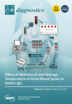
Cover Story (view full-size image): Dried blood spots (DBSs) are a low-cost, non-invasive and convenient alternative to serum/plasma for serological and molecular testing. They can be easily obtained without the necessary equipment for venipuncture or the qualified healthcare personnel. Their use can be beneficial in low- and middle-income countries where it is difficult to obtain and test serum due to the lack of resources, and also in high-income countries to improve sampling by reaching patients not linked to the healthcare system. This manuscript evaluates the utility of DBSs to investigate mumps, measles and rubella IgG. In particular, our work summarizes the effect of hematocrit and different storage conditions (−20 °C, 4 °C and room temperature) for 4 months to perform serological diagnosis using an automated chemiluminescent immunoassay. View this paper
  • Issues are regarded as officially published after their release is announced to the table of contents alert mailing list.
  • You may sign up for e-mail alerts to receive table of contents of newly released issues.
  • PDF is the official format for papers published in both, html and pdf forms. To view the papers in pdf format, click on the "PDF Full-text" link, and use the free Adobe Reader to open them.
Order results
Result details
Section
Select all
Export citation of selected articles as:
5 pages, 14836 KiB  
Interesting Images
Benign Intranodal Thyroid Tissue Similar to Nodal Metastasis of Thyroid Papillary Carcinoma: A Rare Case Report
by Yoo-Na Kang and Jung-Guen Cha
Diagnostics 2023, 13(3), 577; https://doi.org/10.3390/diagnostics13030577 - 03 Feb 2023
Viewed by 1825
Abstract
In patients with thyroid nodules, if the cervical lymph nodes gradually enlarge, a histological confirmation is required to rule out malignancy. Here is a case of benign intranodal thyroid tissue with cystic changes resembling lymph node metastasis of a papillary thyroid carcinoma. A [...] Read more.
In patients with thyroid nodules, if the cervical lymph nodes gradually enlarge, a histological confirmation is required to rule out malignancy. Here is a case of benign intranodal thyroid tissue with cystic changes resembling lymph node metastasis of a papillary thyroid carcinoma. A 47-year-old man received ethanol sclerotherapy because of repeated enlargement of the thyroid gland 2 years prior to presentation. Subsequently, the patient underwent abscess removal from the deep neck and partial lobectomy of the attached left thyroid gland. Two months before the visit, extensive cervical lymphadenopathy was detected on ultrasonography (US) and computed tomography (CT). Total thyroidectomy and cervical lymph node dissection were performed to differentiate between metastatic papillary carcinoma of the thyroid gland and benign thyroid inclusions. Microscopic examination revealed multiple variable-sized nodules of benign thyroid follicles with cystic changes in both thyroid glands and bilateral cervical lymph nodes. An occult papillary microcarcinoma strongly positive for HBME-1 was also observed in the left thyroid lobe. However, the benign intranodal thyroid tissue was negative in both the real-time PCR-based BRAF V600E mutation test and HBME-1 immunohistochemical stain. Similarly, benign intranodal thyroid tissue can be enlarged by multiple cystic changes in a large number of lymph nodes along the neck node chain. For the differentiation of metastatic thyroid papillary carcinoma, real-time PCR-based BRAF V600E mutation test and HBME-1 immunohistochemical staining in addition to histological examination are helpful. Full article
(This article belongs to the Section Medical Imaging and Theranostics)
Show Figures

Figure 1

16 pages, 5950 KiB  
Article
A Medical Image Segmentation Method Based on Improved UNet 3+ Network
by Yang Xu, Shike Hou, Xiangyu Wang, Duo Li and Lu Lu
Diagnostics 2023, 13(3), 576; https://doi.org/10.3390/diagnostics13030576 - 03 Feb 2023
Cited by 12 | Viewed by 3608
Abstract
In recent years, segmentation details and computing efficiency have become more important in medical image segmentation for clinical applications. In deep learning, UNet based on a convolutional neural network is one of the most commonly used models. UNet 3+ was designed as a [...] Read more.
In recent years, segmentation details and computing efficiency have become more important in medical image segmentation for clinical applications. In deep learning, UNet based on a convolutional neural network is one of the most commonly used models. UNet 3+ was designed as a modified UNet by adopting the architecture of full-scale skip connections. However, full-scale feature fusion can result in excessively redundant computations. This study aimed to reduce the network parameters of UNet 3+ while further improving the feature extraction capability. First, to eliminate redundancy and improve computational efficiency, we prune the full-scale skip connections of UNet 3+. In addition, we use the attention module called Convolutional Block Attention Module (CBAM) to capture more essential features and thus improve the feature expression capabilities. The performance of the proposed model was validated by three different types of datasets: skin cancer segmentation, breast cancer segmentation, and lung segmentation. The parameters are reduced by about 36% and 18% compared to UNet and UNet 3+, respectively. The results show that the proposed method not only outperformed the comparison models in a variety of evaluation metrics but also achieved more accurate segmentation results. The proposed models have lower network parameters that enhance feature extraction and improve segmentation performance efficiently. Furthermore, the models have great potential for application in medical imaging computer-aided diagnosis. Full article
(This article belongs to the Section Machine Learning and Artificial Intelligence in Diagnostics)
Show Figures

Figure 1

24 pages, 6851 KiB  
Article
A Novel Automatic Audiometric System Design Based on Machine Learning Methods Using the Brain’s Electrical Activity Signals
by Mustafa Küçükakarsu, Ahmet Reşit Kavsaoğlu, Fayadh Alenezi, Adi Alhudhaif, Raghad Alwadie and Kemal Polat
Diagnostics 2023, 13(3), 575; https://doi.org/10.3390/diagnostics13030575 - 03 Feb 2023
Cited by 1 | Viewed by 2110
Abstract
This study uses machine learning to perform the hearing test (audiometry) processes autonomously with EEG signals. Sounds with different amplitudes and wavelengths given to the person tested in standard hearing tests are assigned randomly with the interface designed with MATLAB GUI. The person [...] Read more.
This study uses machine learning to perform the hearing test (audiometry) processes autonomously with EEG signals. Sounds with different amplitudes and wavelengths given to the person tested in standard hearing tests are assigned randomly with the interface designed with MATLAB GUI. The person stated that he heard the random size sounds he listened to with headphones but did not take action if he did not hear them. Simultaneously, EEG (electro-encephalography) signals were followed, and the waves created in the brain by the sounds that the person attended and did not hear were recorded. EEG data generated at the end of the test were pre-processed, and then feature extraction was performed. The heard and unheard information received from the MATLAB interface was combined with the EEG signals, and it was determined which sounds the person heard and which they did not hear. During the waiting period between the sounds given via the interface, no sound was given to the person. Therefore, these times are marked as not heard in EEG signals. In this study, brain signals were measured with Brain Products Vamp 16 EEG device, and then EEG raw data were created using the Brain Vision Recorder program and MATLAB. After the data set was created from the signal data produced by the heard and unheard sounds in the brain, machine learning processes were carried out with the PYTHON programming language. The raw data created with MATLAB was taken with the Python programming language, and after the pre-processing steps were completed, machine learning methods were applied to the classification algorithms. Each raw EEG data has been detected by the Count Vectorizer method. The importance of each EEG signal in all EEG data has been calculated using the TF-IDF (Term Frequency-Inverse Document Frequency) method. The obtained dataset has been classified according to whether people can hear the sound. Naïve Bayes, Light Gradient Strengthening Machine (LGBM), support vector machine (SVM), decision tree, k-NN, logistic regression, and random forest classifier algorithms have been applied in the analysis. The algorithms selected in our study were preferred because they showed superior performance in ML and succeeded in analyzing EEG signals. Selected classification algorithms also have features of being used online. Naïve Bayes, Light Gradient Strengthening Machine (LGBM), support vector machine (SVM), decision tree, k-NN, logistic regression, and random forest classifier algorithms were used. In the analysis of EEG signals, Light Gradient Strengthening Machine (LGBM) was obtained as the best method. It was determined that the most successful algorithm in prediction was the prediction of the LGBM classification algorithm, with a success rate of 84%. This study has revealed that hearing tests can also be performed using brain waves detected by an EEG device. Although a completely independent hearing test can be created, an audiologist or doctor may be needed to evaluate the results. Full article
(This article belongs to the Special Issue Application of Deep Learning in the Diagnosis of Brain Diseases)
Show Figures

Figure 1

21 pages, 4929 KiB  
Article
In Search of an Efficient and Reliable Deep Learning Model for Identification of COVID-19 Infection from Chest X-ray Images
by Abul Kalam Azad, Mahabub-A-Alahi, Imtiaz Ahmed and Mosabber Uddin Ahmed
Diagnostics 2023, 13(3), 574; https://doi.org/10.3390/diagnostics13030574 - 03 Feb 2023
Cited by 2 | Viewed by 1732
Abstract
The virus responsible for COVID-19 is mutating day by day with more infectious characteristics. With the limited healthcare resources and overburdened medical practitioners, it is almost impossible to contain this virus. The automatic identification of this viral infection from chest X-ray (CXR) images [...] Read more.
The virus responsible for COVID-19 is mutating day by day with more infectious characteristics. With the limited healthcare resources and overburdened medical practitioners, it is almost impossible to contain this virus. The automatic identification of this viral infection from chest X-ray (CXR) images is now more demanding as it is a cheaper and less time-consuming diagnosis option. To that cause, we have applied deep learning (DL) approaches for four-class classification of CXR images comprising COVID-19, normal, lung opacity, and viral pneumonia. At first, we extracted features of CXR images by applying a local binary pattern (LBP) and pre-trained convolutional neural network (CNN). Afterwards, we utilized a pattern recognition network (PRN), support vector machine (SVM), decision tree (DT), random forest (RF), and k-nearest neighbors (KNN) classifiers on the extracted features to classify aforementioned four-class CXR images. The performances of the proposed methods have been analyzed rigorously in terms of classification performance and classification speed. Among different methods applied to the four-class test images, the best method achieved classification performances with 97.41% accuracy, 94.94% precision, 94.81% recall, 98.27% specificity, and 94.86% F1 score. The results indicate that the proposed method can offer an efficient and reliable framework for COVID-19 detection from CXR images, which could be immensely conducive to the effective diagnosis of COVID-19-infected patients. Full article
(This article belongs to the Section Machine Learning and Artificial Intelligence in Diagnostics)
Show Figures

Figure 1

27 pages, 1566 KiB  
Review
Modern Methods of Diagnostics and Treatment of Neurodegenerative Diseases and Depression
by Natalia Shusharina, Denis Yukhnenko, Stepan Botman, Viktor Sapunov, Vladimir Savinov, Gleb Kamyshov, Dmitry Sayapin and Igor Voznyuk
Diagnostics 2023, 13(3), 573; https://doi.org/10.3390/diagnostics13030573 - 03 Feb 2023
Cited by 9 | Viewed by 3677
Abstract
This paper discusses the promising areas of research into machine learning applications for the prevention and correction of neurodegenerative and depressive disorders. These two groups of disorders are among the leading causes of decline in the quality of life in the world when [...] Read more.
This paper discusses the promising areas of research into machine learning applications for the prevention and correction of neurodegenerative and depressive disorders. These two groups of disorders are among the leading causes of decline in the quality of life in the world when estimated using disability-adjusted years. Despite decades of research, the development of new approaches for the assessment (especially pre-clinical) and correction of neurodegenerative diseases and depressive disorders remains among the priority areas of research in neurophysiology, psychology, genetics, and interdisciplinary medicine. Contemporary machine learning technologies and medical data infrastructure create new research opportunities. However, reaching a consensus on the application of new machine learning methods and their integration with the existing standards of care and assessment is still a challenge to overcome before the innovations could be widely introduced to clinics. The research on the development of clinical predictions and classification algorithms contributes towards creating a unified approach to the use of growing clinical data. This unified approach should integrate the requirements of medical professionals, researchers, and governmental regulators. In the current paper, the current state of research into neurodegenerative and depressive disorders is presented. Full article
Show Figures

Figure 1

6 pages, 871 KiB  
Case Report
Hyperammonemic Encephalopathy in a Patient with Pancreatic Neuroendocrine Tumor and Portosystemic Shunt
by Marcel Zorgdrager, Frans J. C. Cuperus and Robbert J. de Haas
Diagnostics 2023, 13(3), 572; https://doi.org/10.3390/diagnostics13030572 - 03 Feb 2023
Viewed by 1408
Abstract
Hyperammonemia can lead to encephalopathy and may be accompanied by a diagnostic dilemma. Imaging as well as biochemical analyses are the cornerstone for identifying possible underlying causes such as severe liver disease or urea cycle defect. We report a case of a patient [...] Read more.
Hyperammonemia can lead to encephalopathy and may be accompanied by a diagnostic dilemma. Imaging as well as biochemical analyses are the cornerstone for identifying possible underlying causes such as severe liver disease or urea cycle defect. We report a case of a patient that presented with neurological deficits based on hyperammonemia in the presence of a large pancreatic neuroendocrine tumor (PNET) and portosystemic shunts in the liver. Prior cases are rather scarce, and the exact mechanism is not fully understood. The case illustrates the added value of a multimodality imaging approach in patients presenting with hyperammonemia-induced encephalopathy. Full article
(This article belongs to the Special Issue Advances and Novelties in Hepatobiliary and Pancreatic Imaging 2.0)
Show Figures

Figure 1

10 pages, 1628 KiB  
Article
Changes in Artery Diameters and Fetal Growth in Cases of Isolated Single Umbilical Artery
by Elena Contro, Laura Larcher, Jacopo Lenzi, Marina Valeriani, Antonio Farina and Eric Jauniaux
Diagnostics 2023, 13(3), 571; https://doi.org/10.3390/diagnostics13030571 - 03 Feb 2023
Cited by 2 | Viewed by 2222
Abstract
Background—There are conflicting data in the international literature on the risks of abnormal fetal growth in fetuses presenting an isolated single umbilical artery (SUA), and the pathophysiology of this complication is poorly understood. Objective—To evaluate if changes in diameter of the remaining umbilical [...] Read more.
Background—There are conflicting data in the international literature on the risks of abnormal fetal growth in fetuses presenting an isolated single umbilical artery (SUA), and the pathophysiology of this complication is poorly understood. Objective—To evaluate if changes in diameter of the remaining umbilical artery in fetuses presenting an isolated SUA are associated with different fetal growth patterns. Study design—This was a two-center prospective longitudinal observational study including 164 fetuses diagnosed with a SUA at the 20–22-week detailed ultrasound examination and 200 control fetuses with a three-vessel cord. In all cases, the diameters of the cord vessels were measured in a transverse view of the central portion of the umbilical cord, and the number of cord vessels was confirmed at delivery. Logistic regression and nonparametric receiver operating characteristic (ROC) analysis were carried out to evaluate the association of the umbilical artery diameter in a single artery with small for-gestational age (SGA) and with fetal growth restriction (FGR). The impact of artery dimension was adjusted for maternal BMI, parity, ethnicity, side of the remaining umbilical artery and umbilical resistance index (RI) in the regression model. Results—A significantly (p < 0.001) larger mean diameter was found for the remaining artery in fetuses with SUA compared with controls (3.0 ± 0.9 vs. 2.5 ± 0.6 mm). After controlling for BMI and parity, we found no difference in umbilical resistance and side of the remaining umbilical artery between the SUA and control groups. A remaining umbilical artery diameter of >3.1 mm was found to be associated with a lower risk of FGR, but this association failed to be statistical significant (OR = 0.60, 95% CI = 0.33–1.09, p value = 0.089). We also found that the mean vein-to-artery area ratio was significantly (p < 0.001) increased in the SUA group as compared with the controls (2.4 ± 1.8 vs. 1.8 ± 0.9; mean difference = 0.6; Cohen’s d = 0.46). Conclusion—In most fetuses with isolate SUA, the remaining artery diameter at 20-22 weeks is significantly larger than in controls. When there are no changes in the diameter and, in particular, if it remains <3.1 mm, the risk of abnormal fetal growth is higher, and measurements of the diameter of the remaining artery could be used to identify fetuses at risk of FGR later in pregnancy. Full article
Show Figures

Figure 1

11 pages, 3038 KiB  
Article
The Use of 99mTc-Mononuclear Leukocyte Scintigraphy for Necrotizing External Otitis Diagnosis
by Sergio Augusto Lopes de Souza, Roberta Silveira Santos Laurindo, Gabriel Gutfilen-Schlesinger, Felippe Felix, José Luiz de Medeiros Amarante Junior and Bianca Gutfilen
Diagnostics 2023, 13(3), 570; https://doi.org/10.3390/diagnostics13030570 - 03 Feb 2023
Viewed by 1531
Abstract
Background: Necrotizing external otitis (NEO) is a severe infectious disease in the external acoustic meatus (EAM) and mastoid that may extend to the cranial base. Due to the lack of a gold standard examination technique, the diagnosis is often difficult and delayed. This [...] Read more.
Background: Necrotizing external otitis (NEO) is a severe infectious disease in the external acoustic meatus (EAM) and mastoid that may extend to the cranial base. Due to the lack of a gold standard examination technique, the diagnosis is often difficult and delayed. This study aimed to evaluate the sensitivity and specificity of 99mTc-mononuclear leukocyte scintigraphy associated with 99mTc-phytate in suspected NEO compared to 99mTc-MDP and 67Ga-citrate. Methods: A prospective study (32 patients) was conducted between 2011 and 2016. Results: At the end, twenty-four patients remained for the study conduction; nineteen had confirmed NEO diagnosis, one had sarcoma, one had EAM cholesteatoma, one had diffuse simple external otitis, and two had an inconclusive diagnosis. 99mTc-mononuclear leukocyte scintigraphy plus 99mTc-phytate was as sensitive as 99mTc-MDP bone scintigraphy (19/19X9/19), and more sensitive than 67Ga scintigraphy (19/19 x 17/19). Regarding specificity, it was superior to bone scintigraphy, 100% × 40% (5/5 × 2/5), and 67Ga scintigraphy, 100% × 20% (5/5 × 1/5). After the infection resolution, all NEO patients had their leukocyte scintigraphy negativized. To the best of our knowledge, this is the first study that evaluates this technique in patients with suspected NEO. Conclusions: 99mTc-mononuclear leukocyte was revealed to be the best option for NEO because of its specificity. Full article
(This article belongs to the Section Optical Diagnostics)
Show Figures

Figure 1

12 pages, 3689 KiB  
Article
APOBEC3B Is Co-Expressed with PKCα/NF-κB in Oral and Oropharyngeal Squamous Cell Carcinomas
by Galinos Fanourakis, Efthymios Kyrodimos, Vasileios Papanikolaou, Aristeidis Chrysovergis, Georgia Kafiri, Nikolaos Papanikolaou, Mihalis Verykokakis, Konstantinos Tosios and Heleni Vastardis
Diagnostics 2023, 13(3), 569; https://doi.org/10.3390/diagnostics13030569 - 03 Feb 2023
Viewed by 1315
Abstract
The enzymatic activity of APOBEC3B (A3B) has been implicated as a prime source of mutagenesis in head and neck squamous cell carcinoma (HNSCC). The expression of Protein Kinase C α (PKCα) and Nuclear Factor-κΒ p65 (NF-κΒ p65) has been linked to the activation [...] Read more.
The enzymatic activity of APOBEC3B (A3B) has been implicated as a prime source of mutagenesis in head and neck squamous cell carcinoma (HNSCC). The expression of Protein Kinase C α (PKCα) and Nuclear Factor-κΒ p65 (NF-κΒ p65) has been linked to the activation of the classical and the non-canonical NF-κB signaling pathways, respectively, both of which have been shown to lead to the upregulation of A3B. Accordingly, the aim of the present study was to evaluate the expression of PKCα, NF-κΒ p65 and A3B in non-HPV related oral and oropharyngeal squamous cell carcinomas (SCC), by means of immunohistochemistry and in silico methods. PKCα was expressed in 29/36 (80%) cases of oral and oropharyngeal SCCs, with 25 (69%) cases showing a PKCα+/A3B+ phenotype and only 6/36 (17%) cases showing a PKCα-/A3B+ phenotype. Εxpression of NF-κB p65 was seen in 33/35 (94%) cases of oral and oropharyngeal SCCs, with 30/35 (86%) cases showing an NF-κB p65+/A3B+ phenotype and only 2/35 (6%) cases showing an NF-κB p65-/A3B+ phenotype. In addition, mRNA expression analysis, using the UALCAN database, revealed strong expression of all three genes. These findings indicate that the expression of A3B is associated with PKCα/NF-κB p65 expression and suggest a potential role for the PKC/NF-κB signaling pathway in the development of oral and oropharyngeal cancer. Full article
(This article belongs to the Section Pathology and Molecular Diagnostics)
Show Figures

Figure 1

13 pages, 318 KiB  
Review
The Use of Endoscopic Ultrasonography in Inflammatory Bowel Disease: A Review of the Literature
by Răzvan-Cristian Statie, Dan Nicolae Florescu, Dan-Ionuț Gheonea, Bogdan Silviu Ungureanu, Sevastița Iordache, Ion Rogoveanu and Tudorel Ciurea
Diagnostics 2023, 13(3), 568; https://doi.org/10.3390/diagnostics13030568 - 03 Feb 2023
Cited by 2 | Viewed by 1575
Abstract
The diagnosis of inflammatory bowel disease (IBD) can sometimes be challenging. By corroborating clinical, imaging and histological data, the two main entities of IBD, ulcerative colitis and Crohn’s disease (CD), can be differentiated in most cases. However, there remains 10–20% of patients where [...] Read more.
The diagnosis of inflammatory bowel disease (IBD) can sometimes be challenging. By corroborating clinical, imaging and histological data, the two main entities of IBD, ulcerative colitis and Crohn’s disease (CD), can be differentiated in most cases. However, there remains 10–20% of patients where the diagnosis cannot be accurately established, in which case the term “IBD unclassified” is used. The imaging techniques most used to evaluate patients with IBD include colonoscopy, ultrasonography and magnetic resonance imaging. Endoscopic ultrasonography is mainly recommended for the evaluation of perianal CD. Through this work, we aim to identify other uses of this method in the case of patients with IBD. Full article
(This article belongs to the Special Issue Advances in Endoscopic Ultrasound)
13 pages, 30347 KiB  
Article
Tumor Lung Visualization and Localization through Virtual Reality and Thermal Feedback Interface
by Samir Benbelkacem, Nadia Zenati-Henda, Nabil Zerrouki, Adel Oulefki, Sos Agaian, Mostefa Masmoudi, Ahmed Bentaleb and Alex Liew
Diagnostics 2023, 13(3), 567; https://doi.org/10.3390/diagnostics13030567 - 03 Feb 2023
Cited by 3 | Viewed by 1955
Abstract
The World Health Organization estimates that there were around 10 million deaths due to cancer in 2020, and lung cancer was the most common type of cancer, with over 2.2 million new cases and 1.8 million deaths. While there have been advances in [...] Read more.
The World Health Organization estimates that there were around 10 million deaths due to cancer in 2020, and lung cancer was the most common type of cancer, with over 2.2 million new cases and 1.8 million deaths. While there have been advances in the diagnosis and prediction of lung cancer, there is still a need for new, intelligent methods or diagnostic tools to help medical professionals detect the disease. Since it is currently unable to detect at an early stage, speedy detection and identification are crucial because they can increase a patient’s chances of survival. This article focuses on developing a new tool for diagnosing lung tumors and providing thermal touch feedback using virtual reality visualization and thermal technology. This tool is intended to help identify and locate tumors and measure the size and temperature of the tumor surface. The tool uses data from CT scans to create a virtual reality visualization of the lung tissue and includes a thermal display incorporated into a haptic device. The tool is also tested by touching virtual tumors in a virtual reality application. On the other hand, thermal feedback could be used as a sensory substitute or adjunct for visual or tactile feedback. The experimental results are evaluated with the performance comparison of different algorithms and demonstrate that the proposed thermal model is effective. The results also show that the tool can estimate the characteristics of tumors accurately and that it has the potential to be used in a virtual reality application to “touch” virtual tumors. In other words, the results support the use of the tool for diagnosing lung tumors and providing thermal touch feedback using virtual reality visualization, force, and thermal technology. Full article
(This article belongs to the Special Issue Leading Diagnosis on Chest Imaging)
Show Figures

Figure 1

12 pages, 1234 KiB  
Article
PET/CT of the Spleen with Gallium-Oxine-Labeled, Heat-Damaged Red Blood Cells: Clinical Experience and Technical Aspects
by Robert Drescher, Philipp Seifert, Sebastian Gröber, Julia Greiser, Christian Kühnel, Falk Gühne and Martin Freesmeyer
Diagnostics 2023, 13(3), 566; https://doi.org/10.3390/diagnostics13030566 - 03 Feb 2023
Cited by 1 | Viewed by 1287
Abstract
Several scintigraphic techniques have been supplemented or replaced by PET/CT methods because of their superior sensitivity, high resolution, and absolute activity quantification capability. The purpose of this project was the development of a PET tracer for splenic imaging, its radiopharmaceutical validation, and its [...] Read more.
Several scintigraphic techniques have been supplemented or replaced by PET/CT methods because of their superior sensitivity, high resolution, and absolute activity quantification capability. The purpose of this project was the development of a PET tracer for splenic imaging, its radiopharmaceutical validation, and its application in selected patients in whom unclear constellations of findings could not be resolved with established imaging methods. Heat-damaged red blood cells (RBCs) were labeled with [68Ga]gallium-oxine, which was produced from [68Ga]gallium and 8-Hydroxyquinoline (oxine) on an automated synthesizer. Ten patients underwent [68Ga]gallium-oxine-RBC-PET/CT for the classification of eleven unclear lesions (3 intra-, 8 extrapancreatic). [68Ga]gallium-oxine and [68Ga]gallium-oxine-labeled RBCs could be synthesized reproducibly and reliably. The products met GMP quality standards. The tracer showed high accumulation in splenic tissue. Of the 11 lesions evaluated by PET/CT, 3 were correctly classified as non-splenic, 6 as splenic, 1 as equivocal, and 1 lesion as a splenic hypoplasia. All lesions classified as non-splenic were malignant, and all lesions classified as splenic did not show malignant features during follow-up. PET/CT imaging of the spleen with [68Ga]gallium-oxine-labeled, heat-damaged RBCs is feasible and allowed differentiation of splenic from non-splenic tissues, and the diagnosis of splenic anomalies. Full article
(This article belongs to the Special Issue The Impact of PET/CT Imaging in Oncology)
Show Figures

Figure 1

10 pages, 8108 KiB  
Article
[18F]FDG PET-MR in the Evaluation and Follow-Up of Incidental Bone Ischemic Lesions in a Mono-Center Cohort of Pediatric Patients Affected by Hodgkin’s Lymphoma
by Chiara Giraudo, Elisa Carraro, Elena Cavallaro, Monica Zuliani, Liliya Spampinato Gotsyak, Davide Massano, Antonella Modugno, Lara Mussolin, Alessandra Biffi, Diego Cecchin, Marta Pillon and Pietro Zucchetta
Diagnostics 2023, 13(3), 565; https://doi.org/10.3390/diagnostics13030565 - 03 Feb 2023
Cited by 1 | Viewed by 1154
Abstract
Hodgkin’s lymphoma (HL) is one of the neoplasms with the best prognosis in children, adolescents and young adults, but sufferers are burdened by the possibility of developing adverse effects such as Bone Ischemic Lesions (BILs) which are lesions of the bone caused by [...] Read more.
Hodgkin’s lymphoma (HL) is one of the neoplasms with the best prognosis in children, adolescents and young adults, but sufferers are burdened by the possibility of developing adverse effects such as Bone Ischemic Lesions (BILs) which are lesions of the bone caused by the loss of/reduction in blood flow. The main goal of this retrospective study was to evaluate the role of [18F]FDG-PET-MR in the early detection of BILs in a single-center cohort of uniformly treated pediatric HL patients. BILs were assessed through PET-MR images as the appearance of medullary lesion surrounded by a serpiginous, tortuous border. From 2017 to 2022, 10/53 (18.9%) HL patients developed BILs which were mostly (8/10 patients) multifocal. Overall, 30 lesions were identified in the 10 asymptomatic patients, all with the above-mentioned features at MR and with very low [18F]FDG uptake. BILs were incidentally detected during HL therapy (n = 6) and follow-up (n = 4), especially in the long bones (66.7%). No factors correlated with the occurrence of BIL were identified. No patients developed complications. PET-MR is a sensitive combined-imaging technique for detecting BILs that are asymptomatic and self-limiting micro-ischemic lesions. BILs can be monitored by clinical follow-up alone both during and after therapy. Full article
Show Figures

Figure 1

12 pages, 805 KiB  
Review
Potential Role of PSMA-Targeted PET in Thyroid Malignant Disease: A Systematic Review
by Alessio Rizzo, Manuela Racca, Sara Dall’Armellina, Roberto C. Delgado Bolton, Domenico Albano, Francesco Dondi, Francesco Bertagna, Salvatore Annunziata and Giorgio Treglia
Diagnostics 2023, 13(3), 564; https://doi.org/10.3390/diagnostics13030564 - 03 Feb 2023
Cited by 9 | Viewed by 1911
Abstract
Background: Recently, several studies introduced the potential use of positron emission tomography/computed tomography (PET/CT) with prostate-specific membrane antigen (PSMA)-targeting radiopharmaceuticals in radioiodine-refractory thyroid cancer (TC). Methods: The authors accomplished a comprehensive literature search of original articles concerning the performance of PSMA-targeted PET/CT in [...] Read more.
Background: Recently, several studies introduced the potential use of positron emission tomography/computed tomography (PET/CT) with prostate-specific membrane antigen (PSMA)-targeting radiopharmaceuticals in radioiodine-refractory thyroid cancer (TC). Methods: The authors accomplished a comprehensive literature search of original articles concerning the performance of PSMA-targeted PET/CT in TC patients. Original papers exploring this molecular imaging examination in radioiodine-refractory TC patients undergoing restaging of their disease were included. Results: A total of 6 documents concerning the diagnostic performance of PSMA-targeted PET/CT in TC (49 patients) were included in this systematic review. The included articles reported heterogeneous values of PSMA-targeted PET/CT detection rates in TC, ranging from 25% to 100% and overall inferior to [18F]-fluorodeoxyglucose PET/CT when the two molecular imaging examinations were compared. Two studies reported the administration of [177Lu]PSMA-radioligands with theragnostic purpose in three patients. Conclusions: The available literature data in this setting are limited and heterogeneous. The employment of PET with PSMA-targeting radiopharmaceuticals in this setting did not affect patient management. Nevertheless, prospective multicentric studies are needed to properly assess its potential role in TC patients. Full article
(This article belongs to the Section Medical Imaging and Theranostics)
Show Figures

Figure 1

14 pages, 5904 KiB  
Review
Imaging Characteristics of Diffuse Idiopathic Skeletal Hyperostosis: More Than Just Spinal Bony Bridges
by Iris Eshed
Diagnostics 2023, 13(3), 563; https://doi.org/10.3390/diagnostics13030563 - 03 Feb 2023
Cited by 6 | Viewed by 9989
Abstract
Diffuse idiopathic skeletal hyperostosis (DISH) is a systemic condition characterized by new bone formation and enthesopathies of the axial and peripheral skeleton. The pathogenesis of DISH is not well understood, and it is currently considered a non-inflammatory condition with an underlying metabolic derangement. [...] Read more.
Diffuse idiopathic skeletal hyperostosis (DISH) is a systemic condition characterized by new bone formation and enthesopathies of the axial and peripheral skeleton. The pathogenesis of DISH is not well understood, and it is currently considered a non-inflammatory condition with an underlying metabolic derangement. Currently, DISH diagnosis relies on the Resnick and Niwayama criteria, which encompass end-stage disease with an already ankylotic spine. Imaging characterization of the axial and peripheral skeleton in DISH subjects may potentially help identify earlier diagnostic criteria and provide further data for deciphering the general pathogenesis of DISH and new bone formation. In the current review, we aim to summarize and characterize axial and peripheral imaging findings of the skeleton related to DISH, along with their clinical and pathogenetic relevance. Full article
(This article belongs to the Special Issue Diagnostic Imaging of Spinal Disorders)
Show Figures

Figure 1

5 pages, 2599 KiB  
Interesting Images
Acute Esophageal Necrosis in Acute Pancreatitis—Report of a Case and Endoscopic and Clinical Perspective
by Monica Grigore, Iulia Enache, Mirela Chirvase, Andrada Loredana Popescu, Florentina Ionita-Radu, Mariana Jinga and Sandica Bucurica
Diagnostics 2023, 13(3), 562; https://doi.org/10.3390/diagnostics13030562 - 03 Feb 2023
Cited by 1 | Viewed by 1723
Abstract
Esophageal stroke, also known as acute esophageal necrosis or Gurvits syndrome, is an entity that has gained more and more recognition in the last two decades. It is also named “black esophagus” because of striking black discoloration of the esophageal mucosa, with an [...] Read more.
Esophageal stroke, also known as acute esophageal necrosis or Gurvits syndrome, is an entity that has gained more and more recognition in the last two decades. It is also named “black esophagus” because of striking black discoloration of the esophageal mucosa, with an abrupt transition to normal mucosa at the gastroesophageal junction. Its most common clinical presentation is represented by upper gastrointestinal bleeding and esophagogastroduodenoscopy is the main diagnostic tool. Among the etiopathogenetic and multiple predisposing factors described are hypovolemia, shock state, ischemia, congestive heart failure, acute renal failure, infections, trauma, and diabetes mellitus. Current management of this condition consists of treating the underlying pathology, nil per os, and antacid administration in uncomplicated cases. Although most of the cases have favorable prognosis, complications such as pneumomediastinum or esophageal stricture may occur and fatal cases are a consequence of underlying comorbidities. Full article
(This article belongs to the Special Issue Diagnosis and Prognosis of Gastrointestinal Diseases)
Show Figures

Figure 1

19 pages, 3695 KiB  
Article
An Ensemble Model for the Diagnosis of Brain Tumors through MRIs
by Ehsan Ghafourian, Farshad Samadifam, Heidar Fadavian, Peren Jerfi Canatalay, AmirReza Tajally and Sittiporn Channumsin
Diagnostics 2023, 13(3), 561; https://doi.org/10.3390/diagnostics13030561 - 03 Feb 2023
Cited by 11 | Viewed by 3710
Abstract
Automatic brain tumor detection in MR Images is one of the basic applications of machine vision in medical image processing, which, despite much research, still needs further development. Using multiple machine learning techniques as an ensemble system is one of the solutions that [...] Read more.
Automatic brain tumor detection in MR Images is one of the basic applications of machine vision in medical image processing, which, despite much research, still needs further development. Using multiple machine learning techniques as an ensemble system is one of the solutions that can be effective in achieving this goal. In this paper, a novel method for diagnosing brain tumors by combining data mining and machine learning techniques has been proposed. In the proposed method, each image is initially pre-processed to eliminate its background region and identify brain tissue. The Social Spider Optimization (SSO) algorithm is then utilized to segment the MRI Images. The MRI Images segmentation allows for a more precise identification of the tumor region in the image. In the next step, the distinctive features of the image are extracted using the SVD technique. In addition to removing redundant information, this strategy boosts the speed of the processing at the classification stage. Finally, a combination of the algorithms Naïve Bayes, Support vector machine and K-nearest neighbor is used to classify the extracted features and detect brain tumors. Each of the three algorithms performs feature classification individually, and the final output of the proposed model is created by integrating the three independent outputs and voting the results. The results indicate that the proposed method can diagnose brain tumors in the BRATS 2014 dataset with an average accuracy of 98.61%, sensitivity of 95.79% and specificity of 99.71%. Additionally, the proposed method could diagnose brain tumors in the BTD20 database with an average accuracy of 99.13%, sensitivity of 99% and specificity of 99.26%. These results show a significant improvement compared to previous efforts. The findings confirm that using the image segmentation technique, as well as the ensemble learning, is effective in improving the efficiency of the proposed method. Full article
(This article belongs to the Special Issue Diagnosis of Brain Tumors)
Show Figures

Figure 1

19 pages, 4028 KiB  
Article
Evaluation and Analysis of Absence of Homozygosity (AOH) Using Chromosome Analysis by Medium Coverage Whole Genome Sequencing (CMA-seq) in Prenatal Diagnosis
by Yan Lü, Yulin Jiang, Xiya Zhou, Na Hao, Guizhen Lü, Xiangxue Guo, Ruidong Guo, Wenjie Liu, Chenlu Xu, Jiazhen Chang, Mengmeng Li, Hanzhe Zhang, Jing Zhou, Wei (Victor) Zhang and Qingwei Qi
Diagnostics 2023, 13(3), 560; https://doi.org/10.3390/diagnostics13030560 - 02 Feb 2023
Cited by 1 | Viewed by 2171
Abstract
Objective: Absence of homozygosity (AOH) is a genetic characteristic known to cause human diseases mainly through autosomal recessive or imprinting mechanisms. The importance and necessity of accurate AOH detection has become more clinically significant in recent years. However, it remains a challenging task [...] Read more.
Objective: Absence of homozygosity (AOH) is a genetic characteristic known to cause human diseases mainly through autosomal recessive or imprinting mechanisms. The importance and necessity of accurate AOH detection has become more clinically significant in recent years. However, it remains a challenging task for sequencing-based methods thus far. Methods: In this study, we developed and optimized a new bioinformatic algorithm based on the assessment of minimum sequencing coverage, optimal bin size, the Z-score threshold of four types of allele count and the frequency for accurate genotyping using 28 AOH negative samples, and redefined the AOH detection cutoff value. We showed the performance of chromosome analysis by five-fold coverage whole genome sequencing (CMA-seq) for AOH identification in 27 typical prenatal/postnatal AOH positive samples, which were previously confirmed by chromosomal microarray analysis with single nucleotide polymorphism array (CMA/SNP array). Results: The blinded study indicated that for all three forms of AOH, including whole genomic AOH, single chromosomal AOH and segmental AOH, and all kinds of sample types, including chorionic villus sampling, amniotic fluid, cord blood, peripheral blood and abortive tissue, CMA-seq showed equivalent detection power to that of routine CMA/SNP arrays (750K). The subtle difference between the two methods is that CMA-seq is prone to detect small inconsecutive AOHs, while CMA/SNP array reports it as a whole. Conclusion: Based on our newly developed bioinformatic algorithm, it is feasible to detect clinically significant AOH using CMA-seq in prenatal diagnosis. Full article
(This article belongs to the Special Issue Prenatal Diagnosis: Current Trends and Future Directions)
Show Figures

Figure 1

16 pages, 950 KiB  
Review
SARS-CoV-2 Omicron (B.1.1.529) Variant: A Challenge with COVID-19
by Zeinab Mohseni Afshar, Ali Tavakoli Pirzaman, Bardia Karim, Shiva Rahimipour Anaraki, Rezvan Hosseinzadeh, Elaheh Sanjari Pireivatlou, Arefeh Babazadeh, Dariush Hosseinzadeh, Seyed Rouhollah Miri, Terence T. Sio, Mark J. M. Sullman, Mohammad Barary and Soheil Ebrahimpour
Diagnostics 2023, 13(3), 559; https://doi.org/10.3390/diagnostics13030559 - 02 Feb 2023
Cited by 11 | Viewed by 2863
Abstract
Since the beginning of the coronavirus disease 2019 (COVID-19) pandemic, there have been multiple peaks of the SARS-CoV-2 (severe acute respiratory syndrome coronavirus virus 2) infection, mainly due to the emergence of new variants, each with a new set of mutations in the [...] Read more.
Since the beginning of the coronavirus disease 2019 (COVID-19) pandemic, there have been multiple peaks of the SARS-CoV-2 (severe acute respiratory syndrome coronavirus virus 2) infection, mainly due to the emergence of new variants, each with a new set of mutations in the viral genome, which have led to changes in the pathogenicity, transmissibility, and morbidity. The Omicron variant is the most recent variant of concern (VOC) to emerge and was recognized by the World Health Organization (WHO) on 26 November 2021. The Omicron lineage is phylogenetically distinct from earlier variants, including the previously dominant Delta SARS-CoV-2 variant. The reverse transcription–polymerase chain reaction (RT–PCR) test, rapid antigen assays, and chest computed tomography (CT) scans can help diagnose the Omicron variant. Furthermore, many agents are expected to have therapeutic benefits for those infected with the Omicron variant, including TriSb92, molnupiravir, nirmatrelvir, and their combination, corticosteroids, and interleukin-6 (IL-6) receptor blockers. Despite being milder than previous variants, the Omicron variant threatens many lives, particularly among the unvaccinated, due to its higher transmissibility, pathogenicity, and infectivity. Mounting evidence has reported the most common clinical manifestations of the Omicron variant to be fever, runny nose, sore throat, severe headache, and fatigue. This review summarizes the essential features of the Omicron variant, including its history, genome, transmissibility, clinical manifestations, diagnosis, management, and the effectiveness of existing vaccines against this VOC. Full article
(This article belongs to the Special Issue Monitoring and Detection for SARS-CoV-2 and Its Variants)
Show Figures

Figure 1

28 pages, 2945 KiB  
Systematic Review
Virtual Versus Light Microscopy Usage among Students: A Systematic Review and Meta-Analytic Evidence in Medical Education
by Sabyasachi Maity, Samal Nauhria, Narendra Nayak, Shreya Nauhria, Tamara Coffin, Jadzia Wray, Sepehr Haerianardakani, Ramsagar Sah, Andrew Spruce, Yujin Jeong, Mary C. Maj, Abhimanyu Sharma, Nicole Okpara, Chidubem J. Ike, Reetuparna Nath, Jack Nelson and Anil V. Parwani
Diagnostics 2023, 13(3), 558; https://doi.org/10.3390/diagnostics13030558 - 02 Feb 2023
Cited by 7 | Viewed by 2591
Abstract
Background: The usage of whole-slide images has recently been gaining a foothold in medical education, training, and diagnosis. Objectives: The first objective of the current study was to compare academic performance on virtual microscopy (VM) and light microscopy (LM) for learning pathology, anatomy, [...] Read more.
Background: The usage of whole-slide images has recently been gaining a foothold in medical education, training, and diagnosis. Objectives: The first objective of the current study was to compare academic performance on virtual microscopy (VM) and light microscopy (LM) for learning pathology, anatomy, and histology in medical and dental students during the COVID-19 period. The second objective was to gather insight into various applications and usage of such technology for medical education. Materials and methods: Using the keywords “virtual microscopy” or “light microscopy” or “digital microscopy” and “medical” and “dental” students, databases (PubMed, Embase, Scopus, Cochrane, CINAHL, and Google Scholar) were searched. Hand searching and snowballing were also employed for article searching. After extracting the relevant data based on inclusion and execution criteria, the qualitative data were used for the systematic review and quantitative data were used for meta-analysis. The Newcastle Ottawa Scale (NOS) scale was used to assess the quality of the included studies. Additionally, we registered our systematic review protocol in the prospective register of systematic reviews (PROSPERO) with registration number CRD42020205583. Results: A total of 39 studies met the criteria to be included in the systematic review. Overall, results indicated a preference for this technology and better academic scores. Qualitative analyses reported improved academic scores, ease of use, and enhanced collaboration amongst students as the top advantages, whereas technical issues were a disadvantage. The performance comparison of virtual versus light microscopy meta-analysis included 19 studies. Most (10/39) studies were from medical universities in the USA. VM was mainly used for teaching pathology courses (25/39) at medical schools (30/39). Dental schools (10/39) have also reported using VM for teaching microscopy. The COVID-19 pandemic was responsible for the transition to VM use in 17/39 studies. The pooled effect size of 19 studies significantly demonstrated higher exam performance (SMD: 1.36 [95% CI: 0.75, 1.96], p < 0.001) among the students who used VM for their learning. Students in the VM group demonstrated significantly higher exam performance than LM in pathology (SMD: 0.85 [95% CI: 0.26, 1.44], p < 0.01) and histopathology (SMD: 1.25 [95% CI: 0.71, 1.78], p < 0.001). For histology (SMD: 1.67 [95% CI: −0.05, 3.40], p = 0.06), the result was insignificant. The overall analysis of 15 studies assessing exam performance showed significantly higher performance for both medical (SMD: 1.42 [95% CI: 0.59, 2.25], p < 0.001) and dental students (SMD: 0.58 [95% CI: 0.58, 0.79], p < 0.001). Conclusions: The results of qualitative and quantitative analyses show that VM technology and digitization of glass slides enhance the teaching and learning of microscopic aspects of disease. Additionally, the COVID-19 global health crisis has produced many challenges to overcome from a macroscopic to microscopic scale, for which modern virtual technology is the solution. Therefore, medical educators worldwide should incorporate newer teaching technologies in the curriculum for the success of the coming generation of health-care professionals. Full article
(This article belongs to the Special Issue Digital Pathology: Records of Successful Implementations)
Show Figures

Graphical abstract

13 pages, 2841 KiB  
Article
Validation of a Deep Learning Model for Detecting Chest Pathologies from Digital Chest Radiographs
by Pranav Ajmera, Prashant Onkar, Sanjay Desai, Richa Pant, Jitesh Seth, Tanveer Gupte, Viraj Kulkarni, Amit Kharat, Nandini Passi, Sanjay Khaladkar and V. M. Kulkarni
Diagnostics 2023, 13(3), 557; https://doi.org/10.3390/diagnostics13030557 - 02 Feb 2023
Cited by 3 | Viewed by 2356
Abstract
Purpose: Manual interpretation of chest radiographs is a challenging task and is prone to errors. An automated system capable of categorizing chest radiographs based on the pathologies identified could aid in the timely and efficient diagnosis of chest pathologies. Method: For this retrospective [...] Read more.
Purpose: Manual interpretation of chest radiographs is a challenging task and is prone to errors. An automated system capable of categorizing chest radiographs based on the pathologies identified could aid in the timely and efficient diagnosis of chest pathologies. Method: For this retrospective study, 4476 chest radiographs were collected between January and April 2021 from two tertiary care hospitals. Three expert radiologists established the ground truth, and all radiographs were analyzed using a deep-learning AI model to detect suspicious ROIs in the lungs, pleura, and cardiac regions. Three test readers (different from the radiologists who established the ground truth) independently reviewed all radiographs in two sessions (unaided and AI-aided mode) with a washout period of one month. Results: The model demonstrated an aggregate AUROC of 91.2% and a sensitivity of 88.4% in detecting suspicious ROIs in the lungs, pleura, and cardiac regions. These results outperform unaided human readers, who achieved an aggregate AUROC of 84.2% and sensitivity of 74.5% for the same task. When using AI, the aided readers obtained an aggregate AUROC of 87.9% and a sensitivity of 85.1%. The average time taken by the test readers to read a chest radiograph decreased by 21% (p < 0.01) when using AI. Conclusion: The model outperformed all three human readers and demonstrated high AUROC and sensitivity across two independent datasets. When compared to unaided interpretations, AI-aided interpretations were associated with significant improvements in reader performance and chest radiograph interpretation time. Full article
Show Figures

Figure 1

12 pages, 1522 KiB  
Article
Evaluation of the Efficacy of BBIBP-CorV Inactivated Vaccine Combined with BNT62b2 mRNA Booster Vaccine
by Éva Rákóczi, Gusztáv Magócs, Sára Kovács, Béla Nagy, Jr., Gabriella Szűcs and Zoltán Szekanecz
Diagnostics 2023, 13(3), 556; https://doi.org/10.3390/diagnostics13030556 - 02 Feb 2023
Cited by 3 | Viewed by 1419
Abstract
Objectives: In this prospective study, SARS-CoV−2 spike protein specific total immunoglobulin (Ig) levels were analyzed before and after BNT162 b2 mRNA booster vaccination in individuals previously administered with two doses of BBIBP-CorV vaccine in comparison to immunized participants with three doses of BNT162 [...] Read more.
Objectives: In this prospective study, SARS-CoV−2 spike protein specific total immunoglobulin (Ig) levels were analyzed before and after BNT162 b2 mRNA booster vaccination in individuals previously administered with two doses of BBIBP-CorV vaccine in comparison to immunized participants with three doses of BNT162 b2 vaccination. Methods: Sixty-one Caucasian volunteers (39 females, 22 males) vaccinated by BBIBP-CorV were included (mean age: 63.9 years). Sixty-one patients (41 females, 20 males) as controls were vaccinated with BNT162b2 (mean age: 59.9 years). Both groups received the third booster BNT162b2 vaccine. Total anti-SARS-CoV−2 S1-RBD Ig levels were measured by an immunoassay (Roche Diagnostics) and their calculated ratios after/before booster dose were compared between the two groups. Results: At baseline, significantly lower anti-SARS-CoV−2 S1-RBD total antibody levels were determined after initial immunization by two doses of inactivated BBIBP-CorV compared to BNT62b2 mRNA vaccine (p < 0.001). After BNT162b2 boosters, similarly high total Ig levels were detected in both the heterologous (27,195 [15,604–42,754] BAU/mL, p < 0.001) and the homologous booster cohort (24,492 [13,779−42,671] BAU/mL, p < 0.001) compared to baseline. Hence, the ratio of after/before total Ig levels was significantly higher with heterologous vs homologous immunization (p < 0.001). Conclusion: To address the concept that basic BBIBP-CorV vaccination is not as effective as BNT162b, we analyzed the effect of heterologous vaccination with BNT162b2. Our results suggest that BNT162b2 can successfully boost the effects of two-dose BBIBP-CorV vaccination. Full article
Show Figures

Figure 1

11 pages, 822 KiB  
Article
The Role of Nasal Cytology and Serum Atopic Biomarkers in Paediatric Rhinitis
by Giulia Dodi, Paola Di Filippo, Francesca Ciarelli, Annamaria Porreca, Fiorella Cazzato, Lorena Matonti, Sabrina Di Pillo, Giampiero Neri, Francesco Chiarelli and Marina Attanasi
Diagnostics 2023, 13(3), 555; https://doi.org/10.3390/diagnostics13030555 - 02 Feb 2023
Cited by 1 | Viewed by 1484
Abstract
A Nasal Provocation Test allows the differentiation of allergic and non-allergic rhinitis, but it is difficult and expensive. Therefore, nasal cytology is taking hold as an alternative. We carried out a cross-sectional study, including 29 patients with persistent rhinitis according to ARIA definition [...] Read more.
A Nasal Provocation Test allows the differentiation of allergic and non-allergic rhinitis, but it is difficult and expensive. Therefore, nasal cytology is taking hold as an alternative. We carried out a cross-sectional study, including 29 patients with persistent rhinitis according to ARIA definition and negative skin prick tests. Nasal symptoms were scored from 0 to 5 using a visual analogue scale, and patients underwent blood tests to investigate blood cell count (particularly eosinophilia and basophilia), to analyze serum total and specific IgE and eosinophil cationic protein (ECP), and to perform nasal cytology. We performed a univariate logistical analysis to evaluate the association between total serum IgE, serum eosinophilia, basophils, and ECP and the presence of eosinophils in the nasal mucosa, and a multivariate logistic model in order to weight the single variable on the presence of eosinophils to level of the nasal mucosa. A statistically significant association between serum total IgE levels and the severity of nasal eosinophilic inflammation was found (confidence interval C.I. 1.08–4.65, odds ratio OR 2.24, p value 0.03). For this reason, we imagine a therapeutic trial with nasal steroids and oral antihistamines in patients with suspected LAR and increased total IgE levels, reserving nasal cytology and NPT to non-responders to the first-line therapy. Full article
(This article belongs to the Section Clinical Laboratory Medicine)
Show Figures

Figure 1

12 pages, 1884 KiB  
Article
Machine-Learning-Based Detecting of Eyelid Closure and Smiling Using Surface Electromyography of Auricular Muscles in Patients with Postparalytic Facial Synkinesis: A Feasibility Study
by Jakob Hochreiter, Eric Hoche, Luisa Janik, Gerd Fabian Volk, Lutz Leistritz, Christoph Anders and Orlando Guntinas-Lichius
Diagnostics 2023, 13(3), 554; https://doi.org/10.3390/diagnostics13030554 - 02 Feb 2023
Cited by 1 | Viewed by 1416
Abstract
Surface electromyography (EMG) allows reliable detection of muscle activity in all nine intrinsic and extrinsic ear muscles during facial muscle movements. The ear muscles are affected by synkinetic EMG activity in patients with postparalytic facial synkinesis (PFS). The aim of the present work [...] Read more.
Surface electromyography (EMG) allows reliable detection of muscle activity in all nine intrinsic and extrinsic ear muscles during facial muscle movements. The ear muscles are affected by synkinetic EMG activity in patients with postparalytic facial synkinesis (PFS). The aim of the present work was to establish a machine-learning-based algorithm to detect eyelid closure and smiling in patients with PFS by recording sEMG using surface electromyography of the auricular muscles. Sixteen patients (10 female, 6 male) with PFS were included. EMG acquisition of the anterior auricular muscle, superior auricular muscle, posterior auricular muscle, tragicus muscle, orbicularis oculi muscle, and orbicularis oris muscle was performed on both sides of the face during standardized eye closure and smiling tasks. Machine-learning EMG classification with a support vector machine allowed for the reliable detection of eye closure or smiling from the ear muscle recordings with clear distinction to other mimic expressions. These results show that the EMG of the auricular muscles in patients with PFS may contain enough information to detect facial expressions to trigger a future implant in a closed-loop system for electrostimulation to improve insufficient eye closure and smiling in patients with PFS. Full article
Show Figures

Figure 1

20 pages, 1335 KiB  
Review
Cardiac Magnetic Resonance Imaging in Appraising Myocardial Strain and Biomechanics: A Current Overview
by Alexandru Zlibut, Cosmin Cojocaru, Sebastian Onciul and Lucia Agoston-Coldea
Diagnostics 2023, 13(3), 553; https://doi.org/10.3390/diagnostics13030553 - 02 Feb 2023
Cited by 6 | Viewed by 2241
Abstract
Subclinical alterations in myocardial structure and function occur early during the natural disease course. In contrast, clinically overt signs and symptoms occur during late phases, being associated with worse outcomes. Identification of such subclinical changes is critical for timely diagnosis and accurate management. [...] Read more.
Subclinical alterations in myocardial structure and function occur early during the natural disease course. In contrast, clinically overt signs and symptoms occur during late phases, being associated with worse outcomes. Identification of such subclinical changes is critical for timely diagnosis and accurate management. Hence, implementing cost-effective imaging techniques with accuracy and reproducibility may improve long-term prognosis. A growing body of evidence supports using cardiac magnetic resonance (CMR) to quantify deformation parameters. Tissue-tagging (TT-CMR) and feature-tracking CMR (FT-CMR) can measure longitudinal, circumferential, and radial strains and recent research emphasize their diagnostic and prognostic roles in ischemic heart disease and primary myocardial illnesses. Additionally, these methods can accurately determine LV wringing and functional dynamic geometry parameters, such as LV torsion, twist/untwist, LV sphericity index, and long-axis strain, and several studies have proved their utility in prognostic prediction in various cardiovascular patients. More recently, few yet important studies have suggested the superiority of fast strain-encoded imaging CMR-derived myocardial strain in terms of accuracy and significantly reduced acquisition time, however, more studies need to be carried out to establish its clinical impact. Herein, the current review aims to provide an overview of currently available data regarding the role of CMR in evaluating myocardial strain and biomechanics. Full article
(This article belongs to the Special Issue Advances in Cardiovascular Magnetic Resonance)
Show Figures

Figure 1

14 pages, 1607 KiB  
Article
The Additive Value of Radiomics Features Extracted from Baseline MR Images to the Barcelona Clinic Liver Cancer (BCLC) Staging System in Predicting Transplant-Free Survival in Patients with Hepatocellular Carcinoma: A Single-Center Retrospective Analysis
by Mohammad Mirza-Aghazadeh-Attari, Bharath Ambale Venkatesh, Mounes Aliyari Ghasabeh, Alireza Mohseni, Seyedeh Panid Madani, Ali Borhani, Haneyeh Shahbazian, Golnoosh Ansari and Ihab R. Kamel
Diagnostics 2023, 13(3), 552; https://doi.org/10.3390/diagnostics13030552 - 02 Feb 2023
Cited by 2 | Viewed by 1888
Abstract
Background: To study the additive value of radiomics features to the BCLC staging system in clustering HCC patients. Methods: A total of 266 patients with HCC were included in this retrospective study. All patients had undergone baseline MR imaging, and 95 radiomics features [...] Read more.
Background: To study the additive value of radiomics features to the BCLC staging system in clustering HCC patients. Methods: A total of 266 patients with HCC were included in this retrospective study. All patients had undergone baseline MR imaging, and 95 radiomics features were extracted from 3D segmentations representative of lesions on the venous phase and apparent diffusion coefficient maps. A random forest algorithm was utilized to extract the most relevant features to transplant-free survival. The selected features were used alongside BCLC staging to construct Kaplan–Meier curves. Results: Out of 95 extracted features, the three most relevant features were incorporated into random forest classifiers. The Integrated Brier score of the prediction error curve was 0.135, 0.072, and 0.048 for the BCLC, radiomics, and combined models, respectively. The mean area under the receiver operating curve (ROC curve) over time for the three models was 81.1%, 77.3%, and 56.2% for the combined radiomics and BCLC models, respectively. Conclusions: Radiomics features outperformed the BCLC staging system in determining prognosis in HCC patients. The addition of a radiomics classifier increased the classification capability of the BCLC model. Texture analysis features could be considered as possible biomarkers in predicting transplant-free survival in HCC patients. Full article
(This article belongs to the Special Issue Biomarkers in Diagnosis and Management of Hepatocellular Carcinoma)
Show Figures

Figure 1

28 pages, 4411 KiB  
Article
DTLCx: An Improved ResNet Architecture to Classify Normal and Conventional Pneumonia Cases from COVID-19 Instances with Grad-CAM-Based Superimposed Visualization Utilizing Chest X-ray Images
by Md. Khabir Uddin Ahamed, Md Manowarul Islam, Md. Ashraf Uddin, Arnisha Akhter, Uzzal Kumar Acharjee, Bikash Kumar Paul and Mohammad Ali Moni
Diagnostics 2023, 13(3), 551; https://doi.org/10.3390/diagnostics13030551 - 02 Feb 2023
Cited by 8 | Viewed by 2355
Abstract
COVID-19 is a severe respiratory contagious disease that has now spread all over the world. COVID-19 has terribly impacted public health, daily lives and the global economy. Although some developed countries have advanced well in detecting and bearing this coronavirus, most developing countries [...] Read more.
COVID-19 is a severe respiratory contagious disease that has now spread all over the world. COVID-19 has terribly impacted public health, daily lives and the global economy. Although some developed countries have advanced well in detecting and bearing this coronavirus, most developing countries are having difficulty in detecting COVID-19 cases for the mass population. In many countries, there is a scarcity of COVID-19 testing kits and other resources due to the increasing rate of COVID-19 infections. Therefore, this deficit of testing resources and the increasing figure of daily cases encouraged us to improve a deep learning model to aid clinicians, radiologists and provide timely assistance to patients. In this article, an efficient deep learning-based model to detect COVID-19 cases that utilizes a chest X-ray images dataset has been proposed and investigated. The proposed model is developed based on ResNet50V2 architecture. The base architecture of ResNet50V2 is concatenated with six extra layers to make the model more robust and efficient. Finally, a Grad-CAM-based discriminative localization is used to readily interpret the detection of radiological images. Two datasets were gathered from different sources that are publicly available with class labels: normal, confirmed COVID-19, bacterial pneumonia and viral pneumonia cases. Our proposed model obtained a comprehensive accuracy of 99.51% for four-class cases (COVID-19/normal/bacterial pneumonia/viral pneumonia) on Dataset-2, 96.52% for the cases with three classes (normal/ COVID-19/bacterial pneumonia) and 99.13% for the cases with two classes (COVID-19/normal) on Dataset-1. The accuracy level of the proposed model might motivate radiologists to rapidly detect and diagnose COVID-19 cases. Full article
(This article belongs to the Special Issue Chest X-ray Detection and Classification of Chest Abnormalities)
Show Figures

Figure 1

19 pages, 5908 KiB  
Review
Diagnosis and Management of Pancreatic Cysts: A Comprehensive Review of the Literature
by Ritu R. Singh, Harishankar Gopakumar and Neil R. Sharma
Diagnostics 2023, 13(3), 550; https://doi.org/10.3390/diagnostics13030550 - 02 Feb 2023
Cited by 1 | Viewed by 2948
Abstract
The prevalence of pancreatic cysts has been rising due to the widespread use of cross-sectional imaging (CT scan and MRI) of the abdomen. While most pancreatic cysts are benign and do not require treatment or surveillance, a significant minority are premalignant and rarely [...] Read more.
The prevalence of pancreatic cysts has been rising due to the widespread use of cross-sectional imaging (CT scan and MRI) of the abdomen. While most pancreatic cysts are benign and do not require treatment or surveillance, a significant minority are premalignant and rarely malignant. The risk stratification of these lesions is not straightforward, and individual risk assessment, cyst size, distribution, and alarming morphologic features (when present) can guide the next steps in management. Neoplastic pancreatic cysts are mucinous or non-mucinous. Endoscopic ultrasound with fine-needle aspiration is often required to classify pancreatic cysts into mucinous and non-mucinous cysts and to assess the malignant potential. Advances in endoscopic techniques (confocal laser endomicroscopy, microforceps biopsy) can provide a definitive diagnosis of pancreatic cysts in some cases; however, the use of these techniques involves a higher risk of adverse events. Full article
(This article belongs to the Special Issue Diagnosis and Management of Pancreatic Cysts)
Show Figures

Figure 1

4 pages, 2678 KiB  
Interesting Images
Delayed Aortic Valve Perforation Caused by Blunt Trauma
by Kazuya Tateishi, Chantal Y. Asselin, Elie M. Elmann and Joseph De Gregorio
Diagnostics 2023, 13(3), 549; https://doi.org/10.3390/diagnostics13030549 - 02 Feb 2023
Cited by 1 | Viewed by 1642
Abstract
Traumatic aortic regurgitation (AR) is a rare complication of blunt chest trauma. We described the case of a 35-year-old male who presented to our hospital with shortness of breath 7 years after sustaining blunt chest trauma associated with a motorcycle accident. Transthoracic and [...] Read more.
Traumatic aortic regurgitation (AR) is a rare complication of blunt chest trauma. We described the case of a 35-year-old male who presented to our hospital with shortness of breath 7 years after sustaining blunt chest trauma associated with a motorcycle accident. Transthoracic and transesophageal echocardiogram detected severe AR with two separate jets. The patient was diagnosed with congestive heart failure due to severe AR, and surgical aortic valve replacement was performed. A large perforation of the right coronary cusp likely sustained during the initial blunt chest trauma injury was confirmed surgically. As AR caused by blunt chest trauma can gradually worsen, it is necessary to confirm if there is a history of trauma in patients with severe AR of unknown origin. Full article
(This article belongs to the Special Issue Thoracic Aortic Disease: From Bench to Bedside)
Show Figures

Figure 1

15 pages, 3218 KiB  
Article
Local-Ternary-Pattern-Based Associated Histogram Equalization Technique for Cervical Cancer Detection
by Saravanan Srinivasan, Aravind Britto Karuppanan Raju, Sandeep Kumar Mathivanan, Prabhu Jayagopal, Jyothi Chinna Babu and Aditya Kumar Sahu
Diagnostics 2023, 13(3), 548; https://doi.org/10.3390/diagnostics13030548 - 02 Feb 2023
Cited by 7 | Viewed by 3098
Abstract
Every year, cervical cancer is a leading cause of mortality in women all over the world. This cancer can be cured if it is detected early and patients are treated promptly. This study proposes a new strategy for the detection of cervical cancer [...] Read more.
Every year, cervical cancer is a leading cause of mortality in women all over the world. This cancer can be cured if it is detected early and patients are treated promptly. This study proposes a new strategy for the detection of cervical cancer using cervigram pictures. The associated histogram equalization (AHE) technique is used to improve the edges of the cervical image, and then the finite ridgelet transform is used to generate a multi-resolution picture. Then, from this converted multi-resolution cervical picture, features such as ridgelets, gray-level run-length matrices, moment invariant, and enhanced local ternary pattern are retrieved. A feed-forward backward propagation neural network is used to train and test these extracted features in order to classify the cervical images as normal or abnormal. To detect and segment cancer regions, morphological procedures are applied to the abnormal cervical images. The cervical cancer detection system’s performance metrics include 98.11% sensitivity, 98.97% specificity, 99.19% accuracy, a PPV of 98.88%, an NPV of 91.91%, an LPR of 141.02%, an LNR of 0.0836, 98.13% precision, 97.15% FPs, and 90.89% FNs. The simulation outcomes show that the proposed method is better at detecting and segmenting cervical cancer than the traditional methods. Full article
(This article belongs to the Section Machine Learning and Artificial Intelligence in Diagnostics)
Show Figures

Figure 1

Previous Issue
Back to TopTop