Next Issue
Volume 8, March
Previous Issue
Volume 8, January


Cells, Volume 8, Issue 2 (February 2019) – 121 articles

Cover Story (view full-size image): 
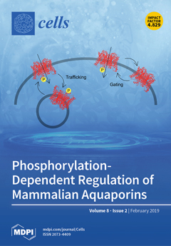
Membrane-bound water channels known as aquaporins facilitate water transport across cellular membranes along an osmotic gradient. In eukaryotes, aquaporins have evolved to be post-translationally regulated through gating or trafficking, allowing cells to adapt the membrane water flux in response to cellular or environmental triggers. A common way of controlling aquaporin regulation is through phosphorylation at specific sites. In mammals, this allows the 13 different aquaporin isoforms to be independently regulated in a tissue-dependent manner. This review provides a summary of what is currently known about phosphorylation-dependent regulation of mammalian aquaporins by both gating and trafficking, dissecting the roles of individual phosphorylation sites and of phosphorylation-dependent protein–protein interactions. View this paper.
  • Issues are regarded as officially published after their release is announced to the table of contents alert mailing list.
  • You may sign up for e-mail alerts to receive table of contents of newly released issues.
  • PDF is the official format for papers published in both, html and pdf forms. To view the papers in pdf format, click on the "PDF Full-text" link, and use the free Adobe Reader to open them.
Order results
Result details
Section
Select all
Export citation of selected articles as:
13 pages, 1041 KiB  
Review
Histone Modifications and the Maintenance of Telomere Integrity
by Meagan Jezek and Erin M. Green
Cells 2019, 8(2), 199; https://doi.org/10.3390/cells8020199 - 25 Feb 2019
Cited by 27 | Viewed by 10052
Abstract
Telomeres, the nucleoprotein structures at the ends of eukaryotic chromosomes, play an integral role in protecting linear DNA from degradation. Dysregulation of telomeres can result in genomic instability and has been implicated in increased rates of cellular senescence and many diseases, including cancer. [...] Read more.
Telomeres, the nucleoprotein structures at the ends of eukaryotic chromosomes, play an integral role in protecting linear DNA from degradation. Dysregulation of telomeres can result in genomic instability and has been implicated in increased rates of cellular senescence and many diseases, including cancer. The integrity of telomeres is maintained by a coordinated network of proteins and RNAs, such as the telomerase holoenzyme and protective proteins that prevent the recognition of the telomere ends as a DNA double-strand breaks. The structure of chromatin at telomeres and within adjacent subtelomeres has been implicated in telomere maintenance pathways in model systems and humans. Specific post-translational modifications of histones, including methylation, acetylation, and ubiquitination, have been shown to be necessary for maintaining a chromatin environment that promotes telomere integrity. Here we review the current knowledge regarding the role of histone modifications in maintaining telomeric and subtelomeric chromatin, discuss the implications of histone modification marks as they relate to human disease, and highlight key areas for future research. Full article
(This article belongs to the Special Issue The Role of Telomere Biology in Aging and Human Disease)
Show Figures

Figure 1

17 pages, 5381 KiB  
Article
Iron Exposure and the Cellular Mechanisms Linked to Neuron Degeneration in Adult Mice
by Lin-Bo Li, Rui Chai, Shuai Zhang, Shuang-Feng Xu, Yan-Hui Zhang, Hai-Long Li, Yong-Gang Fan and Chuang Guo
Cells 2019, 8(2), 198; https://doi.org/10.3390/cells8020198 - 24 Feb 2019
Cited by 55 | Viewed by 5650
Abstract
Although the causal relationship between Alzheimer’s disease (AD) and iron overload remains unclear, iron dyshomeostasis or improper transport mechanisms are speculated to lead to the accumulation of this neurotoxic metal in the hippocampal formation and other cerebral areas related to neurodegenerative diseases, resulting [...] Read more.
Although the causal relationship between Alzheimer’s disease (AD) and iron overload remains unclear, iron dyshomeostasis or improper transport mechanisms are speculated to lead to the accumulation of this neurotoxic metal in the hippocampal formation and other cerebral areas related to neurodegenerative diseases, resulting in the formation of reactive oxygen species (ROS) and, ultimately, cell death. In this study, exposure to high dietary iron (HDI) revealed no significant difference in the number of iron-positive cells and iron content in the cortex and hippocampal region between wild-type (WT) and APP/PS1 mice; however, compared with the control mice, the HDI-treated mice exhibited upregulated divalent metal transporter 1 (DMT1) and ferroportin (Fpn) expression, and downregulated transferrin receptor (TFR) expression. Importantly, we confirmed that there were significantly fewer NeuN-positive neurons in both APP/PS1 and WT mice given HDI, than in the respective controls. Moreover, this iron-induced neuron loss may involve increased ROS and oxidative mitochondria dysfunction, decreased DNA repair, and exacerbated apoptosis and autophagy. Although HDI administration might trigger protective antioxidant, anti-apoptosis, and autophagy signaling, especially in pathological conditions, these data clearly indicate that chronic iron exposure results in neuronal loss due to apoptosis, autophagy, and ferroptosis, hence increasing the risk for developing AD. Full article
Show Figures

Figure 1

12 pages, 5362 KiB  
Article
AQP1 and AQP4 Contribution to Cerebrospinal Fluid Homeostasis
by José Luis Trillo-Contreras, Juan José Toledo-Aral, Miriam Echevarría and Javier Villadiego
Cells 2019, 8(2), 197; https://doi.org/10.3390/cells8020197 - 24 Feb 2019
Cited by 47 | Viewed by 5334
Abstract
Aquaporin 1 (AQP1), expressed in epithelial cells of the choroid plexus, and aquaporin 4 (AQP4) present in ependymal cells and glia limitants have been proposed to play a significant role in cerebrospinal fluid (CSF) production and homeostasis. However, the specific contribution of each [...] Read more.
Aquaporin 1 (AQP1), expressed in epithelial cells of the choroid plexus, and aquaporin 4 (AQP4) present in ependymal cells and glia limitants have been proposed to play a significant role in cerebrospinal fluid (CSF) production and homeostasis. However, the specific contribution of each water channel to these functions remains unknown, being a subject of debate during the last years. Here, we analyzed in detail how AQP1 and AQP4 participate in different aspects of the CSF homeostasis such as the load and drainage of ventricles, and further explored if these proteins play a role in the ventricular compliance. To do that, we carried out records of intraventricular pressure and CSF outflow, and evaluated ventricular volume by magnetic resonance imaging in AQP1−/−, AQP4−/−, double AQP1−/−-AQP4−/− knock out and wild type mice controls. The analysis performed clearly showed that both AQPs have a significant participation in the CSF production, and additionally revealed that the double AQP1-AQP4 mutation alters the CSF drainage and the ventricular compliance. The data reported here indicate a significant extra-choroidal CSF formation mediated by AQP4, supporting the idea of an important and constant CSF production/absorption process, sustained by efflux/influx of water between brain capillaries and interstitial fluid. Moreover, our results suggest the participation of AQPs in structural functions also related with CSF homeostasis such as the distensibility capacity of the ventricular system. Full article
(This article belongs to the Special Issue Aquaporins)
Show Figures

Figure 1

12 pages, 1909 KiB  
Communication
VAS2870 Inhibits Histamine-Induced Calcium Signaling and vWF Secretion in Human Umbilical Vein Endothelial Cells
by Pavel V. Avdonin, Elena Yu. Rybakova, Piotr P. Avdonin, Sergei K. Trufanov, Galina Yu. Mironova, Alexandra A. Tsitrina and Nikolay V. Goncharov
Cells 2019, 8(2), 196; https://doi.org/10.3390/cells8020196 - 23 Feb 2019
Cited by 11 | Viewed by 4258
Abstract
In this study, we investigated the effects of NAD(P)H oxidase (NOX) inhibitor VAS2870 (3-benzyl-7-(2-benzoxazolyl)thio-1,2,3-triazolo[4,5-d]pyrimidine) on the histamine-induced elevation of free cytoplasmic calcium concentration ([Ca2+]i) and the secretion of von Willebrand factor (vWF) in human umbilical vein endothelial cells (HUVECs) [...] Read more.
In this study, we investigated the effects of NAD(P)H oxidase (NOX) inhibitor VAS2870 (3-benzyl-7-(2-benzoxazolyl)thio-1,2,3-triazolo[4,5-d]pyrimidine) on the histamine-induced elevation of free cytoplasmic calcium concentration ([Ca2+]i) and the secretion of von Willebrand factor (vWF) in human umbilical vein endothelial cells (HUVECs) and on relaxation of rat aorta in response to histamine. At 10 μM concentration, VAS2870 suppressed the [Ca2+]i rise induced by histamine. Inhibition was not competitive, with IC50 3.64 and 3.22 μM at 1 and 100 μM concentrations of histamine, respectively. There was no inhibition of [Ca2+]i elevation by VAS2870 in HUVECs in response to the agonist of type 1 protease-activated receptor SFLLRN. VAS2870 attenuated histamine-induced secretion of vWF and did not inhibit basal secretion. VAS2870 did not change the degree of histamine-induced relaxation of rat aortic rings constricted by norepinephrine. We suggest that NOX inhibitors might be used as a tool for preventing thrombosis induced by histamine release from mast cells without affecting vasorelaxation. Full article
Show Figures

Figure 1

14 pages, 1534 KiB  
Review
Tankyrase (PARP5) Inhibition Induces Bone Loss through Accumulation of Its Substrate SH3BP2
by Tomoyuki Mukai, Shunichi Fujita and Yoshitaka Morita
Cells 2019, 8(2), 195; https://doi.org/10.3390/cells8020195 - 22 Feb 2019
Cited by 19 | Viewed by 6022
Abstract
There is considerable interest in tankyrase because of its potential use in cancer therapy. Tankyrase catalyzes the ADP-ribosylation of a variety of target proteins and regulates various cellular processes. The anti-cancer effects of tankyrase inhibitors are mainly due to their suppression of Wnt [...] Read more.
There is considerable interest in tankyrase because of its potential use in cancer therapy. Tankyrase catalyzes the ADP-ribosylation of a variety of target proteins and regulates various cellular processes. The anti-cancer effects of tankyrase inhibitors are mainly due to their suppression of Wnt signaling and inhibition of telomerase activity, which are mediated by AXIN and TRF1 stabilization, respectively. In this review, we describe the underappreciated effects of another substrate, SH3 domain-binding protein 2 (SH3BP2). Specifically, SH3BP2 is an adaptor protein that regulates intracellular signaling pathways. Additionally, in the human genetic disorder cherubism, the gain-of-function mutations in SH3BP2 enhance osteoclastogenesis. The pharmacological inhibition of tankyrase in mice induces bone loss through the accumulation of SH3BP2 and the subsequent increase in osteoclast formation. These findings reveal the novel functions of tankyrase influencing bone homeostasis, and imply that tankyrase inhibitor treatments in a clinical setting may be associated with adverse effects on bone mass. Full article
(This article belongs to the Special Issue Molecular Role of PARP in Health and Disease)
Show Figures

Figure 1

17 pages, 6930 KiB  
Article
Anti-Inflammatory Effect of Sulforaphane on LPS-Activated Microglia Potentially through JNK/AP-1/NF-κB Inhibition and Nrf2/HO-1 Activation
by Lalita Subedi, Jae Hyuk Lee, Silvia Yumnam, Eunhee Ji and Sun Yeou Kim
Cells 2019, 8(2), 194; https://doi.org/10.3390/cells8020194 - 22 Feb 2019
Cited by 182 | Viewed by 11661
Abstract
Sulforaphane (SFN), a potent nuclear factor erythroid 2-related factor 2 (Nrf2) activator, is present in the species of the Brassicaceae, especially in broccoli sprouts. In this study, the effects of SFN against microglial activation and inflammation, and the potential mechanisms involved, were analyzed. [...] Read more.
Sulforaphane (SFN), a potent nuclear factor erythroid 2-related factor 2 (Nrf2) activator, is present in the species of the Brassicaceae, especially in broccoli sprouts. In this study, the effects of SFN against microglial activation and inflammation, and the potential mechanisms involved, were analyzed. As mitogen-activated protein kinase (MAPK) signaling plays a key role in microglial activation and inflammation, we focused on the role of SFN in regulating the MAPK signaling regulation of the inflammatory and anti-inflammatory cascades in lipopolysaccharide (LPS)-activated microglia. The anti-inflammatory and immunomodulatory effects of SFN were explored by evaluating the expression and secretion of inflammatory proteins, cytokines, nuclear factor kappa-light-chain-enhancer of activated B cells (NF-κB), and activator protein-1 (AP-1) under pre- and post-treatment conditions. Under the SFN pre- and post-treatment conditions, the MAPK phosphorylation levels were significantly reduced in both acutely and chronically activated microglial cells. SFN also reduced the c-Jun N-terminal kinase (JNK) phosphorylation levels, which subsequently reduced NF-κB and AP-1 signaling. As a result, the expression of the inflammatory mediators (iNOS, COX-2, NO, and PGE2) and proinflammatory cytokines (TNF-α, IL-6, and IL-1β) was decreased. At the same time, SFN increased the expression of Nrf2 and heme oxygenase-1 (HO-1) as well as the production of the anti-inflammatory cytokines IL-10 and IL-4. In conclusion, this study demonstrated that SFN exerts an anti-neuroinflammatory effect on microglia through JNK/AP-1/NF-κB pathway inhibition and Nrf2/HO-1 pathway activation. Full article
(This article belongs to the Section Cell Signaling)
Show Figures

Graphical abstract

24 pages, 2766 KiB  
Review
Cellular and Molecular Therapeutic Targets in Inflammatory Bowel Disease—Focusing on Intestinal Barrier Function
by Ida Schoultz and Åsa V. Keita
Cells 2019, 8(2), 193; https://doi.org/10.3390/cells8020193 - 22 Feb 2019
Cited by 127 | Viewed by 10647
Abstract
The human gut relies on several cellular and molecular mechanisms to allow for an intact and dynamical intestinal barrier. Normally, only small amounts of luminal content pass the mucosa, however, if the control is broken it can lead to enhanced passage, which might [...] Read more.
The human gut relies on several cellular and molecular mechanisms to allow for an intact and dynamical intestinal barrier. Normally, only small amounts of luminal content pass the mucosa, however, if the control is broken it can lead to enhanced passage, which might damage the mucosa, leading to pathological conditions, such as inflammatory bowel disease (IBD). It is well established that genetic, environmental, and immunological factors all contribute in the pathogenesis of IBD, and a disturbed intestinal barrier function has become a hallmark of the disease. Genetical studies support the involvement of intestinal barrier as several susceptibility genes for IBD encode proteins with key functions in gut barrier and homeostasis. IBD patients are associated with loss in bacterial diversity and shifts in the microbiota, with a possible link to local inflammation. Furthermore, alterations of immune cells and several neuro-immune signaling pathways in the lamina propria have been demonstrated. An inappropriate immune activation might lead to mucosal inflammation, with elevated secretion of pro-inflammatory cytokines that can affect the epithelium and promote a leakier barrier. This review will focus on the main cells and molecular mechanisms in IBD and how these can be targeted in order to improve intestinal barrier function and reduce inflammation. Full article
Show Figures

Graphical abstract

25 pages, 43893 KiB  
Article
In-Depth Proteome Analysis Highlights HepaRG Cells as a Versatile Cell System Surrogate for Primary Human Hepatocytes
by Georg Tascher, Audrey Burban, Sandrine Camus, Marine Plumel, Stéphanie Chanon, Remy Le Guevel, Valery Shevchenko, Alain Van Dorsselaer, Etienne Lefai, Christiane Guguen-Guillouzo and Fabrice Bertile
Cells 2019, 8(2), 192; https://doi.org/10.3390/cells8020192 - 21 Feb 2019
Cited by 45 | Viewed by 7422
Abstract
Of the hepatic cell lines developed for in vitro studies of hepatic functions as alternatives to primary human hepatocytes, many have lost major liver-like functions, but not HepaRG cells. The increasing use of the latter worldwide raises the need for establishing the reference [...] Read more.
Of the hepatic cell lines developed for in vitro studies of hepatic functions as alternatives to primary human hepatocytes, many have lost major liver-like functions, but not HepaRG cells. The increasing use of the latter worldwide raises the need for establishing the reference functional status of early biobanked HepaRG cells. Using deep proteome and secretome analyses, the levels of master regulators of the hepatic phenotype and of the structural elements ensuring biliary polarity were found to be close to those in primary hepatocytes. HepaRG cells proved to be highly differentiated, with functional mitochondria, hepatokine secretion abilities, and an adequate response to insulin. Among differences between primary human hepatocytes and HepaRG cells, the factors that possibly support HepaRG transdifferentiation properties are discussed. The HepaRG cell system thus appears as a robust surrogate for primary hepatocytes, which is versatile enough to study not only xenobiotic detoxification, but also the control of hepatic energy metabolism, secretory function and disease-related mechanisms. Full article
Show Figures

Figure 1

17 pages, 3344 KiB  
Article
The MEK-ERK-MST1 Axis Potentiates the Activation of the Extrinsic Apoptotic Pathway during GDC-0941 Treatment in Jurkat T Cells
by Jana Nováková, Pavel Talacko, Petr Novák and Karel Vališ
Cells 2019, 8(2), 191; https://doi.org/10.3390/cells8020191 - 21 Feb 2019
Cited by 8 | Viewed by 3898
Abstract
The discrete activation of individual caspases is essential during T-cell development, activation, and apoptosis. Humans carrying nonfunctional caspase-8 and caspase-8 conditional knockout mice exhibit several defects in the progression of naive CD4+ T cells to the effector stage. MST1, a key kinase [...] Read more.
The discrete activation of individual caspases is essential during T-cell development, activation, and apoptosis. Humans carrying nonfunctional caspase-8 and caspase-8 conditional knockout mice exhibit several defects in the progression of naive CD4+ T cells to the effector stage. MST1, a key kinase of the Hippo signaling pathway, is often presented as a substrate of caspases, and its cleavage by caspases potentiates its activity. Several studies have focused on the involvement of MST1 in caspase activation and also reported several defects in the immune system function caused by MST1 deficiency. Here, we show the rapid activation of the MEK-ERK-MST1 axis together with the cleavage and activation of caspase-3, -6, -7, -8, and -9 after PI3K signaling blockade by the selective inhibitor GDC-0941 in Jurkat T cells. We determined the phosphorylation pattern of MST1 using a phosphoproteomic approach and identified two amino acid residues phosphorylated in an ERK-dependent manner after GDC-0941 treatment together with a novel phosphorylation site at S21 residue, which was extensively phosphorylated in an ERK-independent manner during PI3K signaling blockade. Using caspase inhibitors and the inhibition of MST1 expression using siRNA, we identified an exclusive role of the MEK-ERK-MST1 axis in the activation of initiator caspase-8, which in turn activates executive caspase-3/-7 that finally potentiate MST1 proteolytic cleavage. This mechanism forms a positive feed-back loop that amplifies the activation of MST1 together with apoptotic response in Jurkat T cells during PI3K inhibition. Altogether, we propose a novel MEK-ERK-MST1-CASP8-CASP3/7 apoptotic pathway in Jurkat T cells and believe that the regulation of this pathway can open novel possibilities in systemic and cancer therapies. Full article
Show Figures

Graphical abstract

26 pages, 6435 KiB  
Review
Targeting mTOR in Acute Lymphoblastic Leukemia
by Carolina Simioni, Alberto M. Martelli, Giorgio Zauli, Elisabetta Melloni and Luca M. Neri
Cells 2019, 8(2), 190; https://doi.org/10.3390/cells8020190 - 21 Feb 2019
Cited by 43 | Viewed by 7107
Abstract
Acute Lymphoblastic Leukemia (ALL) is an aggressive hematologic disorder and constitutes approximately 25% of cancer diagnoses among children and teenagers. Pediatric patients have a favourable prognosis, with 5-years overall survival rates near 90%, while adult ALL still correlates with poorer survival. However, during [...] Read more.
Acute Lymphoblastic Leukemia (ALL) is an aggressive hematologic disorder and constitutes approximately 25% of cancer diagnoses among children and teenagers. Pediatric patients have a favourable prognosis, with 5-years overall survival rates near 90%, while adult ALL still correlates with poorer survival. However, during the past few decades, the therapeutic outcome of adult ALL was significantly ameliorated, mainly due to intensive pediatric-based protocols of chemotherapy. Mammalian (or mechanistic) target of rapamycin (mTOR) is a conserved serine/threonine kinase belonging to the phosphatidylinositol 3-kinase (PI3K)-related kinase family (PIKK) and resides in two distinct signalling complexes named mTORC1, involved in mRNA translation and protein synthesis and mTORC2 that controls cell survival and migration. Moreover, both complexes are remarkably involved in metabolism regulation. Growing evidence reports that mTOR dysregulation is related to metastatic potential, cell proliferation and angiogenesis and given that PI3K/Akt/mTOR network activation is often associated with poor prognosis and chemoresistance in ALL, there is a constant need to discover novel inhibitors for ALL treatment. Here, the current knowledge of mTOR signalling and the development of anti-mTOR compounds are documented, reporting the most relevant results from both preclinical and clinical studies in ALL that have contributed significantly into their efficacy or failure. Full article
(This article belongs to the Special Issue mTOR Signaling in Metabolism and Cancer)
Show Figures

Figure 1

12 pages, 5019 KiB  
Article
Preclinical Evaluation of the Pan-FGFR Inhibitor LY2874455 in FRS2-Amplified Liposarcoma
by Robert Hanes, Else Munthe, Iwona Grad, Jianhua Han, Ida Karlsen, Emmet McCormack, Leonardo A. Meza-Zepeda, Eva Wessel Stratford and Ola Myklebost
Cells 2019, 8(2), 189; https://doi.org/10.3390/cells8020189 - 21 Feb 2019
Cited by 16 | Viewed by 4955
Abstract
Background: FGFR inhibition has been proposed as treatment for dedifferentiated liposarcoma (DDLPS) with amplified FRS2, but we previously only demonstrated transient cytostatic effects when treating FRS2-amplified DDLPS cells with NVP-BGJ398. Methods: Effects of the more potent FGFR inhibitor LY2874455 were investigated [...] Read more.
Background: FGFR inhibition has been proposed as treatment for dedifferentiated liposarcoma (DDLPS) with amplified FRS2, but we previously only demonstrated transient cytostatic effects when treating FRS2-amplified DDLPS cells with NVP-BGJ398. Methods: Effects of the more potent FGFR inhibitor LY2874455 were investigated in three DDLPS cell lines by measuring effects on cell growth and apoptosis in vitro and also testing efficacy in vivo. Genome, transcriptome and protein analyses were performed to characterize the signaling components in the FGFR pathway. Results: LY2874455 induced a stronger, longer-lasting growth inhibitory effect and moderate level of apoptosis for two cell lines. The third cell line, did not respond to FGFR inhibition, suggesting that FRS2 amplification alone is not sufficient to predict response. Importantly, efficacy of LY2874455 was confirmed in vivo, using an independent FRS2-amplified DDLPS xenograft model. Expression of FRS2 was similar in the responding and non-responding cell lines and we could not find any major difference in downstream FGFR signaling. The only FGF expressed by unstimulated non-responding cells was the intracellular ligand FGF11, whereas the responding cell lines expressed extracellular ligand FGF2. Conclusion: Our study supports LY2874455 as a better therapy than NVP-BGJ398 for FRS2-amplified liposarcoma, and a clinical trial is warranted. Full article
(This article belongs to the Special Issue Fibroblast Growth Factor Receptor (FGFR) Signaling Pathway in Tumor)
Show Figures

Graphical abstract

12 pages, 1245 KiB  
Review
Cellular Metabolic Regulation in the Differentiation and Function of Regulatory T Cells
by Ye Chen, Jacob Colello, Wael Jarjour and Song Guo Zheng
Cells 2019, 8(2), 188; https://doi.org/10.3390/cells8020188 - 21 Feb 2019
Cited by 28 | Viewed by 5066
Abstract
Regulatory T cells (Tregs) are essential for maintaining immune tolerance and preventing autoimmune and inflammatory diseases. The activity and function of Tregs are in large part determined by various intracellular metabolic processes. Recent findings have focused on how intracellular metabolism can shape the [...] Read more.
Regulatory T cells (Tregs) are essential for maintaining immune tolerance and preventing autoimmune and inflammatory diseases. The activity and function of Tregs are in large part determined by various intracellular metabolic processes. Recent findings have focused on how intracellular metabolism can shape the development, trafficking, and function of Tregs. In this review, we summarize and discuss current research that reveals how distinct metabolic pathways modulate Tregs differentiation, phenotype stabilization, and function. These advances highlight numerous opportunities to alter Tregs frequency and function in physiopathologic conditions via metabolic manipulation and have important translational implications. Full article
(This article belongs to the Special Issue mTOR Signaling in Metabolism and Cancer)
Show Figures

Figure 1

23 pages, 7597 KiB  
Review
Neutral Lipid Storage Diseases as Cellular Model to Study Lipid Droplet Function
by Sara Missaglia, Rosalind A. Coleman, Alvaro Mordente and Daniela Tavian
Cells 2019, 8(2), 187; https://doi.org/10.3390/cells8020187 - 21 Feb 2019
Cited by 60 | Viewed by 7522
Abstract
Neutral lipid storage disease with myopathy (NLSDM) and with ichthyosis (NLSDI) are rare autosomal recessive disorders caused by mutations in the PNPLA2 and in the ABHD5/CGI58 genes, respectively. These genes encode the adipose triglyceride lipase (ATGL) and α-β hydrolase domain 5 (ABHD5) proteins, [...] Read more.
Neutral lipid storage disease with myopathy (NLSDM) and with ichthyosis (NLSDI) are rare autosomal recessive disorders caused by mutations in the PNPLA2 and in the ABHD5/CGI58 genes, respectively. These genes encode the adipose triglyceride lipase (ATGL) and α-β hydrolase domain 5 (ABHD5) proteins, which play key roles in the function of lipid droplets (LDs). LDs, the main cellular storage sites of triacylglycerols and sterol esters, are highly dynamic organelles. Indeed, LDs are critical for both lipid metabolism and energy homeostasis. Partial or total PNPLA2 or ABHD5/CGI58 knockdown is characteristic of the cells of NLSD patients; thus, these cells are natural models with which one can unravel LD function. In this review we firstly summarize genetic and clinical data collected from NLSD patients, focusing particularly on muscle, skin, heart, and liver damage due to impaired LD function. Then, we discuss how NLSD cells were used to investigate and expand the current structural and functional knowledge of LDs. Full article
(This article belongs to the Section Intracellular and Plasma Membranes)
Show Figures

Figure 1

13 pages, 1328 KiB  
Review
Targeting Telomeres and Telomerase: Studies in Aging and Disease Utilizing CRISPR/Cas9 Technology
by Andrew C. Brane and Trygve O. Tollefsbol
Cells 2019, 8(2), 186; https://doi.org/10.3390/cells8020186 - 21 Feb 2019
Cited by 18 | Viewed by 8527
Abstract
Telomeres and telomerase provide a unique and important avenue of study in improving both life expectancy and quality of life due to their close association with aging and disease. While major advances in our understanding of these two biological mediators have characterized the [...] Read more.
Telomeres and telomerase provide a unique and important avenue of study in improving both life expectancy and quality of life due to their close association with aging and disease. While major advances in our understanding of these two biological mediators have characterized the last two decades, previous studies have been limited by the inability to affect change in real time within living cells. The last three years, however, have witnessed a huge step forward to overcome this limitation. The advent of the clustered regularly interspaced short palindromic repeats/CRISPR-associated (CRISPR/Cas) system has led to a wide array of targeted genetic studies that are already being employed to modify telomeres and telomerase, as well as the genes that affect them. In this review, we analyze studies utilizing the technology to target and modify telomeres, telomerase, and their closely associated genes. We also discuss how these studies can provide insight into the biology and mechanisms that underlie aging, cancer, and other diseases. Full article
(This article belongs to the Special Issue The Role of Telomere Biology in Aging and Human Disease)
Show Figures

Figure 1

14 pages, 828 KiB  
Review
Involvement of CXCR4 in Normal and Abnormal Development
by Nanako Kawaguchi, Ting-Ting Zhang and Toshio Nakanishi
Cells 2019, 8(2), 185; https://doi.org/10.3390/cells8020185 - 20 Feb 2019
Cited by 101 | Viewed by 12916
Abstract
CXC motif chemokine receptor type 4 (CXCR4) is associated with normal and abnormal development, including oncogenesis. The ligand of CXCR4 is stromal cell-derived factor (SDF), also known as CXC motif ligand (CXCL) 12. Through the SDF-1/CXCR4 axis, both homing and migration of hematopoietic [...] Read more.
CXC motif chemokine receptor type 4 (CXCR4) is associated with normal and abnormal development, including oncogenesis. The ligand of CXCR4 is stromal cell-derived factor (SDF), also known as CXC motif ligand (CXCL) 12. Through the SDF-1/CXCR4 axis, both homing and migration of hematopoietic (stem) cells are regulated through niches in the bone marrow. Outside of the bone marrow, however, SDF-1 can recruit CXCR4-positive cells from the bone marrow. SDF/CXCR4 has been implicated in the maintenance and/or differentiation of stemness, and tissue-derived stem cells can be associated with SDF-1 and CXCR4 activity. CXCR4 plays a role in multiple pathways involved in carcinogenesis and other pathologies. Here, we summarize reports detailing the functions of CXCR4. We address the molecular signature of CXCR4 and how this molecule and cells expressing it are involved in either normal (maintaining stemness or inducing differentiation) or abnormal (developing cancer and other pathologies) events. As a constituent of stem cells, the SDF-1/CXCR4 axis influences downstream signal transduction and the cell microenvironment. Full article
(This article belongs to the Section Stem Cells)
Show Figures

Figure 1

27 pages, 1014 KiB  
Review
Astrocytes Maintain Glutamate Homeostasis in the CNS by Controlling the Balance between Glutamate Uptake and Release
by Shaimaa Mahmoud, Marjan Gharagozloo, Camille Simard and Denis Gris
Cells 2019, 8(2), 184; https://doi.org/10.3390/cells8020184 - 20 Feb 2019
Cited by 351 | Viewed by 22507
Abstract
Glutamate is one of the most prevalent neurotransmitters released by excitatory neurons in the central nervous system (CNS); however, residual glutamate in the extracellular space is, potentially, neurotoxic. It is now well-established that one of the fundamental functions of astrocytes is to uptake [...] Read more.
Glutamate is one of the most prevalent neurotransmitters released by excitatory neurons in the central nervous system (CNS); however, residual glutamate in the extracellular space is, potentially, neurotoxic. It is now well-established that one of the fundamental functions of astrocytes is to uptake most of the synaptically-released glutamate, which optimizes neuronal functions and prevents glutamate excitotoxicity. In the CNS, glutamate clearance is mediated by glutamate uptake transporters expressed, principally, by astrocytes. Interestingly, recent studies demonstrate that extracellular glutamate stimulates Ca2+ release from the astrocytes’ intracellular stores, which triggers glutamate release from astrocytes to the adjacent neurons, mostly by an exocytotic mechanism. This released glutamate is believed to coordinate neuronal firing and mediate their excitatory or inhibitory activity. Therefore, astrocytes contribute to glutamate homeostasis in the CNS, by maintaining the balance between their opposing functions of glutamate uptake and release. This dual function of astrocytes represents a potential therapeutic target for CNS diseases associated with glutamate excitotoxicity. In this regard, we summarize the molecular mechanisms of glutamate uptake and release, their regulation, and the significance of both processes in the CNS. Also, we review the main features of glutamate metabolism and glutamate excitotoxicity and its implication in CNS diseases. Full article
(This article belongs to the Special Issue Astrocytes in Space and Time)
Show Figures

Graphical abstract

21 pages, 2033 KiB  
Review
Autophagy as a Therapeutic Target to Enhance Aged Muscle Regeneration
by David E. Lee, Akshay Bareja, David B. Bartlett and James P. White
Cells 2019, 8(2), 183; https://doi.org/10.3390/cells8020183 - 20 Feb 2019
Cited by 43 | Viewed by 9240
Abstract
Skeletal muscle has remarkable regenerative capacity, relying on precise coordination between resident muscle stem cells (satellite cells) and the immune system. The age-related decline in skeletal muscle regenerative capacity contributes to the onset of sarcopenia, prolonged hospitalization, and loss of autonomy. Although several [...] Read more.
Skeletal muscle has remarkable regenerative capacity, relying on precise coordination between resident muscle stem cells (satellite cells) and the immune system. The age-related decline in skeletal muscle regenerative capacity contributes to the onset of sarcopenia, prolonged hospitalization, and loss of autonomy. Although several age-sensitive pathways have been identified, further investigation is needed to define targets of cellular dysfunction. Autophagy, a process of cellular catabolism, is emerging as a key regulator of muscle regeneration affecting stem cell, immune cell, and myofiber function. Muscle stem cell senescence is associated with a suppression of autophagy during key phases of the regenerative program. Macrophages, a key immune cell involved in muscle repair, also rely on autophagy to aid in tissue repair. This review will focus on the role of autophagy in various aspects of the regenerative program, including adult skeletal muscle stem cells, monocytes/macrophages, and corresponding age-associated dysfunction. Furthermore, we will highlight rejuvenation strategies that alter autophagy to improve muscle regenerative function. Full article
(This article belongs to the Special Issue Autophagy in Tissue Injury and Homeostasis)
Show Figures

Figure 1

16 pages, 2520 KiB  
Article
Apolipoprotein E4 Alters Astrocyte Fatty Acid Metabolism and Lipid Droplet Formation
by Brandon C. Farmer, Jude Kluemper and Lance A. Johnson
Cells 2019, 8(2), 182; https://doi.org/10.3390/cells8020182 - 20 Feb 2019
Cited by 95 | Viewed by 12857
Abstract
Lipid droplets (LDs) serve as energy rich reservoirs and have been associated with apolipoprotein E (APOE) and neurodegeneration. The E4 allele of APOE (E4) is the strongest genetic risk factor for the development of late onset Alzheimer’s disease (AD). Since both [...] Read more.
Lipid droplets (LDs) serve as energy rich reservoirs and have been associated with apolipoprotein E (APOE) and neurodegeneration. The E4 allele of APOE (E4) is the strongest genetic risk factor for the development of late onset Alzheimer’s disease (AD). Since both E4 carriers and individuals with AD exhibit a state of cerebral lipid dyshomeostasis, we hypothesized that APOE may play a role in regulating LD metabolism. We found that astrocytes expressing E4 accumulate significantly more and smaller LDs compared to E3 astrocytes. Accordingly, expression of perilipin-2, an essential LD protein component, was higher in E4 astrocytes. We then probed fatty acid (FA) metabolism and found E4 astrocytes to exhibit decreased uptake of palmitate, and decreased oxidation of exogenously supplied oleate and palmitate. We then measured oxygen consumption rate, and found E4 astrocytes to consume more oxygen for endogenous FA oxidation and accumulate more LD-derived metabolites due to incomplete oxidation. Lastly, we found that E4 astrocytes are more sensitive to carnitine palmitoyltransferase-1 inhibition than E3 astrocytes. These findings offer the potential for further studies investigating the link between astrocyte lipid storage, utilization, and neurodegenerative disease as a function of APOE genotype. Full article
Show Figures

Graphical abstract

13 pages, 1220 KiB  
Article
Wound Healing Fluid Reflects the Inflammatory Nature and Aggressiveness of Breast Tumors
by Roberto Agresti, Tiziana Triulzi, Marianna Sasso, Cristina Ghirelli, Piera Aiello, Ilona Rybinska, Manuela Campiglio, Lucia Sfondrini, Elda Tagliabue and Francesca Bianchi
Cells 2019, 8(2), 181; https://doi.org/10.3390/cells8020181 - 19 Feb 2019
Cited by 18 | Viewed by 4473
Abstract
Wound healing fluid that originates from breast surgery increases the aggressiveness of cancer cells that remain after the surgery. We determined the effects of the extent of surgery and tumor-driven remodeling of the surrounding microenvironment on the ability of wound-healing to promote breast [...] Read more.
Wound healing fluid that originates from breast surgery increases the aggressiveness of cancer cells that remain after the surgery. We determined the effects of the extent of surgery and tumor-driven remodeling of the surrounding microenvironment on the ability of wound-healing to promote breast cancer progression. In our analysis of a panel of 34 cytokines, chemokines, and growth factors in wound healing fluid, obtained from 27 breast carcinoma patients after surgery, the levels of several small molecules were associated with the extent of cellular damage that was induced by surgery. In addition, the composition of the resulting wound healing fluid was associated with molecular features of the removed tumor. Specifically, IP-10, IL-6, G-CSF, osteopontin, MIP-1a, MIP-1b, and MCP1-MCAF were higher in more aggressive tumors. Altogether, our findings indicate that the release of factors that are induced by removal of the primary tumor and subsequent wound healing is influenced by the extent of damage due to surgery and the reactive stroma that is derived from the continuously evolving network of interactions between neoplastic cells and the microenvironment, based on the molecular characteristics of breast carcinoma cells. Full article
(This article belongs to the Special Issue Extracellular Matrix Remodeling)
Show Figures

Graphical abstract

11 pages, 6117 KiB  
Article
The Centriolar Adjunct–Appearance and Disassembly in Spermiogenesis and the Potential Impact on Fertility
by Anastasiia S. Garanina, Irina B. Alieva, Elizaveta E. Bragina, Emmanuelle Blanchard, Brigitte Arbeille, Fabrice Guerif, Svetlana Uzbekova and Rustem E. Uzbekov
Cells 2019, 8(2), 180; https://doi.org/10.3390/cells8020180 - 19 Feb 2019
Cited by 25 | Viewed by 3960
Abstract
During spermiogenesis, the proximal centriole forms a special microtubular structure: the centriolar adjunct. This structure appears at the spermatid stage, which is characterized by a condensed chromatin nucleus. We showed that the centriolar adjunct disappears completely in mature porcine spermatozoa. In humans, the [...] Read more.
During spermiogenesis, the proximal centriole forms a special microtubular structure: the centriolar adjunct. This structure appears at the spermatid stage, which is characterized by a condensed chromatin nucleus. We showed that the centriolar adjunct disappears completely in mature porcine spermatozoa. In humans, the centriolar adjunct remnants are present in a fraction of mature spermatids. For the first time, the structure of the centriolar adjunct in the cell, and its consequent impact on fertility, were examined. Ultrastructural analysis using transmission electron microscopy was performed on near 2000 spermatozoa per person, in two patients with idiopathic male sterility (IMS) and five healthy fertile donors. We measured the average length of the “proximal centriole + centriolar adjunct” complex in sections, where it had parallel orientation in the section plane, and found that it was significantly longer in the spermatozoa of IMS patients than in the spermatozoa of healthy donors. This difference was independent of chromatin condensation deficiency, which was also observed in the spermatozoa of IMS patients. We suggest that zygote arrest may be related to an incompletely disassembled centriolar adjunct in a mature spermatozoon. Therefore, centriolar adjunct length can be potentially used as a complementary criterion for the immaturity of spermatozoa in the diagnostics of IMS patients. Full article
(This article belongs to the Collection Cilia and Flagella: Structure, Function and Beyond)
Show Figures

Figure 1

14 pages, 960 KiB  
Review
Switching on Endogenous Metal Binding Proteins in Parkinson’s Disease
by Fleur A. McLeary, Alexandre N. Rcom-H’cheo-Gauthier, Michael Goulding, Rowan A. W. Radford, Yuho Okita, Peter Faller, Roger S. Chung and Dean L. Pountney
Cells 2019, 8(2), 179; https://doi.org/10.3390/cells8020179 - 19 Feb 2019
Cited by 22 | Viewed by 5197
Abstract
The formation of cytotoxic intracellular protein aggregates is a pathological signature of multiple neurodegenerative diseases. The principle aggregating protein in Parkinson’s disease (PD) and atypical Parkinson’s diseases is α-synuclein (α-syn), which occurs in neural cytoplasmic inclusions. Several factors have been found to trigger [...] Read more.
The formation of cytotoxic intracellular protein aggregates is a pathological signature of multiple neurodegenerative diseases. The principle aggregating protein in Parkinson’s disease (PD) and atypical Parkinson’s diseases is α-synuclein (α-syn), which occurs in neural cytoplasmic inclusions. Several factors have been found to trigger α-syn aggregation, including raised calcium, iron, and copper. Transcriptional inducers have been explored to upregulate expression of endogenous metal-binding proteins as a potential neuroprotective strategy. The vitamin-D analogue, calcipotriol, induced increased expression of the neuronal vitamin D-dependent calcium-binding protein, calbindin-D28k, and this significantly decreased the occurrence of α-syn aggregates in cells with transiently raised intracellular free Ca, thereby increasing viability. More recently, the induction of endogenous expression of the Zn and Cu binding protein, metallothionein, by the glucocorticoid analogue, dexamethasone, gave a specific reduction in Cu-dependent α-syn aggregates. Fe accumulation has long been associated with PD. Intracellularly, Fe is regulated by interactions between the Fe storage protein ferritin and Fe transporters, such as poly(C)-binding protein 1. Analysis of the transcriptional regulation of Fe binding proteins may reveal potential inducers that could modulate Fe homoeostasis in disease. The current review highlights recent studies that suggest that transcriptional inducers may have potential as novel mechanism-based drugs against metal overload in PD. Full article
(This article belongs to the Special Issue Emerging Trends in Metal Biochemistry)
Show Figures

Figure 1

20 pages, 1927 KiB  
Review
Essential Roles for the Non-Canonical IκB Kinases in Linking Inflammation to Cancer, Obesity, and Diabetes
by Chong Hyun Shin and Doo-Sup Choi
Cells 2019, 8(2), 178; https://doi.org/10.3390/cells8020178 - 19 Feb 2019
Cited by 37 | Viewed by 8179
Abstract
Non-canonical IκB kinases (IKKs) TBK1 and IKKε have essential roles as regulators of innate immunity and cancer. Recent work has also implicated these kinases in distinctively controlling glucose homeostasis and repressing adaptive thermogenic and mitochondrial biogenic response upon obesity-induced inflammation. Additionally, TBK1 and [...] Read more.
Non-canonical IκB kinases (IKKs) TBK1 and IKKε have essential roles as regulators of innate immunity and cancer. Recent work has also implicated these kinases in distinctively controlling glucose homeostasis and repressing adaptive thermogenic and mitochondrial biogenic response upon obesity-induced inflammation. Additionally, TBK1 and IKKε regulate pancreatic β-cell regeneration. In this review, we summarize current data on the functions and molecular mechanisms of TBK1 and IKKε in orchestrating inflammation to cancer, obesity, and diabetes. Full article
Show Figures

Figure 1

19 pages, 5112 KiB  
Article
Circular RNA circHIPK3 Promotes the Proliferation and Differentiation of Chicken Myoblast Cells by Sponging miR-30a-3p
by Biao Chen, Jiao Yu, Lijin Guo, Mary Shannon Byers, Zhijun Wang, Xiaolan Chen, Haiping Xu and Qinghua Nie
Cells 2019, 8(2), 177; https://doi.org/10.3390/cells8020177 - 19 Feb 2019
Cited by 82 | Viewed by 4999
Abstract
Circular RNAs and microRNAs widely exist in various species and play crucial roles in multiple biological processes. It is essential to study their roles in myogenesis. In our previous sequencing data, both miR-30a-3p and circular HIPK3 (circHIPK3) RNA, which are produced by the [...] Read more.
Circular RNAs and microRNAs widely exist in various species and play crucial roles in multiple biological processes. It is essential to study their roles in myogenesis. In our previous sequencing data, both miR-30a-3p and circular HIPK3 (circHIPK3) RNA, which are produced by the third exon of the HIPK3 gene, were differentially expressed among chicken skeletal muscles at 11 embryo age (E11), 16 embryo age (E16), and 1-day post-hatch (P1). Here, we investigated their potential roles in myogenesis. Proliferation experiment showed that miR-30a-3p could inhibit the proliferation of myoblast. Through dual-luciferase assay and Myosin heavy chain (MYHC) immunofluorescence, we found that miR-30a-3p could inhibit the differentiation of myoblast by binding to Myocyte Enhancer Factor 2 C (MEF2C), which could promote the differentiation of myoblast. Then, we found that circHIPK3 could act as a sponge of miR-30a-3p and exerted a counteractive effect of miR-30a-3p by promoting the proliferation and differentiation of myoblasts. Taking together, our data suggested that circHIPK3 could promote the chicken embryonic skeletal muscle development by sponging miR-30a-3p. Full article
(This article belongs to the Special Issue Regulatory microRNA)
Show Figures

Figure 1

19 pages, 658 KiB  
Review
The Role of Androgen Receptor Signaling in Ovarian Cancer
by Taichi Mizushima and Hiroshi Miyamoto
Cells 2019, 8(2), 176; https://doi.org/10.3390/cells8020176 - 19 Feb 2019
Cited by 46 | Viewed by 7252
Abstract
Emerging evidence has suggested that androgen receptor signaling plays an important role in ovarian cancer outgrowth. Specifically, androgen receptor activation appears to be associated with increased risks of developing ovarian cancer and inducing tumor progression. However, conflicting findings have also been reported. This [...] Read more.
Emerging evidence has suggested that androgen receptor signaling plays an important role in ovarian cancer outgrowth. Specifically, androgen receptor activation appears to be associated with increased risks of developing ovarian cancer and inducing tumor progression. However, conflicting findings have also been reported. This review summarizes and discusses the available data indicating the involvement of androgens as well as androgen receptor and related signals in ovarian carcinogenesis and cancer growth. Although the underlying molecular mechanisms for androgen receptor functions in ovarian cancer remain far from being fully understood, current observations may offer effective chemopreventive and therapeutic approaches, via modulation of androgen receptor activity, against ovarian cancer. Indeed, several clinical trials have been conducted to determine the efficacy of androgen deprivation therapy in patients with ovarian cancer. Full article
(This article belongs to the Collection Functions of Nuclear Receptors)
Show Figures

Figure 1

17 pages, 6212 KiB  
Review
Lessons from the Discovery of Mitochondrial Fragmentation (Fission): A Review and Update
by Dmitry B. Zorov, Ivan A. Vorobjev, Vasily A. Popkov, Valentina A. Babenko, Ljubava D. Zorova, Irina B. Pevzner, Denis N. Silachev, Savva D. Zorov, Nadezda V. Andrianova and Egor Y. Plotnikov
Cells 2019, 8(2), 175; https://doi.org/10.3390/cells8020175 - 19 Feb 2019
Cited by 63 | Viewed by 9649
Abstract
Thirty-five years ago, we described fragmentation of the mitochondrial population in a living cell into small vesicles (mitochondrial fission). Subsequently, this phenomenon has become an object of general interest due to its involvement in the process of oxidative stress-related cell death and having [...] Read more.
Thirty-five years ago, we described fragmentation of the mitochondrial population in a living cell into small vesicles (mitochondrial fission). Subsequently, this phenomenon has become an object of general interest due to its involvement in the process of oxidative stress-related cell death and having high relevance to the incidence of a pathological phenotype. Tentatively, the key component of mitochondrial fission process is segregation and further asymmetric separation of a mitochondrial body yielding healthy (normally functioning) and impaired (incapable to function in a normal way) organelles with subsequent decomposition and removal of impaired elements through autophagy (mitophagy). We speculate that mitochondria contain cytoskeletal elements, which maintain the mitochondrial shape, and also are involved in the process of intramitochondrial segregation of waste products. We suggest that perturbation of the mitochondrial fission/fusion machinery and slowdown of the removal process of nonfunctional mitochondrial structures led to the increase of the proportion of impaired mitochondrial elements. When the concentration of malfunctioning mitochondria reaches a certain threshold, this can lead to various pathologies, including aging. Overall, we suggest a process of mitochondrial fission to be an essential component of a complex system controlling a healthy cell phenotype. The role of reactive oxygen species in mitochondrial fission is discussed. Full article
(This article belongs to the Special Issue Mitochondrial Biology in Health and Disease)
Show Figures

Figure 1

12 pages, 1282 KiB  
Article
The Role of Osteopontin as a Diagnostic and Prognostic Biomarker in Sepsis and Septic Shock
by Luigi Mario Castello, Marco Baldrighi, Luca Molinari, Livia Salmi, Vincenzo Cantaluppi, Rosanna Vaschetto, Greta Zunino, Marco Quaglia, Mattia Bellan, Francesco Gavelli, Paolo Navalesi, Gian Carlo Avanzi and Annalisa Chiocchetti
Cells 2019, 8(2), 174; https://doi.org/10.3390/cells8020174 - 18 Feb 2019
Cited by 33 | Viewed by 4591
Abstract
Sepsis is a life-threatening organ dysfunction caused by a dysregulated host-response to infections. Osteopontin (OPN) is an extracellular matrix protein involved in the inflammatory response. Our aim was to evaluate the diagnostic and prognostic performance in sepsis of a single OPN determination in [...] Read more.
Sepsis is a life-threatening organ dysfunction caused by a dysregulated host-response to infections. Osteopontin (OPN) is an extracellular matrix protein involved in the inflammatory response. Our aim was to evaluate the diagnostic and prognostic performance in sepsis of a single OPN determination in the Emergency Department (ED). We conducted a single-centre prospective observational study in an Italian ED where we enrolled 102 consecutive patients presenting with suspected infection and qSOFA ≥ 2. OPN plasma concentration was found to be an independent predictor of sepsis (OR = 1.020, 95% CI 1.002–1.039, p = 0.031) and the diagnostic receiver operating characteristic (ROC) curve resulted in an area under the curve (AUC) of 0.878. OPN levels were positively correlated to plasma creatinine (r = 0.401 with p = 0.0001), but this relation was not explained by the development of acute kidney injury (AKI), since no difference was found in OPN concentration between AKI and non-AKI patients. The analysis of 30-days mortality showed no significant difference in OPN levels between alive and dead patients (p = 0.482). In conclusion, a single determination of OPN concentration helped to identify patients with sepsis in the ED, but it was not able to predict poor prognosis in our cohort of patients. Full article
(This article belongs to the Special Issue Adding New Pieces in the Osteopontin Puzzle)
Show Figures

Figure 1

21 pages, 6712 KiB  
Article
Decidua Basalis Mesenchymal Stem Cells Favor Inflammatory M1 Macrophage Differentiation In Vitro
by Mohamed H. Abumaree, Seham Al Harthy, Abdullah M. Al Subayyil, Manal A. Alshabibi, Fawaz M. Abomaray, Tanvier Khatlani, Bill Kalionis, Mohammed F. El- Muzaini, Mohammed A. Al Jumah, Dunia Jawdat, Abdullah O. Alawad and Ahmed S. AlAskar
Cells 2019, 8(2), 173; https://doi.org/10.3390/cells8020173 - 18 Feb 2019
Cited by 16 | Viewed by 3832
Abstract
Placental mesenchymal stem cells from maternal decidua basalis tissue (DBMSCs) are promising cells for tissue repair because of their multilineage differentiation and ability to protect endothelial cells from injury. Here, we examined DBMSC interaction with macrophages and whether this interaction could modulate the [...] Read more.
Placental mesenchymal stem cells from maternal decidua basalis tissue (DBMSCs) are promising cells for tissue repair because of their multilineage differentiation and ability to protect endothelial cells from injury. Here, we examined DBMSC interaction with macrophages and whether this interaction could modulate the characteristics and functions of these macrophages. We induced monocytes to differentiate into M1-like macrophages in the presence of DBMSCs. DBMSC effects on differentiation were evaluated using microscopy, flow cytometry, and ELISA. DBMSC effects on M1-like macrophage induction of T cell function were also examined. The culture of DBMSCs with monocytes did not inhibit monocyte differentiation into M1-like inflammatory macrophages. This was confirmed by the morphological appearance of M1-like macrophages, increased expression of inflammatory molecules, and reduced expression of anti-inflammatory molecules. In addition, DBMSCs did not interfere with M1-like macrophage phagocytic activity; rather, they induced stimulatory effects of M1-like macrophages on CD4+ T cell proliferation and subsequent secretion of inflammatory molecules by T cells. We showed that DBMSCs enhanced the differentiation of M1-like inflammatory macrophages, which function as antitumor cells. Therefore, our findings suggest that DBMSCs are inflammatory cells that could be useful in cancer treatment via the enhancement of M1- like macrophages. Full article
(This article belongs to the Special Issue Immunomodulation by Mesenchymal Stem Cells)
Show Figures

Figure 1

18 pages, 3138 KiB  
Article
Chronic Hypoxia Enhances β-Oxidation-Dependent Electron Transport via Electron Transferring Flavoproteins
by Dominik C. Fuhrmann, Catherine Olesch, Nina Kurrle, Frank Schnütgen, Sven Zukunft, Ingrid Fleming and Bernhard Brüne
Cells 2019, 8(2), 172; https://doi.org/10.3390/cells8020172 - 18 Feb 2019
Cited by 15 | Viewed by 5252
Abstract
Hypoxia poses a stress to cells and decreases mitochondrial respiration, in part by electron transport chain (ETC) complex reorganization. While metabolism under acute hypoxia is well characterized, alterations under chronic hypoxia largely remain unexplored. We followed oxygen consumption rates in THP-1 monocytes during [...] Read more.
Hypoxia poses a stress to cells and decreases mitochondrial respiration, in part by electron transport chain (ETC) complex reorganization. While metabolism under acute hypoxia is well characterized, alterations under chronic hypoxia largely remain unexplored. We followed oxygen consumption rates in THP-1 monocytes during acute (16 h) and chronic (72 h) hypoxia, compared to normoxia, to analyze the electron flows associated with glycolysis, glutamine, and fatty acid oxidation. Oxygen consumption under acute hypoxia predominantly demanded pyruvate, while under chronic hypoxia, fatty acid- and glutamine-oxidation dominated. Chronic hypoxia also elevated electron-transferring flavoproteins (ETF), and the knockdown of ETF–ubiquinone oxidoreductase lowered mitochondrial respiration under chronic hypoxia. Metabolomics revealed an increase in citrate under chronic hypoxia, which implied glutamine processing to α-ketoglutarate and citrate. Expression regulation of enzymes involved in this metabolic shunting corroborated this assumption. Moreover, the expression of acetyl-CoA carboxylase 1 increased, thus pointing to fatty acid synthesis under chronic hypoxia. Cells lacking complex I, which experienced a markedly impaired respiration under normoxia, also shifted their metabolism to fatty acid-dependent synthesis and usage. Taken together, we provide evidence that chronic hypoxia fuels the ETC via ETFs, increasing fatty acid production and consumption via the glutamine-citrate-fatty acid axis. Full article
(This article belongs to the Special Issue Metabolomics in Physiology and Diseases)
Show Figures

Figure 1

17 pages, 2218 KiB  
Article
RHOG Activates RAC1 through CDC42 Leading to Tube Formation in Vascular Endothelial Cells
by Oula El Atat, Amira Fakih and Mirvat El-Sibai
Cells 2019, 8(2), 171; https://doi.org/10.3390/cells8020171 - 18 Feb 2019
Cited by 29 | Viewed by 6360
Abstract
Angiogenesis is a hallmark of cancer cell malignancy. The role of the RHO family GTPase RHOG in angiogenesis in vascular endothelial cells has recently been elucidated. However, the regulation of RHOG during this process, as well as its cross-talk with other RHO GTPases, [...] Read more.
Angiogenesis is a hallmark of cancer cell malignancy. The role of the RHO family GTPase RHOG in angiogenesis in vascular endothelial cells has recently been elucidated. However, the regulation of RHOG during this process, as well as its cross-talk with other RHO GTPases, have yet to be fully examined. In this study, we found that siRNA-mediated depletion of RHOG strongly inhibits tube formation in vascular endothelial cells (ECV cells), an effect reversed by transfecting dominant active constructs of CDC42 or RAC1 in the RHOG-depleted cells. We also found CDC42 to be upstream from RAC1 in these cells. Inhibiting either Phosphatidyl inositol (3) kinase (PI3K) with Wortmannin or the mitogen-activated protein kinase extracellular-regulated kinase (MAPK ERK) with U0126 leads to the inhibition of tube formation. While knocking down either RHO, GTPase did not affect p-AKT levels, and p-ERK decreased in response to the knocking down of RHOG, CDC42 or RAC1. Recovering active RHO GTPases in U0126-treated cells also did not reverse the inhibition of tube formation, placing ERK downstream from PI3K-RHOG-CDC42-RAC1 in vascular endothelial cells. Finally, RHOA and the Rho activated protein kinases ROCK1 and ROCK2 positively regulated tube formation independently of ERK, while RHOC seemed to inhibit the process. Collectively, our data confirmed the essential role of RHOG in angiogenesis, shedding light on a potential new therapeutic target for cancer malignancy and metastasis. Full article
(This article belongs to the Collection Rho GTPases in Health and Disease)
Show Figures

Figure 1

9 pages, 1377 KiB  
Review
Leukocyte–Cancer Cell Fusion—Genesis of a Deadly Journey
by Greggory S. Laberge, Eric Duvall, Kay Haedicke and John Pawelek
Cells 2019, 8(2), 170; https://doi.org/10.3390/cells8020170 - 18 Feb 2019
Cited by 25 | Viewed by 4673
Abstract
According to estimates from the International Agency for Research on Cancer, by the year 2030 there will be 22 million new cancer cases and 13 million deaths per year. The main cause of cancer mortality is not the primary tumor itself but metastasis [...] Read more.
According to estimates from the International Agency for Research on Cancer, by the year 2030 there will be 22 million new cancer cases and 13 million deaths per year. The main cause of cancer mortality is not the primary tumor itself but metastasis to distant organs and tissues, yet the mechanisms of this process remain poorly understood. Leukocyte–cancer cell fusion and hybrid formation as an initiator of metastasis was proposed more than a century ago by the German pathologist Prof. Otto Aichel. This proposal has since been confirmed in more than 50 animal models and more recently in one patient with renal cell carcinoma and two patients with malignant melanoma. Leukocyte–tumor cell fusion provides a unifying explanation for metastasis. While primary tumors arise in a wide variety of tissues representing not a single disease but many different diseases, metastatic cancer may be only one disease arising from a common, nonmutational event: Fusion of primary tumor cells with leukocytes. From the findings to date, it would appear that such hybrid formation is a major pathway for metastasis. Studies on the mechanisms involved could uncover new targets for therapeutic intervention. Full article
(This article belongs to the Special Issue Tumor-Cell Fusion—The Dark Matter Phenomenon)
Show Figures

Figure 1

Previous Issue
Next Issue
Back to TopTop