Next Issue
Volume 56, March
Previous Issue
Volume 56, January


Medicina, Volume 56, Issue 2 (February 2020) – 49 articles

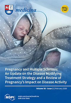
Cover Story (view full-size image): Pregnancy rates are rapidly increasing among women of reproductive age diagnosed with multiple sclerosis (MS). Pregnancy substantially reduces disease activity, although, for some patients, severe relapses could appear whilst pregnant. However, pregnancy is not always compatible with the available highly effective MS treatments. This article explores the aspects and outcomes of pregnancy, such as the impact on disease activity and the management of relapses during pregnancy and the breastfeeding period, and discusses available disease modifying treatment strategies during pregnancy. View Paper here.
  • Issues are regarded as officially published after their release is announced to the table of contents alert mailing list.
  • You may sign up for e-mail alerts to receive table of contents of newly released issues.
  • PDF is the official format for papers published in both, html and pdf forms. To view the papers in pdf format, click on the "PDF Full-text" link, and use the free Adobe Reader to open them.
Order results
Result details
Section
Select all
Export citation of selected articles as:
22 pages, 1672 KiB  
Review
MiRNA-Based Inspired Approach in Diagnosis of Prostate Cancer
by Vlad Cristian Munteanu, Raluca Andrada Munteanu, Anca Onaciu, Ioana Berindan-Neagoe, Bogdan Petrut and Ioan Coman
Medicina 2020, 56(2), 94; https://doi.org/10.3390/medicina56020094 - 24 Feb 2020
Cited by 11 | Viewed by 3877
Abstract
Prostate cancer is one of the most encountered cancer diseases in men worldwide and in consequence it requires the improvement of therapeutic strategies. For the clinical diagnosis, the standard approach is represented by solid biopsy. From a surgical point of view, this technique [...] Read more.
Prostate cancer is one of the most encountered cancer diseases in men worldwide and in consequence it requires the improvement of therapeutic strategies. For the clinical diagnosis, the standard approach is represented by solid biopsy. From a surgical point of view, this technique represents an invasive procedure that may imply several postoperative complications. To overcome these impediments, many trends are focusing on developing liquid biopsy assays and on implementing them in clinical practice. Liquid samples (blood, urine) are rich in analytes, especially in transcriptomic information provided by genetic markers. Additionally, molecular characterization regarding microRNAs content reveals outstanding prospects in understanding cancer progression mechanisms. Moreover, these analytes have great potential for prostate cancer early detection, more accurate prostate cancer staging and also for decision making respecting therapy schemes. However, there are still questionable topics and more research is needed to standardize liquid biopsy-based techniques. Full article
Show Figures

Figure 1

11 pages, 663 KiB  
Article
Comparison of a Five-Year Survival and Cancer Recurrence between Laparoscopically Assisted and Open Colonic Resections due to Adenocarcinoma—A Single Centre Experience
by Jurij Janež and Armand D Škapin
Medicina 2020, 56(2), 93; https://doi.org/10.3390/medicina56020093 - 24 Feb 2020
Cited by 1 | Viewed by 1780
Abstract
Background and objectives: When resecting colon adenocarcinoma, surgeons decide between the use of laparoscopically assisted and open surgery. Laparoscopic resection is known to have short-term benefits over an open operation. However, researchers are not as unified about the long-term findings. The aim of [...] Read more.
Background and objectives: When resecting colon adenocarcinoma, surgeons decide between the use of laparoscopically assisted and open surgery. Laparoscopic resection is known to have short-term benefits over an open operation. However, researchers are not as unified about the long-term findings. The aim of this research is to elaborate on five-year post-operative differences in survival and cancer recurrence between these two different approaches. Materials and methods: 74 enrolled patients were evaluated five years after a primary operation. We collected dates of deaths of deceased patients and time after operation of possible recurrences. Carcinoma staging was done by a pathologist after operation. Blood samples were taken before surgery in order to measure tumor markers (CA19-9 and CEA). Results: Survival after colonic adenocarcinoma surgery did not differ between the two different surgical approaches (p = 0.151). Recurrence of cancer was not associated with the type of operation (p = 0.532). Patients with recurrence had a 37.6 times greater hazard ratio of dying (95% CI: [12.0, 118]; p < 0.001). Advanced age adversely affected survival: patients aged <65 and ≥65 years had a 97%, and 57% survival rate, respectively. Patients with elevated tumor markers at operation had a 19.1 greater hazard ratio of dying (95% CI: [5.16, 70.4]; p < 0.001). Patients with different TNM stages did not have any statistically significant differences in survival (HRII = 2.49; 95% CI: [0.67, 9.30]; pII = 0.173) (HRIII = 2.18; 95% CI: [0.58, 8.12]; pIII = 0.246) or recurrence (p = 0.097). Conclusion: The obtained results suggest that laparoscopic resection of colon cancer is not inferior from an oncologic point of view and results in a similar long-term survival and disease-free interval. Recurrence of carcinoma, older age at initial operation and elevated tumor markers, above a pre-set threshold at operation, were found to be independent factors of lower survival. We believe that the obtained results will be of benefit when choosing treatment for colon adenocarcinoma. Full article
Show Figures

Figure 1

10 pages, 1012 KiB  
Article
Could the Age Difference of a Single Calendar Year between Patients Undergoing IVF at 34, 35 or at 36 Years Old Affect the IVF Outcome? A Retrospective Data Analysis
by Konstantinos Pantos, Konstantinos Sfakianoudis, Sokratis Grigoriadis, Evangelos Maziotis, Petroula Tsioulou, Anna Rapani, Polina Giannelou, Anastasios Atzampos, Sevasti Koulouraki, Michael Koutsilieris, Nikolaos Vlahos, George Mastorakos and Mara Simopoulou
Medicina 2020, 56(2), 92; https://doi.org/10.3390/medicina56020092 - 24 Feb 2020
Cited by 6 | Viewed by 3174
Abstract
Background and Objectives: Clinicians are called to overcome age-related challenges in decision making during In Vitro Fertilization (IVF) treatment. The aim of this study was to investigate the possible impact of a single calendar year difference among patients aged 34, 35 and 36 [...] Read more.
Background and Objectives: Clinicians are called to overcome age-related challenges in decision making during In Vitro Fertilization (IVF) treatment. The aim of this study was to investigate the possible impact of a single calendar year difference among patients aged 34, 35 and 36 on IVF outcomes. Materials and Methods: Medical records between 2008 and 2019 were analyzed retrospectively. The study group consisted of women diagnosed with tubal factor infertility. Sample size was divided in three categories at 34, 35 and 36 years of age. Embryo transfer including two blastocysts was performed for every patient. Comparisons were performed regarding hormonal profile, response to stimulation, quality of transferred embryos, positive hCG test and clinical pregnancy rate. Results: A total of 706 women were eligible to participate. Two-hundred and forty-eight women were 34, 226 were 35 while the remaining 232 were 36 years old. Regarding the hormonal profile, the number of accumulated oocytes and the quality of embryos transferred, no statistically significant difference was documented between the three age groups. Women aged 34 and 35 years old indicated a significantly increased positive hCG rate in comparison to women aged 36 years old (p-value = 0.009, p-value = 0.023, respectively). Women aged 34 and 35 years old presented with a higher clinical pregnancy rate in comparison to those aged 36 years old (p-value = 0.04, p-value = 0.05, respectively). Conclusion: A calendar year difference between patients undergoing IVF treatment at 34 or 35 years of age does not appear to exert any influence regarding outcome. When treatment involves patients above the age of 35, then a single calendar year may exert considerable impact on IVF outcome. This observation indicates that age 35 may serve as a valid cut-off point regarding IVF outcome. Full article
Show Figures

Figure 1

9 pages, 281 KiB  
Article
Dietary Attitude of Adults with Type 2 Diabetes Mellitus in the Kingdom of Saudi Arabia: A Cross-Sectional Study
by Waqas Sami, Khalid M Alabdulwahhab, Mohd Rashid Ab Hamid, Tariq A. Alasbali, Fahd Al Alwadani and Mohammad Shakil Ahmad
Medicina 2020, 56(2), 91; https://doi.org/10.3390/medicina56020091 - 24 Feb 2020
Cited by 4 | Viewed by 3269
Abstract
Background and Objectives: There is a paucity of literature on the dietary attitude (DA) of patients with type 2 diabetes in the Kingdom of Saudi Arabia (KSA). Although the prevalence of diabetes mellitus (DM) is high in Gulf countries, there remains a lack [...] Read more.
Background and Objectives: There is a paucity of literature on the dietary attitude (DA) of patients with type 2 diabetes in the Kingdom of Saudi Arabia (KSA). Although the prevalence of diabetes mellitus (DM) is high in Gulf countries, there remains a lack of understanding of the importance of dietary behavior in diabetes management among patients. Understanding the behavior of patients with diabetes towards the disease requires knowledge of their DA. Therefore, this study aimed to assess and evaluate the DA of type 2 diabetes patients, and it is the first of its kind in the KSA. Material and Methods: An analytical cross-sectional study was conducted among 350 patients with type 2 diabetes. A self-administered DA questionnaire was used to collect the data. Psychometric properties of the questionnaire were assessed by face validity, content validity, exploratory factor analysis, and internal consistency reliability. The data were collected using a systematic random sampling technique. Results: The overall DA of the patients was inappropriate (p = 0.014). Patients had an inappropriate DA towards food selection (p = 0.003), healthy choices (p = 0.005), food restraint (p < 0.001), health impact (p < 0.001), and food categorization (p = 0.033). A poor DA was also observed in relation to the consumption of red meat (p <0.001), rice (p < 0.001), soup and sauces (p = 0.040), dairy products (p = 0.015), and junk food (p < 0.001). Conclusions: It is highly recommended that patients with diabetes receive counseling with an empowerment approach, as this can bring about changes in their dietary behavior, which is deeply rooted in their daily routine. Healthcare providers should also be well-informed about patients’ attitudes and beliefs towards diabetes to design tailored educational and salutary programs for this specific community. Diabetes self-management educational programs should also be provided on a regular basis with a special emphasis on diet and its related components. Full article
(This article belongs to the Special Issue Interdisciplinary Medicine)
13 pages, 7236 KiB  
Article
Stevia Eupatoria and Stevia Pilosa Extracts Inhibit the Proliferation and Migration of Prostate Cancer Cells
by Elizabeth Martínez-Rojo, Raquel Cariño-Cortés, Laura Cristina Berumen, Guadalupe García-Alcocer and Jesica Escobar-Cabrera
Medicina 2020, 56(2), 90; https://doi.org/10.3390/medicina56020090 - 23 Feb 2020
Cited by 11 | Viewed by 4150
Abstract
Background and Objectives: Prostate cancer is the second most harmful disease in men worldwide and the number of cases is increasing. Therefore, new natural agents with anticancer potential should be examined and the response of existing therapeutic drugs must be enhanced. Stevia [...] Read more.
Background and Objectives: Prostate cancer is the second most harmful disease in men worldwide and the number of cases is increasing. Therefore, new natural agents with anticancer potential should be examined and the response of existing therapeutic drugs must be enhanced. Stevia pilosa and Stevia eupatoria are two species that have been widely used in traditional medicine, but their effectiveness on cancer cells and their interaction with antineoplastic drugs have not been studied. The aim of this study was to evaluate the anticancer activity of Stevia pilosa methanolic root extract (SPME) and Stevia eupatoria methanolic root extract (SEME) and their effect, combined with enzalutamide, on prostate cancer cells. Materials and Methods: The study was conducted on a human fibroblast cell line, and on androgen-dependent (LNCaP) and androgen-independent (PC-3) prostate cancer cell lines. The cell viability was evaluated using a Trypan Blue exclusion test for 48 h, and the migration by a wound-healing assay for 24, 48, and 72 h. Results: The results indicate that SPME and SEME were not cytotoxic at concentrations less than 1000 μg/mL in the human fibroblasts. SPME and SEME significantly reduced the viability and migration of prostate cancer cells in all concentrations evaluated. The antiproliferative effect of the Stevia extracts was higher in cancer cells than in normal cells. The enzalutamide decreased the cell viability in all concentrations tested (10–50 µM). The combination of the Stevia extracts and enzalutamide produced a greater effect on the inhibition of the proliferation and migration of cancer cells than the Stevia extracts alone, but not of the enzalutamide alone. Conclusion: The results indicate that SPME and SEME have an inhibitory effect on the viability and migration of prostate cancer cells and do not interfere with the enzalutamide anticancer effect. The data suggest that Stevia extracts may be a potential source of molecules for cancer treatment. Full article
Show Figures

Figure 1

12 pages, 353 KiB  
Review
State of the Art in Fertility Preservation for Female Patients Prior to Oncologic Therapies
by Călin Bogdan Chibelean, Răzvan-Cosmin Petca, Dan Cristian Radu and Aida Petca
Medicina 2020, 56(2), 89; https://doi.org/10.3390/medicina56020089 - 23 Feb 2020
Cited by 4 | Viewed by 2550
Abstract
Quality of life improvement stands as one of the main goals of the medical sciences. Increasing cancer survival rates associated with better early detection and extended therapeutic options led to the specific modeling of patients’ choices, comprising aspects of reproductive life that correlated [...] Read more.
Quality of life improvement stands as one of the main goals of the medical sciences. Increasing cancer survival rates associated with better early detection and extended therapeutic options led to the specific modeling of patients’ choices, comprising aspects of reproductive life that correlated with the evolution of modern society, and requires better assessment. Of these, fertility preservation and ovarian function conservation for pre-menopause female oncologic patients pose a contemporary challenge due to procreation age advance in evolved societies and to the growing expectations regarding cancer treatment. Progress made in cell and tissue-freezing technologies brought hope and shed new light on the onco-fertility field. Additionally, crossing roads with general fertility and senescence studies proved highly beneficial due to the enlarged scope and better synergies and funding. We here strive to bring attention to this domain of care and to sensitize all medical specialties towards a more cohesive approach and to better communication among caregivers and patients. Full article
(This article belongs to the Special Issue Interdisciplinary Medicine)
15 pages, 1302 KiB  
Review
Nutritional Status and the Influence of the Vegan Diet on the Gut Microbiota and Human Health
by Hercules Sakkas, Petros Bozidis, Christos Touzios, Damianos Kolios, Georgia Athanasiou, Eirini Athanasopoulou, Ioanna Gerou and Constantina Gartzonika
Medicina 2020, 56(2), 88; https://doi.org/10.3390/medicina56020088 - 22 Feb 2020
Cited by 97 | Viewed by 30069
Abstract
The human gut microbiota is considered a well-known complex ecosystem composed of distinct microbial populations, playing a significant role in most aspects of human health and wellness. Several factors such as infant transitions, dietary habits, age, consumption of probiotics and prebiotics, use of [...] Read more.
The human gut microbiota is considered a well-known complex ecosystem composed of distinct microbial populations, playing a significant role in most aspects of human health and wellness. Several factors such as infant transitions, dietary habits, age, consumption of probiotics and prebiotics, use of antibiotics, intestinal comorbidities, and even metabolic diseases may continously alter microbiota diversity and function. The study of vegan diet–microbiota interactions is a rapidly evolving field, since plenty of research has been focused on the potential effects of plant-based dietary patterns on the human gut microbiota. It has been reported that well-planned vegan diets and their associated components affect both the bacterial composition and metabolic pathways of gut microbiota. Certain benefits associated with medical disorders but also limitations (including nutritional deficiencies) have been documented. Although the vegan diet may be inadequate in calorific value, it is rich in dietary fiber, polyphenols, and antioxidant vitamins. The aim of the present study was to provide an update of the existing knowledge on nutritional status of vegan diets and the influence of their food components on the human gut microbiota and health. Full article
Show Figures

Figure 1

17 pages, 591 KiB  
Review
The Role of Blood Biomarkers for Magnetic Resonance Imaging Diagnosis of Traumatic Brain Injury
by John K. Yue, Pavan S. Upadhyayula, Lauro N. Avalos, Hansen Deng and Kevin K. W. Wang
Medicina 2020, 56(2), 87; https://doi.org/10.3390/medicina56020087 - 22 Feb 2020
Cited by 27 | Viewed by 4895
Abstract
Background and Objectives: The annual global incidence of traumatic brain injury (TBI) is over 10 million. An estimated 29% of TBI patients with negative computed tomography (CT−) have positive magnetic resonance imaging (MRI+) findings. Judicious use of serum biomarkers with MRI may [...] Read more.
Background and Objectives: The annual global incidence of traumatic brain injury (TBI) is over 10 million. An estimated 29% of TBI patients with negative computed tomography (CT−) have positive magnetic resonance imaging (MRI+) findings. Judicious use of serum biomarkers with MRI may aid in diagnosis of CT-occult TBI. The current manuscript aimed to evaluate the diagnostic, therapeutic and risk-stratification utility of known biomarkers and intracranial MRI pathology. Materials and Methods: The PubMed database was queried with keywords (plasma OR serum) AND (biomarker OR marker OR protein) AND (brain injury/trauma OR head injury/trauma OR concussion) AND (magnetic resonance imaging/MRI) (title/abstract) in English. Seventeen articles on TBI biomarkers and MRI were included: S100 calcium-binding protein B (S100B; N = 6), glial fibrillary acidic protein (GFAP; N = 3), GFAP/ubiquitin carboxyl-terminal hydrolase-L1 (UCH-L1; N = 2), Tau (N = 2), neurofilament-light (NF-L; N = 2), alpha-synuclein (N = 1), and alpha-amino-3-hydroxy-5-methyl-4-isoxazolepropionic acid receptor peptide (AMPAR; N = 1). Results: Acute GFAP distinguished CT−/MRI+ from CT−/MRI− (AUC = 0.777, 0.852 at 9–16 h). GFAP discriminated CT−/diffuse axonal injury (DAI+) from controls (AUC = 0.903). Tau correlated directly with number of head strikes and inversely with white matter fractional anisotropy (FA), and a cutoff > 1.5 pg/mL discriminated between DAI+ and DAI− (sensitivity = 74%/specificity = 69%). NF-L had 100% discrimination of DAI in severe TBI and correlated with FA. Low alpha-synuclein was associated with poorer functional connectivity. AMPAR cutoff > 0.4 ng/mL had a sensitivity of 91% and a specificity of 92% for concussion and was associated with minor MRI findings. Low/undetectable S100B had a high negative predictive value for CT/MRI pathology. UCH-L1 showed no notable correlations with MRI. Conclusions: An acute circulating biomarker capable of discriminating intracranial MRI abnormalities is critical to establishing diagnosis for CT-occult TBI and can triage patients who may benefit from outpatient MRI, surveillance and/or follow up with TBI specialists. GFAP has shown diagnostic potential for MRI findings such as DAI and awaits further validation. Tau shows promise in detecting DAI and disrupted functional connectivity. Candidate biomarkers should be evaluated within the context of analytical performance of the assays used, as well as the post-injury timeframe for blood collection relative to MRI abnormalities. Full article
(This article belongs to the Special Issue Traumatic Brain Injury: Current Efforts in Research and Clinical Care)
Show Figures

Figure 1

8 pages, 1384 KiB  
Article
Timing between Breast Reconstruction and Oncologic Mastectomy—One Center Experience
by Adelaida Avino, Laura Răducu, Lăcrămioara Aurelia Brînduşe, Cristian-Radu Jecan and Ioan Lascăr
Medicina 2020, 56(2), 86; https://doi.org/10.3390/medicina56020086 - 20 Feb 2020
Cited by 6 | Viewed by 3474
Abstract
Background and objectives: Breast cancer is the most common cancer in women. The immunohistochemical profile, but also the stage of the tumor determines the therapeutic management, which varies from conservative surgery to mastectomy associated with chemotherapy, hormonal and biological therapy and/or radiotherapy. Mastectomy [...] Read more.
Background and objectives: Breast cancer is the most common cancer in women. The immunohistochemical profile, but also the stage of the tumor determines the therapeutic management, which varies from conservative surgery to mastectomy associated with chemotherapy, hormonal and biological therapy and/or radiotherapy. Mastectomy remains one of the most radical surgical intervention for women, having great consequences on quality of life, which can be improved by realizing immediate or delayed breast reconstruction. The objective of the study was to evaluate the period of time between the mastectomy and the breast reconstruction. Material and methods: We performed a retrospective study on 57 female patients admitted to the Plastic Surgery Department of the Clinical Emergency Hospital “Prof. Dr. Agrippa Ionescu”, Bucharest, Romania. All the patients underwent immediate or delayed breast reconstruction after mastectomy for confirmed breast cancer. Descriptive data analysis was realized with evaluation of type of breast reconstruction considering the staging of the tumor, the invaded lymph nodes, and the necessity of adjuvant chemoradiotherapy. Moreover, the median period between mastectomy and reconstruction was evaluated. Results: The immediate breast reconstruction was performed in patients with stage I, in patients with stage II, delayed reconstruction was performed after minimum six months, and the patients with stage III had the breast reconstructed with free flap (50%), 8–43 months post-mastectomy. Radiotherapy determines the type of breast reconstruction, in most of the cases the latissimus dorsi flap was used with implant (22.6%). Conclusions: Breast reconstruction is an important step in increasing the quality of life for women who underwent mastectomy after breast cancer. The proper timing for breast reconstruction must be settled by a team formed by the patient, the plastic surgeon, and the oncologist. Full article
(This article belongs to the Special Issue Interdisciplinary Medicine)
Show Figures

Figure 1

10 pages, 2096 KiB  
Article
Bedside Sonographic Duplex Technique as a Monitoring Tool in Patients after Decompressive Craniectomy: A Single Centre Experience
by Habib Bendella, Joachim Spreer, Alexander Hartmann, Alhadi Igressa, Marc Maegele, Rolf Lefering and Makoto Nakamura
Medicina 2020, 56(2), 85; https://doi.org/10.3390/medicina56020085 - 19 Feb 2020
Cited by 1 | Viewed by 2031
Abstract
Background and objectives: Bedside sonographic duplex technique (SDT) may be used as an adjunct to cranial computed tomography (CCT) to monitor brain-injured patients after decompressive craniectomy (DC). The present study aimed to assess the value of SDT in repeated measurements of ventricle dimensions [...] Read more.
Background and objectives: Bedside sonographic duplex technique (SDT) may be used as an adjunct to cranial computed tomography (CCT) to monitor brain-injured patients after decompressive craniectomy (DC). The present study aimed to assess the value of SDT in repeated measurements of ventricle dimensions in patients after DC by comparing both techniques. Materials and Methods: Retrospective assessment of 20 consecutive patients after DC for refractory intracranial pressure (ICP) increase following subarachnoid hemorrhage (SAH), bleeding and trauma which were examined by SDT and CCT in the context of routine clinical practice. Whenever a repeated CCT was clinically indicated SDT examinations were performed within 24 hours and correlated via measurement of the dimensions of all four cerebral ventricles. Basal cerebral arteries including pathologies such as vasospasms were also evaluated in comparison to selected digital subtraction angiography (DSA). Results: Repeated measurements of all four ventricle diameters showed high correlation between CCT and SDT (right lateral r = 0.997, p < 0.001; left lateral r = 0.997, p < 0.001; third r = 0.991, p < 0.001, fourth ventricle r = 0.977, p < 0.001). SDT performed well in visualizing basal cerebral arteries including pathologies (e.g., vasospasms) as compared to DSA. Conclusions: Repeated SDT measurements of the dimensions of all four ventricles in patients after DC for refractory ICP increase delivered reproducible results comparable to CCT. SDT may be considered as a valuable bedside monitoring tool in patients after DC. Full article
(This article belongs to the Special Issue Traumatic Brain Injury: Current Efforts in Research and Clinical Care)
Show Figures

Figure 1

9 pages, 1185 KiB  
Article
Comparison of Lateral Abdominal Musculature Activation during Expiration with an Expiratory Flow Control Device Versus the Abdominal Drawing-in Maneuver in Healthy Women: A Cross-Sectional Observational Pilot Study
by Vanesa Abuín-Porras, Paula Maldonado-Tello, Mónica de la Cueva-Reguera, David Rodríguez-Sanz, César Calvo-Lobo, Daniel López-López, Emmanuel Navarro-Flores and Carlos Romero-Morales
Medicina 2020, 56(2), 84; https://doi.org/10.3390/medicina56020084 - 19 Feb 2020
Cited by 6 | Viewed by 3062
Abstract
Background and Objectives: The purpose of the present study was to quantify and compare lateral abdominal musculature thickness, including the transverse abdominis (TrA), internal oblique (IO), and external oblique (EO) muscles, via rehabilitative ultrasound imaging (RUSI) during the use of the expiratory flow [...] Read more.
Background and Objectives: The purpose of the present study was to quantify and compare lateral abdominal musculature thickness, including the transverse abdominis (TrA), internal oblique (IO), and external oblique (EO) muscles, via rehabilitative ultrasound imaging (RUSI) during the use of the expiratory flow control device (EFCD) versus the classic abdominal drawing-in maneuver (ADIM). Materials and Methods: A cross-sectional observational pilot study. Twenty-one women were recruited and assessed the thickness of each muscle (TrA, IO, and EO) by ultrasound imaging at rest, during the ADIM, and during expiration with the EFCD. Waist circumference was also measured under the same circumstances. Results: Statistically significant differences were observed between ADIM, EFCD, and at rest condition for the thickness of the TrA (p = 0.001) and IO (p = 0.039). Moreover, statistically significant differences for TrAb at rest compared with the ADIM (p = 0.001, Cohen’s d = 2.183) and at rest and with the EFCD (p = 0.001, Cohen’s d = 2.843). In addition, between ADIM and EFCD were not statistically significant, although a moderate effect size was found (p = 0.055, Cohen’s d = 0.694). For the IO muscle thickness, significant differences were reported between the EFCD and at rest (p = 0.038), Cohen’s d = 0.081). Conclusions: Significant differences in the increase of the thickness of the TrA and IO muscles during the use of the EFCD and the ADIM with respect to rest. In addition, for the TrA, statistically significant differences were found during expiration with the EFCD with respect to the ADIM. Expiration with EFCD can be a useful method for the activation of the TrA. Full article
(This article belongs to the Special Issue Ultrasound in Sports Medicine: Current Concepts and Advances)
Show Figures

Figure 1

11 pages, 1099 KiB  
Article
Inter-and Intra-Examiner Reliability of Supraspinatus Muscle Tendon Palpation: A Cross-Sectional Study by Ultrasonography
by Fermin Naranjo-Cinto, Giezi Falcon-Machado, Alejandro Garrido-Marin, Francisco Jose Senin-Camargo, Maria Amalia Jacome-Pumar, Ruben Fernandez-Matias, Daniel Pecos-Martin and Tomas Gallego-Izquierdo
Medicina 2020, 56(2), 83; https://doi.org/10.3390/medicina56020083 - 18 Feb 2020
Cited by 1 | Viewed by 4510
Abstract
Background and objectives: Palpation is an inherent and basic skill of health professionals—particularly manual therapists who base their diagnosis and treatment in a clinical environment. Many authors have previously described multiple palpation positions for supraspinatus muscle tendon (SMT); however, there are no [...] Read more.
Background and objectives: Palpation is an inherent and basic skill of health professionals—particularly manual therapists who base their diagnosis and treatment in a clinical environment. Many authors have previously described multiple palpation positions for supraspinatus muscle tendon (SMT); however, there are no current studies that evaluate palpation concordance reliability for the SMT in this particular position. This study aimed to investigate the intra- and inter-rater reliabilities of supraspinatus muscle tendon (SMT) palpation. Materials and Methods: Thirty-six healthy participants (14 females; aged 22–35 years) were recruited. Musculoskeletal ultrasound was used to measure the SMT localization after two physiotherapists performed the SMT palpation at two different times. The distance between the two marked points was used to analyze the analysis of true agreement between examiners. Finally, we analyzed if the demographic variables influenced the palpation procedure. Results: The intra-examiner reliability showed a high percentage of concordance for examiner 1 (E.1) (first palpation procedure (P.1) = 91.7%: second palpation procedure (P.2) = 95.8%) and examiner 2 (E.2) (P.1 = 91.6%; P.2 = 97.2%) and high percentage of inter-palpation agreement for E.1 (87.5%) and E.2 (88.9%). The inter-examiner reliability showed a high total concordance for the right shoulder (E.1 = 94.4%; E.2 = 95.8%) and left shoulder (E.1 = 93.05%; E.2 = 95.05%). The agreement (%) according to both examiners was 93.05% for the right shoulder and 94.4% for the left shoulder. The agreement between both examiners and the ultrasound (% of true agreement) was 92.9% for the right shoulder and 92.8% for the left shoulder. A statistically significant association (p = 0.02) was found for weight regarding concordance reliability; this was not seen for dominant arm, age, gender, body mass index, height, and tendon depth (p > 0.05). Conclusions: The SMT palpation technique showed a high level of concordance and reproducibility. Full article
(This article belongs to the Special Issue Ultrasound in Sports Medicine: Current Concepts and Advances)
Show Figures

Figure 1

7 pages, 1554 KiB  
Article
Cortical Bone Trajectory Instrumentation with Vertebroplasty for Osteoporotic Thoracolumbar Compression Fracture
by Wei-Lin Hsu, Yu-Hsiang Lin, Hao-Yu Chuang, Han-Chung Lee, Der-Cherng Chen, Yen-Tse Chu, Der-Yang Cho and Chao-Hsuan Chen
Medicina 2020, 56(2), 82; https://doi.org/10.3390/medicina56020082 - 17 Feb 2020
Cited by 3 | Viewed by 2390
Abstract
Background: Osteoporotic spinal fractures commonly occur in elderly patients with low bone mineral density. In these cases, percutaneous vertebroplasty or percutaneous kyphoplasty can provide significant pain relief and improve mobility. However, studies have reported both the recurrence of vertebral compression fractures at the [...] Read more.
Background: Osteoporotic spinal fractures commonly occur in elderly patients with low bone mineral density. In these cases, percutaneous vertebroplasty or percutaneous kyphoplasty can provide significant pain relief and improve mobility. However, studies have reported both the recurrence of vertebral compression fractures at the index level after vertebroplasty and the development of new vertebral fractures at the adjacent level that occur without any additional trauma. Pedicle screw fixation combined with percutaneous vertebroplasty has been proposed as an effective procedure for addressing osteoporotic thoracolumbar fractures. However, in osteoporotic populations, pedicle screws can loosen, pullout, or migrate. Currently, the efficacy of cortical bone trajectory screw fixation for osteoporotic fractures remains unclear. Thus, we assessed the effects of using cortical bone trajectory instrumentation with vertebroplasty on patient outcomes. Method: We retrospectively reviewed data from 12 consecutively sampled osteoporotic thoracolumbar fracture patients who underwent cortical bone trajectory instrumentation with vertebroplasty. Patients were enrolled beginning in October 2015 and were followed for >24 months. Result: The average age was 74 years, and the average dual-energy x-ray absorptiometry T-score was −3.6. The average visual analog scale pain scores improved from 8 to 2.5 after surgery. The average blood loss was 36.25 mL. All patients regained ambulation and experienced reduced pain post-surgery. No recurrent fractures or instrument failures were recorded during follow-up. Conclusions: Our findings suggest that cortical bone trajectory instrumentation combined with percutaneous vertebroplasty may be a good option for treating osteoporotic thoracolumbar fractures, as it can prevent recurrent vertebral fractures or related kyphosis in sagittal alignment. Full article
Show Figures

Figure 1

12 pages, 1402 KiB  
Article
Combined Effect of HPV and Several Gene SNPs in Laryngeal Cancer
by Aušra Stumbrytė-Kaminskienė, Živilė Gudlevičienė, Daiva Dabkevičienė and Irina Mackevičienė
Medicina 2020, 56(2), 81; https://doi.org/10.3390/medicina56020081 - 17 Feb 2020
Cited by 6 | Viewed by 2230
Abstract
Background and objectives: Laryngeal squamous cell carcinoma (LSCC) is one of the most common head and neck tumors. The molecular mechanism of LSCC remains unclear. The aim of this study was to evaluate the prevalence of Human papillomavirus (HPV) and single nucleotide polymorphisms [...] Read more.
Background and objectives: Laryngeal squamous cell carcinoma (LSCC) is one of the most common head and neck tumors. The molecular mechanism of LSCC remains unclear. The aim of this study was to evaluate the prevalence of Human papillomavirus (HPV) and single nucleotide polymorphisms (SNPs) of TP53, MDM2, MDM4, MTHFR, CASP8, and CCR5 genes in LSCC, and to assess their correlations with patient survival. Materials and Methods: 49 LSCC patients were enrolled in this study. PCR and qRT-PCR were used to detect, identify, and quantify HPV. SNPs were genotyped using PCR and PCR-RFLP. Results: By analyzing the interactions of the SNPs of the genes with clinical parameters, the majority of patients with lymph node status (N1,2) were identified as carriers of MDM2 T/G, CASP8 ins/del, CCR5 wt/wt SNP. Cluster analysis showed that patients with MDM2 T/T SNP survive longer than patients identified as CASP8 ins/ins, MTHFR C/C, and MDM4 A/A variant carriers; meanwhile, LSCC patients with MDM2 T/T polymorphic variant had the best survival. Multivariate analysis showed that HPV-positive patients without metastasis in regional lymph nodes (N0) and harboring CASP8 ins/del variant had the best survival. Meanwhile, HPV-negative patients with identified metastasis in lymph nodes (N1 and N2) and CASP8 ins/del variant had poor survival. Conclusions: This finding suggests patients survival prognosis and tumor behavior are different according HPV status, SNP variants, and clinical characteristics of the LSCC. Full article
Show Figures

Figure 1

13 pages, 2900 KiB  
Article
CPAP Effect on Cardiopulmonary Exercise Testing Performance in Patients with Moderate-Severe OSA and Cardiometabolic Comorbidities
by Ioana Mădălina Zota, Cristian Stătescu, Radu Andy Sascău, Mihai Roca, Radu Sebastian Gavril, Teodor Flaviu Vasilcu, Daniela Boișteanu, Alexandra Maștaleru, Alexandra Jitaru, Maria Magdalena Leon Constantin and Florin Mitu
Medicina 2020, 56(2), 80; https://doi.org/10.3390/medicina56020080 - 15 Feb 2020
Cited by 6 | Viewed by 3287
Abstract
Background and Objectives: Obstructive sleep apnea (OSA) is associated with daytime somnolence, cognitive impairment and high cardiovascular morbidity and mortality. Obesity, associated cardiovascular comorbidities, accelerated erythropoiesis and muscular mitochondrial energetic dysfunctions negatively influence exercise tolerance in moderate-severe OSA patients. The cardiopulmonary exercise testing [...] Read more.
Background and Objectives: Obstructive sleep apnea (OSA) is associated with daytime somnolence, cognitive impairment and high cardiovascular morbidity and mortality. Obesity, associated cardiovascular comorbidities, accelerated erythropoiesis and muscular mitochondrial energetic dysfunctions negatively influence exercise tolerance in moderate-severe OSA patients. The cardiopulmonary exercise testing (CPET) offers an integrated assessment of the individual’s aerobic capacity and helps distinguish the main causes of exercise limitation. The purpose of this study is to evaluate the aerobic capacity of OSA patients, before and after short-term continuous positive airway pressure (CPAP). Materials and Methods: Our prospective study included 64 patients with newly diagnosed moderate-severe OSA (apnea hypopnea index (AHI) 39.96 ± 19.04 events/h) who underwent CPET before and after CPAP. Thirteen patients were unable to tolerate CPAP or were lost during follow-up. Results: 49.29% of our patients exhibited a moderate or severe decrease in functional capacity (Weber C or D). CPET performance was influenced by gender but not by apnea severity. Eight weeks of CPAP induced significant improvements in maximal exercise load (Δ = 14.23 W, p = 0.0004), maximum oxygen uptake (Δ = 203.87 mL/min, p = 0.004), anaerobic threshold (Δ = 316.4 mL/min, p = 0.001), minute ventilation (Δ = 5.1 L/min, p = 0.01) and peak oxygen pulse (Δ = 2.46, p = 0.007) as well as a decrease in basal metabolic rate (BMR) (Δ = −8.3 kCal/24 h, p = 0.04) and average Epworth score (Δ = −4.58 points, p < 0.000001). Conclusions: Patients with moderate-severe OSA have mediocre functional capacity. Apnea severity (AHI) was correlated with basal metabolic rate, resting heart rate and percent predicted maximum effort but not with anaerobic threshold or maximum oxygen uptake. Although CPET performance was similar in the two apnea severity subgroups, short-term CPAP therapy significantly improved most CPET parameters, suggesting that OSA per se has a negative influence on effort capacity. Full article
(This article belongs to the Special Issue Obstructive Sleep Apnea: Epidemiology, Pathomechanism and Treatment)
Show Figures

Figure 1

6 pages, 255 KiB  
Article
Cost-Effectiveness of Titanium Elastic Nail (TEN) in the Treatment of Forearm Fractures in Children
by Ovidiu Adam, Vlad Laurentiu David, Florin George Horhat and Eugen Sorin Boia
Medicina 2020, 56(2), 79; https://doi.org/10.3390/medicina56020079 - 15 Feb 2020
Cited by 3 | Viewed by 2055
Abstract
Background and objectives: There are various methods in the management of forearm fractures in children. Elastic stable intramedullary nailing using Titanium Elastic Nail (TEN) is nowadays employed in diaphysis fractures of children, with clear benefits over other treatment options. However, in the [...] Read more.
Background and objectives: There are various methods in the management of forearm fractures in children. Elastic stable intramedullary nailing using Titanium Elastic Nail (TEN) is nowadays employed in diaphysis fractures of children, with clear benefits over other treatment options. However, in the case of TEN versus other treatment methods of forearm fractures in children, cost is an important issue. This report will focus on the cost assessment of using TEN versus other therapeutic means in the treatment of forearm fractures in children. Materials and Methods: We performed a retrospective longitudinal study of 173 consecutive patients with forearm fractures treated in a single institution during 2017. We calculated the cost for each patient by summing up direct costs plus indirect costs, calculated at an aggregate level. Hospital income data were extracted from the Diagnosis Related Groups database. Results: A total of 173 patients with forearm fractures were treated, 44 using TEN, 86 using K-wire, and 46 using closed reduction and cast. There were 66 radius fractures, 1 ulna fracture, and 106 that were both radius and ulna fractures. Mean treatment cost were $632.76 for TEN, $499.50 for K-wire, and $451.30 for closed reduction and cast. Costs for TEN were higher than for K-wire insertion (p = 0.00) and higher than closed reduction and cast ($182.42; p = 0.00). Reimbursement per patient was higher with TEN versus K-wire patients; $497.88 vs. $364.64 /patient (p = 0.00), and higher than for patients treated with closed reduction and cast (p = 0.00). Conclusions: The treatment of upper extremity fractures using TEN was more expensive than the other methods. In Romania, because the reimbursement for TEN is higher as well, there are no differences in the financial burden when treating forearm fractures with TEN versus K-wire. Non-surgical treatment has the lowest cost but also the lowest reimbursement. Full article
14 pages, 906 KiB  
Article
Preliminary Results of an Exercise Program After Laparoscopic Resective Colorectal Cancer Surgery in Non-Metastatic Adenocarcinoma: A Pilot Study of a Randomized Control Trial
by Gabriele Mascherini, Maria Novella Ringressi, Jorge Castizo-Olier, Georgian Badicu, Alfredo Irurtia, Laura Stefani, Giorgio Galanti and Antonio Taddei
Medicina 2020, 56(2), 78; https://doi.org/10.3390/medicina56020078 - 14 Feb 2020
Cited by 6 | Viewed by 2831
Abstract
Background and objectives: Performing physical exercise after a colorectal cancer diagnosis is associated with lower mortality related to the tumor itself. In order to improve physical recovery after elective surgery, there are no specific exercise protocols after discharge from the hospital. The purpose [...] Read more.
Background and objectives: Performing physical exercise after a colorectal cancer diagnosis is associated with lower mortality related to the tumor itself. In order to improve physical recovery after elective surgery, there are no specific exercise protocols after discharge from the hospital. The purpose of this study is to show the preliminary results of an exercise program after colorectal cancer surgery. Materials and Methods: Six patients with non-metastatic colorectal adenocarcinoma addressed to respective laparoscopic were randomly assigned to a mixed supervised/home-based exercise program for six months and compared to a control group without exercise. To assess the effectiveness of the program, functional and body composition parameters were evaluated. Results: Three months after surgery, the exercise group increased flexibility (p <0.01, ES = 0.33), strength of lower limbs (p <0.01, ES = 0.42) and aerobic capacity (p <0.01, ES = 0.28). After surgery, the six patients experienced a significant reduction in body mass index (BMI) and free fat mass. More specifically, fat mass reached the lowest values, with a concomitant increase in cell mass after six months (p <0.01, ES = 0.33). This did not occur in the control group. Conclusions: Colorectal cancer treatment induces a reduction in physical function, particularly during the first six months after treatment. A mixed exercise approach appears promising in countering this process after colorectal cancer surgery. Full article
(This article belongs to the Special Issue Molecular and Therapeutic Landscapes in Colorectal Carcinoma)
Show Figures

Figure 1

10 pages, 1429 KiB  
Article
Circulating Soluble Urokinase-Type Plasminogen Activator Receptor in Obstructive Sleep Apnoea
by Renata Marietta Bocskei, Martina Meszaros, Adam Domonkos Tarnoki, David Laszlo Tarnoki, Laszlo Kunos, Zsofia Lazar and Andras Bikov
Medicina 2020, 56(2), 77; https://doi.org/10.3390/medicina56020077 - 14 Feb 2020
Cited by 4 | Viewed by 2828
Abstract
Background and Objectives: Obstructive sleep apnoea (OSA) is associated with heightened systemic inflammation and a hypercoagulation state. Soluble urokinase-type plasminogen activator receptor (suPAR) plays a role in fibrinolysis and systemic inflammation. However, suPAR has not been investigated in OSA. Materials and Methods [...] Read more.
Background and Objectives: Obstructive sleep apnoea (OSA) is associated with heightened systemic inflammation and a hypercoagulation state. Soluble urokinase-type plasminogen activator receptor (suPAR) plays a role in fibrinolysis and systemic inflammation. However, suPAR has not been investigated in OSA. Materials and Methods: A total of 53 patients with OSA and 15 control volunteers participated in the study. Medical history was taken and in-hospital sleep studies were performed. Plasma suPAR levels were determined by ELISA. Results: There was no difference in plasma suPAR values between patients with OSA (2.198 ± 0.675 ng/mL) and control subjects (2.088 ± 0.976 ng/mL, p = 0.62). Neither was there any difference when patients with OSA were divided into mild (2.134 ± 0.799 ng/mL), moderate (2.274 ± 0.597 ng/mL) and severe groups (2.128 ± 0.744 ng/mL, p = 0.84). There was no significant correlation between plasma suPAR and indices of OSA severity, blood results or comorbidities, such as hypertension, diabetes, dyslipidaemia or cardiovascular disease. Plasma suPAR levels were higher in women when all subjects were analysed together (2.487 ± 0.683 vs. 1.895 ± 0.692 ng/mL, p < 0.01), and also separately in controls (2.539 ± 0.956 vs. 1.411 ± 0.534 ng/mL, p = 0.02) and patients (2.467 ± 0.568 vs. 1.991 ± 0.686 ng/mL, p < 0.01). Conclusions: Our results suggest that suPAR does not play a significant role in the pathophysiology of OSA. The significant gender difference needs to be considered when conducting studies on circulating suPAR. Full article
(This article belongs to the Special Issue Obstructive Sleep Apnea: Epidemiology, Pathomechanism and Treatment)
Show Figures

Figure 1

9 pages, 3468 KiB  
Case Report
De novo NIPBL Mutations in Vietnamese Patients with Cornelia de Lange Syndrome
by Duong Chi Thanh, Can Thi Bich Ngoc, Ngoc-Lan Nguyen, Chi Dung Vu, Nguyen Van Tung and Huy Hoang Nguyen
Medicina 2020, 56(2), 76; https://doi.org/10.3390/medicina56020076 - 14 Feb 2020
Cited by 2 | Viewed by 7894
Abstract
Cornelia de Lange Syndrome (CdLS) is a rare congenital genetic disease causing abnormal unique facial phenotypes, several defects in organs and body parts, and mental disorder or intellectual disorder traits. Main causes of CdLS have been reported as variants in cohesin complex genes, [...] Read more.
Cornelia de Lange Syndrome (CdLS) is a rare congenital genetic disease causing abnormal unique facial phenotypes, several defects in organs and body parts, and mental disorder or intellectual disorder traits. Main causes of CdLS have been reported as variants in cohesin complex genes, in which mutations in the NIPBL gene have been estimated to account for up to 80%. Our study included three Vietnamese patients with typical CdLS phenotypes. Whole exome sequencing revealed two known heterozygous mutations c.6697G>A (p.Val2233Met) and c.2602C>T (p.Arg868X), and a novel heterozygous mutation c.4504delG (p.Val1502fsX87) in the NIPBL gene of the three patients. In silico analyses of the identified mutations predicted possible damaging and truncating effects on the NIPBL protein. Inherited analyses in the patients’ families showed that all of the mutations are de novo. Our results lead a definitive diagnosis of patients with CdLS and expand the spectrum of mutations in the NIPBL gene. These findings also confirm whole exome sequencing is an efficient tool for genetic screening of CdLS. Full article
Show Figures

Figure 1

12 pages, 2959 KiB  
Article
Sinus Augmentation with Simultaneous, Non-Submerged, Implant Placement Using a Minimally Invasive Hydraulic Technique
by Liat Chaushu, Gavriel Chaushu, Hadar Better, Sarit Naishlos, Roni Kolerman, Juan Manuel Aragoneses, José Luis Calvo-Guirado and Joseph Nissan
Medicina 2020, 56(2), 75; https://doi.org/10.3390/medicina56020075 - 13 Feb 2020
Cited by 8 | Viewed by 2989
Abstract
Background and objectives: To evaluate whether sinus augmentation, using a minimally invasive implant device, via a non-submerged surgical approach, might negatively influence the outcome. Materials and Methods: A retrospective cohort study was conducted by evaluating patients’ files, classifying them into two groups. [...] Read more.
Background and objectives: To evaluate whether sinus augmentation, using a minimally invasive implant device, via a non-submerged surgical approach, might negatively influence the outcome. Materials and Methods: A retrospective cohort study was conducted by evaluating patients’ files, classifying them into two groups. Fifty patients (22 men 28 women) were included in the study, 25 in each group. The use of an implant device based on residual alveolar ridge height for sinus augmentation, radiographic evaluation, insertion torque, membrane perforation, post-operative healing, and a minimum of 12 months follow-up were evaluated. Results: The mean residual alveolar ridge height was 5.4 mm for the non-submerged group and 4.2 mm for the submerged group. There were no intraoperative or postoperative complications (including membrane perforations). The mean insertion torque was 45 N/cm for the study group and 20 N/cm for the control group. Complete soft tissue healing was observed within three weeks. Mean bone gain height was 8 mm for the study and 9.3 mm for the control group. All implants osseointegrated after 6–9 months of healing time. Mean follow-up was 17.5 months, range 12–36 months. Marginal bone loss at last follow-up was not statistically significantly different: 1 mm in the non-submerged vs. 1.2 mm in the submerged group. Conclusions: Submerged and non-submerged healing following maxillary sinus augmentation was comparable provided residual alveolar ridge height >5 mm and insertion torque >25 N/cm. Full article
(This article belongs to the Section Dentistry)
Show Figures

Figure 1

9 pages, 580 KiB  
Article
Vegetarian Diets along with Regular Exercise: Impact on High-Density Lipoprotein Cholesterol Levels among Taiwanese Adults
by Shu-Lin Chang, Kuan-Jung Lee, Oswald Ndi Nfor, Pei-Hsin Chen, Wen-Yu Lu, Chien Chang Ho, Chia-Chi Lung, Ming-Chih Chou and Yung-Po Liaw
Medicina 2020, 56(2), 74; https://doi.org/10.3390/medicina56020074 - 13 Feb 2020
Cited by 3 | Viewed by 2655
Abstract
Background and objectives: High-density lipoprotein cholesterol (HDL-C) is important for improving risk estimates of atherosclerotic cardiovascular disease. We investigated the effect of omnivore and diverse vegetarian diets in connection with exercise on HDL-C. Materials and Methods: Historical data of 9588 biobank participants (4025 [...] Read more.
Background and objectives: High-density lipoprotein cholesterol (HDL-C) is important for improving risk estimates of atherosclerotic cardiovascular disease. We investigated the effect of omnivore and diverse vegetarian diets in connection with exercise on HDL-C. Materials and Methods: Historical data of 9588 biobank participants (4025 exercisers and 5563 non-exercisers) aged 30–70 years were categorized as omnivores (n = 8589), former vegetarians (n = 544), lacto-ovo vegetarians (n = 417), and strict vegetarians (n = 38). We used multiple linear regression for analyses. Results: HDL-C levels were higher in exercisers compared to non-exercisers. Compared with omnivores, strict vegetarians had decreased levels of HDL-C (β = −5.705; p = 0.001) followed by lacto-ovo vegetarians (β = −3.900; p < 0.001) and former vegetarians (β = −0.329; p = 0.475). The test for trend was significant (p < 0.001). After categorization by exercise modalities, the β-value was −13.984 for strict vegetarians, −4.419 for lacto-ovo vegetarians, and −1.864 for former vegetarians, respectively (p < 0.05). There was an interaction between diet and exercise (p = 0.009). Omnivores who exercised regularly had significantly higher HDL-C, whereas strict vegetarians who exercised regularly had significantly lower HDL-C. Conclusions: In summary, strict vegetarian diets in conjunction with regular exercise might not serve as healthful behaviors to be implemented in everyday life considering the negative impact on HDL-C. Full article
Show Figures

Figure 1

11 pages, 975 KiB  
Article
Predicting the Risk of Ischemic Stroke among Patients with Rheumatoid Arthritis Using a Simplified RA-CHADSV Score Based on the CHA2DS2-VASc Score
by Chia-Wen Hsu, Khai-Jing Ng, Ming-Chi Lu and Malcolm Koo
Medicina 2020, 56(2), 73; https://doi.org/10.3390/medicina56020073 - 12 Feb 2020
Viewed by 2226
Abstract
Background and Objectives: The aim of this retrospective cohort study was to develop a new score (RA-CHADSV) (rheumatoid arthritis - congestive heart failure, hypertension, age ≥75 years, diabetes mellitus, stroke/transient ischemic attack/thromboembolism, and vascular disease), modified from the CHA2DS2-VASc [...] Read more.
Background and Objectives: The aim of this retrospective cohort study was to develop a new score (RA-CHADSV) (rheumatoid arthritis - congestive heart failure, hypertension, age ≥75 years, diabetes mellitus, stroke/transient ischemic attack/thromboembolism, and vascular disease), modified from the CHA2DS2-VASc score (congestive heart failure, hypertension, age ≥75 years (doubled), diabetes mellitus, stroke/transient ischemic attack (doubled), vascular disease, age 65–74 years, and female), in predicting the risk of ischemic stroke in rheumatoid arthritis (RA) patients without atrial fibrillation (AF). Materials and Methods: Using the Taiwan’s National Health Insurance Research Database, 592 patients with RA diagnosed between 2000 and 2002 were identified and followed until first occurrence of ischemic stroke or the last available date in the database. Incidence rate ratios (IRR) of ischemic stroke for the CHA2DS2-VASc score were calculated using Poisson regression models. A new prediction score RA-CHADSV was developed using multiple logistic regression analysis with bootstrap validation. Results: The area under the receiver operating characteristic curve of the newly developed RA-CHADSV score and the CHA2DS2-VASc score were 0.73 (95% confidence interval (CI) 0.64–0.82) and 0.70 (95% CI 0.61–0.79), respectively. The RA-CHADSV score was significantly associated with a higher ischemic stroke incidence in the patients who scored ≥1 (adjusted IRR 7.39, p < 0.001). Conclusions: A simplified RA-CHADSV score, with comparable efficiency as the CHA2DS2-VASc score, but easier to use clinically was developed for predicting the risk of ischemic stroke among non-AF RA patients. Full article
Show Figures

Figure 1

9 pages, 1291 KiB  
Communication
Bmi-1 Immunohistochemical Expression in Endometrial Carcinoma is Correlated with Prognostic Activity
by Kayo Horie, Chihiro Iseki, Moe Kikuchi, Keita Miyakawa, Mao Yoshizaki, Haruhiko Yoshioka and Jun Watanabe
Medicina 2020, 56(2), 72; https://doi.org/10.3390/medicina56020072 - 12 Feb 2020
Cited by 4 | Viewed by 2092
Abstract
Background and objectives: B-lymphoma Mo-MLV insertion region 1 (Bmi-1) is a stem cell factor that is overexpressed in various human cancer tissues. It has been implicated in cancer cell proliferation, cell invasion, distant metastasis, and chemosensitivity, and is associated with patient survival. Several [...] Read more.
Background and objectives: B-lymphoma Mo-MLV insertion region 1 (Bmi-1) is a stem cell factor that is overexpressed in various human cancer tissues. It has been implicated in cancer cell proliferation, cell invasion, distant metastasis, and chemosensitivity, and is associated with patient survival. Several reports have also identified Bmi-1 protein overexpression in endometrial carcinoma; however, the relationship between Bmi-1 expression and its significance as a clinicopathological parameter is still insufficiently understood. Accordingly, the present study aimed to clarify whether immunohistochemical staining for Bmi-1 in human endometrial carcinoma and normal endometrial tissues can be used as a prognostic and cell proliferation marker. Materials and Methods: Bmi-1 expression was assessed in endometrioid carcinoma (grade 1–3) and normal endometrial tissues (in the proliferative and secretory phases) by immunohistochemistry; protein expression was evaluated using the nuclear labeling index (%) in the hot spot. Furthermore, we examined other independent prognostic and proliferation markers, including the protein levels of Ki-67, p53, and cyclin A utilizing semi-serial sections of endometrial carcinoma tissues. Results: The expression of the Bmi-1 protein was significantly higher in all grades of endometrial carcinoma than in the secretory phase of normal tissues. Moreover, Bmi-1 levels tended to be higher in G2 and G3 tissues than in G1 tissue, without reaching significance. Bmi-1 expression showed no notable differences among International Federation of Gynecology and Obstetrics (FIGO) stages in endometrial carcinoma. Furthermore, we observed a significant positive relationship between Bmi-1 and Ki-67, cyclin A, or p53 by Spearman’s rank correlation test, implying that high Bmi-1 expression can be an independent prognostic marker in endometrial carcinoma. Conclusions: Our study suggests that Bmi-1 levels in endometrial carcinoma tissues may be useful as a reliable proliferation and prognostic biomarker. Recently, the promise of anti-Bmi-1 strategies for the treatment of endometrial carcinoma has been detected. Our results provide fundamental data regarding this anti-Bmi-1 strategy. Full article
(This article belongs to the Section Obstetrics and Gynecology)
Show Figures

Figure 1

14 pages, 1146 KiB  
Review
Immunological Effects of a Single Hemodialysis Treatment
by Andrea Angeletti, Fulvia Zappulo, Chiara Donadei, Maria Cappuccilli, Giulia Di Certo, Diletta Conte, Giorgia Comai, Gabriele Donati and Gaetano La Manna
Medicina 2020, 56(2), 71; https://doi.org/10.3390/medicina56020071 - 12 Feb 2020
Cited by 18 | Viewed by 3352
Abstract
Immune disorders, involving both innate and adaptive response, are common in patients with end-stage renal disease under chronic hemodialysis. Endogenous and exogenous factors, such as uremic toxins and the extracorporeal treatment itself, alter the immune balance, leading to chronic inflammation and higher risk [...] Read more.
Immune disorders, involving both innate and adaptive response, are common in patients with end-stage renal disease under chronic hemodialysis. Endogenous and exogenous factors, such as uremic toxins and the extracorporeal treatment itself, alter the immune balance, leading to chronic inflammation and higher risk of cardiovascular events. Several studies have previously described the immune effects of chronic hemodialysis and the possibility to modulate inflammation through more biocompatible dialyzers and innovative techniques. On the other hand, very limited data are available on the possible immunological effects of a single hemodialysis treatment. In spite of the lacking information about the immunological reactivity related to a single session, there is evidence to indicate that mediators of innate and adaptive response, above all complement cascade and T cells, are implicated in immune system modulation during hemodialysis treatment. Expanding our understanding of these modulations represents a necessary basis to develop pro-tolerogenic strategies in specific conditions, like hemodialysis in septic patients or the last session prior to kidney transplant in candidates for receiving a graft. Full article
(This article belongs to the Special Issue Dialysis and Kidney Transplantation)
Show Figures

Figure 1

9 pages, 613 KiB  
Article
Oxidant/Antioxidant Status of Breast Cancer Patients in Pre- and Post-Operative Periods
by Janina Didžiapetrienė, Birutė Kazbarienė, Renatas Tikuišis, Audrius Dulskas, Daiva Dabkevičienė, Vaida Lukosevičienė, Eglė Kontrimavičiūtė, Kęstutis Sužiedėlis and Valerijus Ostapenko
Medicina 2020, 56(2), 70; https://doi.org/10.3390/medicina56020070 - 11 Feb 2020
Cited by 15 | Viewed by 2224
Abstract
Background and Objectives: The purpose of this study is to evaluate the level of oxidative stress before and after breast cancer surgery. Materials and Methods: Malondialdehyde (MDA) level was tested using a thiobarbituric acid (TBA) assay based on the release of a color [...] Read more.
Background and Objectives: The purpose of this study is to evaluate the level of oxidative stress before and after breast cancer surgery. Materials and Methods: Malondialdehyde (MDA) level was tested using a thiobarbituric acid (TBA) assay based on the release of a color complex due to TBA reaction with MDA. The glutathione S-transferase (GST) activity was evaluated by enzymatic conjugation of reduced glutathione (GSH) with 1-chloro-2,4-dinitrobenzene. The level of total glutathione (reduced GSH and oxidized GSSG) was detected using a recycling system by 5,5-dithiobis(2-nitrobenzoic acid). The levels of the indices were determined in the serum of 52 patients before surgery, two hours and five days after surgery, and in 42 healthy women. Results: In the patients over 50 years old the level of MDA was higher after surgery in comparison with before surgery, and GST activity was lower in comparison with the control. The GSH + GSSG level in both ages groups after surgery was lower than in the control. Significant differences of MDA level were detected in patients with stage III after surgery compared to the control. The level of GSH + GSSG was significantly lower in the patients with I–III stages compared to the control. Conclusion: The most expressed changes demonstrate the significance of MDA as a marker to evaluate oxidative stress in breast cancer patients. The degree of oxidative stress depends on the patient’s age and stage of disease. (1) Malondialdehyde can be used as an oxidative stress marker; (2) A higher stage of the disease and older age correspond to a higher rise of malondialdehyde, suggesting more intensive oxidative stress. Full article
Show Figures

Figure 1

14 pages, 336 KiB  
Review
Markers of Subclinical Cardiovascular Disease in Patients with Adrenal Incidentaloma
by Magdalena Szychlińska, Angelika Baranowska-Jurkun, Wojciech Matuszewski, Katarzyna Wołos-Kłosowicz and Elżbieta Bandurska-Stankiewicz
Medicina 2020, 56(2), 69; https://doi.org/10.3390/medicina56020069 - 10 Feb 2020
Cited by 8 | Viewed by 2424
Abstract
Due to the growing availability of imaging examinations the percentage of patients with incidentally diagnosed adrenal tumors has increased. The vast majority of these lesions are benign, non-functioning adenomas, although according to various estimates even up to 30%–50% of patients with adrenal incidentaloma [...] Read more.
Due to the growing availability of imaging examinations the percentage of patients with incidentally diagnosed adrenal tumors has increased. The vast majority of these lesions are benign, non-functioning adenomas, although according to various estimates even up to 30%–50% of patients with adrenal incidentaloma may present biochemical hypercortisolemia, without typical clinical features of Cushing’s syndrome. Adrenal adenomas secreting small amounts of glucocorticoids may cause morphological and functional changes in the myocardium and blood vessels. Early stages of cardiovascular remodeling may be observed among asymptomatic patients with adrenal adenoma. Vascular changes precede the development of cardiovascular diseases and can increase morbidity and mortality in patients with adrenal incidentaloma. This risk may result not only from the traditional risk factors. Seemingly hormonally inactive adrenal tumors can indeed produce small amounts of glucocorticoids that have metabolic implications. Therefore, evaluation of patients with incidental adrenal findings presenting with subclinical cardiovascular disease seems of particular importance. Full article
11 pages, 601 KiB  
Article
Electrocardiographic Changes in Liver Cirrhosis—Clues for Cirrhotic Cardiomyopathy
by Letitia Toma, Adriana Mercan Stanciu, Anca Zgura, Nicolae Bacalbasa, Camelia Diaconu and Laura Iliescu
Medicina 2020, 56(2), 68; https://doi.org/10.3390/medicina56020068 - 10 Feb 2020
Cited by 8 | Viewed by 2865
Abstract
Background and Objectives: Cirrhotic cardiomyopathy is a chronic cardiac dysfunction associated with liver cirrhosis, in patients without previous heart disease, irrespective of the etiology of cirrhosis. Electrocardiography (ECG) is an important way to evaluate patients with cirrhosis and may reveal significant changes [...] Read more.
Background and Objectives: Cirrhotic cardiomyopathy is a chronic cardiac dysfunction associated with liver cirrhosis, in patients without previous heart disease, irrespective of the etiology of cirrhosis. Electrocardiography (ECG) is an important way to evaluate patients with cirrhosis and may reveal significant changes associated with liver disease. Our study aimed to evaluate ECG changes in patients with diagnosed liver cirrhosis and compare them to patients with chronic hepatitis. Materials and Methods: We evaluated laboratory findings and ECG tracings in 63 patients with cirrhosis and 54 patients with chronic hepatitis of viral etiology. The end points of the study were prolonged QT interval, QRS hypovoltage and T-peak-to-T-end decrease. We confirmed the diagnosis of cirrhotic cardiomyopathy using echocardiography data. Results: Advanced liver disease was associated with prolonged QT intervals. Also, QRS amplitude was lower in patients with decompensated cirrhosis than in patients with compensated liver disease. We found an accentuated deceleration of the T wave in patients with cirrhosis. These findings correlated to serum levels of albumin, cholesterol and ammonia. Conclusions: ECG changes in liver cirrhosis are frequently encountered and are important noninvasive markers for the presence of cirrhotic cardiomyopathy. Full article
Show Figures

Figure 1

8 pages, 290 KiB  
Review
Safety and Tolerability of Ketamine Use in Treatment-Resistant Bipolar Depression Patients with Regard to Central Nervous System Symptomatology: Literature Review and Analysis
by Adam Włodarczyk and Wiesław Jerzy Cubała
Medicina 2020, 56(2), 67; https://doi.org/10.3390/medicina56020067 - 09 Feb 2020
Cited by 16 | Viewed by 3646
Abstract
The current psychopharmacological treatment approaches for major depression focus on monoaminergic interventions, which are ineffective in a large proportion of patients. Globally, treatment-resistant bipolar depression (TRBD) affects up to 33% of depressive patients receiving treatment. Certain needs are still unmet and require new [...] Read more.
The current psychopharmacological treatment approaches for major depression focus on monoaminergic interventions, which are ineffective in a large proportion of patients. Globally, treatment-resistant bipolar depression (TRBD) affects up to 33% of depressive patients receiving treatment. Certain needs are still unmet and require new approaches. Many studies are in favor of treatments with ketamine, an N-methyl-D-aspartate (NMDA) receptor antagonist, even in single use, whose effects emerge in minutes to hours post administration. However, little data are available on ketamine performance in TRBD patients with somatic comorbidities, including highly prevalent ones, i.e., cardiovascular disease (heart failure, hypertension, post-myocardial infarct, arrhythmias, etc.) diabetes, and obesity, and depression-associated comorbidities such as stroke, epilepsy, as well as in the elderly population. The literature shows that treatment with ketamine is efficacious and safe, and the majority of adverse drug reactions are mild and tend to mostly disappear within 30 min to 2 h of ketamine administration. Full article
7 pages, 288 KiB  
Article
Quality of Life in Patients with Surgically Removed Skin Tumors
by Laura Răducu, Adelaida Avino, Raluca Purnichescu Purtan, Andra-Elena Balcangiu-Stroescu, Daniela Gabriela Bălan, Delia Timofte, Dorin Ionescu and Cristian-Radu Jecan
Medicina 2020, 56(2), 66; https://doi.org/10.3390/medicina56020066 - 09 Feb 2020
Cited by 10 | Viewed by 2443
Abstract
Background and Objectives: Skin cancer is one of the most frequently diagnosed malignancies. The main goal of the therapeutic management is total excision with the prevention of recurrence and metastasis. The quality of life of the patients with skin cancer is affected [...] Read more.
Background and Objectives: Skin cancer is one of the most frequently diagnosed malignancies. The main goal of the therapeutic management is total excision with the prevention of recurrence and metastasis. The quality of life of the patients with skin cancer is affected by the morbidity risk, surgery, and cosmetic or functional aspects. The aim of this study was to evaluate the quality of life of patients with skin cancer prior to and post surgical intervention. Material and methods: We performed a prospective study on 247 patients with skin tumors. Quality of life was evaluated through an initial questionnaire that was given to all consenting patients. This was used to determine patients’ mobility, selfcare, normal activities, pain, and despair, using a five-point Likert scale. The general autoperceived health state was also recorded using a 100-point scale. The study included the responses of all patients at hospital admission, after one month of surgery, and after one year of surgery. Results: In patients with squamous cell carcinoma (SCC), the general health state indicator statistically significantly decreased one month after surgery and increased at one-year follow-up. In malignant melanoma (MM) patients, mobility, selfcare, normal activities, and discomfort presented a decrease in values one year after surgery, compared to the values registered at hospital admission. In patients with basal cell carcinoma (BCC), all indicators of quality of life presented an impaired value one year after surgery, after a decreasing trend. The general health state indicator statistically significantly increased one month after surgery and after one year. Conclusions: Surgery is one of the main steps in treating skin cancer. It has a great impact on patients’ quality of life because of pain andthe effect on mobility and normal activities. Skin cancers influence the quality of life of patients both psychologicallyand physically. Full article
(This article belongs to the Special Issue Interdisciplinary Medicine)
9 pages, 1900 KiB  
Article
Evaluation of Pain Management after Surgery: An Observational Study
by Regina Sierżantowicz, Jolanta Lewko, Dorota Bitiucka, Karolina Lewko, Bianka Misiak and Jerzy Robert Ładny
Medicina 2020, 56(2), 65; https://doi.org/10.3390/medicina56020065 - 07 Feb 2020
Cited by 8 | Viewed by 3606
Abstract
Background and Objectives: Choosing a pain management strategy is essential for improving recovery after surgery. Effective pain management reduces the stress response, facilitates mobilization, and improves the quality of the postoperative period. The aim of the study was to assess the effectiveness of [...] Read more.
Background and Objectives: Choosing a pain management strategy is essential for improving recovery after surgery. Effective pain management reduces the stress response, facilitates mobilization, and improves the quality of the postoperative period. The aim of the study was to assess the effectiveness of pain management in patients after surgery. Materials and Methods: The study included 216 patients operated on in the following surgical wards: the Department of Cardiosurgery and the Department of General and Endocrine Surgery. Patients were hospitalized on average for 6 ± 4.5 days. Patients were randomly selected for the study using a questionnaire technique with a numerical rating scale. Results: Immediately after surgery, pre-emptive analgesia, multimodal analgesia, and analgosedation were used significantly more frequently than other methods (p < 0.001). In the subsequent postoperative days, the method of administering drugs on demand was used most often. Patients with confirmed complications during postoperative wound healing required significantly more frequent use of drugs from Steps 2 and 3 of the World Health Organization (WHO) analgesic ladder compared with patients without complications. Conclusion: The mode of patient admission for surgery significantly affected the level of pain perception. Different pain management methods were used and not every method was effective. Full article
Show Figures

Figure 1

Previous Issue
Next Issue
Back to TopTop