

NeuroSci 2023, 4(1), 31-42; https://doi.org/10.3390/neurosci4010004 - 16 Jan 2023
Cited by 7 | Viewed by 3580
Abstract
►
Show Figures
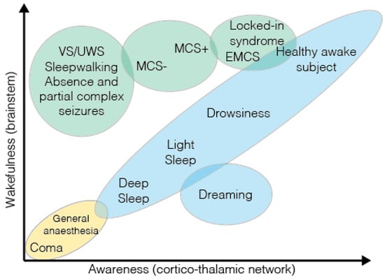
The new science called Sentiomics aims to identify the dynamic patterns that endow living systems with the capacity to feel and become conscious. One of the most promising fields of investigation in Sentiomics is the development and ‘education’ of human brain organoids to
[...] Read more.
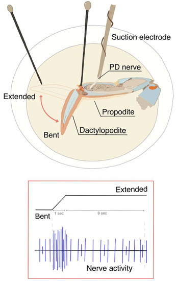
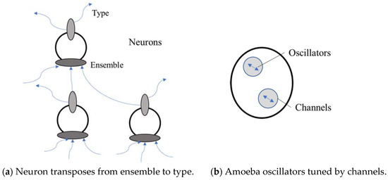
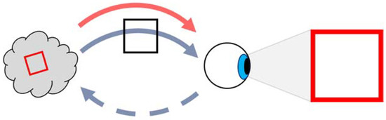
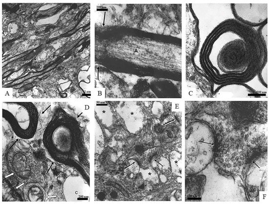
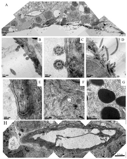
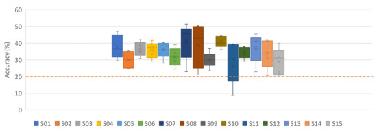
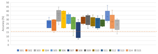
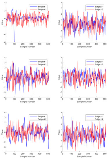
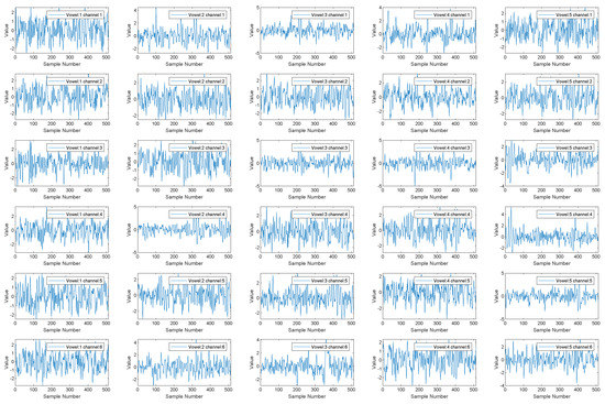
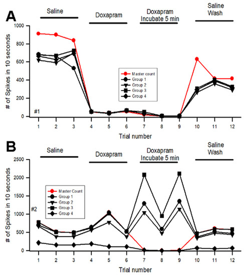
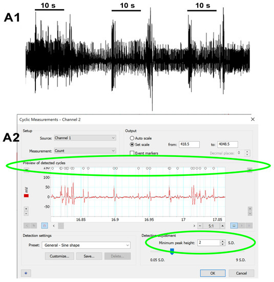
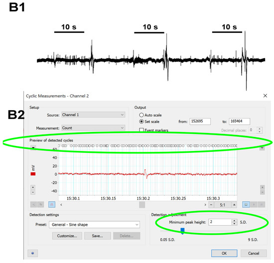
The new science called Sentiomics aims to identify the dynamic patterns that endow living systems with the capacity to feel and become conscious. One of the most promising fields of investigation in Sentiomics is the development and ‘education’ of human brain organoids to become sentient and useful for the promotion of human health in the (also new) field of Regenerative Neuromedicine. Here, we discuss the type of informational-rich input necessary to make a brain organoid sentient in experimental settings. Combining this research with the ecological preoccupation of preserving ways of sentience in the Amazon Rainforest, we also envisage the development of a new generation of biosensors to capture dynamic patterns from the forest, and use them in the ‘education’ of brain organoids to afford them a ‘mental health’ quality that is likely to be important in future advances in ‘post-humanist’ procedures in regenerative medicine. This study is closely related to the psychophysical approach to human mental health therapy, in which we have proposed the use of dynamic patterns in electric and magnetic brain stimulation protocols, addressing electrochemical waves in neuro-astroglial networks.
Full article
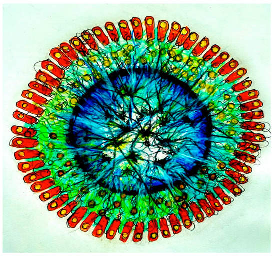
Figure 1
