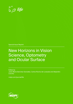
New Horizons in Vision Science, Optometry and Ocular Surface

A special issue of Life (ISSN 2075-1729). This special issue belongs to the section "Physiology and Pathology".

Deadline for manuscript submissions: closed (31 July 2023) | Viewed by 20893

Printed Edition Available!
A printed edition of this Special Issue is available here.

Special Issue Editors


E-Mail Website
Guest Editor
Department of Physics of Condensed Matter, Optics Area, University of Seville, 41012 Seville, Spain
Interests: optometry; vision science; contact lens; binocular vision
Special Issues, Collections and Topics in MDPI journals

E-Mail Website
Guest Editor
1. Qvision, Opththalmology Department, VITHAS Almería Hospital, 04120 Almería, Spain
2. Ophthalmology Department, VITHAS Málaga, 29016 Málaga, Spain
3. Hospital Regional, Universitario de Málaga, Plaza del Hospital Civil, S/N. 29009 Malaga, Spain
4. Departamento de Cirugía, Universidad de Sevilla, Área de Oftalmología, Doctor Fedriani, S/N, 41009 Seville, Spain
Interests: anterior segment surgery; refractive surgery; presbyopia and cataracts
Special Issues, Collections and Topics in MDPI journals

E-Mail Website
Guest Editor
Department of Optics & Optometry & Vision Sciences, University of Valencia, 46010 València, Spain
Interests: physiological optics; ocular accommodation; ocular autofluorescence; ocular surface; refractive correction
Special Issues, Collections and Topics in MDPI journals

Special Issue Information

Dear Colleagues,

Currently research in optometry and ocular surface covers fields ranging from binocular vision, accommodation, and ocular surface to contactology or refractive surgery. Ophthalmologists and optometrists need the latest and most up-to-date information available to improve their processes in daily clinical practice. However, on some occasions, daily care work does not allow time to review the latest developments in clinical optometry and ocular surface. Therefore, this Special Issue aims to share with professionals the latest news and new horizons in this discipline.

Research is the present of our profession, and it must also be the future. This will enable us to achieve a more optimized clinical practice, as research is the result of our knowledge power and a source of new inspiration and progress.

This Special Issue invites original research and review articles on recent achievements in advanced and clinical optometry and ocular innovative studies.

Dr. José-María Sánchez-González
Dr. Carlos Rocha-de-Lossada
Dr. Alejandro Cerviño
Guest Editors

Manuscript Submission Information

Manuscripts should be submitted online at www.mdpi.com by registering and logging in to this website. Once you are registered, click here to go to the submission form. Manuscripts can be submitted until the deadline. All submissions that pass pre-check are peer-reviewed. Accepted papers will be published continuously in the journal (as soon as accepted) and will be listed together on the special issue website. Research articles, review articles as well as short communications are invited. For planned papers, a title and short abstract (about 100 words) can be sent to the Editorial Office for announcement on this website.

Submitted manuscripts should not have been published previously, nor be under consideration for publication elsewhere (except conference proceedings papers). All manuscripts are thoroughly refereed through a single-blind peer-review process. A guide for authors and other relevant information for submission of manuscripts is available on the Instructions for Authors page. Life is an international peer-reviewed open access monthly journal published by MDPI.

Please visit the Instructions for Authors page before submitting a manuscript. The Article Processing Charge (APC) for publication in this open access journal is 2600 CHF (Swiss Francs). Submitted papers should be well formatted and use good English. Authors may use MDPI's English editing service prior to publication or during author revisions.

Keywords

  • clinical optometry
  • binocular vision
  • ocular surface
  • refractive surgery
  • contactology and dry eye disease
Related Special Issue

Published Papers (13 papers)

Order results
Result details
Select all
Export citation of selected articles as:

Editorial

Jump to: Research, Review

5 pages, 244 KiB  
Editorial
Exploring the Cutting Edge of Vision Science: New Developments in Diagnostics and Treatment of Ocular Surface in Dry Eye Disease
by José-María Sánchez-González, Carlos Rocha-de-Lossada and Alejandro Cerviño
Life 2023, 13(7), 1584; https://doi.org/10.3390/life13071584 - 19 Jul 2023
Viewed by 1103
Abstract
The ocular surface refers to the outermost layer of the eye, which includes the cornea, conjunctiva and eyelids [...] Full article
(This article belongs to the Special Issue New Horizons in Vision Science, Optometry and Ocular Surface)

Research

Jump to: Editorial, Review

15 pages, 5331 KiB  
Article
Visual Quality, Motility Behavior, and Retinal Changes Associated with Reading Tasks Performed on Electronic Devices
by Elvira Orduna-Hospital, María Munarriz-Escribano and Ana Sanchez-Cano
Life 2023, 13(8), 1777; https://doi.org/10.3390/life13081777 - 20 Aug 2023
Cited by 1 | Viewed by 959
Abstract
Background: The purpose of this study was to objectively evaluate visual discomfort using an eye tracker and aberrometer after a 21-min reading session on an iPad and an Ebook. Additionally, retinal changes were analyzed using optical coherence tomography (OCT). Methods: A total of [...] Read more.
Background: The purpose of this study was to objectively evaluate visual discomfort using an eye tracker and aberrometer after a 21-min reading session on an iPad and an Ebook. Additionally, retinal changes were analyzed using optical coherence tomography (OCT). Methods: A total of 31 young subjects (24 ± 4 years) participated in this study. They read for 21 min on an Ebook and for another 21 min on an iPad under controlled lighting conditions while their eye movements were monitored using an eye tracker. Aberrometry and retinal OCT measurements were taken before and after each reading session. Parameters such as pupil diameter, fixations, saccades, blinks, total aberration, high-order aberration, low-order aberration, and central and peripheral retinal thickness in the nine early treatment diabetic retinopathy study (ETDRS) areas were measured for each reading situation. Statistical analysis was performed on the collected data. Results: No statistically significant differences (p > 0.05) between the two devices were observed in terms of the different types of eye movements or the changes in retinal thickness. However, the aberrometric analysis showed variations in post-reading situations depending on the device used. Conclusion: Reading speed and visual discomfort resulting from electronic device usage can be objectively assessed using an eye tracker and aberrometer. Additionally, changes found in central and peripheral retinal thickness between the two devices and the baseline measurements were not significant and remained relatively stable. Full article
(This article belongs to the Special Issue New Horizons in Vision Science, Optometry and Ocular Surface)
Show Figures

Figure 1

16 pages, 1890 KiB  
Article
Assessment of Visual Quality Improvement as a Result of Spectacle Personalization
by Fruzsina Benyó, Lilla István, Huba Kiss, Andrea Gyenes, Gábor Erdei, Éva Juhász, Natalia Vlasak, Claudia Unger, Tamás Andorfi, Kata Réz, Illés Kovács and Zoltán Zsolt Nagy
Life 2023, 13(8), 1707; https://doi.org/10.3390/life13081707 - 08 Aug 2023
Viewed by 1149
Abstract
Personalized spectacles customized according to an individual’s facial anatomy were developed to provide enhanced visual performance and overall comfort when compared to standard spectacles. In this comparative crossover trial, each subject was randomly assigned to wear either personalized spectacles or standard spectacles for [...] Read more.
Personalized spectacles customized according to an individual’s facial anatomy were developed to provide enhanced visual performance and overall comfort when compared to standard spectacles. In this comparative crossover trial, each subject was randomly assigned to wear either personalized spectacles or standard spectacles for two weeks and then tried the second pair for another two weeks. Visual acuity and reading speed were measured, and visual quality and comfort were assessed using specific questionnaires. The correlation of the wearing parameters with the subjects’ satisfaction was calculated. According to our results, the subjects wearing personalized glasses reported significantly less experience of swaying and significantly higher overall satisfaction compared to those wearing the control spectacles. At the end of the study, 62% of subjects preferred the personalized spectacles, and visual quality was the primary reason for their spectacle preference followed by wearing comfort. The difference from the ideal cornea–vertex distance was significantly lower when wearing the personalized spectacles compared to the control frames. In addition, the absolute value of the difference from the ideal cornea–vertex distance was significantly correlated with patient satisfaction. These results suggest that personalized spectacles, customized according to an individual’s facial anatomy for the ideal wearing parameters, result in both visual and comfort advantages for wearers. Full article
(This article belongs to the Special Issue New Horizons in Vision Science, Optometry and Ocular Surface)
Show Figures

Figure 1

10 pages, 1249 KiB  
Article
The Relationship between Fixation Stability and Retinal Structural Parameters in Children with Anisometropic, Strabismic and Mixed Amblyopia
by Raquel Mompart-Martínez, Marc Argilés, Genis Cardona, Lluís Cavero-Roig, Lluís González-Sanchís and Maria Soledad Pighin
Life 2023, 13(7), 1517; https://doi.org/10.3390/life13071517 - 06 Jul 2023
Viewed by 1271
Abstract
(1) Background: Amblyopia is an ocular condition leading to structural and functional changes. The relationship between these changes is complex and remains poorly understood. (2) Methods: Participants included 31 children aged 5 to 9 years with strabismic (n = 9), anisometropic ( [...] Read more.
(1) Background: Amblyopia is an ocular condition leading to structural and functional changes. The relationship between these changes is complex and remains poorly understood. (2) Methods: Participants included 31 children aged 5 to 9 years with strabismic (n = 9), anisometropic (n = 16) and mixed (n = 6) unilateral amblyopia, and 14 age-matched non-amblyopic children. The 95% and 63% Bivariate Contour Ellipse Area (BCEA), axial length, Foveal Avascular Zone (FAZ) area, center macular thickness and volume were assessed. The relationship between these parameters was explored. (3) Results: Statistically significant differences were found among the four groups in best corrected distance visual acuity (BCVA) (p < 0.001), BCEA 95% (p = 0.002) and BCEA 63% (p = 0.002), but not in the FAZ area, central macular thickness, central macular volume and axial length. Eyes with amblyopia had poorer BCVA and larger fixation instability than controls. Inter-ocular differences were more significant in patients with strabismic amblyopia, particularly in BCVA (p = 0.003), central macular thickness (p < 0.001) and central macular volume (p = 0.002). In amblyopic eyes, BCEA 95% and 63% were correlated with BCVA, but not with the FAZ area. (4) Conclusion: Amblyopia is associated with a reduction in fixation stability and BCVA, although there is a general lack of correlation with structural changes, suggesting a complex interaction between anatomy and function in amblyopia. Full article
(This article belongs to the Special Issue New Horizons in Vision Science, Optometry and Ocular Surface)
Show Figures

Figure 1

10 pages, 595 KiB  
Article
Effectiveness of SMILE Combined with Micro-Monovision in Presbyopic Patients: A Pilot Study
by Joaquín Fernández, Federico Alonso-Aliste, Noemí Burguera, Julia Hernández-Lucena, Jonatan Amián-Cordero and Manuel Rodríguez-Vallejo
Life 2023, 13(3), 838; https://doi.org/10.3390/life13030838 - 20 Mar 2023
Cited by 2 | Viewed by 1609
Abstract
Binocular summation along all defocus range after a micro-monovision procedure has scarcely been studied. The aim of this pilot study was to evaluate the efficacy of SMILE combined with different levels of micro-monovision in presbyopic patients and to assess the binocular summation effect [...] Read more.
Binocular summation along all defocus range after a micro-monovision procedure has scarcely been studied. The aim of this pilot study was to evaluate the efficacy of SMILE combined with different levels of micro-monovision in presbyopic patients and to assess the binocular summation effect on contrast sensitivity defocus curves (CSDC) at the 6-month follow-up. Efficacy was assessed on the basis of visual acuity (VA) and stereopsis at far, intermediate, and near distances. Patient-reported outcomes (PROs) and binocular CSDC were also evaluated. Six patients completed the study with a programmed median anisometropia of 0.81 Diopter. The median binocular uncorrected VA was better than 0 logMAR at the three evaluated distances, and stereopsis was not impaired in any patient, achieving a median of ≤119 arcsec at any distance. CSDC increased binocularly after surgery, significantly in the range of −2 to −3 D (p < 0.05). No clinically relevant changes were observed in PROs compared with the preoperative period, and all patients achieved spectacle independence at intermediate/near distance and were likely or very likely to undergo the same surgery. In conclusion, micro-monovision with SMILE could be an effective procedure, with results that might be comparable to other laser correction techniques specifically designed for presbyopia correction. Full article
(This article belongs to the Special Issue New Horizons in Vision Science, Optometry and Ocular Surface)
Show Figures

Figure 1

12 pages, 1501 KiB  
Article
Key Factors in Early Diagnosis of Myopia Progression within Ocular Biometric Parameters by Scheimpflug Technology
by Alfredo López-Muñoz, Beatriz Gargallo-Martínez, María Carmen Sánchez-González, Raúl Capote-Puente, Concepción De-Hita-Cantalejo, Marta Romero-Luna, Juan-José Conejero-Domínguez and José-María Sánchez-González
Life 2023, 13(2), 447; https://doi.org/10.3390/life13020447 - 04 Feb 2023
Viewed by 1280
Abstract
The aim of this study was to evaluate the relationship between myopia and ocular biometric variables using the Pentacam AXL® single rotation Scheimpflug camera. This prospective, cross-sectional, single-center study was performed in fifty Caucasian patients aged between 18 and 30 years (24.84 [...] Read more.
The aim of this study was to evaluate the relationship between myopia and ocular biometric variables using the Pentacam AXL® single rotation Scheimpflug camera. This prospective, cross-sectional, single-center study was performed in fifty Caucasian patients aged between 18 and 30 years (24.84 ± 3.04 years). The measured variables included maximum and minimum keratometry (K1 and K2, respectively), anterior chamber depth (ACD), corneal horizontal diameter or white to white (WTW), central corneal thickness (CCT), corneal asphericity (Q), and axial length (AXL). The tomographic and biometric measurements were considered optimal when the quality factor was greater than 95% according to the manufacturer’s software instructions. The AXL presented a significant correlation with the spherical equivalent without cycloplegia (SE without CP), age at onset of myopia (r = −0.365, p = 0.012), mean keratometry (Km) (r = −0.339, p = 0.016), ACD (r = 0.304, p = 0.032), and WTW (r = 0.406, p = 0.005). The eyes with AXL higher than 25 mm had earlier onset; higher SE without CP, AXL, and Q; and a flatter Km. AXL is the biometric variable with the greatest influence on the final refractive state in the adult myopic eye. Ophthalmologists and optometric management must consider these biometric differences in order to identify the most appropriate correction techniques in each case. The use of the Pentacam AXL in ocular biometric measurement is effective, reproducible, and non-invasive. Full article
(This article belongs to the Special Issue New Horizons in Vision Science, Optometry and Ocular Surface)
Show Figures

Figure 1

11 pages, 1171 KiB  
Article
Accommodation Response Variations in University Students under High Demand for Near-Vision Activity
by Concepción De-Hita-Cantalejo, María-de-los-Ángeles Benítez-Rodríguez, María Carmen Sánchez-González, María-José Bautista-Llamas and José-María Sánchez-González
Life 2022, 12(11), 1837; https://doi.org/10.3390/life12111837 - 09 Nov 2022
Cited by 1 | Viewed by 1260
Abstract
The objective of this study was to investigate accommodation changes and visual discomfort in a university student population after a period of high demand for near-vision activity. A total of 50 university students aged between 20 and 22 years were recruited. The tests [...] Read more.
The objective of this study was to investigate accommodation changes and visual discomfort in a university student population after a period of high demand for near-vision activity. A total of 50 university students aged between 20 and 22 years were recruited. The tests performed involved positive relative accommodation (PRA), negative relative accommodation (NRA), accommodation amplitude (AA), and monocular and binocular accommodative facility (MAF and BAF). Visual discomfort was measured on a scale involving a visual discomfort questionnaire (VDQ). All accommodative variables underwent changes during the exam period; specifically, regarding NRA and PRA, 30.4% and 15.1% of the studied population, respectively, appeared to be below average. Moreover, 42.3% of the population exhibited values below average in the second measure of AA. On the other hand, a small percentage of the population was below average in MAF and BAF measurements: 3% in the monocular right eye test, 6% in the left eye test, and 9.1% in the binocular facility test. Finally, the VDQ score did not reveal a statistically significant difference between the two measurements. Prolonged near-distance work, such as a university exams period, changed all accommodation systems (amplitude of accommodation, relative accommodation, and accommodation facility). These changes influence an accommodation excess that results in blurred vision, headache, and problems with focusing. Full article
(This article belongs to the Special Issue New Horizons in Vision Science, Optometry and Ocular Surface)
Show Figures

Figure 1

10 pages, 1017 KiB  
Article
Pre-Lens Tear Meniscus Height, Lipid Layer Pattern and Non-Invasive Break-Up Time Short-Term Changes with a Water Gradient Silicone Hydrogel Contact Lens
by Raúl Capote-Puente, María-José Bautista-Llamas, Caterina Manzoni and José-María Sánchez-González
Life 2022, 12(11), 1710; https://doi.org/10.3390/life12111710 - 26 Oct 2022
Cited by 4 | Viewed by 1739
Abstract
To evaluate pre-lens tear film volume, stability and lipid interferometry patterns with a silicone hydrogel water content contact lens, a novel, noninvasive, ocular-surface-analyzer technology was used. A prospective, longitudinal, single-center, self-control study was performed in daily or monthly replacement silicone hydrogel contact lens [...] Read more.
To evaluate pre-lens tear film volume, stability and lipid interferometry patterns with a silicone hydrogel water content contact lens, a novel, noninvasive, ocular-surface-analyzer technology was used. A prospective, longitudinal, single-center, self-control study was performed in daily or monthly replacement silicone hydrogel contact lens wearers. A tear film analysis was achieved with the Integrated Clinical Platform (ICP) Ocular Surface Analyzer (OSA) from SBM System. The subjects were reassessed, with the contact lens, after 30 min of wearing to quantify the volume, stability and lipid pattern of the short-term pre-lens tear film. Lipid layer thickness decreased from 2.05 ± 1.53 to 1.90 ± 1.73 Guillon patterns (p = 0.23). First pre-lens NIBUT decreased from 5.03 ± 1.04 to 4.63 ± 0.89 s (p = 0.01). Mean pre-lens NIBUT significantly increased from 15.19 ± 9.54 to 21.27 ± 11.97 s (p < 0.01). Lid opening time significantly increased from 26.36 ± 19.72 to 38.58 ± 21.78 s (p < 0.01). The silicone hydrogel contact lens with water gradient technology significantly increased the mean pre-lens NIBUT and lid opening time. Lehfilcon A suggested an improvement in contact lens wearers with tear film instability or decreased subjective symptoms of dry eye disease. Full article
(This article belongs to the Special Issue New Horizons in Vision Science, Optometry and Ocular Surface)
Show Figures

Figure 1

9 pages, 2078 KiB  
Article
Prevalence of Dry Eyes Symptoms in Association with Contact Lenses and Refractive Status in Portugal
by Miguel Ángel Sánchez-Tena, Clara Martinez-Perez, Cristina Alvarez-Peregrina and Núcleo de Investigação Aplicada em Ótica e Optometria
Life 2022, 12(10), 1656; https://doi.org/10.3390/life12101656 - 20 Oct 2022
Viewed by 1790
Abstract
Background: Determine whether the presence of ocular symptoms in soft-contact-lens wearers changes depending on the refractive status. Methods: During the months of January to March 2022, the CLDEQ-8 questionnaire was administered to soft-contact-lens wearers. The statistical analysis was carried out using the SPSS [...] Read more.
Background: Determine whether the presence of ocular symptoms in soft-contact-lens wearers changes depending on the refractive status. Methods: During the months of January to March 2022, the CLDEQ-8 questionnaire was administered to soft-contact-lens wearers. The statistical analysis was carried out using the SPSS 27.0 computer program (SPSS Inc., Chicago, IL, USA). Results: A total of 251 subjects participated in the study, with a higher percentage of myopes than hyperopes (82.1% versus 16.7%; p < 0.001). Out of all total participants, 21.5% suffered from dry-eye symptoms. It was noted that hyperopes presented a higher rate of dry-eye symptoms (p = 0.041). At the same time, the spherical equivalent was more positive in the participants with dry-eye symptoms (p = 0.014). Significant differences were found based on the symptoms present with contact lenses and the degree of myopia. The intensity of visual disturbances was higher in the participants with medium myopia (median [IQR]: 1/5 [2]) compared to those with low (median [IQR]: 0/5 [2]) and high myopia (median [IQR]: 0/5 [1]) (p = 0.009). Conclusions: Contact-lens wearers with hyperopia showed a higher rate of ocular dryness than those with myopia. In turn, wearing daily-replacement lenses could be one of the reasons for the lesser presence of ocular dryness compared to monthly-replacement lenses. Full article
(This article belongs to the Special Issue New Horizons in Vision Science, Optometry and Ocular Surface)
Show Figures

Figure 1

Review

Jump to: Editorial, Research

12 pages, 390 KiB  
Review
Promising High-Tech Devices in Dry Eye Disease Diagnosis
by Andrea De Luca, Alessandro Ferraro, Chiara De Gregorio, Mariateresa Laborante, Marco Coassin, Roberto Sgrulletta and Antonio Di Zazzo
Life 2023, 13(7), 1425; https://doi.org/10.3390/life13071425 - 21 Jun 2023
Cited by 1 | Viewed by 1678
Abstract
Background: Dry eye disease (DED) is a common and debilitating condition that affects millions of people worldwide. Despite its prevalence, the diagnosis and management of DED can be challenging, as the condition is multifactorial and symptoms can be nonspecific. In recent years, there [...] Read more.
Background: Dry eye disease (DED) is a common and debilitating condition that affects millions of people worldwide. Despite its prevalence, the diagnosis and management of DED can be challenging, as the condition is multifactorial and symptoms can be nonspecific. In recent years, there have been significant advancements in diagnostic technology for DED, including the development of several new devices. Methods: A literature review of articles on the dry eye syndrome and innovative diagnostic devices was carried out to provide an overview of some of the current high-tech diagnostic tools for DED, specifically focusing on the TearLab Osmolarity System, DEvice Hygrometer, IDRA, Tearcheck, Keratograph 5M, Cornea Dome Lens Imaging System, I-PEN Osmolarity System, LipiView II interferometer, LacryDiag Ocular Surface Analyzer, Tearscope-Plus, and Cobra HD Camera. Conclusions: Despite the fact that consistent use of these tools in clinical settings could facilitate diagnosis, no diagnostic device can replace the TFOS algorithm. Full article
(This article belongs to the Special Issue New Horizons in Vision Science, Optometry and Ocular Surface)
Show Figures

Figure 1

18 pages, 1932 KiB  
Review
Dynamic Aspects of Pre-Soft Contact Lens Tear Film and Their Relation to Dry Eye: Basic Science and Clinical Relevance
by Norihiko Yokoi, Petar Eftimov and Georgi As. Georgiev
Life 2023, 13(4), 859; https://doi.org/10.3390/life13040859 - 23 Mar 2023
Cited by 5 | Viewed by 2109
Abstract
Soft contact lens (SCL) perturbs the intimate connection between the pre-lens tear film (PLTF) and the ocular surface in various ways, i.e., (i) decrease in tear meniscus radius and aqueous tear thickness, (ii) attenuation of tear film lipid layer spread, (iii) limited wettability [...] Read more.
Soft contact lens (SCL) perturbs the intimate connection between the pre-lens tear film (PLTF) and the ocular surface in various ways, i.e., (i) decrease in tear meniscus radius and aqueous tear thickness, (ii) attenuation of tear film lipid layer spread, (iii) limited wettability of SCL surface, (iv) increased friction with eyelid wiper, etc. This often results in SCL-related dry eye (SCLRDE) manifested as PLTF instability and contact lens discomfort (CLD). In this review, the individual contributions of factors (i–iv) to PLTF breakup patterns (BUP) and CLD are considered via the tear film-oriented diagnosis framework adopted by the Asia Dry Eye Society from a clinical and basic science perspective. It is shown that SCLRDE (due to aqueous deficiency, increased evaporation, or decreased wettability) and BUP of PLTF classify within the same types as the ones observed for the precorneal tear film. The analysis of PLTF dynamics reveals that the inclusion of SCL enhances the manifestation of BUP associated with (i) decreased thickness of PLTF aqueous layer and (ii) limited SCL wettability as shown by the rapid expansion of BUP area. PLTF thinness and instability result in increased blink-related friction and lid wiper epitheliopathy as major contributor to CLD. Full article
(This article belongs to the Special Issue New Horizons in Vision Science, Optometry and Ocular Surface)
Show Figures

Figure 1

12 pages, 19178 KiB  
Review
Complications of Small Aperture Intracorneal Inlays: A Literature Review
by María Carmen Sánchez-González, Estanislao Gutiérrez-Sánchez, José-María Sánchez-González, Concepción De-Hita-Cantalejo, Ana-María Pinero-Rodríguez, Timoteo González-Cruces and Raúl Capote-Puente
Life 2023, 13(2), 312; https://doi.org/10.3390/life13020312 - 22 Jan 2023
Viewed by 1385
Abstract
Presbyopia can be defined as the refractive state of the eye in which, due to a physiological decrease in the ability to accommodate, it is not possible to sustain vision without fatigue in a prolonged manner, along with difficulty focusing near vision. It [...] Read more.
Presbyopia can be defined as the refractive state of the eye in which, due to a physiological decrease in the ability to accommodate, it is not possible to sustain vision without fatigue in a prolonged manner, along with difficulty focusing near vision. It is estimated that its prevalence in 2030 will be approximately 2.1 billion people. Corneal inlays are an alternative in the correction of presbyopia. They are implanted beneath a laser-assisted in situ keratomileusis (LASIK) flap or in a pocket in the center of the cornea of the non-dominant eye. The purpose of this review is to provide information about intraoperative and postoperative KAMRA inlay complications in the available scientific literature. A search was conducted on PubMed, Web of Science, and Scopus with the following search strategy: ("KAMRA inlay" OR “KAMRA” OR “corneal inlay pinhole” OR “pinhole effect intracorneal” OR “SAICI” OR “small aperture intracorneal inlay”) AND (“complication” OR “explantation” OR “explanted” OR “retired”). The bibliography consulted shows that the insertion of a KAMRA inlay is an effective procedure that improves near vision with a slight decrease in distance vision. However, postoperative complications such as corneal fibrosis, epithelial iron deposits, and stromal haze are described. Full article
(This article belongs to the Special Issue New Horizons in Vision Science, Optometry and Ocular Surface)
Show Figures

Figure 1

13 pages, 7129 KiB  
Review
Ophthalmological Approach for the Diagnosis of Dry Eye Disease in Patients with Sjögren’s Syndrome
by Robinson T. Barrientos, Fernando Godín, Carlos Rocha-De-Lossada, Matias Soifer, José-María Sánchez-González, Esteban Moreno-Toral, Ana-Luisa González, Mike Zein, Pablo Larco, Jr., Carolina Mercado and Maria-Adelaida Piedrahita
Life 2022, 12(11), 1899; https://doi.org/10.3390/life12111899 - 15 Nov 2022
Cited by 8 | Viewed by 2228
Abstract
Dry eye has two basic subdivisions: aqueous deficient dry eye (ADDE), with SS a major cause; and evaporative dry eye (EDE), due to either intrinsic or extrinsic factors. SS is a chronic inflammatory disorder defined by dysfunction of the exocrine glands leading to [...] Read more.
Dry eye has two basic subdivisions: aqueous deficient dry eye (ADDE), with SS a major cause; and evaporative dry eye (EDE), due to either intrinsic or extrinsic factors. SS is a chronic inflammatory disorder defined by dysfunction of the exocrine glands leading to dry eye and dry mouth. The objective of this article was to carry out a systematic and critical review of several scientific publications on dry eye disease, with the aim of providing general recommendations to distinguish dry eye and its different variants in patients with SS, during the period 1979 to 2020, using search engines for articles indexed in Scopus, Latindex, Scielo, Clinical Trials, Medline, Embase, and Cochrane, allowing the analysis of 132 articles published in indexed journals on the subject of dry eye disease and SS, evidencing its conceptualization, prevalence, risk factors, etiopathogenesis, clinical manifestations, diagnosis, and treatment. Full article
(This article belongs to the Special Issue New Horizons in Vision Science, Optometry and Ocular Surface)
Show Figures

Figure 1

Back to TopTop