Dentistry, Oral Health and Maxillofacial Surgery
A topical collection in Healthcare (ISSN 2227-9032). This collection belongs to the section "Chronic Care".
Viewed by 70245
Share This Topical Collection
Editor
 Dr. Saturnino Marco Lupi
Dr. Saturnino Marco Lupi
E-Mail
Website
Collection Editor
Department of Clinico-Surgical, Diagnostic and Pediatric Sciences, Dental Clinic, University of Pavia, P.le Golgi 2, 27100 Pavia, Italy
Interests: dentistry; oral surgery; maxillofacial surgery; tissue regeneration; bone regeneration; dental implants; implant surface treatments; dental radiology; oral pathology; tooth anatomy; community dentistry; caries prevention; oral cancer; dental prosthesis; gnathology; dental material; laser
Special Issues, Collections and Topics in MDPI journals
Topical Collection Information
Dear Colleagues,
In recent years, numerous innovations have had an impact on dentistry and maxillofacial surgery. Treatments require a multidisciplinary approach, and greater attention must be placed on pre- and post-operative patient management; specific attention must be paid to the fragile patient; to digital procedures, such as intraoral and extraoral scanners or 3D printing, which modify the diagnostic and therapeutic approach, making treatments increasingly personalized; to the prevention of pathologies affecting the head and neck area, as well as the possibility of early diagnosis; to the impact of the work on caregivers or medical students; and most recently, to the impact of the COVID-19 pandemic on the medical and dental field.
The Topical Collection on “Dentistry, Oral Health and Maxillofacial Surgery” invites worldwide investigators as well as clinicians to submit their most interesting overviews, reviews, and original articles that may provide novel insights regarding the topics indicated. Original research articles, systematic reviews, meta-analyses, case series, and case reports are welcome if they provide new sound scientific evidence in these fields.
Dr. Saturnino Marco Lupi
Collection Editor
Manuscript Submission Information
Manuscripts should be submitted online at www.mdpi.com by registering and logging in to this website. Once you are registered, click here to go to the submission form. Manuscripts can be submitted until the deadline. All submissions that pass pre-check are peer-reviewed. Accepted papers will be published continuously in the journal (as soon as accepted) and will be listed together on the collection website. Research articles, review articles as well as short communications are invited. For planned papers, a title and short abstract (about 100 words) can be sent to the Editorial Office for announcement on this website.
Submitted manuscripts should not have been published previously, nor be under consideration for publication elsewhere (except conference proceedings papers). All manuscripts are thoroughly refereed through a single-blind peer-review process. A guide for authors and other relevant information for submission of manuscripts is available on the Instructions for Authors page. Healthcare is an international peer-reviewed open access semimonthly journal published by MDPI.
Please visit the Instructions for Authors page before submitting a manuscript.
The Article Processing Charge (APC) for publication in this open access journal is 2700 CHF (Swiss Francs).
Submitted papers should be well formatted and use good English. Authors may use MDPI's
English editing service prior to publication or during author revisions.
Keywords
- dentistry
- maxillofacial surgery
- dental materials
- restorative dentistry
- prosthodontics
- oral surgery
- implantology
- pediatric dentistry
- orthodontics
- gnathology
- CAD/CAM
- microbiome
- nanomaterials
- periodontics
- digital workflow
- fragile patient
- intraoral and extraoral scanner
- 3D printing
- perioperative care
- COVID-19
Published Papers (31 papers)
Open AccessCase Report
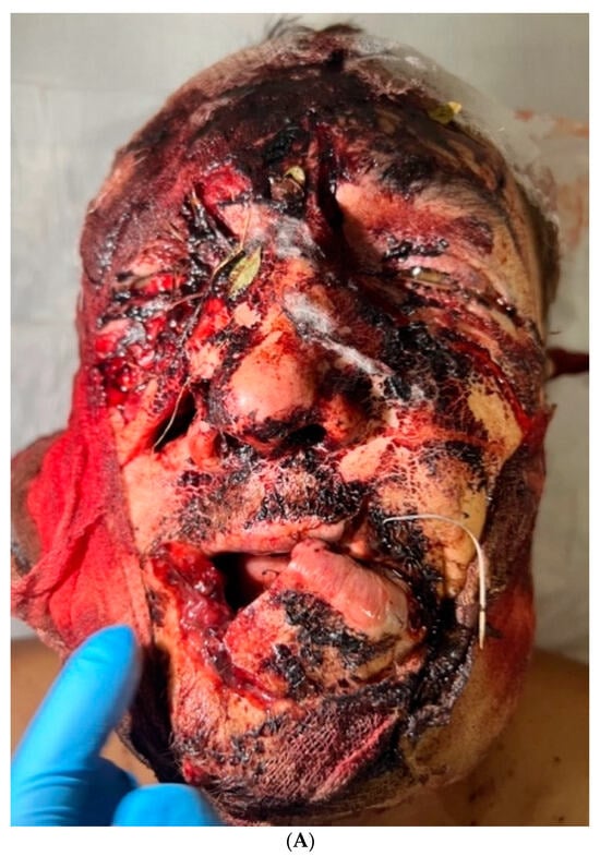
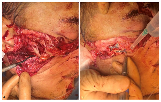
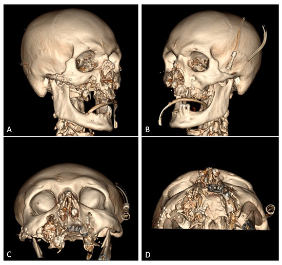
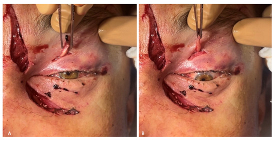
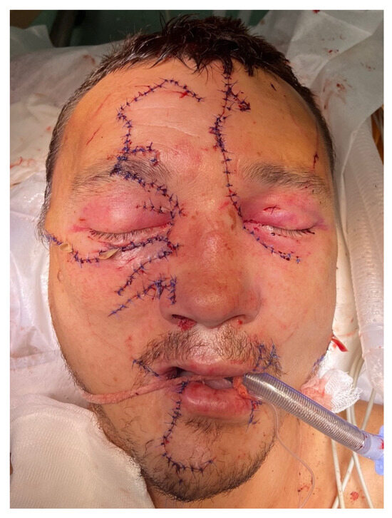
Polytrauma Caused by a Bear Attacking a Human with a Benign Outcome
by
Ruslan Mellin, Ellina Velichko, Larisa Maltseva, Sergey Dydykin and Yuriy Vasil’ev
Viewed by 2209
Abstract
Injuries to humans caused by wild animals, particularly bears, are rarely mentioned in the literature. Such injuries are frequent in Siberia, which is a territory surrounded by dense forests inhabited by brown bears. In the last 4 months alone (September–December 2023), four bear
[...] Read more.
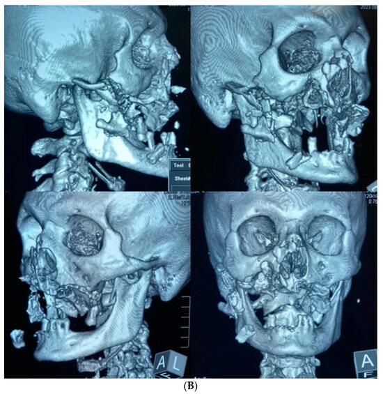
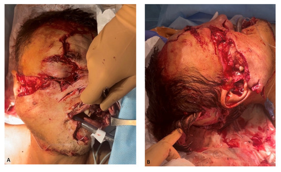
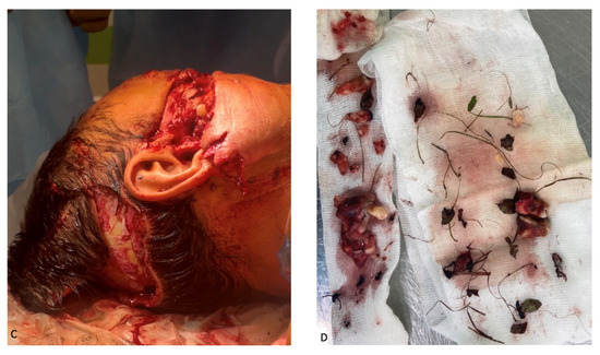
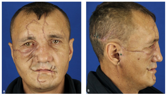
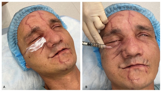
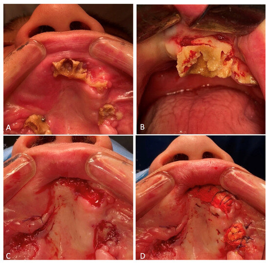
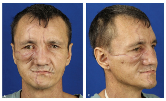
Injuries to humans caused by wild animals, particularly bears, are rarely mentioned in the literature. Such injuries are frequent in Siberia, which is a territory surrounded by dense forests inhabited by brown bears. In the last 4 months alone (September–December 2023), four bear attacks on humans were registered in Khakassia, Russia. This article presents a clinical case of rehabilitating a patient after a bear attack, who suffered multiple fragmentary fractures of the facial skeleton with displaced bone fragments, subcutaneous emphysema of the soft tissues of the face, damage to the parietal and right occipital regions and paranasal sinus hemorrhage on the left side. The nature of the injuries was enhanced by trauma to the upper extremity caused by the patient defending himself against the animal. In addition to the damage to his face, the bear tried to open his cranium, as evidenced by four furrows caused by its canines, including two each on the frontal and occipital bones of the skull. The patient’s complex treatment included both maxillofacial and reconstructive surgeries, and outpatient treatment involved the formation of normotrophic scars using a neodymium laser and injections of a heterogeneous composition consisting of microparticles of “crosslinked” collagen of animal origin placed in a gel identical to the natural extracellular matrix.
Full article
►▼
Show Figures
Open AccessArticle
Retrospective Evaluation of 20 Years of Outpatient Dental Services to Adults with Disabilities at the Dental Hospitals of the Medical University of Innsbruck, Austria
by
Dagmar Schnabl, Matthias Michael Strohm, Pit Eugene Schummer, Lukas Sigwart and Ines Kapferer-Seebacher
Viewed by 568
Abstract
Disabled persons’ chairside dentistry is challenging. We aimed for a retrospective breakdown of dental services delivered to disabled patients by dental students and to discuss feasibility of a chairside approach. Consecutive patients, who received scheduled dental treatment by dental students from 2002 to
[...] Read more.
Disabled persons’ chairside dentistry is challenging. We aimed for a retrospective breakdown of dental services delivered to disabled patients by dental students and to discuss feasibility of a chairside approach. Consecutive patients, who received scheduled dental treatment by dental students from 2002 to 2021, were included. Demographic data, medical diagnoses, number of treatment sessions, performed treatments, and treatment break-offs were collected and analyzed with descriptive statistics. In total, 224 individuals with various disabilities (mean age 36.4 ± 14.6 years) received dental services in 2282 sessions altogether (10.3 ± 11. sessions per patient). Professional tooth cleaning was the most frequently provided treatment (55.8% of sessions). A total of 654 teeth were restored with fillings, 97 teeth were extracted, 56 teeth had endodontic treatment, and 25 removable dentures were fitted. Treatment break-off due to incompliance and referral to dental general anesthesia occurred in 74 patients (33%). Chairside treatment of disabled persons by dental students is feasible in many cases. Our study may serve as an incentive for clinicians/researchers to report on treatment modalities and outcomes of chairside dentistry in patients with special oral health care needs, preferably by the use of prospective study designs, to contribute data and strategies in the fight for control of oral health inadequacies.
Full article
►▼
Show Figures
Open AccessSystematic Review
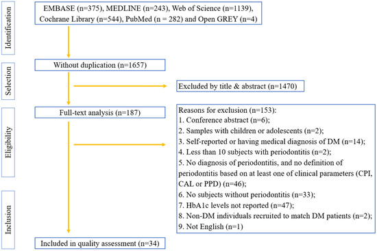
Association between Periodontitis and HbA1c Levels in Non-Diabetic Patients: A Systematic Review and Meta-Analysis
by
Dan Zhao, Yangyang Sun, Xin Li, Xiaoxiao Wang, Lijie Lu, Chen Li, Yaping Pan and Songlin Wang
Viewed by 1064
Abstract
Background: A high detection rate of diabetes among dental visitors has been reported recently. This systematic review aimed to evaluate the association between periodontitis and glycated hemoglobin (HbA1c) levels among non-diabetic individuals. Methods: The EMBASE, MEDLINE, Web of Science, Cochrane Library, PubMed, and
[...] Read more.
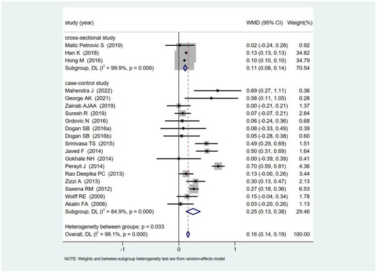
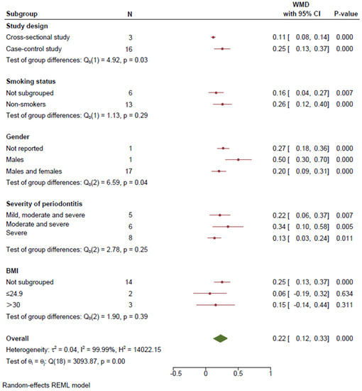
Background: A high detection rate of diabetes among dental visitors has been reported recently. This systematic review aimed to evaluate the association between periodontitis and glycated hemoglobin (HbA1c) levels among non-diabetic individuals. Methods: The EMBASE, MEDLINE, Web of Science, Cochrane Library, PubMed, and Open GREY databases were searched, and observational studies published until 1st June 2023 were identified. A methodological quality assessment was conducted based on the original and modified versions of the Newcastle–Ottawa scale. Cohort, case–control, and cross-sectional studies that performed clinical periodontal examinations and measured HbA1c levels in non-diabetic adults were included. A meta-analysis was conducted to estimate the weighted mean difference (WMD) between individuals with and without periodontitis. Results: In total, 29 case–control and 5 cross-sectional studies were selected from 2583 potentially eligible articles. Among them, sixteen case–control and three cross-sectional studies with moderate to high quality were selected for the meta-analyses. The HbA1c levels in periodontitis patients were significantly higher than those in individuals with healthy periodontal conditions (WMD = 0.16;
p < 0.001) among the non-diabetic populations. Conclusions: This study reveals a significant association between periodontitis and HbA1c levels in non-diabetic populations. Thus, HbA1c screening may be recommended to detect potential hyperglycemia in non-diabetic periodontitis patients.
Full article
►▼
Show Figures
Open AccessArticle
Lip and Oral Cavity Cancer Incidence and Mortality Rates Associated with Smoking and Chewing Tobacco Use and the Human Development Index in 172 Countries Worldwide: An Ecological Study 2019–2020
by
Antonio Hernández-Morales, Blanca Silvia González-López, Rogelio José Scougall-Vilchis, Josué Roberto Bermeo-Escalona, Ulises Velázquez-Enríquez, Rosalina Islas-Zarazúa, Sonia Márquez-Rodríguez, Taurino Amílcar Sosa-Velasco, Carlo Eduardo Medina-Solís and Gerardo Maupomé
Cited by 10 | Viewed by 2010
Abstract
Tobacco use is associated with diseases worldwide, including cancer. This is one of the major public health problems globally, causing more than 19 million new cases in 2020. Lip and oral cavity cancer (LOCC) is neoplastic growth in the tongue, gums, and lips.
[...] Read more.
Tobacco use is associated with diseases worldwide, including cancer. This is one of the major public health problems globally, causing more than 19 million new cases in 2020. Lip and oral cavity cancer (LOCC) is neoplastic growth in the tongue, gums, and lips. The objective of this ecological study was to quantify the strength of the association between incidence and mortality of LOCC, with tobacco use and with the Human Development Index (HDI). Incidence and mortality data on LOCC were obtained for 172 countries in 2020, from the Global Cancer Observatory (GLOBOCAN). The prevalence of tobacco smoking and chewing was obtained from reports conducted in 2019. The inequality in human development was estimated using the HDI from the United Nations Development Program, Human Development Report (2019). Statistically significant correlations were observed between the incidence of LOCC and tobacco smoking and chewing prevalence, except for negative correlations between the prevalence of tobacco smoking LOCC mortality in women, just as in the case of the HDI. No statistically significant differences were found between the prevalence of tobacco chewing only and the incidence of LOCC overall and by sex. A higher LOCC incidence overall and by sex was associated with higher HDI. In conclusion, the present study found positive correlations for various HDI socioeconomic indicators and tobacco use with the incidence and mortality of LOCC, but also a few inverse correlations.
Full article
Open AccessArticle
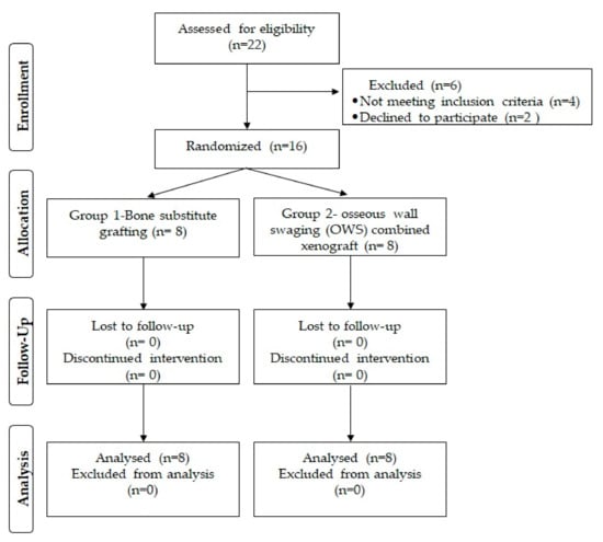
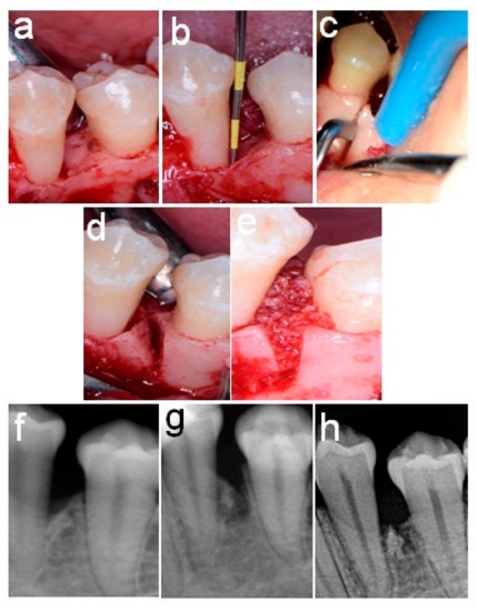
A Novel Approach of Periodontal Osseous Wall Piezosplitting and Sequential Bone Expansion in Management of Localized Intra-Bony Defects with Wide Angulation—A Randomized Controlled Trial
by
Mahmoud Taha El-Destawy, Mohamed Fekry Khedr, Mostafa Mohamed Hosny, Ahmed Mohamed Bilal, Ahmed Mohamed Elshamy, Ibrahim Sabry El sayed, Abd el-latif galal Borhamy, Abd al-aziz kamal Aboamo and Ahmed Yousef Gamal
Cited by 1 | Viewed by 2263
Abstract
Piezoelectric surgical instruments with various mini-sized tips and cutting technology offer a precise and thin cutting line that could allow the wider use of periodontal osseous wall swaging. This randomized controlled trial was designed to investigate the use of a minimally invasive piezo
[...] Read more.
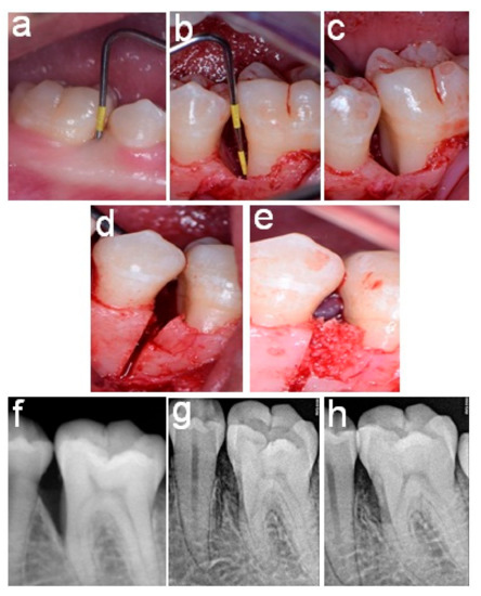
Piezoelectric surgical instruments with various mini-sized tips and cutting technology offer a precise and thin cutting line that could allow the wider use of periodontal osseous wall swaging. This randomized controlled trial was designed to investigate the use of a minimally invasive piezo knife to harvest vascularized interseptal bone pedicles in treating intra-bony defects. Sixteen non-smoking patients (mean age 39.6 ± 3.9) with severe chronic periodontitis were randomly assigned into one of two groups (N = 8). The Group 1 (control) patients were treated by bone substitute grafting of the intra-bony defect, whereas the Group 2 patients were treated by intra-bony defect osseous wall swaging (OWS) combined with xenograft filling of the space created by bone tilting. In both groups, the root surfaces were treated with a neutral 24% EDTA gel followed by saline irrigation. Clinical and radiographic measurements were obtained at baseline and 6 months after surgery. The sites treated with osseous wall swaging showed a statistically significant probing-depth reduction and increase in clinical attachment compared with those of the Group 1 patients. The defect base level was significantly reduced for the OWS group compared to that of the Group 1 control. By contrast, the crestal bone level was significantly higher in the OWS group compared to Group 1. The crestal interseptal bone width was significantly higher in Group 2 at 6 months compared to the baseline value and to that of Group 1 (<0.001). The osseous wall swaging effectively improved the clinical hard- and soft-tissue parameters. The use of mini inserts piezo-cutting, sequential bone expanders for osseous wall redirection, and root surface EDTA etching appears to be a reliable approach that could allow the use of OWS at any interproximal dimension.
Full article
►▼
Show Figures
Open AccessArticle
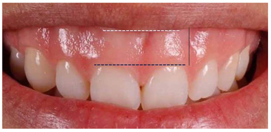
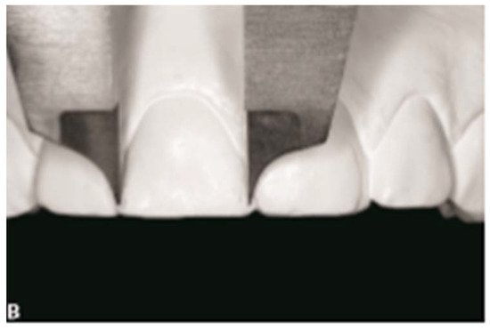
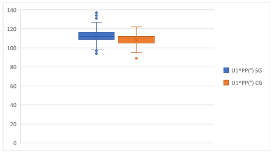
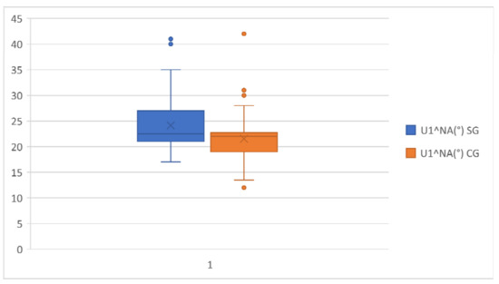
Is There a Correlation between Gingival Display and Incisal Inclination in a Gummy Smile? Study on Cephalometric Parameters
by
Alessandra Impellizzeri, Raissa Palmigiani, Martina Horodynski, Tiziana D’alfonso, Antonella Polimeni, Adriana De Stefano and Gabriella Galluccio
Viewed by 2482
Abstract
Background: Excessive gingival display or “gummy smile” is a clinical condition where a maxillary gum shows between the inferior line of the superior lip and the gingival line of the incisive superior during a spontaneous smile. The aim of this research was to
[...] Read more.
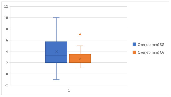
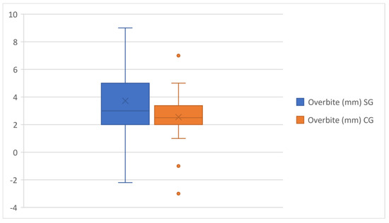
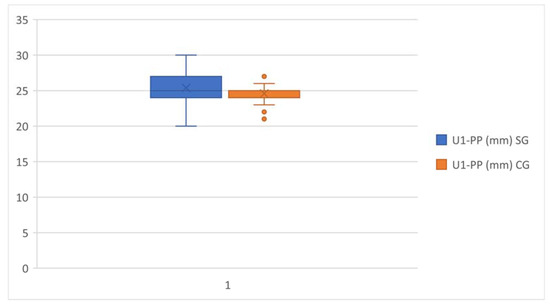
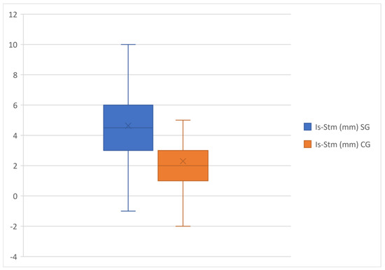
Background: Excessive gingival display or “gummy smile” is a clinical condition where a maxillary gum shows between the inferior line of the superior lip and the gingival line of the incisive superior during a spontaneous smile. The aim of this research was to understand the various skeletal and dentoalveolar components contributing to a gummy smile in a sample of 120 patients. Material and Methods: This retrospective case-control study had the primary objectives of analyzing the existence of a correlation between the presence of gingival exposure and the alteration of the inclination of the upper incisors with respect to the Frankfurt plane, the Palatine plane (bi-spinal) and to the NA line in a sample of orthodontic patients, and also evaluating the association with skeletal, dental, and aesthetic cephalometric parameters. Result and Conclusions: In our study, it’s emerged a correlation between the gingival exposure and the presence of alterations to incisal torque in the vestibular direction and the quantity of maxillary gingiva evident during the smile, which is correlated in particular to the Is–Sts distance, overjet and overbite. The major indicative data, therefore, are related to the vertical position of the upper incisors, in particular with respect to the upper lip and to the sagittal position.
Full article
►▼
Show Figures
Open AccessArticle
Comparative Analysis of Edentulism in a Sample of Mexican Adults with and without Type 2 Diabetes
by
Rosalina Islas-Zarazúa, Mariana Mora-Acosta, José de Jesús Navarrete-Hernández, Josefina Reynoso-Vázquez, Juan José Villalobos-Rodelo, Laura Rojas-Ortega, Taurino Amilcar Sosa-Velazco, María de Lourdes Márquez-Corona, Carlo Eduardo Medina-Solís and Gerardo Maupomé
Cited by 4 | Viewed by 1497
Abstract
The objective of the present study was to compare the prevalence of edentulism in Mexican adults with and without a diagnosis of type 2 diabetes mellitus (T2DM) when they are seeking dental care. A cross-sectional study was conducted on 1921 medical records of
[...] Read more.
The objective of the present study was to compare the prevalence of edentulism in Mexican adults with and without a diagnosis of type 2 diabetes mellitus (T2DM) when they are seeking dental care. A cross-sectional study was conducted on 1921 medical records of Mexican adults 40 years of age and older who sought dental care at clinics of a public university in Mexico. The dependent variable was edentulism, clinically determined through an oral examination. The main independent variable was the self-report of previous T2DM diagnosis made by a physician. Sociodemographic, socioeconomic and behavioral covariates were included in a multivariate binary logistic regression model. Overall edentulism prevalence was 8.4% (95% CI = 7.1–9.6). The prevalence of T2DM was 14.3% (n = 274). The prevalence of edentulism among individuals with T2DM was 13.1%, but only 7.6% among individuals without T2DM. In the multivariate binary logistic regression model, a previous T2DM diagnosis increased the probability of being edentulous 1.61 times (95% CI = 1.03–2.50). For each year a person’s age increased, the likelihood of being edentulous increased by 12% (95% CI = 10–14%). In summary, a higher prevalence of edentulism was present in Mexican adults with T2DM and in those of older age. This information may be used by dental care providers and health policymakers to improve approaches to preventive care, as well as to characterize and anticipate care needs more accurately for the adult and older adult populations.
Full article
Open AccessArticle
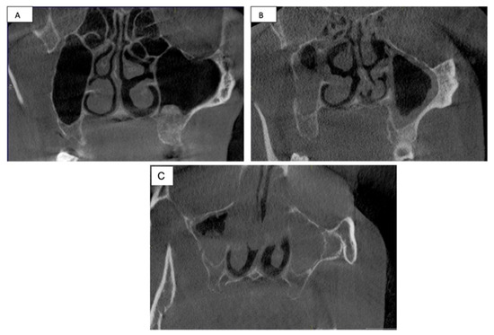
Association between Periodontitis and Chronic Rhinosinusitis Involving Maxillary Sinus Measured by Lund Mackay Staging System
by
Khalid Gufran, Abdulaziz Mohammad Alsakr, Abdullah Saad Alqahtani, Nasser Raqe Alqhtani, Dhafer Alasmari, Faisal Fahad Alzamil, Nawaf Munawir Alotaibi, Hamid Mohammed Alhamid and Ashwag Saleem Aldafiri
Viewed by 1488
Abstract
This study aimed to evaluate the association between periodontitis and chronic rhinosinusitis (CRS) via cone-beam-computed tomography (CBCT) using the Lund–Mackay staging system. CBCT images from different departments of the school of dentistry, at Prince Sattam University were evaluated for the presence of rhinosinusitis.
[...] Read more.
This study aimed to evaluate the association between periodontitis and chronic rhinosinusitis (CRS) via cone-beam-computed tomography (CBCT) using the Lund–Mackay staging system. CBCT images from different departments of the school of dentistry, at Prince Sattam University were evaluated for the presence of rhinosinusitis. All the CBCT scans were exposed for multiple indications, and no patients had a scan exposed solely for this study. The Lund–Mackay staging system was used to measure the CRS in the CBCT. Descriptive statistics for the frequencies and percentages were used to summarize the data. Logistic regression was used to examine the associations between periodontitis and CRS. Each variable was assessed individually by using multivariable analysis. Collinearity issues among the variables were solved to select a limited set of factors using a stepwise variable selection procedure. A total of 399 CBCT images were included in the current research. Logistic regression showed that only gender was significantly associated (
p = 0.0001) with the presence of CRS. However, a stepwise variable selection procedure included gender and bone loss as significantly associated with CRS. No significant difference was observed between unilateral vs. bilateral CRS in gender, bone loss, medical status, and periodontitis. However, only gender showed a significant difference in both bilateral vs. no CRS and unilateral vs. no CRS. Periodontitis is not associated with CRS. However, gender has a significant influence on CRS.
Full article
►▼
Show Figures
Open AccessArticle
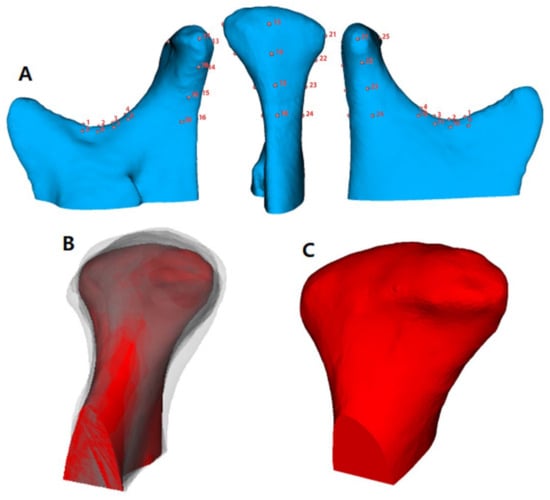
Assessment of Morphologic Change of Mandibular Condyle in Temporomandibular Joint Osteoarthritis Patients with Stabilization Splint Therapy: A Pilot Study
by
Tae-Hoon Kim, Youn Joong Kim, Yun-Heon Song, Ilho Tae, Ho-Kyung Lim and Seok-Ki Jung
Cited by 2 | Viewed by 1473
Abstract
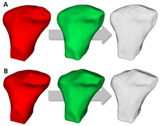
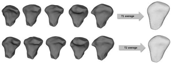
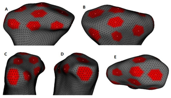
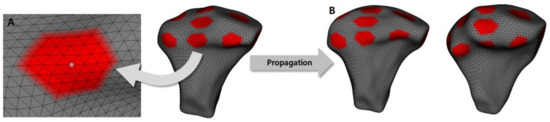
(1) Background: The purpose of this study was to evaluate the 3-dimensional bony changes of the mandibular condyle in temporomandibular joints-osteoarthritis (TMJ-OA) patients treated with stabilization splint (SS) therapy using shape correspondence analysis. (2) Methods: A total of 27 adult patients (2 men
[...] Read more.
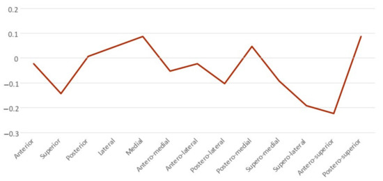
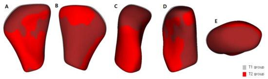
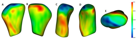
(1) Background: The purpose of this study was to evaluate the 3-dimensional bony changes of the mandibular condyle in temporomandibular joints-osteoarthritis (TMJ-OA) patients treated with stabilization splint (SS) therapy using shape correspondence analysis. (2) Methods: A total of 27 adult patients (2 men and 25 women) with a mean age of 24.6 ± 3.9 years were included in this study. All patients were diagnosed with TMJ-OA and were treated with an SS. Cone-beam computed tomography data of the condylar head before and after SS therapy from 42 condyles (15 bilateral and 12 unilateral TMJ-OA) were used for the analysis. For the performance shape correspondence analysis (SPHARM-PDM), statistical differences were performed using the one-way analysis of variance and Scheffe post hoc tests. (3) Results: After SS treatment in TMJ-OA patients, bone resorption of the condyle head surface was predominant in the anterosuperior, superolateral, and superior areas, and bone formation was superior in the lateral, medial, posterosuperior, and posteromedial areas. The change in the condylar volume between the two groups was not statistically significant. (4) Conclusions: After SS treatment in TMJ-OA patients, there was both bone resorption and bone formation on the mandibular condyle head surface, which induced morphological changes in the condyle head.
Full article
►▼
Show Figures
Open AccessArticle
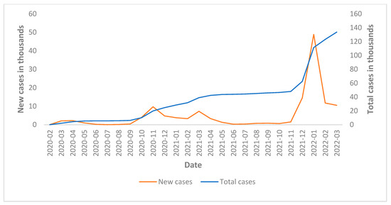
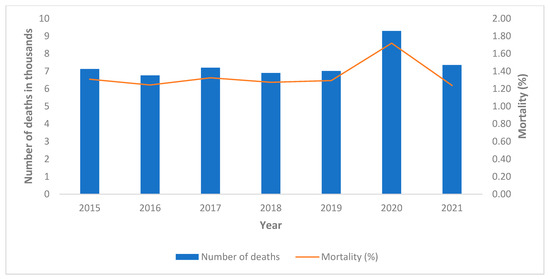
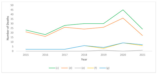
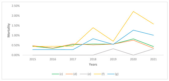
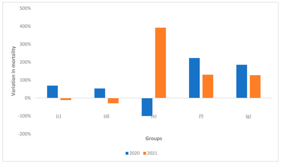
Excess Mortality among Physicians and Dentists during COVID-19 in Italy: A Cross-Sectional Study Related to a High-Risk Territory
by
Saturnino Marco Lupi, Claudia Todaro, Domenico Camassa, Silvana Rizzo, Stefano Storelli and Ruggero Rodriguez y Baena
Cited by 6 | Viewed by 1470
Abstract
Background: Many studies previously reported epidemiological data on mortality due to COVID-19 among health workers. All these studies included a partial sample of the population with a substantial selection bias. The present study evaluates the trend of mortality among physicians and dentists operating
[...] Read more.
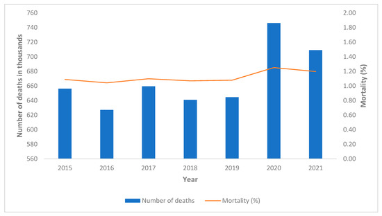
Background: Many studies previously reported epidemiological data on mortality due to COVID-19 among health workers. All these studies included a partial sample of the population with a substantial selection bias. The present study evaluates the trend of mortality among physicians and dentists operating in an area considered to be at high risk during the COVID-19 pandemic. Methods: Data relating to all physicians and dentists registered in the province of Pavia (Italy), a sample consisting of 5454 doctors in 2020 was analyzed. The mortality rates obtained were compared with those related to the 5-year period preceding the pandemic and with those related to the general population. Results: In the area considered, a mortality rate of 0.83% (+69% compared to 2015–2019) was observed in the entire sample in 2020 and 0.43% (−11% compared to 2015–2019) in 2021; among physicians, there was a mortality rate of 0.76% (+53% compared to 2015-2019) in 2020 and 0.35% (−29% compared to 2015–2019) in 2021; for dentists, there was a mortality rate of 1.27% (+185% compared to 2015–2019) in 2020 and 1.01% (+127% compared to 2015–2019) in 2021. Conclusions: These data report the global impact of the SARS-CoV-2 pandemic on physicians and dentists in a high-risk territory. In 2020, a significant increase in the mortality rate compared to the previous 5 years was observed for both physicians and dentists; in 2021, a significant increase in the mortality rate was observed only for dentists. These data are also significant in evaluating the impact of vaccination on physicians and dentists and indicate that dentists were among the professions most at risk during the pandemic.
Full article
►▼
Show Figures
Open AccessArticle
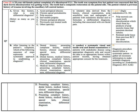
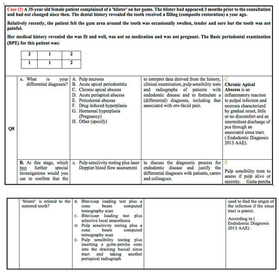
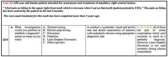
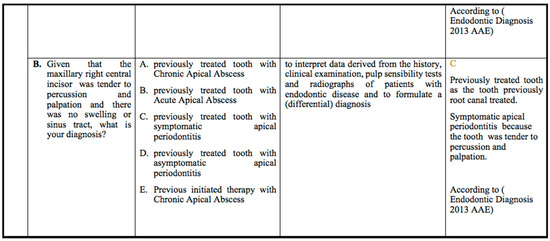
Endodontic Clinical Diagnostic Skills amongst Undergraduate Dental Students: Cross-Sectional Study
by
Mohammed A Alobaoid, Omir Aldowah and Mohmed Isaqali Karobari
Cited by 3 | Viewed by 3416
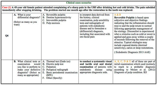
Abstract
The purpose of the study was to evaluate the clinical endodontic diagnostic skills amongst undergraduate dental students at pre-clinical and clinical levels at Cardiff University School of Dentistry. An online questionnaire containing eight questions about endodontic diagnosis and hypothetical clinical scenarios was sent
[...] Read more.
The purpose of the study was to evaluate the clinical endodontic diagnostic skills amongst undergraduate dental students at pre-clinical and clinical levels at Cardiff University School of Dentistry. An online questionnaire containing eight questions about endodontic diagnosis and hypothetical clinical scenarios was sent to all year 3rd, 4th, and 5th-year undergraduate dental students who were divided into G1, G2, and G3 groups. The data were analysed descriptively and reported in percentages. Around 121 students out of 226 responded to the questionnaire with a response rate of 53.5%. The overall correct response from G1 (3rd year) was 31.6% to 65.8%, G2 (4th year) was 73% to 93%, and G3 (5th year) was 73.2% to 92.7%. The study concludes that the 4th and 5th-year undergraduate dental students’ responses to the hypothetical clinical scenarios were higher than the 3rd-year students. However, regarding questions about the endodontic diagnosis, the percentages of correct answers were similar among all the 3rd, 4th, and 5th-year students. Therefore, further studies assessing endodontic diagnostic skills amongst the same cohort of students during their progression in the undergraduate course are recommended.
Full article
►▼
Show Figures
Open AccessArticle
The Radiographic Assessment of Furcation Area in Maxillary and Mandibular First Molars while Considering the New Classification of Periodontal Disease
by
Mohammed Alasqah, Fahad Dhaifallah Alotaibi and Khalid Gufran
Cited by 3 | Viewed by 1640
Abstract
This study aimed to evaluate the radiographic reliability in the diagnosis of furcation involvement in first molars. A total of 52 subjects were included in the current study. Personal history regarding smoking was recorded and a periodontal examination was performed. Pocket depth (PD),
[...] Read more.
This study aimed to evaluate the radiographic reliability in the diagnosis of furcation involvement in first molars. A total of 52 subjects were included in the current study. Personal history regarding smoking was recorded and a periodontal examination was performed. Pocket depth (PD), clinical attachment level (CAL), gingival recession, and furcation involvement in all first molars were assessed for each patient. Periodontal staging and grading were evaluated using the new classification of periodontal disease. Class II and Class III furcation classification were more frequently observed in radiographs than the Class I furcation; however, no significant differences were observed. Radiographic observation of the furcation was seen more when PD and CAL were >5 mm in all molars. The presence of gingival recession and its relation to the radiographic assessment did not reveal any statistically significant association (
p > 0.05) except for tooth #16. The trend of visibility of furcation radiographically was more as the grade of staging was increased. Moreover, the presence of smoking habits and visibility of furcation radiographically did not have any statistical significance. Smoking may not be a factor in the furcation involvement. There is a direct relationship between the staging and grading of the periodontitis and furcation involvement.
Full article
Open AccessArticle
A Prospective Clinical Study Evaluating the Efficacy of Intra-Ligamentary Anesthetic Solutions in Mandibular Molars Diagnosed as Symptomatic Irreversible Pulpitis with Symptomatic Apical Periodontitis
by
Khalid Gufran, Mubashir Baig Mirza, Ali Robaian, Abdullah Saad Alqahtani, Nasser Raqe Alqhtani, Mohammed Alasqah and Abdulaziz Mohammad Alsakr
Cited by 1 | Viewed by 1547
Abstract
Accomplishing painless endodontic treatment, especially in the mandibular molar region, is challenging. Hence, the aim of the study was to compare the efficacy of 2% lidocaine and 4% articaine when used as supplemental intra-ligamentary (IL) anesthesia in mandibular molars having symptomatic irreversible pulpitis
[...] Read more.
Accomplishing painless endodontic treatment, especially in the mandibular molar region, is challenging. Hence, the aim of the study was to compare the efficacy of 2% lidocaine and 4% articaine when used as supplemental intra-ligamentary (IL) anesthesia in mandibular molars having symptomatic irreversible pulpitis with symptomatic apical periodontitis after failed Inferior Alveolar Nerve Block (IANB) injection. In this prospective study, one-hundred and forty-seven adult patients diagnosed with irreversible pulpitis in a mandibular tooth were included who received IANB with 1.8 mL of 2% lidocaine with 1:100,000 epinephrine. Patients who experienced pain were recorded using the Heft–Parker visual analog scale (HP-VAS score ≥ 55 mm) and received supplement intra-ligament injection with either4% articaine or 2% lidocaine with 1:100,000 epinephrine. Supplementary intra-ligament injections resulted in 82.6% and 91.3% of profound anesthesia in the first molar region for 2% lidocaine and 4% articaine, respectively. Similarly, an additional IL injection of articaine success percent (78.9%) in the second molar region was higher than lidocaine (63.1%). The overall success ratio revealed no significant difference in achieving profound anesthesia of either solution. In this study population, there was no difference in the success ratio of anesthesia between 2% lidocaine and 4% articaine when used as supplemental IL injection.
Full article
Open AccessArticle
Incidence and Risk of Anti-Resorptive Agent-Related Osteonecrosis of the Jaw after Tooth Extraction: A Retrospective Study
by
Rieko Shimizu, Shintaro Sukegawa, Yuka Sukegawa, Kazuaki Hasegawa, Sawako Ono, Tomoya Nakamura, Ai Fujimura, Ayaka Fujisawa, Keisuke Nakano, Kiyofumi Takabatake, Hotaka Kawai, Hitoshi Nagatsuka and Yoshihiko Furuki
Cited by 1 | Viewed by 1515
Abstract
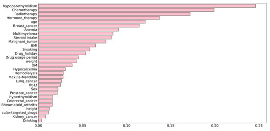
Bone-modifying agents (BMA) such as bisphosphonates and denosumab are frequently used for the treatment of bone metastases, osteoporosis, and multiple myeloma. BMA may lead to anti-resorptive agent-related osteonecrosis of the jaw (ARONJ). This study aimed to clarify the risk factors for and probabilities
[...] Read more.
Bone-modifying agents (BMA) such as bisphosphonates and denosumab are frequently used for the treatment of bone metastases, osteoporosis, and multiple myeloma. BMA may lead to anti-resorptive agent-related osteonecrosis of the jaw (ARONJ). This study aimed to clarify the risk factors for and probabilities of developing ARONJ after tooth extraction in patients undergoing BMA therapy. In this study, the records of 505 target sites of 302 patients undergoing BMA who presented with mandibular fractures at the Department of Oral and Maxillofacial Surgery, Kagawa Prefectural Central Hospital, from March 2014 to January 2022, were retrospectively analyzed for the onset of ARONJ after tooth extraction. The following variables were investigated as attributes: anatomy, health status, and dental treatment. The correlation coefficient was calculated for the success or failure of endodontic surgery for each variable, the odds ratio was calculated for the upper variable, and the factors related to the onset of ARONJ were identified. The incidence rate of ARONJ was found to be 3.2%. Hypoparathyroidism was an important factor associated with ARONJ development. Thus, systemic factors are more strongly related to the onset of ARONJ after tooth extraction than local factors.
Full article
►▼
Show Figures
Open AccessArticle
Dental Treatment under General Anesthesia in Patients with Special Needs Provided by Private and Public Healthcare Services—A Retrospective, Comparative Study
by
Marcello Alves Marinho, Flávia Cristina Teixeira Ramos, Andréa Lanzillotti Cardoso, Geraldo Oliveira Silva-Junior, Marcelo Daniel Brito Faria, Luciana Freitas Bastos, Arkadiusz Dziedzic and Bruna Lavinas Sayed Picciani
Cited by 1 | Viewed by 2140
Abstract
In special care dentistry, general anesthesia (GA) is considered as an alternative option to facilitate treatment for uncooperative patients with special needs (PSN) who require invasive dental interventions. Objective: to evaluate the profile of dental treatment procedures performed and the characteristics of PSN
[...] Read more.
In special care dentistry, general anesthesia (GA) is considered as an alternative option to facilitate treatment for uncooperative patients with special needs (PSN) who require invasive dental interventions. Objective: to evaluate the profile of dental treatment procedures performed and the characteristics of PSN who underwent dental treatment under GA, provided by private and public healthcare providers. Methods: A retrospective, observational study involving a sample of 100 PSN treated in hospital and specialist secondary care settings. Demographic data and clinical information were collected. The analysis of data was performed using descriptive analysis and frequency statistical tests. Results: out of 100 participants, 63% of the PSN who received care in the private sector and the remaining 37% of PSN registered with public-funded care providers, aged 6 to 80 years old, were treated under GA. Autistic spectrum disorder was the most common medical diagnosis recorded (33%). More than half (52%) of the PSN treated by private care providers sought specialist care in an outpatient setting prior to GA vs. 5% of the PSN treated by public-funded providers. The utilization of sedation prior to GA was more common in the private sector. A vast majority (86%) of all subjects underwent multiple dental extractions (86%) and restorations (62%). Conclusions: comprehensive dental care under GA, which composes an integral part of special care dentistry, can be safely provided in a hospital setting, in both private and public sectors. While early intervention using sedation and behavioral management partially mitigates the need for dental care under GA, the vast majority of PSN may require dental treatment under GA in future to facilitate complex dental care.
Full article
Open AccessArticle
Variation in Hemodynamic Characteristics during Periodontal Crown-Lengthening Surgical Procedure: An Uncontrolled Cohort Study
by
Abdullah Saad Alqahtani, Rajashekhara Bhari Sharanesha, Khalid Gufran, Nasser Raqe Alqhtani, Alwaleed Abushanan, Mohammed Alasqah, Abdulaziz Mohammad Alsakr and Hassan Alkharaan
Cited by 1 | Viewed by 1329
Abstract
(1) Background: The purpose of this prospective study was to determine the changes in primary hemodynamic parameters and oxygen saturation in systemically healthy patients during the surgical procedure involving crown lengthening. (2) Methods: A total of 44 patients who required a crown-lengthening procedure
[...] Read more.
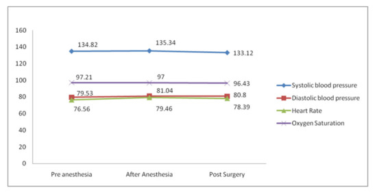
(1) Background: The purpose of this prospective study was to determine the changes in primary hemodynamic parameters and oxygen saturation in systemically healthy patients during the surgical procedure involving crown lengthening. (2) Methods: A total of 44 patients who required a crown-lengthening procedure in a single tooth in the maxillary arch were included in this study. Heart rate (HR), blood pressure (BP) and oxygen saturation (SpO
2) were measured in all the subjects at three different intervals: before injecting the anesthetic (T1), after the anesthetic injection (T2) and after the procedure (T3). Descriptive statistics were computed, and observations were recorded as mean and standard deviation (SD). Analysis of variance (ANOVA) was used to compare the mean observation within parameters at different time intervals. (3) Results: All primary hemodynamic parameters were increased in the T2 phase over T1 and decreased in the T3 phase over T2. However, SpO
2 decreased in both the T2 and T3 phases compared to the initial T1 phase. No significant differences were observed among the primary hemodynamic variables. However, SpO
2 showed a significant difference (
p = 0.013) among the T1, T2 and T3 phases. (4) Conclusions: Further study with larger sample size is required in order to analyze the accurate hemodynamic alterations.
Full article
►▼
Show Figures
Open AccessArticle
Oral Tissue Involvement and Probable Factors in Post-COVID-19 Mucormycosis Patients: A Cross-Sectional Study
by
Neelam Chandwani, Sandeep Dabhekar, Kalai Selvi, Roshan Noor Mohamed, Shahabe Saquib Abullais, Muhamood Moothedath, Ganesh Jadhav, Jaya Chandwani, Mohmed Isaqali Karobari and Ajinkya M. Pawar
Cited by 4 | Viewed by 2230
Abstract
The primary goal of this study was to assess the prevalence of oral involvement and, secondarily, the likely variables in patients with confirmed COVID-19 accompanied by mucormycosis infection. The study design was a cross-sectional descriptive sort that was performed at a tertiary centre.
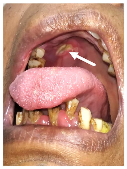
[...] Read more.
The primary goal of this study was to assess the prevalence of oral involvement and, secondarily, the likely variables in patients with confirmed COVID-19 accompanied by mucormycosis infection. The study design was a cross-sectional descriptive sort that was performed at a tertiary centre. The non-probability convenience sampling approach was used to determine the sample size. Between May 2021 and July 2021, all patients who presented to our tertiary care centre with suspected mucormycosis were considered for the investigation. The research only included individuals with proven mucormycosis after COVID-19. The features of the patients, the frequency of intraoral signs/symptoms, and the possible variables were all noted. Of the 333 COVID-19-infected patients, 47 (14%) were diagnosed with confirmed mucormycosis. The mean (SD) age of the patients was 59.7 (11.9) years. Of the 47 patients with confirmed mucormycosis, 34% showed sudden tooth mobility, 34% expressed toothache, 8.5% reported palatal eschar, 34% presented with jaw pain, 8.5% had tongue discoloration, and 17% had temporomandibular pain. About 53% of the patients were known cases of type 2 diabetes mellitus, 89% of patients had a history of hospitalization due to COVID-19 infection, 89.3% underwent oxygen support therapy, and 89.3% were administered intravenous steroids during hospitalization due to COVID-19 infection. About 14% of the suspected cases attending the mucormycosis out-patient department (OPD) had been confirmed with definite mucormycosis. Oral involvement was seen in 45% of cases of CAM (COVID-associated mucormycosis). The most frequent oral symptoms presented in CAM were sudden tooth mobility and toothache. Diabetes and steroids were the likely contributing factors associated with CAM.
Full article
►▼
Show Figures
Open AccessReview
Evaluation of Adjuvant Systems in Non-Surgical Peri-Implant Treatment: A Literature Review
by
Andrea Butera, Carolina Maiorani, Simone Gallo, Maurizio Pascadopoli, Adith Venugopal, Anand Marya and Andrea Scribante
Cited by 26 | Viewed by 2057
Abstract
Can the use of lasers, ozone, probiotics, glycine and/or erythritol, and chlorhexidine in combination with non-surgical peri-implant treatment have additional beneficial effects on the clinical parameters?
Objectives: The non-surgical treatment of peri-implant pathologies is based on mechanical debridement to eliminate bacterial biofilm
[...] Read more.
Can the use of lasers, ozone, probiotics, glycine and/or erythritol, and chlorhexidine in combination with non-surgical peri-implant treatment have additional beneficial effects on the clinical parameters?
Objectives: The non-surgical treatment of peri-implant pathologies is based on mechanical debridement to eliminate bacterial biofilm and reduce tissue inflammation; some additional therapies have been studied to achieve more detailed clinical results.
Materials and methods: A literature search for publications until January 2022 was conducted. The research question is formulated following the Problem, Intervention, Comparison/Control, and Outcome. Studies investigating adjunctive therapies were included.
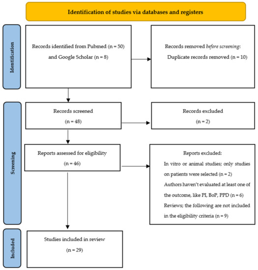
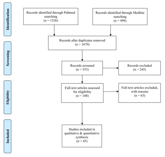
Results: In total, 29 articles were included. Most of the studies did not show any additional benefit of these therapies in the evaluation of bleeding on probing, probing pocket depth, or plaque index; among the proposed treatments, the use of laser was the one most studied in the literature, with the achievement of a reduction of bleeding and pocket depth. More studies would be needed to assess the benefit of other therapies.
Conclusions: This review showed no significant improvements in the state of health in support of mechanical debridement therapy. However, the few benefits found would deserve to be considered in new clinical studies.
Full article
►▼
Show Figures
Open AccessArticle
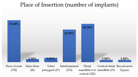
Clinically Based Classification and Positioning Indication for Single-Piece Compressive Implants Placement in Regard to Extraction Socket
by
Mmehul Jani, Vivek Gaur, Anita Gala Doshi, Kiran Patel and Łukasz Pałka
Viewed by 2041
Abstract
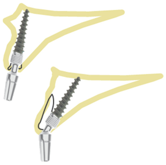
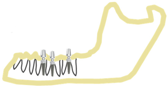
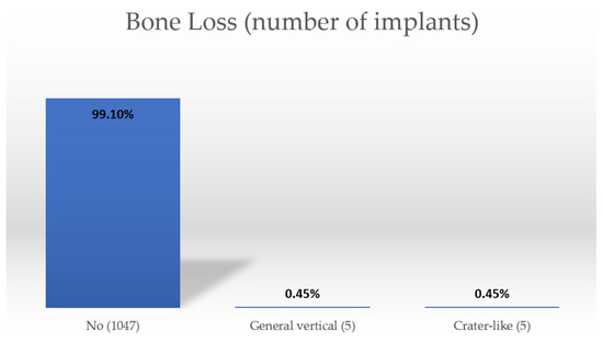
(1) Background: Dental implantology has been rapidly developing over the last decades. The introduction of new materials, surface modifications and implant designs has brought the need to rethink and systematize our knowledge regarding dental implants. Thus, the aim of this paper is to
[...] Read more.
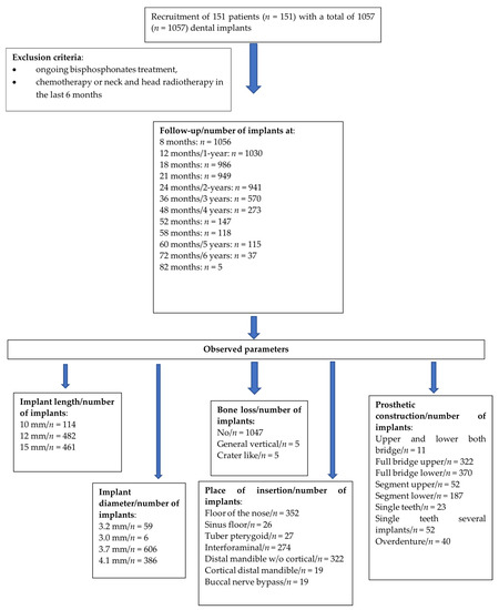
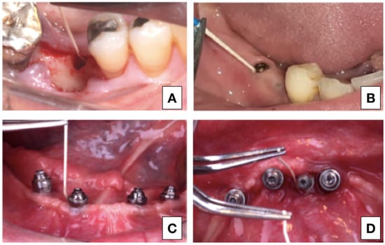
(1) Background: Dental implantology has been rapidly developing over the last decades. The introduction of new materials, surface modifications and implant designs has brought the need to rethink and systematize our knowledge regarding dental implants. Thus, the aim of this paper is to introduce a new classification and implant positioning indications that can be used to maximize the survival rate and the aesthetic outcome of single-piece compressive screw implants. (2) Materials and methods: This classification was based on a multicenter clinical and radiological observation of 151 patients, in whom 1057 implants were placed with a success rate of 98.5% (1041). The follow-up period was up to 82 months with a mean of 22.34 months. (3) Results: it seems that, in the case of single-piece implants, diameter and length of the implant have influence on their survival rate, whereas smoking and hypertension do not. (4) Conclusions: this paper provides clinicians with comprehensive information about the rationale, criteria and implementation of the new classifications based on a large number of implants and long-term observations.
Full article
►▼
Show Figures
Open AccessArticle
Satisfaction Factors with a Dental Unit Chair System in South Korea: A Dentist’s Perspective
by
Keunbada Son, Young-Tak Son, Myoung-Uk Jin and Kyu-Bok Lee
Cited by 1 | Viewed by 3106
Abstract
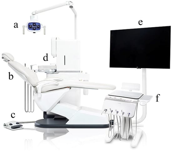
This study aimed to survey users’ satisfaction with a dental unit chair in order to highlight the elements affecting the dentist’s satisfaction. The questionnaire items were drawn up with seven components that constitute a dental unit chair, including the light, patient seat, foot
[...] Read more.
This study aimed to survey users’ satisfaction with a dental unit chair in order to highlight the elements affecting the dentist’s satisfaction. The questionnaire items were drawn up with seven components that constitute a dental unit chair, including the light, patient seat, foot controller, water fountain and cuspidor, monitor, bracket table and controller, and dentist chair. With these questionnaire elements, a pilot experiment was conducted to test the reliability, and reliability analysis was conducted. The scale reliability was checked using Cronbach’s alpha coefficient. Bartlett’s test of sphericity, the Kaiser-Meyer-Olkin (KMO) measure, and factor analysis were performed to test whether the items would constitute appropriate questionnaire items for the survey. The survey was conducted with 26 dentists with more than three years of clinical experience. A correlation analysis was conducted using Pearson’s correlation coefficient (PCC) (α = 0.05) to analyze the impact of the factors on the overall satisfaction with the dental unit chair. The items that were strongly correlated with the overall satisfaction score of the dental unit chair were the design and appearance quality of the dental unit chair (PCC = 0.781), its maintenance (PCC = 0.784), and the overall satisfaction with the water fountain and cuspidor (PCC = 0.703) (
p < 0.05). Most of the questionnaire items could affect the overall satisfaction with the dental unit chair. Additionally, because the design and appearance quality, maintenance, and overall satisfaction with the water fountain and cuspidor may have the greatest impact on the overall satisfaction with the dental unit chair, the improvement of these elements may bring about the enhancement of the overall satisfaction.
Full article
►▼
Show Figures
Open AccessArticle
White Spots Prevalence and Tooth Brush Habits during Orthodontic Treatment
by
Çeljana Toti, Agron Meto, Gerta Kaçani, Etleva Droboniku, Dorjan Hysi, Michele Tepedino, Edlira Zaja, Luca Fiorillo, Aida Meto, Denada Buci and Olja Tanellari
Cited by 7 | Viewed by 1671
Abstract
White spots (WS) are one of the most undesirable side effects in patients undergoing orthodontic therapy and are usually located around bracket bases and even detected under the molar bands. The aim of the present cross-sectional study was to evaluate the WS lesion
[...] Read more.
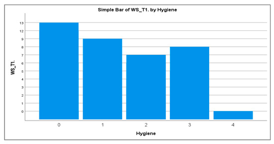
White spots (WS) are one of the most undesirable side effects in patients undergoing orthodontic therapy and are usually located around bracket bases and even detected under the molar bands. The aim of the present cross-sectional study was to evaluate the WS lesion during orthodontic therapy and the correlation between WS and oral hygiene habits. Patients requiring orthodontic treatment with a fixed appliance were screened for the inclusion/exclusion criteria, and 74 subjects were finally enrolled. Each patient received three examinations: at T0, the day of the application of the fixed appliance; at T1, three months later; and at T2, six months after treatment start. After calculating descriptive statistics, differences between groups were evaluated with an independent sample
t-test. The first type error was set as
p ≤ 0.01. The observed prevalence of WS lesions was 59.5% on T1 and 60.8% on T2. The most affected teeth result to be upper molars, lower left first molar, upper right central incisor and upper left lateral incisor, upper right canine, upper left first premolar, and lower right first molar. A higher frequency of daily tooth brushing was accompanied by a lower prevalence of WS. No significant effect of sex was observed.
Full article
►▼
Show Figures
Open AccessArticle
Oral-Health-Related Quality of Life (OHRQoL) and Anterior Open Bite in Adult Patients: A Case-Control Study
by
Adrián Curto, Alberto Albaladejo and Alfonso Alvarado-Lorenzo
Cited by 4 | Viewed by 2255
Abstract
Oral-health-related quality of life (OHRQoL) is defined as the impact of oral health on activities of daily living. Malocclusions are a public health problem with a high prevalence. Different studies have concluded that malocclusions negatively affect OHRQoL in patients of all ages. The
[...] Read more.
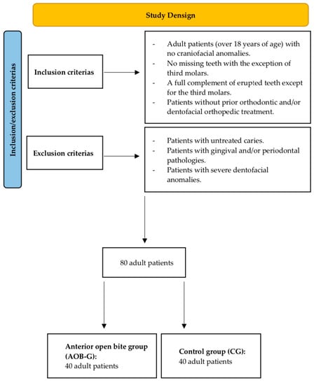
Oral-health-related quality of life (OHRQoL) is defined as the impact of oral health on activities of daily living. Malocclusions are a public health problem with a high prevalence. Different studies have concluded that malocclusions negatively affect OHRQoL in patients of all ages. The aim of this study was to analyze the influence of having an anterior open bite on the OHRQoL of adult patients. Materials and Methods: A case-control study (1:1) was carried out with a sample size of 80 adults at the University of Salamanca in 2021. The case group (
n = 40) was made up of patients with an anterior open bite, and the control group (
n = 40) contained patients without an anterior open bite. OHRQoL was assessed using the Oral Health Impact Profile-14 (OHIP-14) questionnaire. The influences of gender and age on the OHRQoL of the patients were also analyzed. Results: There were no significant differences in gender or age between the case and control groups. An anterior open bite was not found to influence the OHRQoL of adult patients. Age was not shown to significantly influence OHRQoL. Female patients with an anterior open bite had higher scores in the handicap domain of the OHIP-14 questionnaire compared with male patients (
p < 0.05). Conclusions: Anterior open bite can influence the OHRQoL of orthodontic patients. Gender can be considered an influencing factor.
Full article
►▼
Show Figures
Open AccessArticle
Oral Hygiene Practice among Hospitalized Patients: An Assessment by Dental Hygiene Students
by
Saturnino Marco Lupi, Maurizio Pascadopoli, Carolina Maiorani, Camilla Preda, Benedetto Trapani, Alessandro Chiesa, Francesca Esposito, Andrea Scribante and Andrea Butera
Cited by 8 | Viewed by 3056
Abstract
Aim: An epidemiological study was carried out, in hospital wards, with the aim of assessing the oral health status of patients subjected to multiple medical treatments. Material and Methods: The study was conducted at Fondazione IRCCS Policlinico San Matteo (Pavia, Italy). A questionnaire
[...] Read more.
Aim: An epidemiological study was carried out, in hospital wards, with the aim of assessing the oral health status of patients subjected to multiple medical treatments. Material and Methods: The study was conducted at Fondazione IRCCS Policlinico San Matteo (Pavia, Italy). A questionnaire was submitted to patients for the evaluation of oral hygiene devices used; then, a clinical examination was conducted to collect Decayed Missing Filled Teeth (DMFT) index, Plaque Index (PI), and Marginal Gingival Index (MGI) values. Results: Manual toothbrushes were used by a wide range of the sample study (65–100% among hospital wards), together with mouthwash (20–80%); interproximal aids were used by few patients (the lowest recorded value was 33.3%). Conclusion: dental hygienists could be integrated into hospital wards as oral hygiene procedure instructors, for the improvement of the oral health conditions of hospitalized patients.
Full article
Open AccessCase Report
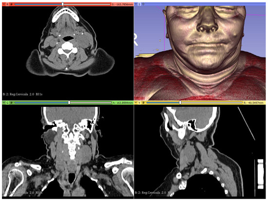
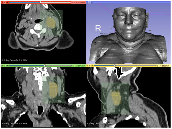
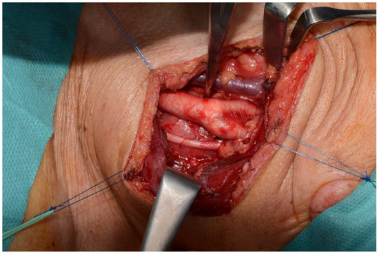
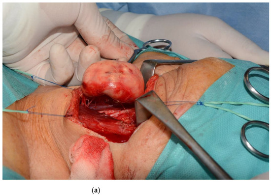
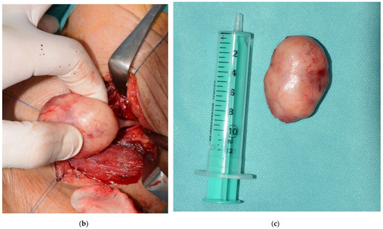
A Predictable Approach of a Rare and Frequently Misdiagnosed Entity: Laryngeal Nerve Schwannoma
by
Iulian Filipov, Lucian Chirila, Mihai Sandulescu and Corina Marilena Cristache
Cited by 2 | Viewed by 1714
Abstract
(1) Background: Schwannoma, a mesenchymal neoplasm derived from Schwann cells that line peripheral nerve sheaths, has a challenging diagnosis, due to the non-specific medical history and clinical examination. Nowadays, virtual reality (VR) is increasingly more used for enhancing diagnosis and for preoperative planning
[...] Read more.
(1) Background: Schwannoma, a mesenchymal neoplasm derived from Schwann cells that line peripheral nerve sheaths, has a challenging diagnosis, due to the non-specific medical history and clinical examination. Nowadays, virtual reality (VR) is increasingly more used for enhancing diagnosis and for preoperative planning of surgical procedures. With VR, the surgeon can interact, before any surgery, with a virtual environment that is completely generated by a computer, offering them a real experience inside a virtual 3D model. (2) Methods and Results: The aim of the present paper was to present a case of surgically removal of a schwannoma, which originated from the fibers of the superior laryngeal nerve, in a predictable and minimally invasive fashion, upon using VR for diagnosis and surgical procedure planning. (3) Conclusions: The current clinical report attracted the attention of including schwannoma in the possible differential diagnosis of a swelling in the anterior cervical region, mainly when a nonspecific radiological appearance is noticed, even with the use of multiple imaging modalities. Virtual reality can increase the predictability and success rate of the surgical procedure, being in the meantime a good tool for communication with the patient.
Full article
►▼
Show Figures
Open AccessArticle
Correlation between the Mandibular Lingula Position and Some Anatomical Landmarks in Cone Beam CT
by
Saturnino Marco Lupi, Jessica Landini, Giorgia Olivieri, Claudia Todaro, Andrea Scribante and Ruggero Rodriguez y Baena
Cited by 9 | Viewed by 3715
Abstract
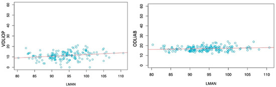
Background: the position of the mandibular lingula (Li) affects the success rate of the inferior alveolar nerve block (IANB) and ramus osteotomies. This study evaluated the position of the Li, to investigate the anatomical relationship between the Li and some anatomical measurements using
[...] Read more.
Background: the position of the mandibular lingula (Li) affects the success rate of the inferior alveolar nerve block (IANB) and ramus osteotomies. This study evaluated the position of the Li, to investigate the anatomical relationship between the Li and some anatomical measurements using cone beam computed tomography (CBCT). Methods: 201 hemimandibular CBCTs of 111 patients (43 males and 68 females; 18 to 88 years old) were retrospectively evaluated. The Li location was determined from the lingula tip to: the occlusal plane, the anterior and posterior borders of the mandibular ramus, the lower border of the mandible, the distal surface of the mandibular second molar, and the mandibular notch. We evaluated the correlations between the Li and the anteroposterior diameter of the mandibular ramus; the vertical distance between condyle and mandibular angle; the mesial–distal diameter of the first, second, and third mandibular molar, the intercanine distance, the intermolar distances among the first, second, and third mandibular molars; the distance between the intermolar line of the first molar and midline, and the length of the mandibular body. Results: the vertical distance of the Li from the occlusal plane was 11.22 ± 4.27 mm. Some parameters significantly correlated with the anatomical measurements taken into consideration. Conclusions: the present study provides new information concerning the Li and mandibular anatomy in the Italian population. Moreover, by correlating some anatomic measurements to the Li position, the localization of the Li is made possible, indirectly through the measurement of some distances between anatomical landmarks.
Full article
►▼
Show Figures
Open AccessArticle
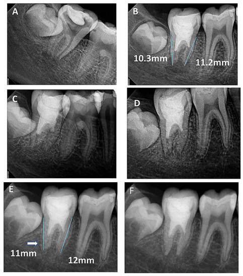
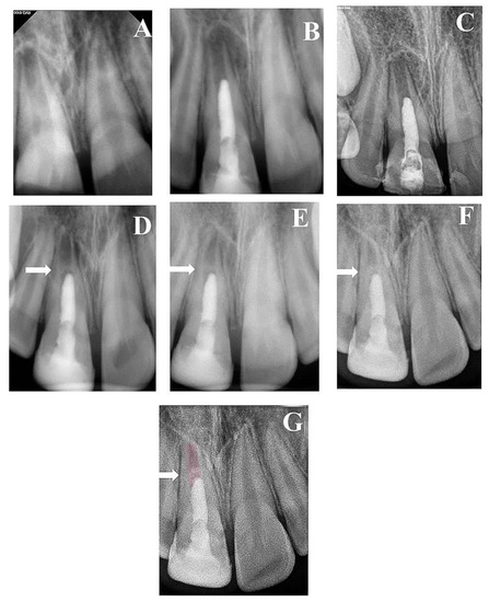
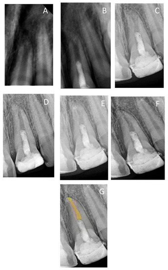
A Prospective Study of Long-Term Regenerative Endodontics Outcomes of Necrotic Immature Permanent Teeth: An 8-Year Follow-Up
by
Sawsan T. Abu Zeid, Ruaa A. Alamoudi, Osama S. Alothmani, Abeer A. Mokeem Saleh and Amna Y. Siddiqui
Cited by 3 | Viewed by 2771
Abstract
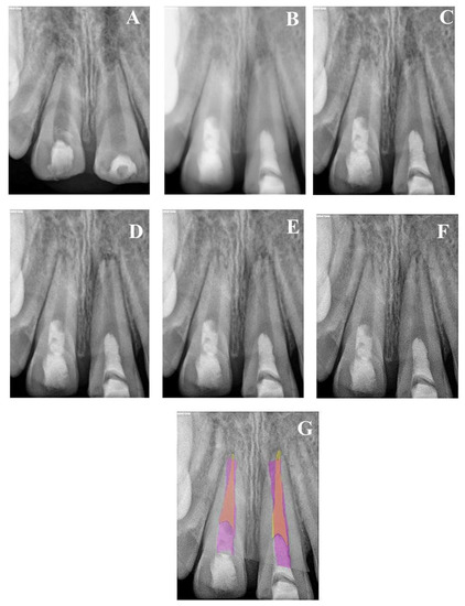
For the management of necrotic immature teeth, regenerative endodontics offers the advantage of further root lengthening, thickening of dentin wall, and apical closure. This prospective study aimed to evaluate the long-term outcome of regenerative endodontics in immature necrotic permanent teeth. A total of
[...] Read more.
For the management of necrotic immature teeth, regenerative endodontics offers the advantage of further root lengthening, thickening of dentin wall, and apical closure. This prospective study aimed to evaluate the long-term outcome of regenerative endodontics in immature necrotic permanent teeth. A total of 23 immature roots were medicated by triple antibiotic paste. After 21 days, bleeding was induced by over-instrumentation, and then mineral trioxide aggregate and coronal restoration were applied. Patients were scheduled for clinical and radiographic follow-up for 8 years. The radiographic changes of root dimensions were assessed using the ImageJ Plugin and statistically analyzed by Kruskal–Wallis test at a 95% confidence level. For qualitative evaluation, images were overlapped and analyzed using Photoshop software. All teeth were asymptomatic one month after the treatment. All teeth (
n = 18) with preoperative periapical radiolucency showed complete resolution within 6–9 months. Recall rate at two, three, and eight years was 69.6%, 56.5%, and 34.8%, respectively. Continuous root development with a significant increase in root length and thickening of dentin wall accompanied by a significant decrease in apical canal diameter was seen at the end of the observation period (
p < 0.001). In conclusion, the long-term outcome of regenerative endodontics revealed successful clinical and radiographic results with appropriate case selection.
Full article
►▼
Show Figures
Open AccessEditor’s ChoiceReview
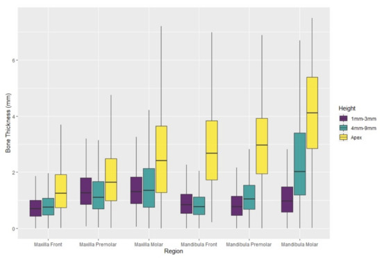
Buccal Bone Thickness in Anterior and Posterior Teeth—A Systematic Review
by
Diana Heimes, Eik Schiegnitz, Robert Kuchen, Peer W. Kämmerer and Bilal Al-Nawas
Cited by 10 | Viewed by 5479
Abstract
(1) Background: Immediate dental implant placement has been a subject of great interest over the last decade. Here, information regarding the anatomy and bone thickness of the jaw prior to dental implant placement is crucial to increase the surgery’s success and the patient’s
[...] Read more.
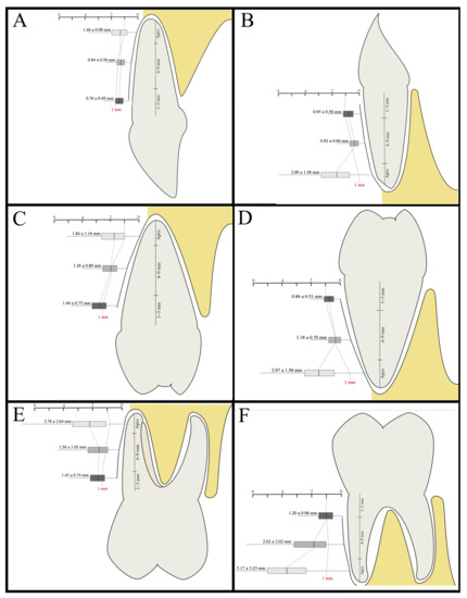
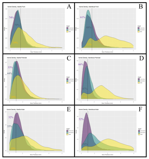
(1) Background: Immediate dental implant placement has been a subject of great interest over the last decade. Here, information regarding the anatomy and bone thickness of the jaw prior to dental implant placement is crucial to increase the surgery’s success and the patient’s safety. The clinical premises for this approach have been controversially discussed. One of those heavily discussed premises is a buccal bone thickness of at least 1 mm thickness. This meta-analysis aims to systematically review buccal bone thickness (BBT) in healthy patients. Thus, the feasibility of immediate dental implant placement in daily practice can be assessed. (2) Methods: A search in the electronic databases was performed to identify articles reporting on BBT that was measured by computed tomography in adults. (3) Results: We were able to find 45 studies, including 4324 patients with 25,452 analyzed teeth. The analysis showed a BBT at the alveolar crest of 0.76 ± 0.49 mm in the maxillary frontal and of 1.42 ± 0.74 mm in the maxillary posterior region. In the mandible, the average measured values were similar to those in the maxilla (front: 0.95 ± 0.58 mm; posterior: 1.20 ± 0.96 mm). In the maxillary frontal region 74.4% and in the mandibular frontal region 61.2% of the crestal buccal bones showed widths <1 mm. (4) Conclusions: In more than 60% of the cases, the BBT at the alveolar crest is <1 mm in maxillary and mandibular frontal regions. This anatomic data supports careful pre-surgical assessment, planning of a buccal graft, and critical selection of indication for immediate implant placement, especially in the maxillary and mandibular frontal and premolar region.
Full article
►▼
Show Figures
Open AccessCase Report
Minimally Invasive Two-Staged Surgery in the Treatment of Large Cystic Lesions of the Jaw
by
Andreea Irimia, Liliana Moraru, Diana Alina Ciubotaru, Constantin Caruntu, Alexandru-Titus Farcasiu and Ana Caruntu
Cited by 3 | Viewed by 2928
Abstract
Background: Cystic lesions of the jaw are commonly found in clinical practice. Large, expansive cysts raise challenges for the clinician from both diagnostic and surgical perspectives. The aim of our work is to present a combined, two-staged surgical approach in histologically confirmed non-aggressive
[...] Read more.
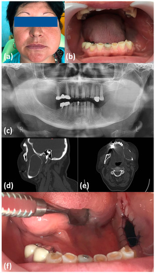
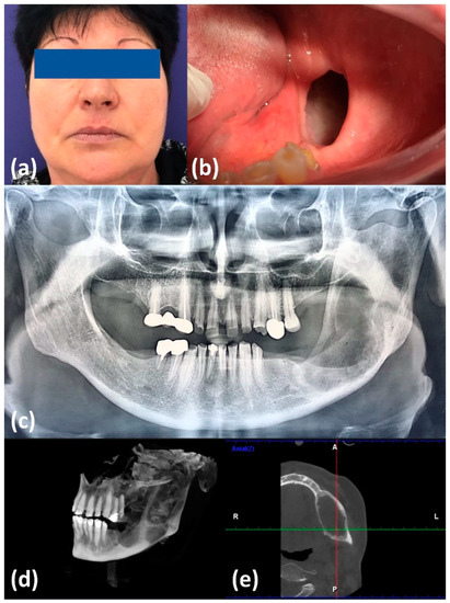
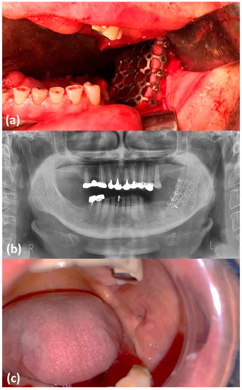
Background: Cystic lesions of the jaw are commonly found in clinical practice. Large, expansive cysts raise challenges for the clinician from both diagnostic and surgical perspectives. The aim of our work is to present a combined, two-staged surgical approach in histologically confirmed non-aggressive cystic lesions of the jaw. Methods and Results: We report the case of an extensive mandibular cyst, associating a high risk of bone fracture, that is treated in the initial stage by cystic decompression through marsupialization with concomitant histological diagnostic confirmation, followed in the second stage by radical excision and mandibular reconstruction with titanium mesh, with the purpose of prevention for oro-cystic chronic fistula formation. Conclusions: Large odontogenic mandibular cysts imply a meticulously conducted assessment and treatment. Marsupialization should be taken into consideration for the treatment of large cystic lesions, followed by secondary enucleation, with minimal risks for the patient. The soft tissue healing process can be optimized with the use of titanium meshes, as an alternative for other reconstructive techniques, in the management of large cystic lesions.
Full article
►▼
Show Figures
Open AccessArticle
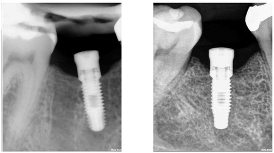
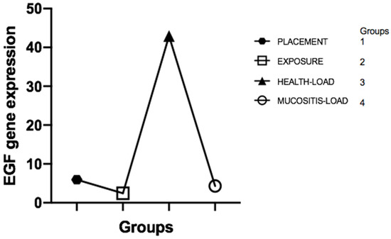
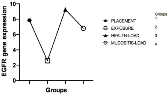
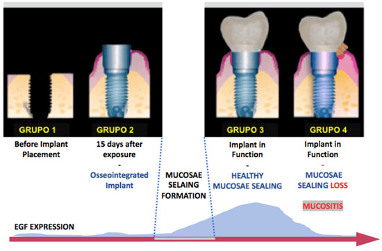
Epidermal Growth Factor Is Associated with Loss of Mucosae Sealing and Peri-Implant Mucositis: A Pilot Study
by
José Jorge Schoichet, Carlos Fernando de Almeida Barros Mourão, Edgard de Mello Fonseca, Carlos Ramirez, Ricardo Villas-Boas, Juliana Prazeres, Valquiria Quinelato, Telma Regina Aguiar, Marina Prado, Angelo Cardarelli, Rafael Mello-Machado and Priscila Casado
Cited by 2 | Viewed by 1706
Abstract
This study aimed to evaluate the correlation between epidermal growth factor (EGF) and receptor (EGFR) levels in different clinical stages of dental implant rehabilitation and trace mucositis development’s biological profile. Thirty-six participants from the Specialization in Implant Dentistry, Universidade Federal Fluminense, Brazil, were
[...] Read more.
This study aimed to evaluate the correlation between epidermal growth factor (EGF) and receptor (EGFR) levels in different clinical stages of dental implant rehabilitation and trace mucositis development’s biological profile. Thirty-six participants from the Specialization in Implant Dentistry, Universidade Federal Fluminense, Brazil, were included in the study and underwent sample collection: inside the alveolar socket, immediately before implant placement (Group 1,
n = 10); at the peri-implant crevicular fluid (PICF) during reopening (Group 2,
n = 10); PICF from healthy peri-implant in function (Group 3,
n = 8); and PICF from mucositis sites (Group 4,
n = 18). Quantitative polymerase chain reaction (PCR) evaluated EGF/EGFR gene expression using the SYBR Green Master Mix detection system. The results showed that EGF expression in the peri-implant crevicular fluid was statistically different. There was a higher EGF expression for group C (peri-implant health) (
p = 0.04) than for the other groups. Regarding EGFR, there was no statistical difference among the groups (
p = 0.56). It was concluded that low levels of EGF gene expression in the peri-implant crevicular fluid are related to the development of peri-implant mucositis and the absence of mucosae sealing. There was no correlation between EGFR gene expression with health or mucositis.
Full article
►▼
Show Figures
Open AccessCase Report
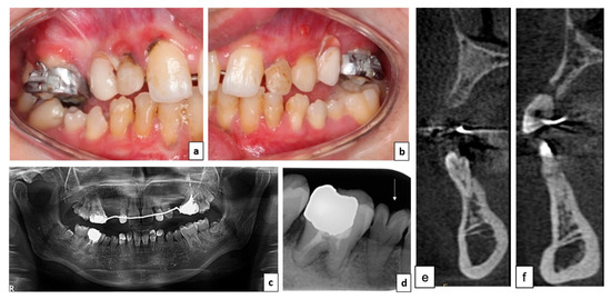
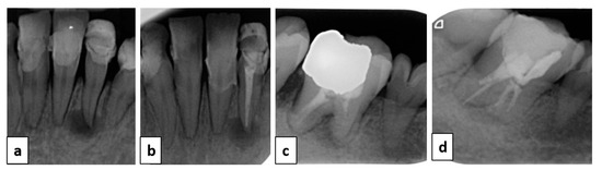
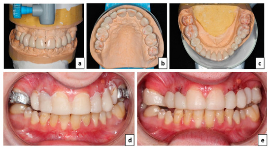
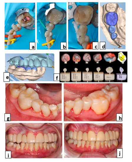
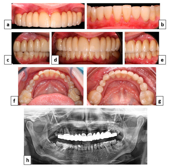
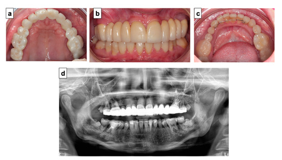
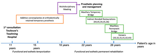
From Child to Adulthood, a Multidisciplinary Approach of Multiple Microdontia Associated with Hypodontia: Case Report Relating a 15 Year-Long Management and Follow-Up
by
Charlotte Thomas, Frédéric Vaysse, Teva Courset, Karim Nasr, Bruno Courtois, Arnaud L’Homme, Nicolas Chassaing, Alexia Vinel, Isabelle Bailleul-Forestier, Luc Raynaldy and Sara Laurencin-Dalicieux
Cited by 2 | Viewed by 2346
Abstract
Oral rehabilitation of patients presenting multiple microdontia is a real therapeutic challenge. These alterations in size, often associated with other dental anomalies, have aesthetic and functional repercussions for patients and can lead to significant psycho-social consequences. We report here the case of an
[...] Read more.
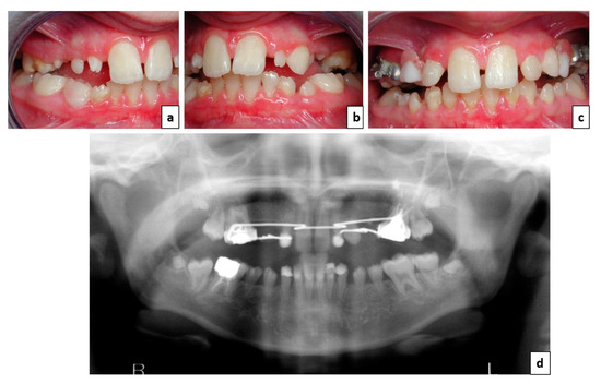
Oral rehabilitation of patients presenting multiple microdontia is a real therapeutic challenge. These alterations in size, often associated with other dental anomalies, have aesthetic and functional repercussions for patients and can lead to significant psycho-social consequences. We report here the case of an 11-year-old patient with bilateral sectorial microdontia and agenesis of teeth numbers 13 and 23. She also presented staturo-ponderal delay and a history of acute coronary syndrome with a lower coronary occlusion of unknown aetiology. At first, additive coronoplasties and an orthodontically retained interim prosthesis answered the aesthetic and functional need during childhood and adolescence. Once she reached adulthood, a multidisciplinary meeting was conducted and a treatment plan was established. The decision was made to rehabilitate the upper arch with a permanent bridge and the lower arch with indirect adhesive restorations. This solution solved the problem of the bilateral lateral infraocclusions and tooth agenesis, restoring both aesthetics and function. This paper presents 15 years of management and treatment of a patient presenting multiple microdontia associated with hypodontia. Both the multidisciplinary approach and coordination between the different medical team members was essential to maintain the existing dentition while preparing, planning, and carrying out a personalized treatment plan once maxillofacial growth was complete.
Full article
►▼
Show Figures
Open AccessArticle
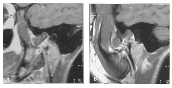
Analysis of Earlier Temporomandibular Joint Disorders in JIA Patients: A Clinical Report
by
Alessandro Polizzi, Vincenzo Quinzi, Simona Santonocito, Giuseppe Palazzo, Giuseppe Marzo and Gaetano Isola
Cited by 1 | Viewed by 2236
Abstract
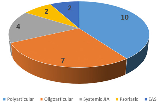
The aim of this study was to analyse the structural characteristics of the temporo-mandibular joint (TMJ) and the dysfunctional consequences induced by disease in subjects with juvenile idiopathic arthritis (JIA). The study was conducted in 25 patients with JIA (median age (IQR), 13.9
[...] Read more.
The aim of this study was to analyse the structural characteristics of the temporo-mandibular joint (TMJ) and the dysfunctional consequences induced by disease in subjects with juvenile idiopathic arthritis (JIA). The study was conducted in 25 patients with JIA (median age (IQR), 13.9 (10.9–15.3)) and 26 healthy controls (median age (IQR), 14.3 (11.6–17.2)) years. All enrolled patients were subjected to anamnestic evaluation, laboratory parameters, JIA subclass, and type of therapy for the disease. A clinical-gnathological evaluation, anamnestic and dysfunctional index (Ai and Di), and magnetic resonance imaging of TMJs were performed in all patients. The test group showed a significant reduction (
p < 0.001) regarding the clinical findings such as maximal mouth opening, left and rightward laterotrusion and protrusion, and a significant difference in the reported symptoms (TMJ sounds, reduced mouth opening and pain), and Ai and Di (
p < 0.001) compared to healthy patients. Correlation analysis showed a significant correlation between the median duration of disease and the maximum mouth opening and between visual analogue scale (VAS) score and maximum mouth opening, leftward laterotrusion, rightward laterotrusion, and protrusion. The results obtained in this study suggest that patients with JIA presented a cohort of symptoms in TMJs in comparison with healthy controls. Moreover, a careful TMJs evaluation and an early diagnosis of TMJs dysfunction and regular follow-ups are recommended in order to prevent and reduce functional and chewing problems in patients with JIA.
Full article
►▼
Show Figures


