
Assessment of Morphologic Change of Mandibular Condyle in Temporomandibular Joint Osteoarthritis Patients with Stabilization Splint Therapy: A Pilot Study
 , and
, and 
Abstract
:1. Introduction
2. Materials and Methods
2.1. Participants
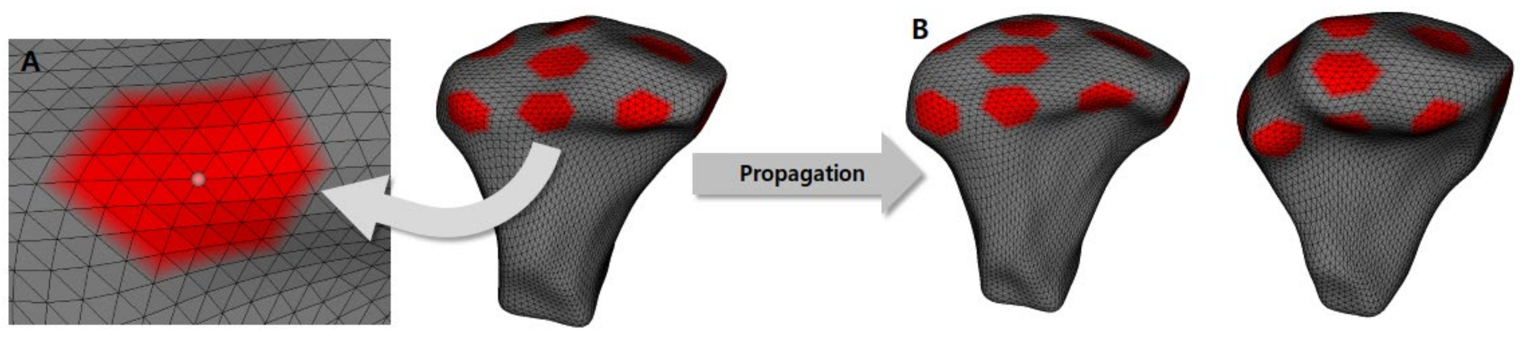
2.2. Image Preparation
2.3. Measurements
2.4. Statistical Analysis
3. Results
4. Discussion
5. Conclusions
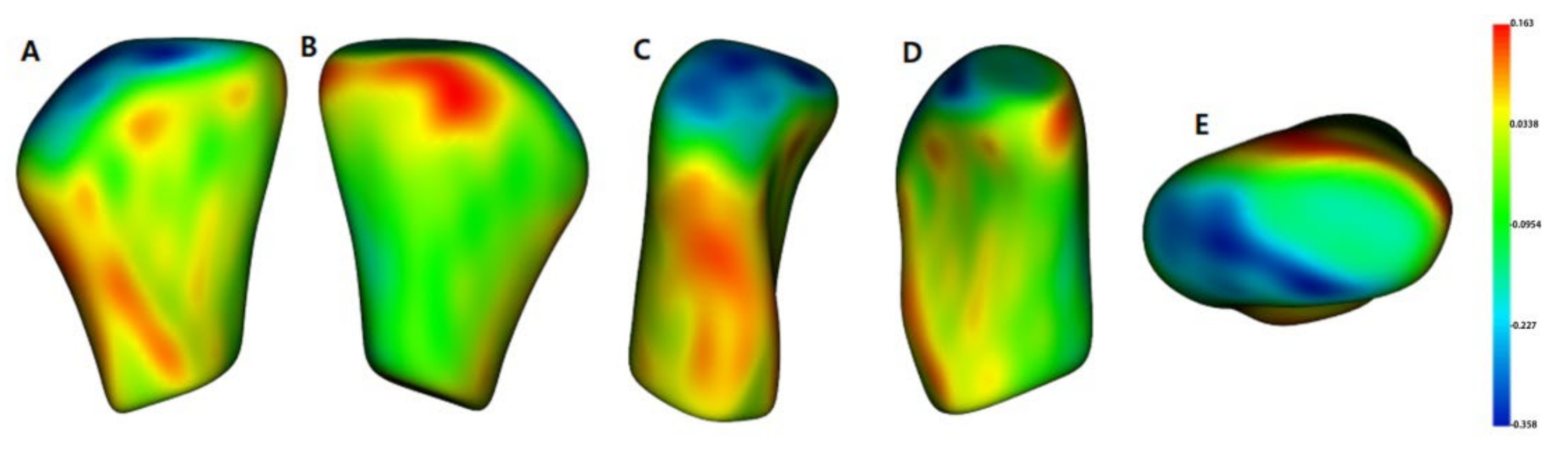
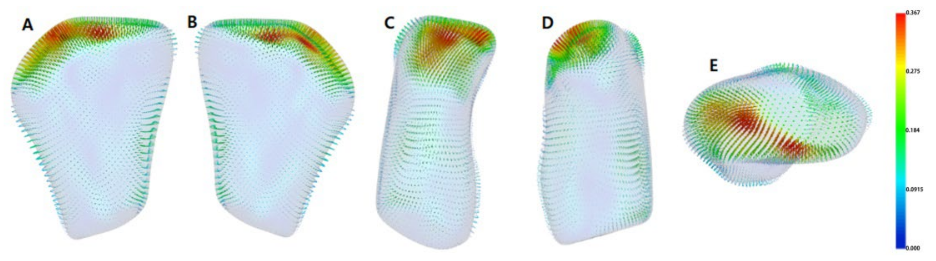
- After SS treatment in TMJ-OA patients, there was both bone resorption and bone formation on the mandibular condyle head surface, which induced morphological changes in the condyle head.
- Bone resorption of the mandibular condyle head surface was predominant in the AS, SL, and S areas, while bone formation was superior in the L, M, PS, and PM areas.
- The mandibular condyle volume changes before treatment (T1) and after SS treatment (T2) were not statistically significant.
Author Contributions
Funding
Institutional Review Board Statement
Informed Consent Statement
Data Availability Statement
Conflicts of Interest
References
- De Leeuw, R. American Academy of Orofacial Pain Guidelines for Assessment, Diagnosis, and Management, 4th ed.; Quintessence: Chicago, IL, USA, 2008. [Google Scholar]
- LeResche, L. Epidemiology of temporomandibular disorders: Implications for the investigation of etiologic factors. Crit. Rev. Oral Biol. Med. 1997, 8, 291–305. [Google Scholar] [CrossRef] [PubMed]
- Cascone, P.; Gennaro, P.; Gabriele, G.; Chisci, G.; Mitro, V.; De Caris, F.; Iannetti, G. Temporomandibular synovial chondromatosis with numerous nodules. J. Craniofac. Surg. 2014, 25, 1114–1115. [Google Scholar] [CrossRef] [PubMed]
- Helenius, L.; Tervahartiala, P.; Helenius, I.; Al-Sukhun, J.; Kivisaari, L.; Suuronen, R.; Kautiainen, H.; Hallikainen, D.; Lindqvist, C.; Leirisalo-Repo, M. Clinical, radiographic and MRI findings of the temporomandibular joint in patients with different rheumatic diseases. Int. J. Oral Maxillofac. Surg. 2006, 35, 983–989. [Google Scholar] [CrossRef] [PubMed]
- Milam, S.B. Pathogenesis of degenerative temporomandibular joint arthritides. Odontology 2005, 93, 7–15. [Google Scholar] [CrossRef]
- Tanaka, E.; Detamore, M.S.; Mercuri, L.G. Degenerative disorders of the temporomandibular joint: Etiology, diagnosis, and treatment. J. Dent. Res. 2008, 87, 296–307. [Google Scholar] [CrossRef]
- Pereira, F.J., Jr.; Lundh, H.; Westesson, P.L.; Carlsson, L.E. Clinical findings related to morphologic changes in TMJ autopsy specimens. Oral Surg. Oral Med. Oral. Pathol. 1994, 78, 288–295. [Google Scholar] [CrossRef]
- Machon, V.; Hirjak, D.; Lukas, J. Therapy of the osteoarthritis of the temporomandibular joint. J. Craniomaxillofac. Surg. 2011, 39, 127–130. [Google Scholar] [CrossRef]
- Manfredini, D.; Guarda-Nardini, L.; Ferronato, G. Single-needle temporomandibular joint arthrocentesis with hyaluronic acid injections. Preliminary data after a five-injection protocol. Minerva Stomatol. 2009, 58, 471–478. [Google Scholar]
- Mejersjö, C.; Wenneberg, B. Diclofenac sodium and occlusal splint therapy in TMJ osteoarthritis: A randomized controlled trial. J. Oral Rehabil. 2008, 35, 729–738. [Google Scholar] [CrossRef]
- Kuttila, M.; Le Bell, Y.; Savolainen-Niemi, E.; Kuttila, S.; Alanen, P. Efficiency of occlusal appliance therapy in secondary otalgia and temporomandibular disorders. Acta Odontol. Scand. 2002, 60, 248–254. [Google Scholar] [CrossRef]
- Al-Ani, M.Z.; Davies, S.J.; Gray, R.J.; Sloan, P.; Glenny, A. Stabilisation splint therapy for temporomandibular pain dysfunction syndrome. Cochrane Database Syst. Rev. 2004, 1, CD002778. [Google Scholar]
- Liu, M.Q.; Chen, H.M.; Yap, A.U.; Fu, K.Y. Condylar remodeling accompanying splint therapy: A cone-beam computerized tomography study of patients with temporomandibular joint disk displacement. Oral Surg. Oral Med. Oral Pathol. Oral Radiol. 2012, 114, 259–265. [Google Scholar] [CrossRef]
- Ok, S.M.; Lee, J.H.; Kim, Y.I.; Lee, J.Y.; Kim, K.B.; Jeong, S.H. Anterior condylar remodeling observed in stabilization splint therapy for temporomandibular joint osteoarthritis. Oral Surg. Oral Med. Oral Pathol. Oral Radiol. 2014, 118, 363–370. [Google Scholar] [CrossRef] [PubMed]
- Ahmad, M. Research diagnostic criteria for temporomandibular disorders (RDC/TMD): Development of image analysis criteria and examiner reliability for image analysis. Oral Surg. Oral Med. Oral Pathol. Oral Radiol. Endod. 2009, 107, 844–860. [Google Scholar] [CrossRef] [PubMed] [Green Version]
- Paniagua, B.; Cevidanes, L.; Walker, D.; Zhu, H.; Guo, R.; Styner, M. Clinical application of SPHARM-PDM to quantify temporomandibular joint osteoarthritis. Comput. Med. Imaging Graph. 2011, 35, 345–352. [Google Scholar] [CrossRef] [Green Version]
- Cevidanes, L.H.; Hajati, A.-K.; Paniagua, B.; Lim, P.F.; Walker, D.G.; Palconet, G.; Nackley, A.G.; Styner, M.; Ludlow, J.B.; Zhu, H.; et al. Quantification of condylar resorption in temporomandibular joint osteoarthritin. Oral Surg. Oral Med. Oral Pathol. Oral Radiol. Endod. 2010, 110, 110–117. [Google Scholar] [CrossRef] [Green Version]
- Yushkevich, P.A.; Piven, J.; Hazlett, H.C.; Smith, R.G.; Ho, S.; Gee, J.C.; Gerig, G. User-guided 3D active contour segmentation of anatomical structures: Significantly improved efficiency and reliability. Neuroimage 2006, 31, 1116–1128. [Google Scholar] [CrossRef] [Green Version]
- Cevidanes, L.H.S.; Gomes, L.R.; Jung, B.T.; Gomes, M.R.; Ruellas, A.C.O.; Goncalves, J.R.; Schilling, J.; Styner, M.; Nguyen, T.; Kapila, S.; et al. 3D superimposition and understanding temporomandibular joint arthritis. Orthod. Craniofac. Res. 2015, 18, 18–28. [Google Scholar] [CrossRef] [Green Version]
- Hollender, L.; Ridell, A. Radiography of the temporomandibular joint after oblique sliding osteotomy of the mandibular rami. Scand. J. Dent. Res. 1974, 82, 466–469. [Google Scholar] [CrossRef]
- Hollender, L.; Lindahl, L. Radiographic study of articular remodeling in the temporomandibular joint after condylar fractures. Scand. J. Dent. Res. 1974, 82, 462–465. [Google Scholar] [CrossRef]
- Paulsen, H.U. Morphological changes of the TMJ condyles of 100 patients treated with the Herbst appliance in the period of puberty to adulthood: A long-term radiographic study. Eur. J. Orthod. 1997, 19, 657–668. [Google Scholar] [CrossRef] [PubMed]
- Lundh, H.; Westesson, P.L. Long-term follow-up after occlusal treatment to correct abnormal temporomandibular joint disk position. Oral Surg. Oral Med. Oral Pathol. 1989, 67, 2–10. [Google Scholar] [CrossRef]
- Suei, Y.; Tanimoto, K.; Ogawa, I.; Wada, T. Formation of double contour in the human mandibular condyle. A case report. Oral Surg. Oral Med. Oral Pathol. 1994, 77, 327–330. [Google Scholar] [CrossRef]
- Pereira, F.J.; Lundh, H.; Eriksson, L.; Westesson, P.L. Microscopic changes in the retrodiscal tissues of painful temporomandibular joints. J. Oral Maxillofac. Surg. 1996, 54, 461–469. [Google Scholar] [CrossRef]
- Hasegawa, Y.; Kakimoto, N.; Tomita, S.; Honda, K.; Tanaka, Y.; Yagi, K.; Kondo, J.; Nagashima, T.; Ono, T.; Maeda, Y. Movement of the mandibular condyle and articular disc on placement of an occlusal splint. Oral Surg. Oral Med. Oral Pathol. Oral Radiol. Endod. 2011, 112, 640–647. [Google Scholar] [CrossRef]
- Ekberg, E.C.; Sabet, M.E.; Petersson, A.; Nilner, M. Occlusal appli- ance therapy in a short-term perspective in patients with tem- poromandibular disorders correlated to condyle position. Int. J. Prosthodont. 1998, 11, 263–268. [Google Scholar]
- Demling, A.; Fauska, K.; Ismail, F.; Stiesch, M. A comparison of change in condylar position in asymptomatic volunteers utilizing a stabilization and a pivot appliance. Cranio 2009, 27, 54–61. [Google Scholar] [CrossRef]
- O’Ryan, F.; Epker, B.N. Temporomandibular joint function and morphology: Observations on the spectra of normalcy. Oral Surg. Oral Med. Oral Pathol. 1984, 58, 272–279. [Google Scholar] [CrossRef]
- Elguy, D.; Elguy, M.; FiGekcioLlu, E.; Dolekog˘lu, S.; Ersan, N. Articular eminence inclination, height, and condyle morphology on cone beam computed tomography. Sci. World J. 2014, 6, 761714. [Google Scholar]
- Ok, S.-M.; Jeong, S.-H.; Ahn, Y.-W.; Kim, Y.-I. Effect of stabilization splint therapy on glenoid fossa remodeling in temporomandibular joint osteoarthritis. J. Pros. Res. 2016, 60, 301–307. [Google Scholar] [CrossRef]
- Brooks, S.L.; Brand, J.W.; Gibbs, S.; Hollender, L.; Lurie, A.G.; Omnell, K.; Westesson, P.-L.; White, S. Imaging of the temporomandibular joint: A position paper of the American Academy of Oral and Maxillofacial Radiology. Oral Surg. Oral Med. Oral Pathol. Oral Radiol. Endod. 1997, 83, 609–618. [Google Scholar] [CrossRef]
- Crow, H.C.; Parks, E.; Campbell, J.H.; Stucki, D.S.; Daggy, J. The utility of panoramic radiography in temporomandibular joint assessment. Dentomaxillofac. Radiol. 2005, 34, 91–95. [Google Scholar] [CrossRef] [PubMed]
- Ludlow, J.B.; Davies-Ludlow, L.E.; Brooks, S.L.; Howerton, W.B. Dosimetry of 3 CBCT devices for oral and maxillofacial radiology: CB Mercuray, NewTom 3G and i-CAT. Dentomaxillofac. Radiol. 2006, 35, 219–226. [Google Scholar] [CrossRef]
- Nguyen, T.; Cevidanes, L.; Paniagua, B.; Zhu, H.; Koerich, L.; De Clerck, H. Use of shape correspondence analysis to quantify skeletal changes associated with bone-anchored Class III correction. Angle Orthod. 2014, 84, 329–336. [Google Scholar] [CrossRef] [PubMed]


| Variables | Values |
|---|---|
| Gender | |
| Female | 25 |
| Male | 2 |
| TMJ OA | |
| Bilateral | 15 |
| Unilateral | 12 |
| Age (years) | |
| Mean (range) | 24.6 ± 3.9 (20.0–31.1) |
| Treatment duration (years) | |
| Mean (range) | 2.5 ± 1.3 (1.3–7.9) |
| Condylar Morphologic Changes (mm) | |||||||||||||
|---|---|---|---|---|---|---|---|---|---|---|---|---|---|
| A | S | P | L | M | AM | AL | PL | PM | SM | SL | AS | PS | |
| Mean | −0.02 | −0.14 | 0.01 | 0.05 | 0.09 | −0.05 | −0.02 | −0.1 | 0.05 | −0.09 | −0.19 | −0.22 | 0.09 |
| SD | 0.32 | 0.4 | 0.24 | 0.25 | 0.27 | 0.3 | 0.32 | 0.23 | 0.24 | 0.41 | 0.44 | 0.44 | 0.5 |
| Min | −1.4 | −1.57 | −0.77 | −0.90 | −0.62 | −1.5 | −1.71 | −1.04 | −0.61 | −1.14 | −1.84 | −1.84 | −2.2 |
| Max | 0.62 | 0.55 | 0.65 | 1.02 | 1.08 | 0.53 | 0.65 | 0.32 | 0.92 | 0.61 | 0.72 | 0.72 | 0.54 |
| Percentile | |||||||||||||
| 25th | −0.12 | −0.33 | −0.12 | −0.08 | −0.08 | −0.19 | −0.13 | −0.17 | −0.09 | −0.11 | −0.36 | −0.28 | −0.06 |
| 75th | 0.19 | 0.07 | 0.13 | 0.18 | 0.25 | 0.14 | 0.16 | 0.03 | 0.17 | 0.05 | 0.08 | 0.07 | 0.22 |
| Group | 1 | 2 | 3 | 4 | 5 | 6 | 7 | 8 |
|---|---|---|---|---|---|---|---|---|
| AS | −0.22 | |||||||
| SL | −0.19 | −0.19 | ||||||
| S | −0.14 | −0.14 | ||||||
| PL | −0.10 | −0.10 | ||||||
| SM | −0.09 | |||||||
| AM | −0.05 | −0.05 | ||||||
| A | −0.02 | −0.02 | ||||||
| AL | −0.02 | −0.02 | ||||||
| P | 0.01 | 0.01 | ||||||
| PM | 0.05 | 0.05 | ||||||
| L | 0.05 | 0.05 | ||||||
| M | 0.09 | |||||||
| PS | 0.09 | |||||||
| Sig. | 0.882 | 0.264 | 0.561 | 0.208 | 0.905 | 0.898 | 0.428 | 0.305 |
| Variable | T1 (Average ± SD) | T2 (Average ± SD) | p Value |
|---|---|---|---|
| Condylar volume, mm3 | 1182.64 ± 329.73 | 1149.54 ± 320.53 | 0.642 |
Publisher’s Note: MDPI stays neutral with regard to jurisdictional claims in published maps and institutional affiliations. |
© 2022 by the authors. Licensee MDPI, Basel, Switzerland. This article is an open access article distributed under the terms and conditions of the Creative Commons Attribution (CC BY) license (https://creativecommons.org/licenses/by/4.0/).
Share and Cite
Kim, T.-H.; Kim, Y.J.; Song, Y.-H.; Tae, I.; Lim, H.-K.; Jung, S.-K. Assessment of Morphologic Change of Mandibular Condyle in Temporomandibular Joint Osteoarthritis Patients with Stabilization Splint Therapy: A Pilot Study. Healthcare 2022, 10, 1939. https://doi.org/10.3390/healthcare10101939
Kim T-H, Kim YJ, Song Y-H, Tae I, Lim H-K, Jung S-K. Assessment of Morphologic Change of Mandibular Condyle in Temporomandibular Joint Osteoarthritis Patients with Stabilization Splint Therapy: A Pilot Study. Healthcare. 2022; 10(10):1939. https://doi.org/10.3390/healthcare10101939
Chicago/Turabian StyleKim, Tae-Hoon, Youn Joong Kim, Yun-Heon Song, Ilho Tae, Ho-Kyung Lim, and Seok-Ki Jung. 2022. "Assessment of Morphologic Change of Mandibular Condyle in Temporomandibular Joint Osteoarthritis Patients with Stabilization Splint Therapy: A Pilot Study" Healthcare 10, no. 10: 1939. https://doi.org/10.3390/healthcare10101939