

Dermatopathology 2024, 11(2), 142-146; https://doi.org/10.3390/dermatopathology11020014 - 19 Apr 2024
Abstract
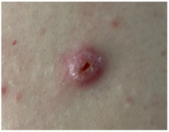
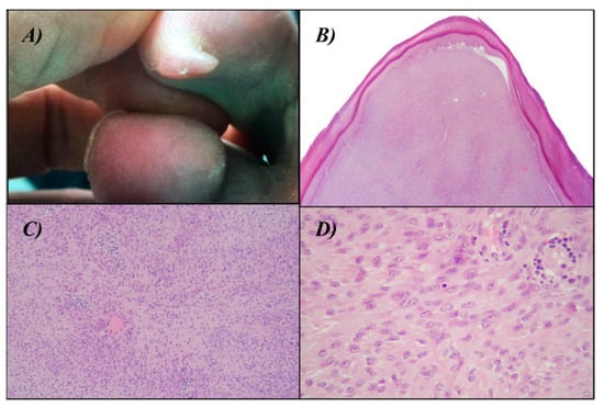
The intratarsal keratinous cyst (IKC) is a recently described entity, often clinically misdiagnosed as a chalazion. We report a case of a 61-year-old male patient with a chief complaint of a small lesion on the upper eyelid that evolved over six months. On
[...] Read more.
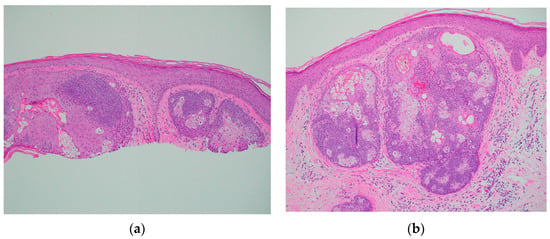
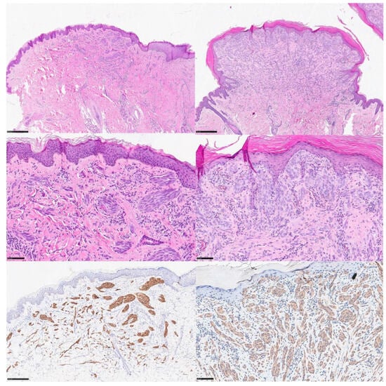
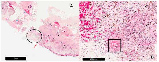
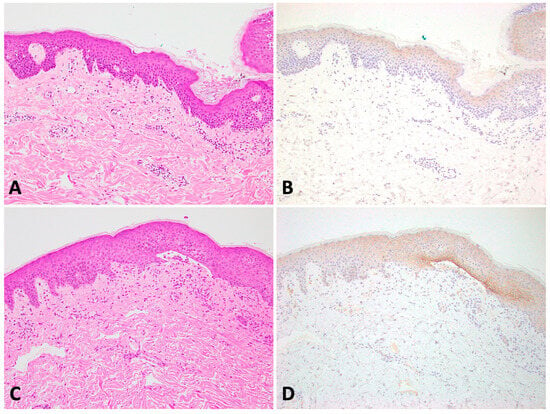
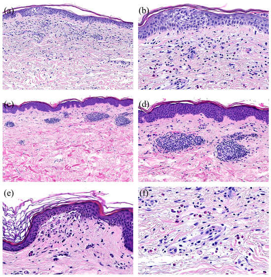
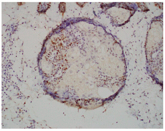
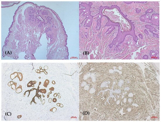
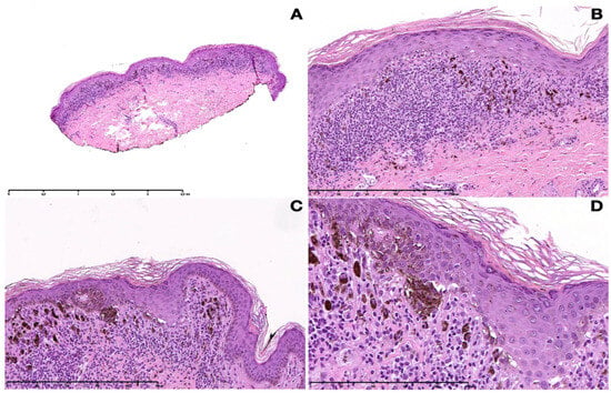
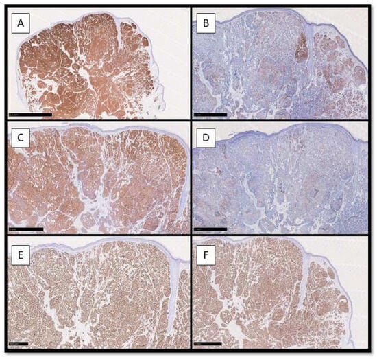
The intratarsal keratinous cyst (IKC) is a recently described entity, often clinically misdiagnosed as a chalazion. We report a case of a 61-year-old male patient with a chief complaint of a small lesion on the upper eyelid that evolved over six months. On physical examination, an asymptomatic, firm nodule was identified on the left upper eyelid. The patient reported no history of trauma. A provisional diagnosis of chalazion was established, and an excisional biopsy was performed. Histopathologically, the lesion was lined with a stratified squamous epithelium, with a corrugated epithelial surface showing abrupt keratinization without keratohyalin granules, and compact keratinous-appearing material in the cystic lumen. The diagnosis was IKC. No signs of recurrence were observed after one year of follow-up. It is essential to accurately diagnose IKC and distinguish it from chalazion and epidermal inclusion cysts, because IKC requires complete surgical excision and can exhibit multiple recurrences if not properly removed.
Full article
(This article belongs to the Special Issue Educational Case Reports in Dermatopathology)
►
Show Figures
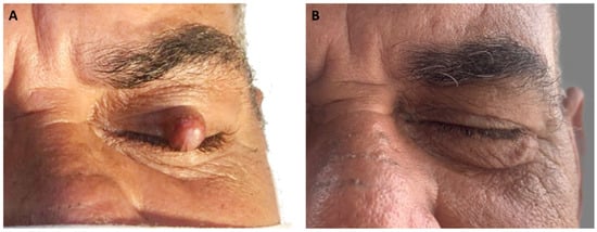
Figure 1
