Review

Diagnosing Cutaneous Melanocytic Tumors in the Molecular Era: Updates and Review of Literature

by
Chelsea Huang
1,*,
Tiffany Wing-See Lau
2 and
Bruce R. Smoller
3
1
Department of Pathology, Loma Linda University Medical Center, Loma Linda, CA 92354, USA
2
Department of Pathology, Queen Mary Hospital, Hong Kong, China
3
Department of Pathology and Laboratory Medicine, University of Rochester Medical Center, Rochester, NY 14642, USA
*
Author to whom correspondence should be addressed.
Dermatopathology 2024, 11(1), 26-51; https://doi.org/10.3390/dermatopathology11010005
Submission received: 31 October 2023 / Revised: 8 January 2024 / Accepted: 9 January 2024 / Published: 18 January 2024

Abstract

:
Over the past decade, molecular and genomic discoveries have experienced unprecedented growth, fundamentally reshaping our comprehension of melanocytic tumors. This review comprises three main sections. The first part gives an overview of the current genomic landscape of cutaneous melanocytic tumors. The second part provides an update on the associated molecular tests and immunohistochemical stains that are helpful for diagnostic purposes. The third section briefly outlines the diverse molecular pathways now utilized for the classification of cutaneous melanomas. The primary goal of this review is to provide a succinct overview of the molecular pathways involved in melanocytic tumors and demonstrate their practical integration into the realm of diagnostic aids. As the molecular and genomic knowledge base continues to expand, this review hopes to serve as a valuable resource for healthcare professionals, offering insight into the evolving molecular landscape of cutaneous melanocytic tumors and its implications for patient care.

1. Introduction

The exponential growth of molecular and genomic discoveries in the past decade has laid the foundation of the molecular landscape for melanocytic tumors. This unraveled ancillary molecular testing methods and the development of immunohistochemical (IHC) markers toward diagnostic and prognostic evaluations. This article reviews the current genomic landscape of cutaneous melanocytic tumors; briefly provides an overview of the molecular pathways of cutaneous melanomas; and reviews the integration of molecular studies into the differential diagnosis, classification, prognosis, and therapeutic responses of melanocytic tumors. We aim to provide a brief molecular overview and illustrate their practical integration as diagnostic aids.

2. Background of the Genomic Landscape
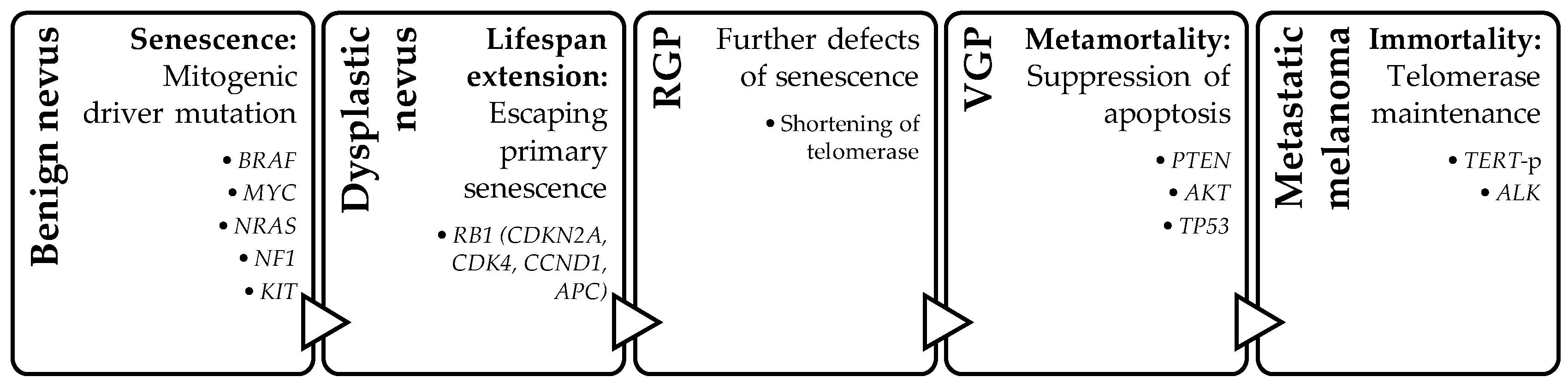
Various genomic studies, including whole-genome sequencing, reveal that the accumulation of multiple genetic alterations contributes to the complex and heterogeneous nature of melanoma development and progression [1]. The genomic landscape of melanoma can vary between individuals and within different subtypes of melanoma. In addition to diagnostic and prognostic purposes, understanding the genomic landscape of melanoma is crucial for developing targeted therapies and personalized treatment approaches.

Four-Step Model

Several genetic and epigenetic driver changes are identified in melanoma. The complex interplay between genetic alterations and epigenetic modifications contributes to the development and progression of melanoma. The proposed genetic model for melanoma progression includes a four-step model: senescence, lifespan extension, apoptosis suppression, and immortality. Histologically, the corresponding morphologies of this four-step sequence are as follows: benign nevus, dysplastic nevus, thin melanoma/radial growth phase (RGP), invasive melanoma/vertical growth phase (VGP), and metastasis [2]. Not all mutations include all four steps or happen in the above-described sequence; however, these four steps represent the most common sequence in the development of metastatic melanoma [2]. The initial step involves the mitogenic driver mutation in the mitogen-activated protein kinases (MAPKs) pathway, which stimulates cell proliferation by mimicking growth factor signaling. This process includes the interaction of genomic, environmental, and host factors that lead to mutations in BRAF, MYC, NRAS, ERBB4, PTPs, NF1, KIT, etc. The second step involves additional mutations that overcome tumor suppressor mechanisms, mainly through disruption of the p16 or RB1 pathway (CDKN2A, CDK4, CCND1, APC, etc.), allowing the cells to escape primary senescence and leading to a lifespan extension. The third step involves mutations that suppress apoptosis (TP53, APAF1, PTEN, PTPs, PREX2, PTKs, AKT, etc.). The fourth and final step allows the tumor cells to achieve immortality through telomerase reverse transcriptase promoter (TERT-p) or ALK alterations, which overcome replicative senescence through longer telomeres [2]. (Figure 1).

3. Genetic Change: Dysregulation of Three Main Oncogenic Signaling Pathways

In melanomas, the three most frequently dysregulated oncogenic signaling pathways are the MAPK, phosphatidylinositol 3-kinase (PI3K)-AKT, and Wnt/β-catenin signaling pathways.

3.1. MAPK Pathway

The MAPK pathway, also known as the RAS-RAF-MEK-ERK signaling pathway, is a classic intracellular pathway that plays a crucial role in the homeostasis of normal cell turnover, cellular proliferation, differentiation, survival, and apoptosis. Aberrant activation of this signaling pathway induces tumorigenesis and was found to be associated with various malignancies. The activation of the MAPK pathway is via growth factors binding to receptor tyrosine kinases (RTKs) on the cell surface. This leads to the activation of sequential downstream components: Ras, Raf kinases, MEK, and ERK [3]. In melanomas, the hyperactivation of the MAPK pathway is mainly through activating mutations in BRAF, NRAS, NF1, and KIT [4]. Alterations in these genes are usually mutually exclusive in untreated melanomas. These alterations can give rise to melanocytic nevi and, therefore, arise early in the progression of melanomas [5]. The most frequently seen primary oncogenic event in melanomas is the BRAF-activating mutation, which leads to permanent activation of the MAPK pathway and subsequently promotes uncontrolled cell proliferation, survival, and other oncogenic processes [6,7].
BRAF mutations are found in around 50% of all melanomas, with p.V600E being the most common mutation, accounting for 80–90% of BRAF-mutated melanomas [8]. Compared with patients without a BRAF mutation, BRAF-mutated melanomas are seen in the younger age group of <40 years old and in anatomical regions with zero-to-low cumulative sun damage (CSD). Histological characteristics of BRAF-mutated melanomas include superficial spreading and/or nodular growth patterns. Other features include pagetoid scatter, nest formation, sharp demarcation, and thickened epidermis. Cytologically, the melanocytes are larger and rounder with more pigment. In contrast, the presence or absence of ulceration, tumor thickness, and mitotic count are not associated with the presence or absence of BRAF mutation [9].
Mutations in NRAS account for approximately 20% of all melanomas [10,11,12,13]. NRAS mutations are associated with an older age group (>55 years) and high CSD, with a preferential occurrence on the upper extremities. Histologically, NRAS-mutated melanomas feature thicker primary tumors (greater Breslow depth), greater levels of mitosis, and infrequent ulceration. Prognostically, compared with the BRAF-mutated NRAS wild-type melanomas, the NRAS-mutated melanomas have been related to more aggressive biological behavior with a higher risk of distant metastasis [14]. Of note, NRAS is the most common mutation seen in nevoid melanoma, especially the papillomatous type [15].
Also associated with an older age group and high CSD are melanomas with somatic NF1 mutations. NF1 mutation is found in 10–17% of cutaneous melanomas [11,13,16,17]. Typically, NF1 mutations are found in melanomas that lack mutations of BRAF and NRAS. In contrast to the triple wild-type (BRAF wild-type, NRAS wild-type, and NF1 wild-type) melanomas and the BRAF- and NRAS-mutated melanomas, NF1-mutated melanomas have a strong association with ultraviolet (UV) damage, as evidenced histologically by a higher degree of background solar elastosis in the dermis [18]. The NF1 loss-of-function mutation is found in 45–95% of desmoplastic melanomas (DMs), which is a subtype of melanoma with severe background solar elastosis. DM is associated with a high propensity for local recurrence and occasional regional lymph node spread [19]. Overall, due to the high CSD and the association with a strong UV signature, NF1-mutated melanomas are found to harbor a greater mutational burden [20]. The high mutational burden in NF1-mutated melanomas demonstrates a favorable outcome for immune checkpoint inhibitor therapy [21].
NRAS-mutated melanomas and NF1-mutated melanomas are both UV-driven but they are different phenotypically and biologically. An NF1 mutation is associated with desmoplastic melanomas, while an NRAS mutation is associated with nevoid melanomas. NRAS-mutated melanomas are aggressive with a high risk of distant metastasis. NF-mutated melanomas are associated with local recurrence and regional lymph node spread.
Activating KIT mutations are found in 20–90% of mucosal (anorectal and oral cavity) melanomas, approximately 36% of acral melanomas (AM), and approximately 28% of high-CSD cutaneous melanomas [22,23]. Even though KIT-mutated melanomas are found to be closely associated with older age [24], there is still inconsistency regarding the association with other clinicopathological features, such as gender, Breslow thickness, histological types, ulceration, mitotic rate, and tumor stages through different studies [25].
In the MAPK pathway, targeted therapies, such as BRAF-inhibitors (e.g., vemurafenib, dabrafenib) and MEK-inhibitors (e.g., trametinib), have been developed to specifically inhibit components of the MAPK pathway in melanomas. These drugs have shown significant efficacy in patients with BRAF-mutant melanomas by blocking the excessive signaling and suppressing tumor growth. While targeted inhibition of BRAF and MEK has resulted in improved survival in patients with BRAF V600E-mutated melanoma, therapeutic resistance is often the end result [26]. The PI3K-AKT pathway plays a significant role in BRAF-/MEK-inhibitor resistance in melanoma patients and may represent a crucial target for combination therapy.

3.2. PI3K-AKT Pathway

The PI3K-AKT pathway is one of the most important signaling networks in cancer. Activation of this pathway plays a significant role in melanomas, frequently in the setting of concurrent activation of the MAPK signaling pathway through upstream RAS oncogene activation [27]. Activation of the RAS oncogene in the MAPK pathway leads to the downstream activation of two different but interconnected pathways: the RAF–MEK–ERK pathway and the PI3K-AKT signaling pathway [28]. The PI3K-AKT pathway is activated by various growth factors and cytokines. It involves the activation of PI3K, leading to the production of phosphatidylinositol (3, 4, 5)-trisphosphate (PIP3). PIP3 recruits and activates AKT (protein kinase B), which then regulates multiple downstream effectors involved in cell survival, growth, and metabolism. Dysregulation of the PI3K-AKT pathway in melanomas is commonly observed through mutations or amplifications in genes such as PTEN, PIK3CA, and AKT1. PTEN is a negative regulator of the pathway, while PIK3CA is responsible for encoding the catalytic subunit of PI3K. Dysregulated PI3K-AKT signaling promotes cell proliferation, survival, and resistance to apoptosis [29].
The common feature in PTEN-altered melanomas is the advanced stage at diagnosis. Loss of PTEN accumulates throughout the disease as it progresses; hence, the association with advanced-stage melanomas. Some survival analysis studies show that PTEN-retained tumors are linked to a better patient outcome, demonstrating the crucial role of this tumor suppressor gene. In contrast, PTEN loss is associated with poor survival [30]. Tumor-infiltrating lymphocytes, T-cells, and cytotoxic T-cells are found to be decreased in number and function in tumors with PTEN loss. Thus, PTEN loss is associated with resistance to immunotherapy in melanomas [31].

3.3. Wnt/β-Catenin Pathway

The Wnt/β-catenin pathway is a key component in embryonic development, tissue homeostasis, and stem cell maintenance. Activation of this pathway occurs when the Wnt ligands bind to Frizzled receptors and co-receptors, leading to β-catenin stabilization and translocation into the nucleus. In the nucleus, β-catenin interacts with transcription factors, regulating the expression of target genes involved in cell fate determination and proliferation [32]. Wnt-signaling plays an important function in the skin by guiding the migration of neural crest cells and multipotent precursor cells and driving them toward a melanocyte fate, including terminal differentiation of melanoblasts to melanocytes [33]. In melanomas, Wnt/β-catenin pathway dysregulation leads to aberrant activation of the pathway through mutations in genes such as CTNNB1 (encoding β-catenin) or APC.
Although dysregulated Wnt/β-catenin signaling promotes cell proliferation, survival, and invasion, the activation of Wnt/β-catenin signaling in melanomas is found to be associated with a lower proliferative index and correlates with a more favorable prognosis [34]. A study on a murine model shows that melanoma cells expressing Wnt3a, a member of the Wnt family, behave similarly to highly differentiated melanocytic cells (high degree of pigmentation, alterations in the cell cycle leading to decreased proliferation, upregulation of melanocytic genes, and formation of smaller tumors) [34]. Another study on mice indicates that Wnt3a expression is linked to decreased metastasis [35]. The murine models reflect the key roles that Wnt3a plays in regulating cell differentiation, proliferation, regeneration, and motility [36].
Wnt-activated deep penetrating/plexiform nevus (DPN) features combined activation of the MAPK and Wnt signaling pathways. Hence, DPN is molecularly defined by the Wnt/β-catenin and MAPK pathway mutations [37]. Mutations of the β-catenin pathway change the phenotype of a BRAF-mutated conventional nevus into that of DPN. Demographically, DPN are typically seen in a younger age group as small, sub-centimeter-sized, solitary, dark-pigmented papules/nodules with a predilection for the head and neck region [38]. Histologically, DPN feature symmetrical, well-demarcated, wedge-shaped, dermal-based lesions that extend down into the deep reticular dermis, and sometimes the subcutis. The lesion comprises fascicular or plexiform nests of large uniform epithelioid to spindled melanocytes with low-grade atypia and near-absent mitoses [39]. Despite the deep-seated nature and occasional mild atypia, most DPN have a good prognosis with benign behavior. Both DPN and DPN-like melanomas share activation of the Wnt/β-catenin pathway, suggesting that some DPN can progress to melanoma. Additional molecular oncogenic alterations (CDKN2A and TERT-p) are necessary for the development of DPN-like melanomas [37]. Morphologically, in addition to the histological features reminiscent of a DPN, DPN-like melanomas show increased cellularity, atypical junctional component with pagetoid spread, poorly circumscribed border and increased depth of lesion, hyperplasia or atrophy of the epidermis, increased tumor cellularity, significant cytological atypia, prominent nuclear pseudo-inclusions, abundant pale cytoplasm, increase mitoses, and associated melanophages [38,40,41]. Other features include ulceration, necrosis, inflammatory reaction, perineural invasion, and lymphovascular invasion [37,42]. Using IHC, DPN and DPN-like melanomas both express β-catenin (cytoplasmic and membranous), cyclinD1 (nuclear and cytoplasmic), and LEF1 [37,43].
These three oncogenic signaling pathways (MAPK, AKT, and Wnt/β-catenin) are interconnected and often coexist in melanomas. They contribute to the dysregulated cellular processes that drive melanoma development, including abnormal cell growth, survival, invasion, and metastasis.

4. Epigenetic Changes: DNA Methylation, Histone Modification, and MicroRNA Dysregulation

While the above-described genetic changes alter the protein end product, epigenetic changes affect gene expression by turning it “on” or “off” and making no changes to the DNA sequence [44]. Epigenetic driver changes further add to the complex interplay between genetic alterations and epigenetic modifications toward melanoma development and progression. The three main epigenetic modifications are DNA methylation changes, histone modifications, and microRNA dysregulation.

4.1. DNA Methylation Changes

DNA methylation is an epigenetic modification that involves the addition of a methyl group to DNA molecules, often leading to gene silencing and affecting the expressions of genes involved. It is considered the underlying primary epigenetic mechanism for melanoma development and progression [45]. Numerous genes were found to be involved with DNA methylation in melanomas. Some of those genes include O6-methylguanine-DNA methyltransferase (MGMT), CDKN2A, Ras-association domain family 1 isoform A (RASSF1A), TBC Domain Family Member 1(TBC1D16), Platelet-Derived Growth Factor D (PDGFD), Thyroid Hormone Receptor Beta (THRB), and Zinc Finger E-Box Binding Homeobox 1 (ZEB1) [46].

4.2. Histone Modifications

Histones are proteins that help to package DNA in the nucleus, and they play a role in gene regulation. Chromosomal DNA is packed into nucleosomes with DNA enveloped around histone protein complexes, which consist of subunits named H2A, H2B, H3, and H4. Modification of the histone subunits either activates or silences gene transcription. Histone subunit methylation and acetylation regulate genetic expression by controlling the DNA accessibility to the transcriptional process and through the participation of additional protein complexes [47]. Abnormal histone modifications, such as acetylation, methylation, or phosphorylation, can impact gene expression and contribute to melanoma development. For example, the loss of histone variant macroH2A, which is generally considered to be transcriptionally repressive, promotes melanoma progression [48]. The overexpression of histone variant H2A.Z.2 promotes cell cycle progression in melanomas and is associated with poor prognoses [49,50].

4.3. MicroRNA Dysregulation

MicroRNAs (miRNA) are small non-coding RNA molecules that play a role in regulating gene expression by binding to target messenger RNAs (mRNAs). The dysregulation of miRNAs observed in melanomas is associated with tumor growth, invasion, and metastasis [51,52,53]. Although there is still limited data on miRNA expression profiles in melanomas, molecular techniques, such as quantitative in situ hybridization (qISH) for the fluorescent detection of candidate miRNAs, reverse transcription quantitative real-time polymerase chain reaction (RT-qPCR), and miRNA microarray, have been used to assess different miRNA expression levels in melanomas by comparing normal melanocytes with benign melanocytic lesions. miRNA expression levels can also be compared between primary and metastatic melanomas [54]. In one study that compared primary melanomas to metastatic melanomas, a low expression of miR-203 through DNA methylation was observed in metastatic melanomas, and it was also associated with poor survival. It was suggested that hypermethylation of the miR-203 promoter is a potential mechanism of tumor metastasis [55]. Several studies suggested the role of miRNAs in regulating sensitivity to BRAF-targeted therapy [56,57].

5. Practical Molecular Knowledge for a Diagnostic Surgical Pathologist

This section reviews the updated available molecular IHC stains and molecular tests that can assist in diagnosis, monitor prognosis, and assess therapeutic response.

5.1. IHC for Assessment of Molecular Alteration

Although molecular genetic diagnostic techniques are useful, IHC remains the most cost-effective and most frequently performed tool as the next step to aid in differential diagnosis. Currently, the available IHCs that can assess genomic events include VE1, NRASQ61R, ALK, ROS1, Pan-TRK, BAP1, PRKAR1A, β-catenin, LEF1, and p16 [58,59,60]. This IHC section is divided into two parts. The first part briefly reviews each IHC method and the second part elaborates on the application for selected melanocytic lesions (Table 1).

5.1.1. Genomic Events That Can Be Assessed Using IHC

VE1

VE1 IHC assesses an alteration in BRAF V600E, which is commonly detected in melanocytic lesions in intermittently sun-exposed skin, such as SSM, conventional nevi, dysplastic nevi, and simple lentigo. However, a positive VE1 stain can also be seen in other melanocytic lesions, such as nevoid melanomas, nodular melanomas, pigmented epithelioid melanocytomas (PEMs), BAP1-inactivated melanocytic lesions, and acral melanocytic lesions. Careful interpretation of VE1 needs to be based on the foundation of histological features. Additional stains can help with difficult cases. For example, BAP1-inactivated melanocytic lesions are positive for VE1 in addition to BAP1 loss, while PEM is positive for both VE1 and PRKAR1A.
For lesions with BRAF fusion, such as Spitz lesions, IHC showing VE1-negative staining cannot specifically detect BRAF fusions because wild-type BRAF can be overexpressed in other melanocytic tumors [61]. Also, because a significant proportion of BRAF fusions are the result of inversions or deletions, a fluorescence in situ hybridization (FISH) break-apart probe may not be sufficient to determine the fusion. Next-generation sequencing (NGS) is the test of choice to produce a more reliable result in such cases.

NRASQ16R

The most common NRAS alteration, accounting for 82.4% of all NRAS mutations, was found to be glutamine-to-arginine substitution at position 61 (NRAS Q61R) through the largest single institution cohort [62]. The development of NRAS Q16R IHC provides an accurate, rapid, and cost-effective method for detecting the presence of an NRAS Q61R mutation in melanomas [63]. Detection of positive IHC suggests melanomas in the high-CSD pathway.

ALK, ROS1, and Pan-TRK

Spitz lesions harbor several alterations, including a few that can be assessed using IHC. Pan-TRK IHC identifies NTRK1, NTRK2, and NTRK3 alterations. ALK (clone 5A4) [64] and ROS1 IHC identify mutations in ALK and ROS1, respectively.

β-Catenin and LEF1

Uniform strong nuclear staining with β-catenin and/or LEF1 IHCs suggests the diagnosis of DPN or DPN-like melanomas, confirming the altered Wnt/β-catenin pathway that is distinct in this group of lesions. Lesions without alteration in the Wnt/β-catenin pathway show a gradient expression that features intense staining superficially and gradual loss of staining with increasing depth [65]. β-catenin and LEF1 IHCs are useful in differentiating challenging melanocytic lesions that mimic DPN, such as blue nevi and cellular blue nevi.

BAP1

BAP1-inactivated nevi or melanocytomas harbor bi-allelic inactivation of BAP1, which can typically be demonstrated through a loss of BAP1 nuclear expression on IHC [66]. However, this IHC may not be fully sensitive for the detection of all BAP1 alterations. BAP1-alteration can occur through pathogenic missense mutations, resulting in an impaired but not fully inactivated function of BAP1 protein [66].

PRKAR1A

A subset of PEMs is characterized by the combination of two molecular alterations, including an initial alteration in the MAPK pathway and a second alteration in the PRKAR1A gene [67]. The PRKAR1A alteration is via a loss-of-function mutation. Using IHC, a positive expression of VE1, in addition to the loss of cytoplasmic staining of PRKAR1A, is helpful to support the diagnosis of PEM. However, there is a subset of pure PEMs with PRKCA fusion instead of loss of function mutation. In PEMs with PRKCA fusion, PRKAR1A expression is retained on IHC [61].

P16

P16 is a tumor suppressor and splice product of CDKN2A. Complete loss of staining in p16 indicates biallelic or homozygotic inactivation of the CDKN2A gene that corresponds to the late molecular event in the oncogenesis in advanced cutaneous melanomas [68]. A strong diffuse or mosaic pattern of p16 indicates no alteration in CDKN2A. P16 is useful in three diagnostic scenarios: the differential diagnosis of nodal nevi from metastatic melanomas, evaluation of suspicious atypical melanocytic lesions for malignancy, and identification of aggressive phenotype acquired in conventional melanomas. In sentinel lymph node biopsies, p16 block negativity is seen in metastatic melanomas, while p16 mosaic staining indicates nodal nevi. In dermal or nodular atypical melanocytic lesions, the identification of p16 loss reflects the biallelic inactivation that is strongly associated with malignancy. P16 loss can be seen in areas in melanomas with additional inactivation of CDKN2A, indicating a more aggressive biology [69,70].
However, several different cohorts from various centers still show variability in the definition of “positive p16” in terms of p16 staining location in the cell (nuclear or cytoplasmic), percentage, and pattern [71]. A reproducible interpretation and criteria that define a positive p16 reactivity for melanocytic lesions still need to be refined.

5.1.2. Selected Melanocytic Lesions

Spitz Family

Spitz melanomas and spitzoid melanomas often share similar and overlapping histologic features; however, the underlying molecular alterations are distinct. A “true” Spitz melanoma shares the same molecular alteration as lesions of the Spitz family, namely, kinase fusion or HRAS aberration. Driver mutations of a conventional melanoma, such as BRAF, NRAS, or NF1, should not be seen in the Spitz family. In melanomas showing Spitz-like features, the presence of VE1 and/or NRAS Q61R IHC helps to confirm the conventional melanoma pathway alteration in BRAF V600E and/or NRAS Q61R, respectively. The appropriate classification of such a lesion should be spitzoid melanoma. On the other hand, IHC with positive pan-TRK, ALK, or ROS1 with the absence of VE1 or NRASQ61R signifies a true Spitz melanoma.

Deep Penetrating Nevus

By the WHO definition, DPN is caused by the combination of MAPK and Wnt/β-catenin signaling pathway activations. Alteration in these two pathways can be detected using positive IHC for VE1, in combination with positive β-catenin, respectively.
As mentioned in the previous section, DPN and DPN-like melanomas both express β-catenin, cyclinD1, and LEF1 due to the shared Wnt/β-catenin pathway alteration. The main difference is that DPN-like melanomas often harbor additional genetic alterations, including TERT-p mutations and inactivation of CDKN2A. While TERT-p is analyzed using sequencing, the assessment of CDKN2A can be tested using p16 IHC.

5.2. Molecular Tests

Molecular assessments for melanomas can be divided into four categories: tests that assess copy number abnormalities, gene expression profiling, mutation analysis, and imaging mass spectrometry (Table 2).

5.2.1. Genomic Copy Number Assessment

Melanomas and benign melanocytic nevi show many overlapping histopathological features and are often misdiagnosed. Using comparative genomic hybridization (CGH), early research discovered significant differences between melanomas and nevi through copy number variations. Melanoma is characterized by an unstable genome with numerous DNA copy number abnormalities (CNA), whereas nevus shows a lack of or very limited CNA [72]. The discovery of the distinct non-overlapping pattern in genomic abnormalities led to the development of diagnostic platforms to differentiate nevi and melanomas using CNA. The two currently available tests are CGH/single nucleotide polymorphism (SNP) array and FISH [60].

CGH/SNP/MIP

CGH is a type of DNA microarray-based technique that provides information on genomic alterations (amplifications or deletions) by comparing the DNA copy number changes between a reference DNA sample and a test sample. The visualization of the intensity ratio of the fluorochromes is used to determine the relative gain or loss of tumor DNA compared with the normal reference at each locus [73]. In melanomas, CGH identifies the genetic alterations by comparing the DNA of melanoma cells with normal DNA samples. The recently emerged SNP microarrays can provide allele frequency data in addition to copy number changes. It is useful to identify selected mutations centering specific SNP and detect copy-neutral loss of heterozygosity events [74]. The uses of CGH/SNP arrays provide an opportunity for diagnostic strategies that help to differentiate melanomas from melanocytic nevi using the non-overlapping pattern of chromosomal aberrations and evaluating DNA copy number alterations [75,76]. One of the challenges in performing CGH/SNP arrays in melanocytic tumors is the low DNA quantities in biopsy samples. Most of the formalin-fixed paraffin-embedded (FFPE) tissue from small biopsies yields a low amount of degraded DNA, resulting in insufficiency for most CGH/SNP array platforms [77]. In the recent decade, novel protocols, such as techniques based on molecular inversion probes (MIP), have improved the ability to analyze degraded DNA [78]. The MIP, with a footprint of only 40 bp that targets SNPs, allows for the evaluation of fragmented DNA from FFPE tissue. The advantage of incorporating MIP is the improved signal-to-noise ratio compared with the conventional CGH/SNP arrays. All the unused MIPs and target DNAs are degraded by exonuclease, leaving behind only the probes. The probes are further amplified and hybridized into a microarray [79].
For melanocytic lesions, a CGH/SNP array is used to aid in the diagnosis of histologically ambiguous melanocytic neoplasms. Most melanomas have an unstable genome with multiple segmental DNA abnormalities, whereas the majority of benign melanocytic nevi have no chromosomal aberrations or have isolated chromosomal gains and losses that are not commonly seen in melanomas. The detection of multiple segmental genomic abnormalities on CGH/SNP suggests a diagnosis of melanoma.
A CGH/SNP array can be used to differentiate spitzoid melanoma from Spitz nevus. The majority of Spitz nevi are found to carry a normal karyotype using a CGH assay; however, 20% of Spitz nevi are found to have an isolated gain in chromosome 11p (HRAS locus) [80,81]. This aberration is almost never found in melanomas or other nevi. Another major distinction between Spitz nevus and spitzoid melanoma lies in the difference in their genomic stability [82]. Assessment of an HRAS mutation using CGH/SNP is a reproducible method to help in the differential diagnosis between Spitz nevus and spitzoid melanoma [83].
While CGH/SNP’s novel protocols based on MIP allow for the analysis of small or degraded tissue samples, this technique still requires >25% tumor purity to yield reliable results [84]. In addition to tumor purity on the tissue sample, 10 unstained slides are usually required for CGH/SNP array testing. Because of these requirements, cases of superficial or in situ melanocytic tumors with background heavy inflammatory infiltrate and cases with only a few slides available will be unsuitable for CGH/SNP array analysis [85].

Fluorescence in Situ Hybridization (FISH)

Fluorescence in situ hybridization (FISH), similar to CGH, allows for the detection of genetic abnormalities in cancer cells and is used as an alternative by evaluating a limited number of genomic loci for CNAs. The advantage of FISH is that it requires only 1–2 tissue sections and it allows for the direct visualization of tumor cells under a fluorescence microscope. This facilitates the analysis of small lesions with limited tissue samples and lesions with associated heavy inflammatory infiltrates. The disadvantage of FISH is that it is a targeted technique that requires prior knowledge of the specific genetic alterations being investigated to detect them, such as chromosomal rearrangements or gene amplifications [86]. The four-probe FISH assay targeting 6p25 (RREB1), 6q23 (MYB), CEP6 (centromere 6), and 11q13 (CCND1) helps to differentiate histologically unequivocal melanomas from benign nevi [86,87,88,89]. FISH positivity can assist in diagnosing the “morphologically intermediate” nevoid melanocytic neoplasms [90]. Subsequently, these sets were modified to 9p21 (CDKN2A), 6p25 (RREB1), 11q13 (CCND1), and 8q24 (MYC), improving the discriminatory power in differentiating melanomas from nevi [91]. More specifically, 11q13 (CCND1) amplification using FISH can be used as a diagnostic marker for histologically undetermined early acral melanoma in situ [92].
The standard four-probe FISH assay should not be applied to tumors for which melanoma arising in blue nevus is a consideration because genomic CNAs typically observed in melanoma arising in blue nevus (GNAQ and GNA11) are different than those of the typical melanoma pathways [59].
Although the standard four-probe FISH shows high sensitivity and specificity for conventional melanomas, it has a low sensitivity for detecting spitzoid melanomas [93,94]. The most commonly lost genomic region in spitzoid melanomas was found to be chromosome 9p from the CGH database [95]. Hence, the adaptation of a five-probe FISH assay targeting against 9p21 (CDKN2A), 11q13 (CCND1), and 8q24 (MYC) has improved the discriminatory power for spitzoid melanomas [93,96,97]. Tetraploidy, which is commonly seen in Spitz nevi, is another false-positive caveat in the interpretation of FISH due to the increase in absolute signal counts, which incorrectly reflects the relative imbalances in the actual tumor genome.
Compared with CGH, FISH analysis seems to be more cost-effective; however, it has a relatively high false-negative rate in analyzing melanocytic lesions. FISH investigates only 4–6 genomic loci. Mutations, small insertions or deletions, and genomic rearrangements cannot be detected using FISH. Therefore, both positive and negative FISH results need to be analyzed in the proper clinicopathological context [90].

5.2.2. Gene Expression Profiling

Gene expression profiling (GEP) assists with the diagnosis and prognosis of melanocytic tumors. The steps of GEP include RNA extraction from the FFPE blocks or tape-stripping from the surface of pigmented lesions, reverse transcription to complementary DNA (cDNA), and then amplification via real-time reverse transcription polymerase chain reaction (RT-PCR) [59,98]. All cells on the tumor tissue, including neoplastic melanocytes, stromal cells, and inflammatory cells, are measured for gene expression. Depending on the different GEP tests used, a proprietary algorithm is used to provide a numerical score range corresponding to the likelihood of the tumor being benign, intermediate, or malignant based on the expression of the genes [99]. The overall sensitivity of the tests is between 90–95% with a variable specificity [100,101]. A few commercially available testing platforms include Myriad myPath test and DecisionDx-Melanoma™.
The Myriad myPath test evaluates the expressions of 23 genes, including the 14 genes involved in melanoma pathogenesis and 9 housekeeping genes. The 14 genes are categorized into three components related to cell differentiation, cell signaling, and immune response. More specifically, one gene related to tumorigenesis (PRAME), eight genes related to immune signaling (CCL5, CD38, CXCL10, CXCL9, IRF1, LCP2, PTPRC, and SSL), and five genes with multifunctional roles (S100A9, S100A7, S100A8, S100A12, and PI3). The performed test generates an algorithmic myPath score ranging from −16.7 to 11.1. Higher positive scores indicate a higher suspicion of malignant disease. The test report classifies scores as “benign” (−16.7 to −2.1), “indeterminate” (−2.0 to −0.1), or “malignant” (0.0 to +11.1) [102]. The myPath test is an add-on ancillary test to the standard histopathology assessment. The aim of myPath is to aid clinicians in assessing the likelihood of malignancy based on tumor molecular profile, providing valuable information for personalized patient management decisions and further diagnostic evaluation.
DecisionDx-Melanoma™, also known as 31-GEP testing, constitutes a signature of 31 genes, comprising 28 discriminating genes and three control genes. Its primary purpose is to measure the risk of metastasis in individuals diagnosed with stage 1 or 2 cutaneous melanoma and stratify tumors into two risk groups, low (class 1) and high risk (class 2), for developing metastasis within 5 years of diagnosis. DecisionDx-Melanoma™ aims to provide an independent prediction of metastatic risk, independent of current risk assessment metrics (Breslow’s thickness, ulceration status, mitotic rate, American Joint Committee on Cancer stage, and sentinel lymph node biopsy status). This independent risk prediction could potentially guide more aggressive surveillance and treatment strategies for individuals with high-risk stage 1 or 2 disease than they would have otherwise received based on the current risk assessment metrics [103].
The American Academy of Dermatology’s (AAD’s) 2019 guidelines on the management of primary cutaneous melanomas acknowledge GEP’s investigational potential in ambiguous melanocytic neoplasms; however, the AAD emphasizes caution and limited application of GEP as a diagnostic tool. Similarly, for prognostic GEP tests, the AAD discourages the use of routine molecular testing, including GEP, for prognostication purposes until better use criteria are defined. The application of molecular information in clinical management is not recommended outside of research settings [104]. Overall, the integration of GEP into clinical practice still needs further research.
Comparing GEP with genomic copy number assessment methods (CGH and FISH), GEP is helpful for understanding dynamic changes in gene expression. It enables high-throughput analysis, quantitative measurement of gene expression patterns, and the identification of biomarkers and pathways. GEP facilitates the analysis of entire biological pathways and networks by examining how multiple genes are co-regulated [105]. CGH and FISH focus on localized genomic structure and specific genomic regions or individual genes. CGH and FISH do not provide the same breadth of information on coordinated gene expression across the entire genome as GEP does. CGH and FISH focus on detecting structural variations in DNA (amplifications or deletions) and are not as directly suited for identifying biomarkers based on gene expression patterns. Although GEP has its advantages, CGH and FISH are still valuable for certain applications, such as studying genomic structural variations and the precise localization of specific genes.

5.2.3. Mutation Analysis: Telomerase Reverse Transcriptase Promoter

Telomerase reverse transcriptase (TERT) encodes the catalytic subunit of the telomerase enzyme, which plays a role in maintaining the length of telomeres to prevent cellular senescence. Mutations in the promoter region of the TERT gene can result in increased TERT expression and telomerase activity, contributing to cellular immortality and tumor growth. The presence of TERT promoter (TERT-p) mutation is assessed using sequencing. The TERT-p mutation is not seen in most nevi, but likely occurs early in the evolution of melanomas. Therefore, the TERT-p mutation status can potentially differentiate between nevi and melanomas with fairly high specificity, but it shows a low sensitivity for detecting melanoma in some studies. Currently, there is insufficient evidence to make recommendations for testing [60]. In cutaneous melanomas, the presence of TERT-p mutations has been associated with lymph nodes and distant metastases [106,107]. It also appears that a combined BRAF and TERT-p mutation has a synergistic effect on tumor metastasis [108].

5.2.4. Imaging Mass Spectrometry

Imaging mass spectrometry (IMS) is a technology that maps proteasome molecules directly from FFPE tissue sections to simultaneously reveal the spatial distribution of molecular signatures without the requirement for special labels [109]. While many types of IMS technologies have been reported in the literature, the most common type for melanoma assessment is matrix-assisted laser desorption/ionization (MALDI) [110]. MALDI IMS is used to evaluate the distribution of peptides, proteins, DNA segments, and lipids directly from tissue sections with visualization through spatial resolution. MALDI IMS requires minimal diagnostic material from FFPE tissue. In situ analysis only needs one single 6 μm section. For comparison, an additional H&E-stained section is used for histopathological annotation and a serial section is used for mass spectrometry data acquisition [99]. The proteomic differences help with differentiating between malignant melanomas from benign nevi (overall accuracy of 93%). It also has a high sensitivity (97%) and specificity (90%) in differentiating spitzoid melanomas from Spitz nevi [111,112]. In patients with metastatic melanomas, MALDI IMS is used in negative lymph node tissue to identify a set of proteins to correlate with patient prognosis [113,114]. A study using IMS detected greater proteomic changes in BRAF- and NRAS-mutated melanomas compared with melanomas without the mutations [115]. This discovery helps with classifying melanomas according to molecular subtypes. However, currently, MALDI IMS is primarily used as a research tool in the field of mass spectrometry and molecular imaging, and it is not widely available as a routine diagnostic tool in clinical settings. The price is high, ranging from approximately 380–500 USD per slide, in addition to data analysis of approximately 110–140 USD per hour from the Harvard Cancer Center [116].
MALDI IMS is a promising diagnostic tool to differentiate malignant melanomas from benign melanocytic nevi, offer potential for the improved characterization of clinical tissues, and suggest biomarker candidates for therapeutic purposes.

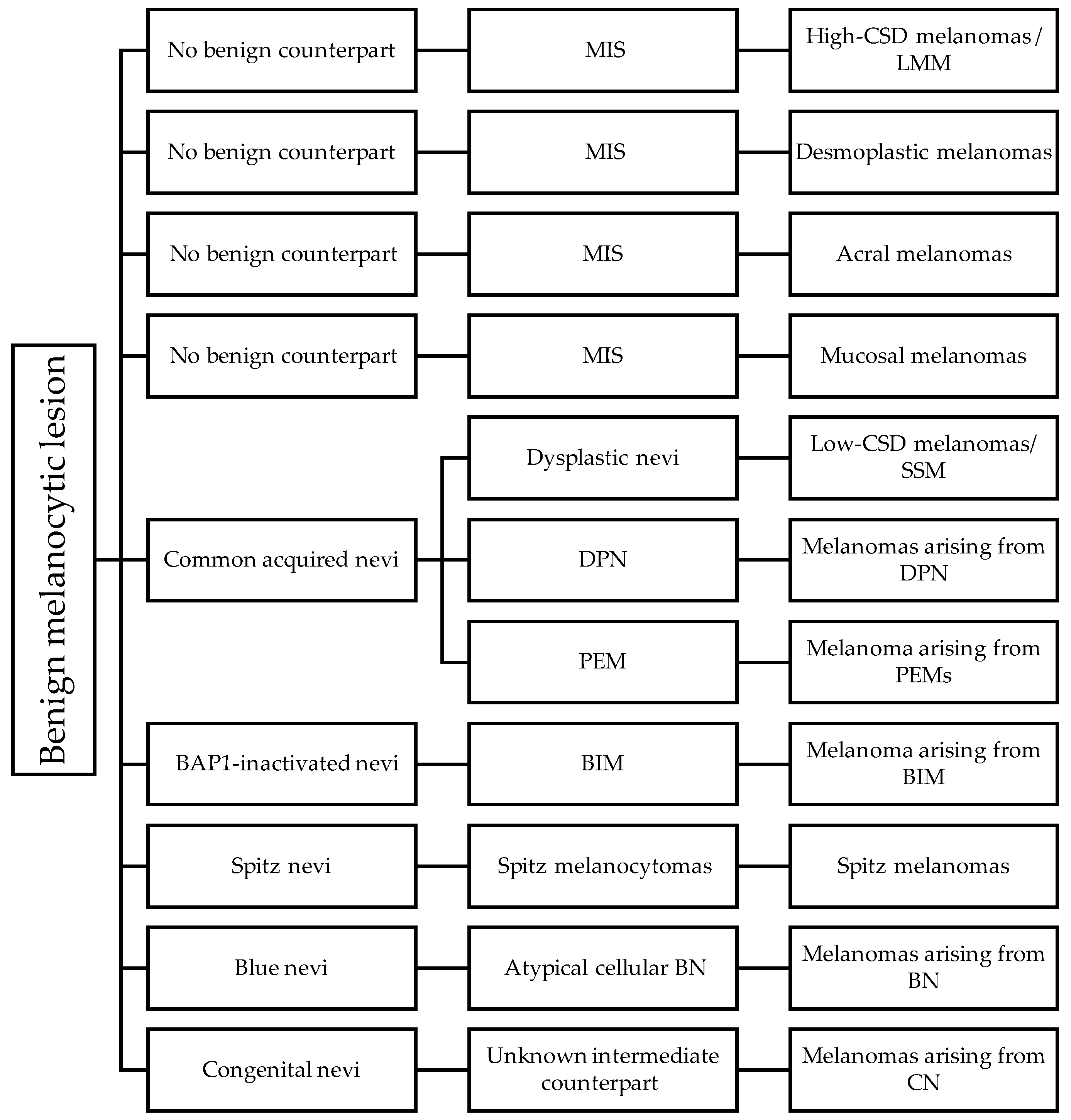
6. The Pathways of Cutaneous Melanomas

The current WHO classification categorizes eight pathways of cutaneous and mucosal melanoma development [100,117] (Figure 2 and Table 3).

6.1. Pathway 1: Low-CSD/Superficial Spreading Melanoma

The melanoma pathway that is the most encountered and best understood is pathway 1, namely, the low-cumulative-sun-damage (low-CSD) pathway. Low-CSD melanoma is the most prevalent form of cutaneous melanomas, accounting for around 41% of all melanomas [52], most of which present as superficial spreading melanomas (SSM). Both the benign conventional melanocytic nevus and the malignant melanoma of pathway 1 typically harbor a BRAF V600E mutation in approximately 80–90% of cases. Melanomas also harbor additional TERT-p mutation and CNAs. Low-CSD melanoma is often found within or adjacent to a precursor of conventional melanocytic nevus [59]. When there are borderline features, molecular assessment for the presence of additional mutations, such as CDKN2A or TERT-p, and chromosomal CNA can be helpful in differentiating low-CSD melanomas from intermediate lesions. The presence of BRAF V600E alteration is helpful in differentiating low-CSD melanomas from melanomas of the Spitz lineage.

6.2. Pathway 2: High-CSD/Lentigo Maligna Melanoma

Unlike low-CSD melanomas, high-CSD melanomas usually arise de novo and often originate from melanoma in situ. This group usually does not have benign or intermediate/low-grade precursor lesions. High-CSD melanomas occur mostly in elderly individuals, especially in heavily sun-exposed populations, including outdoor workers and persons with frequent recreational sun exposure [118,119]. The most common sites are the heavily sun-exposed sites; 90% of the cases involve the head and neck region [119,120]. The hallmark histological features include a single-cell “lentiginous” pattern of melanocytic proliferation within the epidermis with severe background solar elastosis. A pre-existing nevus is typically absent; melanoma in situ is considered the predominant precursor lesion in this group of melanomas [100,101]. The genomic landscape of high-CSD melanomas differs from non- or low-CSD melanomas and they typically do not harbor the signature BRAF V600E mutations. Instead, high-CSD melanoma is characterized by a more miscellaneous set of MAPK pathway mutations, such as BRAF V600K, NRAS, or KIT mutations, or inactivation of the negative regulators of Ras, NF1, or RASA2 [76]. Common specific molecular alterations in this group include bi-allelic inactivating mutations in NF1 (30%) [121], copy number increases of CCND1 (20%) [122], activating mutations of KIT (10%) [22], inactivating mutations of TP53 and ARID2, and TERT-p mutations [119,123,124,125]. The difference in the genomic landscape indicates that high- and low-CSD have distinct genetic profiles that progress through different molecular pathways, and hence, they are different molecular entities. Having predominant UV signature mutations, melanomas of pathway 2 have a very high mutation burden that correlates with their better responsiveness to checkpoint inhibitor immunotherapy [126,127].

6.3. Pathway 3: Desmoplastic Melanoma

Desmoplastic melanomas (DM) account for <4% of primary cutaneous melanomas in the population, with a predominant occurrence in pale-skinned individuals [128]. DMs are regarded as a variant of high-CSD melanoma because they commonly arise in skin with a high burden of UV-induced mutations [117,129]. Even though DM harbors overlapping genomic features with high-CSD melanomas, they are independently classified in the 2018 WHO Classification of Skin Tumors due to sufficient and distinctive histopathological features. Histologically, DMs feature a dermal component of spindled, unpigmented melanocytes interspersed between thick, scar-like, collagen bundles; hence, the “desmoplastic” and spindle cell “VGP” patterns. DMs are not necessarily associated with an RGP or in situ component [100,130]. The signature molecular alteration of DMs is the NF1 bi-allelic inactivating mutation disrupting the “off” state downregulation in the MAPK pathway. Normally, the intact NF1 function catalyzes the hydrolysis of GTP by RAS family members, which accelerates the transition to the “off” state, resulting in the downregulation of the MAPK pathway [131]. Inactivated NF1 allows RAS to remain activated, leading to sustained MAPK pathway activation, thus driving proliferation [132]. Though NF1 mutations are typically seen in melanomas that lack mutations in BRAF or NRAS, nearly 4% of melanomas with mutations in BRAF or NRAS also harbor NF1 mutations. Conversely, the oncogenic mutations, such as BRAF and NRAS, frequently found in other melanomas are generally absent in DMs. Because of the shared high-UV signature, NF1 mutation is also frequently seen in high-CSD melanomas of pathway 2. This showcases the high burden of the UV mutational signature in NF1-mutant melanomas [131].

6.4. Pathway 4: Spitz Melanoma

The Spitz family comprises a spectrum of lesions including Spitz nevus (SN), Spitz melanocytoma (atypical Spitz tumor/AST), and Spitz melanoma (SM). Morphologically, these tumors feature distinctive large spindle and/or epithelioid melanocytes with minimal solar elastosis [133]. Spitz tumors are defined as those harboring activating mutations in HRAS or kinase fusions by the most recent WHO Classification of Skin Tumors [117].
To date, the kinase component of the fusions found in Spitz neoplasms includes ALK, MET, RET, ROS, NTRK1, NTRK3, BRAF, MAP3K3, and MAP3K8 [83,134,135,136,137].
Spitz tumors with ALK and NTRK1 fusions have characteristic morphologic features. Spitz tumors with ALK fusions feature a wedge-shaped and plexiform architecture composed of fascicles of amelanotic wavy to fusiform and spindled melanocytes with mild-to-moderate atypia [64,138,139]. NTRK1-rearranged Spitz tumors are commonly characterized by wedge-shaped growth of lobulated nests or rosettes of small spindle cells with filigree-like rete ridges and the exaggerated maturation of dermal melanocytes, associated Kamino bodies, and epidermal hyperplasia [140]. This growth pattern is suggested to correlate with a greater dependence on epidermal growth factors compared with non-NTRK1-altered Spitz tumors [61].
Spitz melanomas with BRAF and MAP3K8 fusions tend to be epithelioid and high-grade. They often have a sheet-like growth pattern of intermediate to large epithelioid melanocytes with high-grade nuclear atypia and a higher likelihood of showing melanin pigments compared with other fusions [141]. Scattered single cells may be seen within sclerotic stroma [138].
The progression of SN to SM is through the additional genetic inactivation of CDKN2A and the acquisition of TERT-p mutations [96,142]. CDKN2A encodes the p16 protein, which is vital for tumor suppression. The inactivation of CDKN2A results in the loss of function in p16, leading to a promoted cell cycle progression. A strong and diffuse mosaic p16 staining pattern indicates SN, as it suggests an intact CDKN2A function, and hence, the benign nature of the lesion. In contrast, the complete absence or absence in sizable areas of p16 within the tumor correlates with CDKN2A inactivation and is highly suggestive of melanoma [97]. More specifically, the loss of p16 expression indicates a homozygous loss of 9p21, which leads to the alteration of CDKN2A [143,144,145]. The intermediate lesion, namely, AST, can present with either the homozygous or heterozygous loss of 9p21, giving rise to variable staining patterns of p16 IHC.
The term “spitzoid” is applied to tumors that morphologically resemble Spitz tumors but lack defining molecular characteristics. In addition, many of these “spitzoid” tumors harbor BRAF mutations of the conventional melanoma pathway, and thus, should not fall into the Spitz pathway [130,146].

6.5. Pathway 5: Acral Melanoma

Acral melanomas (AMs) occur on glabrous skin that lacks hair and has a thick stratum corneum. Over the years, AM has been observed to be the most common melanoma subtype in dark-skinned populations (African, Asian, and Hispanic) given the relatively lower incidence of other CSD-related cutaneous melanomas particularly in these darker-skinned populations [147,148,149]. AM comprises over 50% of cutaneous melanomas overall and represents the most common subtype in East Asian countries [150].
It was postulated that traumatic events and mechanical stress contribute to the etiology of AM [151]. Histologically, AMs feature a lentiginous single-cell growth pattern with a broad RGP that may be present for many years before progressing to invasive melanomas. The neoplastic cells often extend along eccrine ducts. Recent studies in mice identified melanocytic precursor cells within eccrine glands, suggesting that the secretory portion of eccrine sweat glands of volar surfaces may be the cell of origin for acral lentiginous melanomas [152,153].
AMs have a relatively low burden of point mutations and a high incidence of CNAs with multiple gene amplification, including CCND1 and KIT. The most frequently seen alteration in AMs is BRAF, followed by NRAS and TP53 [154]. Although CCND1 encodes cyclin-D1, the correlation of detecting CCND1 copy number alteration using cyclin-D1 IHC has not found to be consistent [155]. Therefore, cyclin-D1 IHC cannot be used as a surrogate in place of CCND1 FISH in melanomas.

6.6. Pathway 6: Mucosal Melanoma

Mucosal melanomas (MM), which represents 1–3% of all melanomas, describes melanomas of the head and neck mucosa (oral and nasal cavities), anorectal mucosa, and female genital tract mucosa; rarely, MM can also occur in the lower urinary tract and esophageal mucosa [156,157]. An associated nevus is rarely seen in MM. Histologically, invasive components of MM typically comprise sheets and expansive nodules in the submucosa. [158]. Adjacent intraepithelial or superficially invasive components may be present and usually appear as lentiginous melanocytic dysplasia, melanoma in situ, or RGP. This is a histological clue indicating that a lesion is a mucosal primary [158].
The etiology of MM is still unknown, and it shows no association with UV exposure or carcinogens [159]. Cicaresse et al. showed that HPV and EBV DNA were found in 17% and 20% of mucosal and ocular melanomas, suggesting that these viruses may act as cofactors in the development of mucosal and ocular melanomas [160]. MM has a low mutation burden and is characterized by frequent focal amplifications, deletions, and structural rearrangements, similar to AMs [1,161]. NRAS, BRAF, NF1, KIT, TP53, SF3B1, CCND1, CDKN2A, and TERT-p mutations were described in a substantial portion of MMs of various sites [22,159,162]. SPRED1 was recently identified as a tumor suppressor in MM and it is often inactivated in the setting of KIT mutation [22,162]. Even though MM is known to have a poor prognosis, the diverse driver alterations in MM include some that can suggest potential susceptibility to CDK4/6 and/or MEK inhibitors [159].

6.7. Pathway 7: Melanoma Arising from Congenital Nevus

Congenital nevi (CN), especially giant CN, are caused by an alteration of NRAS or BRAF in the MAPK pathway in utero or shortly after birth [163]. Large CN with satellite lesions have a 10–15% risk of developing melanoma in the patient’s lifetime [164]. Most such melanomas are highly aggressive and they present during the first 5 years of life. Melanomas arising in CN typically appear as a discolored area that looks different than the background CN in terms of color and texture. They present as rapidly growing nodules or plaques with associated ulceration. It is common to see synchronous lymph node metastatic disease at presentation. Histologically, in the prepubescent age group, the principal site of melanoma development is the dermis or subcutis, whereas, in adults, the development of melanoma starts at the dermal–epidermal junction. Frank malignant features, such as expansile growth, necrosis, brisk mitosis, and cytological features, are often seen.
Giant CN are most frequently associated with NRAS mutation while medium-to-small CN harbor BRAF V600E as the predominant mutation [165,166]. Up to 95% of giant CN are found to have an NRAS mutation and most melanomas arising in giant CN also exhibit NRAS mutations [167,168]. Several other alterations, such as BRAF, TP53, RAF1, PTEN, KIT, TERT-p, CDKN2A, and PRKCA, were also observed in addition to CNAs [169]. Melanomas arising from small or medium CN often harbor an additional TERT-p mutation [170]. The genetic alterations characterizing melanoma development in CN remain poorly understood.

6.8. Pathway 8: Melanoma Arising from Blue Nevus

Blue nevi (BN) originate from melanocytes that develop from neural crest stem cells [171]. Those melanocytes are not associated with any epithelium. Atypical cellular blue nevus is the intermediate lesion for melanoma arising from blue nevus (MBN). Histologically, the overlying epidermis is usually uninvolved in MBN. The tumor features invasive dense dermal sheets or nodules of large spindled and epithelioid melanocytes that often infiltrate the surrounding adnexal structures. At the periphery of the MBN, pre-existing BN can usually be found. BN do not harbor a UV mutation signature. MBN is caused by somatic mutations activating the Gαq signaling pathway, predominantly in the genes GNAQ and GNA11, rarely in the upstream receptor CYSLTR1, or downstream effector PLB4 [172,173,174]. BAP1 and SF3B1 mutations also contribute to the malignant transformation of BN [175,176].
Loss of nuclear BAP1 IHC strongly supports the diagnosis of MBN in the setting of a suspicious malignant transformation of BN [175].
Distinguishing MBN from other highly pigmented melanomas is often difficult, especially when remnant BN cannot be clearly identified. The presence of an activating Gαq mutation helps to classify lesions into the BN pathway, while the presence of BRAF V600E mutation can help to classify lesions into the low-CSD pathway.

7. Discussion

The elucidation of numerous molecular pathways of melanocytic lesions has given rise to a range of ancillary immunohistochemistry and molecular tests based on their respective molecular alterations. These tests serve four main purposes: to differentiate between nevi and melanomas in histologically ambiguous lesions, to classify melanocytic lesions, to predict prognosis, and to predict response to therapy.
For differentiating between nevi and melanomas in histologically borderline lesions, IHC, such as p16, and molecular assays, such as CGH and FISH, may be employed. In addition, TERT-p mutation analysis, GEP, and IMS play potential roles in differential diagnosis, but more research is needed before clear recommendations can be made.
To classify melanocytic lesions, IHC, such as ALK, ROS1, and pan-NTRK, are surrogate markers for detecting kinase fusions in Spitz tumors. β-catenin is a surrogate marker for CTNNB1-activating mutations in DPN. BAP1 loss is crucial for the diagnosis of BAP1-inactivated melanocytic tumors, while PRKR1A1 loss supports the diagnosis of PEM.
For predicting prognosis in melanomas, several GEP assays have been developed for risk stratification.
For predicting response to systemic therapy in high-stage melanomas, BRAF mutation status must be assessed, either using IHC specific for the V600E mutant protein or using BRAF gene mutation analysis before BRAF-inhibitor and MEK-inhibitor therapy can be initiated.
Overall, the role of molecular assays in characterizing melanocytic lesions is expanding, particularly for the purpose of differentiating between benign and malignant tumors. Such diagnostic molecular tests should only be employed on melanocytic lesions in which a definitive diagnosis cannot be reached using histology. Furthermore, their results should be interpreted in the context of the clinical presentation and histologic features and should not overturn the initial histologic impression. The progression from benign to intermediate and finally the malignant counterpart within each molecular pathway is characterized by a gradual accumulation of molecular alterations, and therefore, the molecular assays demonstrate a variable sensitivity and specificity to differentiate between nevi and melanomas for histologically borderline lesions.

8. Conclusions

The significant advances in our understanding of multiple molecular pathways in the progression of melanocytic lesions have enabled the development of ancillary molecular tests to aid in their diagnosis, classification, prognostication, and prediction of therapeutic response. Nevertheless, a comprehensive diagnostic algorithm integrating the clinical findings, histological features, and molecular alterations is still in development. In addition, more studies are needed to improve the performance of ancillary molecular tests in the characterization and diagnosis of histologically borderline lesions.

Author Contributions

Conceptualization, C.H. and B.R.S.; writing—original draft preparation, C.H.; writing—review and editing, T.W.-S.L. and B.R.S.; visualization, C.H. and T.W.-S.L.; supervision, B.R.S. All authors have read and agreed to the published version of the manuscript.

Funding

This research received no external funding.

Conflicts of Interest

The authors declare no conflicts of interest.

References

  1. Hayward, N.K.; Wilmott, J.S.; Waddell, N.; Johansson, P.A.; Field, M.A.; Nones, K.; Patch, A.M.; Kakavand, H.; Alexandrov, L.B.; Burke, H.; et al. Whole-genome landscapes of major melanoma subtypes. Nature 2017, 545, 175–180. [Google Scholar] [CrossRef]
  2. Bennett, D.C. Genetics of melanoma progression: The rise and fall of cell senescence. Pigment Cell Melanoma Res. 2016, 29, 122–140. [Google Scholar] [CrossRef]
  3. Sullivan, R.; Flaherty, K. MAP kinase signaling and inhibition in melanoma. Oncogene 2013, 32, 2373–2379. [Google Scholar] [CrossRef] [PubMed]
  4. Guo, W.; Wang, H.; Li, C. Signal pathways of melanoma and targeted therapy. Signal Transduct. Target. Ther. 2021, 6, 424. [Google Scholar] [CrossRef]
  5. Platz, A.; Egyhazi, S.; Ringborg, U.; Hansson, J. Human cutaneous melanoma; a review of NRAS and BRAF mutation frequencies in relation to histogenetic subclass and body site. Mol. Oncol. 2008, 1, 395–405. [Google Scholar] [CrossRef]
  6. Hodis, E.; Watson, I.R.; Kryukov, G.V.; Arold, S.T.; Imielinski, M.; Theurillat, J.P.; Nickerson, E.; Auclair, D.; Li, L.; Place, C.; et al. A landscape of driver mutations in melanoma. Cell 2012, 150, 251–263. [Google Scholar] [CrossRef]
  7. Davies, H.; Bignell, G.R.; Cox, C.; Stephens, P.; Edkins, S.; Clegg, S.; Teague, J.; Woffendin, H.; Garnett, M.J.; Bottomley, W.; et al. Mutations of the BRAF gene in human cancer. Nature 2002, 417, 949–954. [Google Scholar] [CrossRef]
  8. COSMIC. BRAF. Available online: https://cancer.sanger.ac.uk/cosmic/gene/analysis?ln=BRAF (accessed on 15 August 2023).
  9. Long, G.V.; Menzies, A.M.; Nagrial, A.M.; Haydu, L.E.; Hamilton, A.L.; Mann, G.J.; Hughes, T.M.; Thompson, J.F.; Scolyer, R.A.; Kefford, R.F. Prognostic and clinicopathologic associations of oncogenic BRAF in metastatic melanoma. J. Clin. Oncol. 2011, 29, 1239–1246. [Google Scholar] [CrossRef]
  10. COSMIC. NRAS. Available online: https://cancer.sanger.ac.uk/cosmic/gene/analysis?ln=NRAS (accessed on 15 August 2023).
  11. Cerami, E.; Gao, J.; Dogrusoz, U.; Gross, B.E.; Sumer, S.O.; Aksoy, B.A.; Jacobsen, A.; Byrne, C.J.; Heuer, M.L.; Larsson, E.; et al. The cBio Cancer Genomics Portal: An Open Platform for Exploring Multidimensional Cancer Genomics Data. Cancer Discov. 2012, 2, 401. [Google Scholar] [CrossRef] [PubMed]
  12. cBioPortal. NRAS. Available online: https://www.cbioportal.org/results/cancerTypesSummary?case_set_id=all&gene_list=NRAS&cancer_study_list=5c8a7d55e4b046111fee2296 (accessed on 15 August 2023).
  13. Gao, J.; Aksoy, B.A.; Dogrusoz, U.; Dresdner, G.; Gross, B.; Sumer, S.O.; Sun, Y.; Jacobsen, A.; Sinha, R.; Larsson, E.; et al. Integrative analysis of complex cancer genomics and clinical profiles using the cBioPortal. Sci. Signal. 2013, 6, pl1. [Google Scholar] [CrossRef] [PubMed]
  14. Devitt, B.; Liu, W.; Salemi, R.; Wolfe, R.; Kelly, J.; Tzen, C.Y.; Dobrovic, A.; McArthur, G. Clinical outcome and pathological features associated with NRAS mutation in cutaneous melanoma. Pigment Cell Melanoma Res. 2011, 24, 666–672. [Google Scholar] [CrossRef] [PubMed]
  15. Cook, M.G.; Massi, D.; Blokx, W.A.M.; Van den Oord, J.; Koljenović, S.; De Giorgi, V.; Kissin, E.; Grant, M.; Mandal, A.; Gremel, G.; et al. New insights into naevoid melanomas: A clinicopathological reassessment. Histopathology 2017, 71, 943–950. [Google Scholar] [CrossRef] [PubMed]
  16. COSMIC. NF1. Available online: https://cancer.sanger.ac.uk/cosmic/gene/analysis?ln=NF1 (accessed on 15 August 2023).
  17. cBioPortal. NF1. Available online: https://www.cbioportal.org/results/cancerTypesSummary?case_set_id=all&gene_list=NF1&cancer_study_list=5c8a7d55e4b046111fee2296 (accessed on 15 August 2023).
  18. Philpott, C.; Tovell, H.; Frayling, I.M.; Cooper, D.N.; Upadhyaya, M. The NF1 somatic mutational landscape in sporadic human cancers. Hum. Genom. 2017, 11, 13. [Google Scholar] [CrossRef]
  19. Wiesner, T.; Kiuru, M.; Scott, S.N.; Arcila, M.; Halpern, A.C.; Hollmann, T.; Berger, M.F.; Busam, K.J. NF1 Mutations Are Common in Desmoplastic Melanoma. Am. J. Surg. Pathol. 2015, 39, 1357–1362. [Google Scholar] [CrossRef]
  20. Cirenajwis, H.; Lauss, M.; Ekedahl, H.; Törngren, T.; Kvist, A.; Saal, L.H.; Olsson, H.; Staaf, J.; Carneiro, A.; Ingvar, C.; et al. NF1-mutated melanoma tumors harbor distinct clinical and biological characteristics. Mol. Oncol. 2017, 11, 438–451. [Google Scholar] [CrossRef]
  21. Thielmann, C.M.; Chorti, E.; Matull, J.; Murali, R.; Zaremba, A.; Lodde, G.; Jansen, P.; Richter, L.; Kretz, J.; Möller, I.; et al. NF1-mutated melanomas reveal distinct clinical characteristics depending on tumor origin and respond favorably to immune checkpoint inhibitors. Eur. J. Cancer 2021, 159, 113–124. [Google Scholar] [CrossRef] [PubMed]
  22. Curtin, J.A.; Busam, K.; Pinkel, D.; Bastian, B.C. Somatic activation of KIT in distinct subtypes of melanoma. J. Clin. Oncol. 2006, 24, 4340–4346. [Google Scholar] [CrossRef] [PubMed]
  23. Satzger, I.; Schaefer, T.; Kuettler, U.; Broecker, V.; Voelker, B.; Ostertag, H.; Kapp, A.; Gutzmer, R. Analysis of c-KIT expression and KIT gene mutation in human mucosal melanomas. Br. J. Cancer 2008, 99, 2065–2069. [Google Scholar] [CrossRef] [PubMed]
  24. Gong, H.Z.; Zheng, H.Y.; Li, J. The clinical significance of KIT mutations in melanoma: A meta-analysis. Melanoma Res. 2018, 28, 259–270. [Google Scholar] [CrossRef]
  25. Pham, D.D.M.; Guhan, S.; Tsao, H. KIT and Melanoma: Biological Insights and Clinical Implications. Yonsei Med. J. 2020, 61, 562–571. [Google Scholar] [CrossRef] [PubMed]
  26. Siroy, A.E.; Davies, M.A.; Lazar, A.J. The PI3K-AKT Pathway in Melanoma. In Genetics of Melanoma. Cancer Genetics; Torres-Cabala, C., Curry, J., Eds.; Springer: New York, NY, USA, 2016. [Google Scholar] [CrossRef]
  27. Campbell, P.M.; Der, C.J. Oncogenic Ras and its role in tumor cell invasion and metastasis. Semin. Cancer Biol. 2004, 14, 105–114. [Google Scholar] [CrossRef] [PubMed]
  28. Paluncic, J.; Kovacevic, Z.; Jansson, P.J.; Kalinowski, D.; Merlot, A.M.; Huang, M.L.; Lok, H.C.; Sahni, S.; Lane, D.J.; Richardson, D.R. Roads to melanoma: Key pathways and emerging players in melanoma progression and oncogenic signaling. Biochim. Biophys. Acta 2016, 1863, 770–784. [Google Scholar] [CrossRef] [PubMed]
  29. Rascio, F.; Spadaccino, F.; Rocchetti, M.T.; Castellano, G.; Stallone, G.; Netti, G.S.; Ranieri, E. The Pathogenic Role of PI3K/AKT Pathway in Cancer Onset and Drug Resistance: An Updated Review. Cancers 2021, 13, 3949. [Google Scholar] [CrossRef] [PubMed]
  30. Bucheit, A.B.; Chen, G.; Siroy, A.; Tetzlaff, M.; Broaddus, R.; Milton, D.; Fox, P.; Bassett, R.; Hwu, P.; Gershenwald, J.E.; et al. Complete loss of PTEN protein expression correlates with shorter time to brain metastasis and survival in stage IIIB/C melanoma patients with BRAFV600 mutations. Clin. Cancer Res. 2014, 20, 5527–5536. [Google Scholar] [CrossRef] [PubMed]
  31. Peng, W.; Chen, J.Q.; Liu, C.; Malu, S.; Creasy, C.; Tetzlaff, M.T.; Xu, C.; McKenzie, J.A.; Zhang, C.; Liang, X.; et al. Loss of PTEN Promotes Resistance to T Cell-Mediated Immunotherapy. Cancer Discov. 2016, 6, 202–216. [Google Scholar] [CrossRef]
  32. Lucero, O.M.; Dawson, D.W.; Moon, R.T.; Chien, A.J. A re-evaluation of the “oncogenic” nature of Wnt/beta-catenin signaling in melanoma and other cancers. Curr. Oncol. Rep. 2010, 12, 314–318. [Google Scholar] [CrossRef]
  33. Regad, T. Molecular and cellular pathogenesis of melanoma initiation and progression. Cell. Mol. Life Sci. 2013, 70, 4055–4065. [Google Scholar] [CrossRef] [PubMed]
  34. Chien, A.J.; Moore, E.C.; Lonsdorf, A.S.; Kulikauskas, R.M.; Rothberg, B.G.; Berger, A.J.; Major, M.B.; Hwang, S.T.; Rimm, D.L.; Moon, R.T. Activated Wnt/beta-catenin signaling in melanoma is associated with decreased proliferation in patient tumors and a murine melanoma model. Proc. Natl. Acad. Sci. USA 2009, 106, 1193–1198. [Google Scholar] [CrossRef]
  35. Biechele, T.L.; Camp, N.D.; Fass, D.M.; Kulikauskas, R.M.; Robin, N.C.; White, B.D.; Taraska, C.M.; Moore, E.C.; Muster, J.; Karmacharya, R.; et al. Chemical-genetic screen identifies riluzole as an enhancer of Wnt/beta-catenin signaling in melanoma. Chem Biol. 2010, 17, 1177–1182. [Google Scholar] [CrossRef]
  36. He, S.; Lu, Y.; Liu, X.; Huang, X.; Keller, E.T.; Qian, C.-N.; Zhang, J. Wnt3a: Functions and implications in cancer. Chin. J. Cancer 2015, 34, 50. [Google Scholar] [CrossRef]
  37. Yeh, I.; Lang, U.E.; Durieux, E.; Tee, M.K.; Jorapur, A.; Shain, A.H.; Haddad, V.; Pissaloux, D.; Chen, X.; Cerroni, L.; et al. Combined activation of MAP kinase pathway and β-catenin signaling cause deep penetrating nevi. Nat. Commun. 2017, 8, 644. [Google Scholar] [CrossRef] [PubMed]
  38. Luzar, B.; Calonje, E. Deep Penetrating Nevus: A Review. Arch. Pathol. Lab. Med. 2011, 135, 321–326. [Google Scholar] [CrossRef] [PubMed]
  39. Robson, A.; Morley-Quante, M.; Hempel, H.; McKee, P.H.; Calonje, E. Deep penetrating naevus: Clinicopathological study of 31 cases with further delineation of histological features allowing distinction from other pigmented benign melanocytic lesions and melanoma. Histopathology 2003, 43, 529–537. [Google Scholar] [CrossRef]
  40. Magro, C.M.; Abraham, R.M.; Guo, R.; Li, S.; Wang, X.; Proper, S.; Crowson, A.N.; Mihm, M. Deep penetrating nevus-like borderline tumors: A unique subset of ambiguous melanocytic tumors with malignant potential and normal cytogenetics. Eur. J. Dermatol. 2014, 24, 594–602. [Google Scholar] [CrossRef] [PubMed]
  41. Massi, G.; LeBoit, P.E. Histological Diagnosis of Nevi and Melanoma; Springer: Berlin/Heidelberg, Germany, 2014; Melanoma with a plexiform pattern; pp. 581–588. [Google Scholar]
  42. Isales, M.C.; Khan, A.U.; Zhang, B.; Compres, E.V.; Kim, D.; Tan, T.L.; Beaubier, N.; Gerami, P. Molecular analysis of atypical deep penetrating nevus progressing to melanoma. J. Cutan. Pathol. 2020, 47, 1150–1154. [Google Scholar] [CrossRef] [PubMed]
  43. Erstine, E.M.; Lazar, A.J. Toward an effective use of β-catenin immunohistochemistry in the evaluation of challenging melanocytic lesions. Virchows Arch. 2019, 474, 535–537. [Google Scholar] [CrossRef]
  44. Mancarella, D.; Plass, C. Epigenetic Signatures in Cancer: Proper Controls, Current Challenges, and the Potential for Clinical Translation. Genome Med. 2021, 13, 23. [Google Scholar] [CrossRef]
  45. Fujiwara, S.; Nagai, H.; Jimbo, H.; Jimbo, N.; Tanaka, T.; Inoie, M.; Nishigori, C. Gene Expression and Methylation Analysis in Melanomas and Melanocytes from the Same Patient: Loss of NPM2 Expression Is a Potential Immunohistochemical Marker for Melanoma. Front. Oncol. 2019, 8, 675. [Google Scholar] [CrossRef]
  46. Aleotti, V.; Catoni, C.; Poggiana, C.; Rosato, A.; Facchinetti, A.; Scaini, M.C. Methylation Markers in Cutaneous Melanoma: Unravelling the Potential Utility of Their Tracking by Liquid Biopsy. Cancers 2021, 13, 6217. [Google Scholar] [CrossRef]
  47. Sutopo, N.C.; Kim, J.H.; Cho, J.Y. Role of histone methylation in skin cancers: Histone methylation-modifying enzymes as a new class of targets for skin cancer treatment. Biochim. Biophys. Acta. Rev. Cancer 2023, 1878, 188865. [Google Scholar] [CrossRef]
  48. Kapoor, A.; Goldberg, M.S.; Cumberland, L.K.; Ratnakumar, K.; Segura, M.F.; Emanuel, P.O.; Menendez, S.; Vardabasso, C.; LeRoy, G.; Vidal, C.I.; et al. The histone variant macroH2A suppresses melanoma progression through regulation of CDK8. Nature 2010, 468, 1105–1109. [Google Scholar] [CrossRef] [PubMed]
  49. Vardabasso, C.; Gaspar-Maia, A.; Hasson, D.; Pünzeler, S.; Valle-Garcia, D.; Straub, T.; Keilhauer, E.C.; Strub, T.; Dong, J.; Panda, T.; et al. Histone variant H2A.Z.2 mediates proliferation and drug sensitivity of malignant melanoma. Mol. Cell 2015, 59, 75–88. [Google Scholar] [CrossRef]
  50. Giunta, E.F.; Arrichiello, G.; Curvietto, M.; Pappalardo, A.; Bosso, D.; Rosanova, M.; Diana, A.; Giordano, P.; Petrillo, A.; Federico, P.; et al. Epigenetic Regulation in Melanoma: Facts and Hopes. Cells 2021, 10, 2048. [Google Scholar] [CrossRef] [PubMed]
  51. Palmieri, G.; Colombino, M.; Casula, M.; Manca, A.; Mandalà, M.; Cossu, A.; Italian Melanoma Intergroup (IMI). Molecular Pathways in Melanomagenesis: What We Learned from Next-Generation Sequencing Approaches. Curr. Oncol. Rep. 2018, 20, 86. [Google Scholar] [CrossRef] [PubMed]
  52. Scatena, C.; Murtas, D.; Tomei, S. Cutaneous Melanoma Classification: The Importance of High-Throughput Genomic Technologies. Front. Oncol. 2021, 11, 635488. [Google Scholar] [CrossRef] [PubMed]
  53. Diana, A.; Gaido, G.; Murtas, D. Microrna Signature in Human Normal and Tumoral Neural Stem Cells. Int. J. Mol. Sci. 2019, 20, 4123. [Google Scholar] [CrossRef] [PubMed]
  54. Gajos-Michniewicz, A.; Czyz, M. Role of miRNAs in Melanoma Metastasis. Cancers 2019, 11, 326. [Google Scholar] [CrossRef]
  55. Lohcharoenkal, W.; Das Mahapatra, K.; Pasquali, L.; Crudden, C.; Kular, L.; Akkaya Ulum, Y.Z.; Zhang, L.; Xu Landén, N.; Girnita, L.; Jagodic, M.; et al. Genome-Wide Screen for MicroRNAs Reveals a Role for miR-203 in Melanoma Metastasis. J. Investig. Dermatol. 2018, 138, 882–892. [Google Scholar] [CrossRef]
  56. Shah, M.Y.; Ferrajoli, A.; Sood, A.K.; Lopez-Berestein, G.; Calin, G.A. microRNA Therapeutics in Cancer—An Emerging Concept. EBioMedicine 2016, 12, 34–42. [Google Scholar] [CrossRef]
  57. Varrone, F.; Caputo, E. The miRNAs Role in Melanoma and in Its Resistance to Therapy. Int. J. Mol. Sci. 2020, 21, 878. [Google Scholar] [CrossRef]
  58. Saleem, A.; Narala, S.; Raghavan, S.S. Immunohistochemistry in melanocytic lesions: Updates with a practical review for pathologists. Semin. Diagn. Pathol. 2022, 39, 239–247. [Google Scholar] [CrossRef] [PubMed]
  59. Yeh, I. Update on classification of melanocytic tumors and the role of immunohistochemistry and molecular techniques. Semin. Diagn. Pathol. 2022, 39, 248–256. [Google Scholar] [CrossRef]
  60. Andea, A.A. Molecular testing in melanoma for the surgical pathologist. Pathology 2023, 55, 245–257. [Google Scholar] [CrossRef] [PubMed]
  61. Quan, V.L.; Panah, E.; Zhang, B.; Shi, K.; Mohan, L.S.; Gerami, P. The role of gene fusions in melanocytic neoplasms. J. Cutan. Pathol. 2019, 46, 878–887. [Google Scholar] [CrossRef] [PubMed]
  62. Jakob, J.A.; Bassett, R.L., Jr.; Ng, C.S.; Curry, J.L.; Joseph, R.W.; Alvarado, G.C.; Rohlfs, M.L.; Richard, J.; Gershenwald, J.E.; Kim, K.B.; et al. NRAS mutation status is an independent prognostic factor in metastatic melanoma. Cancer 2012, 118, 4014–4023. [Google Scholar] [CrossRef]
  63. Massi, D.; Simi, L.; Sensi, E.; Baroni, G.; Xue, G.; Scatena, C.; Caldarella, A.; Pinzani, P.; Fontanini, G.; Carobbio, A.; et al. Immunohistochemistry is highly sensitive and specific for the detection of NRASQ61R mutation in melanoma. Mod. Pathol. 2015, 28, 487–497. [Google Scholar] [CrossRef]
  64. Yeh, I.; de la Fouchardiere, A.; Pissaloux, D.; Mully, T.W.; Garrido, M.C.; Vemula, S.S.; Busam, K.J.; LeBoit, P.E.; McCalmont, T.H.; Bastian, B.C. Clinical, histopathologic, and genomic features of Spitz tumors with ALK fusions. Am. J. Surg. Pathol. 2015, 39, 581–591. [Google Scholar] [CrossRef]
  65. Raghavan, S.S.; Saleem, A.; Wang, J.Y.; Rieger, K.E.; Brown, R.A.; Novoa, R.A. Diagnostic Utility of LEF1 Immunohistochemistry in Differentiating Deep Penetrating Nevi from Histologic Mimics. Am. J. Surg. Pathol. 2020, 44, 1413–1418. [Google Scholar] [CrossRef]
  66. Yeh, I. New and evolving concepts of melanocytic nevi and melanocytomas. Mod. Pathol. 2020, 33 (Suppl. S1), 1–14. [Google Scholar] [CrossRef]
  67. Benton, S.; Zhao, J.; Asadbeigi, S.; Kim, D.; Zhang, B.; Gerami, P. Pigmented Epithelioid Melanocytoma: Morphology and Molecular Drivers. Surg. Pathol. Clin. 2021, 14, 285–292. [Google Scholar] [CrossRef]
  68. Ricci, C.; Dika, E.; Ambrosi, F.; Lambertini, M.; Veronesi, G.; Barbara, C. Cutaneous Melanomas: A Single Center Experience on the Usage of Immunohistochemistry Applied for the Diagnosis. Int. J. Mol. Sci. 2022, 23, 5911. [Google Scholar] [CrossRef] [PubMed]
  69. Straume, O.; Sviland, L.; Akslen, L.A. Loss of nuclear p16 protein expression correlates with increased tumor cell proliferation (Ki-67) and poor prognosis in patients with vertical growth phase melanoma. Clin. Cancer Res. 2000, 6, 1845–1853. [Google Scholar] [PubMed]
  70. Serra, S.; Chetty, R. p16. J. Clin. Pathol. 2018, 71, 853–858. [Google Scholar] [CrossRef] [PubMed]
  71. Koh, S.S.; Cassarino, D.S. Immunohistochemical Expression of p16 in Melanocytic Lesions: An Updated Review and Meta-analysis. Arch. Pathol. Lab. Med. 2018, 142, 815–828. [Google Scholar] [CrossRef]
  72. Balaban, G.; Herlyn, M.; Guerry, D.T.; Bartolo, R.C.; Koprowski, H.; Clark, W.H., Jr.; Nowell, P.C. Cytogenetics of human malignant melanoma and premalignant lesions. Cancer Genet. Cytogenet. 1984, 11, 429–439. [Google Scholar] [CrossRef]
  73. Kallioniemi, A.; Kallioniemi, O.P.; Sudar, D.; Rutovitz, D.; Gray, J.W.; Waldman, F.; Pinkel, D. Comparative genomic hybridization for molecular cytogenetic analysis of solid tumors. Science 1992, 258, 818–821. [Google Scholar] [CrossRef]
  74. Wiszniewska, J.; Bi, W.; Shaw, C.; Stankiewicz, P.; Kang, S.H.; Pursley, A.N.; Lalani, S.; Hixson, P.; Gambin, T.; Tsai, C.H.; et al. Combined array CGH plus SNP genome analyses in a single assay for optimized clinical testing. Eur. J. Hum. Genet. EJHG 2014, 22, 79–87. [Google Scholar] [CrossRef]
  75. Bastian, B.C.; LeBoit, P.E.; Hamm, H.; Bröcker, E.B.; Pinkel, D. Chromosomal gains and losses in primary cutaneous melanomas detected by comparative genomic hybridization. Cancer Res. 1998, 58, 2170–2175. [Google Scholar]
  76. Bastian, B.C.; Olshen, A.B.; LeBoit, P.E.; Pinkel, D. Classifying melanocytic tumors based on DNA copy number changes. Am. J. Pathol. 2003, 163, 1765–1770. [Google Scholar]
  77. Hosein, A.N.; Song, S.; McCart Reed, A.E.; Jayanthan, J.; Reid, L.E.; Kutasovic, J.R.; Cummings, M.C.; Waddell, N.; Lakhani, S.R.; Chenevix-Trench, G.; et al. Evaluating the repair of DNA derived from formalin-fixed paraffin-embedded tissues prior to genomic profiling by SNP–CGH analysis. Lab. Investig. 2013, 93, 701–710. [Google Scholar] [CrossRef]
  78. Wang, Y.; Moorhead, M.; Karlin-Neumann, G.; Wang, N.J.; Ireland, J.; Lin, S.; Chen, C.; Heiser, L.M.; Chin, K.; Esserman, L.; et al. Analysis of molecular inversion probe performance for allele copy number determination. Genome Biol. 2007, 8, R246. [Google Scholar] [CrossRef]
  79. Chandler, W.M.; Rowe, L.R.; Florell, S.R.; Jahromi, M.S.; Schiffman, J.D.; South, S.T. Differentiation of malignant melanoma from benign nevus using a novel genomic microarray with low specimen requirements. Arch. Pathol. Lab. Med. 2012, 136, 947–955. [Google Scholar] [CrossRef] [PubMed]
  80. Bastian, B.C.; Wesselmann, U.; Pinkel, D.; Leboit, P.E. Molecular cytogenetic analysis of Spitz nevi shows clear differences to melanoma. J. Investig. Dermatol. 1999, 113, 1065–1069. [Google Scholar] [CrossRef] [PubMed]
  81. Emanuel, P.O.; Andea, A.A.; Vidal, C.I.; Missall, T.A.; Novoa, R.A.; Bohlke, A.K.; Hughes, S.R.; Hurley, M.Y.; Kim, J. Evidence behind the use of molecular tests in melanocytic lesions and practice patterns of these tests by dermatopathologists. J. Cutan. Pathol. 2018, 45, 839–846. [Google Scholar] [CrossRef] [PubMed]
  82. Jiveskog, S.; Ragnarsson-Olding, B.; Platz, A.; Ringborg, U. N-RAS mutations are common in melanomas from sun-exposed skin of humans but rare in mucosal membranes of unexposed skin. J. Investig. Dermatol. 1998, 111, 757–761. [Google Scholar] [CrossRef]
  83. Bastian, B.C.; LeBoit, P.E.; Pinkel, D. Mutations and copy number increase of HRAS in Spitz nevi with distinctive histopathological features. Am. J. Pathol. 2000, 157, 967–972. [Google Scholar] [CrossRef]
  84. Andea, A.A. Molecular testing for melanocytic tumors: A practical update. Histopathology. 2022, 80, 150–165. [Google Scholar] [CrossRef]
  85. FISH for Melanoma, Multiplex Probe. Available online: https://mlabs.umich.edu/tests/fish-melanoma-multiplex-probe (accessed on 7 January 2023).
  86. Gerami, P.; Jewell, S.S.; Morrison, L.E.; Blondin, B.; Schulz, J.; Ruffalo, T.; Matushek, P.; Legator, M., IV; Jacobson, K.; Dalton, S.R.; et al. Fluorescence in situ hybridization (FISH) as an ancillary diagnostic tool in the diagnosis of melanoma. Am. J. Surg. Pathol. 2009, 33, 1146–1156. [Google Scholar] [CrossRef]
  87. Miedema, J.; Andea, A.A. Through the looking glass and what you find there: Making sense of comparative genomic hybridization and fluorescence in situ hybridization for melanoma diagnosis. Mod. Pathol. 2020, 33, 1318–1330. [Google Scholar] [CrossRef]
  88. Vergier, B.; Prochazkova-Carlotti, M.; de la Fouchardière, A.; Cerroni, L.; Massi, D.; De Giorgi, V.; Bailly, C.; Wesselmann, U.; Karlseladze, A.; Avril, M.F.; et al. Fluorescence in situ hybridization, a diagnostic aid in ambiguous melanocytic tumors: European study of 113 cases. Mod. Pathol. 2011, 24, 613–623. [Google Scholar] [CrossRef]
  89. Pouryazdanparast, P.; Newman, M.; Mafee, M.; Haghighat, Z.; Guitart, J.; Gerami, P. Distinguishing epithelioid blue nevus from blue nevus-like cutaneous melanoma metastasis using fluorescence in situ hybridization. Am. J. Surg. Pathol. 2009, 33, 1396–1400. [Google Scholar] [CrossRef] [PubMed]
  90. Ferrara, G.; De Vanna, A.C. Fluorescence In Situ Hybridization for Melanoma Diagnosis: A Review and a Reappraisal. Am. J. Dermatopathol. 2016, 38, 253–269. [Google Scholar] [CrossRef] [PubMed]
  91. Gerami, P.; Li, G.; Pouryazdanparast, P.; Blondin, B.; Beilfuss, B.; Slenk, C.; Du, J.; Guitart, J.; Jewell, S.; Pestova, K. A highly specific and discriminatory FISH assay for distinguishing between benign and malignant melanocytic neoplasms. Am. J. Surg. Pathol. 2012, 36, 808–817. [Google Scholar] [CrossRef] [PubMed]
  92. Cho-Vega, J.H.; Cao, T.; Ledon, J.; Moller, M.; Avisar, E.; Elgart, G.; Tan, J.H.; Fan, Y.S.; Grichnik, J.M. Diagnostic application of cyclin D1 fluorescent in situ hybridization for histologically undetermined early lesions of acral melanoma in situ: A case series. Ann. Diagn. Pathol. 2021, 50, 151681. [Google Scholar] [CrossRef]
  93. Shahbain, H.; Cooper, C.; Gerami, P. Molecular diagnostics for ambiguous melanocytic tumors. Semin. Cutan. Med. Surg. 2012, 31, 274–278. [Google Scholar] [CrossRef]
  94. Gammon, B.; Beilfuss, B.; Guitart, J.; Gerami, P. Enhanced detection of spitzoid melanomas using fluorescence in situ hybridization with 9p21 as an adjunctive probe. Am. J. Surg. Pathol. 2012, 36, 81–88. [Google Scholar] [CrossRef] [PubMed]
  95. Curtin, J.A.; Fridlyand, J.; Kageshita, T.; Patel, H.N.; Busam, K.J.; Kutzner, H.; Cho, K.H.; Aiba, S.; Bröcker, E.B.; LeBoit, P.E.; et al. Distinct sets of genetic alterations in melanoma. N. Engl. J. Med. 2005, 353, 2135–2147. [Google Scholar] [PubMed]
  96. Gerami, P.; Scolyer, R.A.; Xu, X.; Elder, D.E.; Abraham, R.M.; Fullen, D.; Prieto, V.G.; Leboit, P.E.; Barnhill, R.L.; Cooper, C.; et al. Risk assessment for atypical spitzoid melanocytic neo-plasms using FISH to identify chromosomal copy number aberrations. Am. J. Surg. Pathol. 2013, 37, 676–684. [Google Scholar] [CrossRef]
  97. Cho-Vega, J. A diagnostic algorithm for atypical spitzoid tumors: Guidelines for immunohistochemical and molecular assessment. Mod. Pathol. 2016, 29, 656–670. [Google Scholar] [CrossRef]
  98. Grossman, D.; Kim, C.C.; Hartman, R.I.; Berry, E.; Nelson, K.C.; Okwundu, N.; Curiel-Lewandrowski, C.; Leachman, S.A.; Swetter, S.M. Prognostic gene expression profiling in melanoma: Necessary steps to incorporate into clinical practice. Melanoma Manag. 2019, 6, MMT32. [Google Scholar] [CrossRef]
  99. Clarke, L.E.; Warf, M.B.; Flake, D.D., II; Hartman, A.R.; Tahan, S.; Shea, C.R.; Gerami, P.; Messina, J.; Florell, S.R.; Wenstrup, R.J.; et al. Clinical validation of a gene expression signature that differentiates benign nevi from malignant melanoma. J. Cutan. Pathol. 2015, 42, 244–252. [Google Scholar] [CrossRef] [PubMed]
  100. Yeh, I.; Bastian, B.C. Melanoma pathology: New approaches and classification. Br. J. Dermatol. 2021, 185, 282–293. [Google Scholar] [CrossRef]
  101. Grossman, D.; Okwundu, N.; Bartlett, E.K.; Marchetti, M.A.; Othus, M.; Coit, D.G.; Hartman, R.I.; Leachman, S.A.; Berry, E.G.; Korde, L.; et al. Prognostic Gene Expression Profiling in Cutaneous Melanoma: Identifying the Knowledge Gaps and Assessing the Clinical Benefit. JAMA Dermatol. 2020, 156, 1004–1011. [Google Scholar] [CrossRef] [PubMed]
  102. Clarke, L.E.; Mabey, B.; Flake II, D.D.; Meek, S.; Cassarino, D.S.; Duncan, L.M.; High, W.A.; Napekoski, K.M.; Prieto, V.G.; Tetzlaff, M.T.; et al. Clinical validity of a gene expression signature in diagnostically uncertain neoplasms. Pers. Med. 2020, 17, 361–371. [Google Scholar] [CrossRef] [PubMed]
  103. Greenhaw, B.N.; Covington, K.R.; Kurley, S.J.; Yeniay, Y.; Cao, N.A.; Plasseraud, K.M.; Cook, R.W.; Hsueh, E.C.; Gastman, B.R.; Wei, M.L. Molecular risk prediction in cutaneous melanoma: A meta-analysis of the 31-gene expression profile prognostic test in 1,479 patients. J. Am. Acad. Dermatol. 2020, 83, 745–753. [Google Scholar] [CrossRef] [PubMed]
  104. Swetter, S.M.; Tsao, H.; Bichakjian, C.K.; Curiel-Lewandrowski, C.; Elder, D.E.; Gershenwald, J.E.; Guild, V.; Grant-Kels, J.M.; Halpern, A.C.; Johnson, T.M.; et al. Guidelines of care for the management of primary cutaneous melanoma. J. Am. Acad. Dermatol. 2019, 80, 208–250. [Google Scholar] [CrossRef] [PubMed]
  105. Marks, E.; Jarell, A.; Ludzik, J.; Farberg, A.S.; Rabinovitz, H.S.; Phelps, R.G.; Cockerell, C.J.; Witkowski, A. A Physician’s Guide to the Use of Gene Expression Profile Ancillary Diagnostic Testing for Cutaneous Melanocytic Neoplasms. J. Clin. Aesthetic Dermatol. 2023, 16, 12–20. [Google Scholar]
  106. Heidenreich, B.; Nagore, E.; Rachakonda, P.S.; Garcia-Casado, Z.; Requena, C.; Traves, V.; Becker, J.; Soufir, N.; Hemminki, K.; Kumar, R. Telomerase reverse transcriptase promoter mutations in primary cutaneous melanoma. Nat. Commun. 2014, 5, 3401. [Google Scholar] [CrossRef]
  107. Osella-Abate, S.; Bertero, L.; Senetta, R.; Mariani, S.; Lisa, F.; Coppola, V.; Metovic, J.; Pasini, B.; Puig, S.S.; Fierro, M.T.; et al. TERT Promoter Mutations are Associated with Visceral Spreading in Melanoma of the Trunk. Cancers 2019, 11, 452. [Google Scholar] [CrossRef]
  108. Macerola, E.; Loggini, B.; Giannini, R.; Garavello, G.; Giordano, M.; Proietti, A.; Niccoli, C.; Basolo, F.; Fontanini, G. Coexistence of TERT promoter and BRAF mutations in cutaneous melanoma is associated with more clinicopathological features of aggressiveness. Virchows Arch. 2015, 467, 177–184. [Google Scholar] [CrossRef]
  109. Neumann, E.K.; Djambazova, K.V.; Caprioli, R.M.; Spraggins, J.M. Multimodal Imaging Mass Spectrometry: Next Generation Molecular Mapping in Biology and Medicine. J. Am. Soc. Mass Spectrom. 2020, 31, 2401–2415. [Google Scholar] [CrossRef] [PubMed]
  110. Ryan, D.J.; Spraggins, J.M.; Caprioli, R.M. Protein identification strategies in MALDI imaging mass spectrometry: A brief review. Curr. Opin. Chem. Biol. 2019, 48, 64–72. [Google Scholar] [CrossRef]
  111. Casadonte, R.; Kriegsmann, M.; Kriegsmann, K.; Hauk, I.; Meliß, R.R.; Müller, C.S.L.; Kriegsmann, J. Imaging Mass Spectrometry-Based Proteomic Analysis to Differentiate Melanocytic Nevi and Malignant Melanoma. Cancers 2021, 13, 3197. [Google Scholar] [CrossRef] [PubMed]
  112. Lazova, R.; Seeley, E.H.; Keenan, M.; Gueorguieva, R.; Caprioli, R.M. Imaging mass spectrometry—A new and promising method to differentiate Spitz nevi from Spitzoid malignant melanomas. Am. J. Dermatopathol. 2012, 34, 82–90. [Google Scholar] [CrossRef] [PubMed]
  113. Hardesty, W.M.; Caprioli, R.M. In situ molecular imaging of proteins in tissues using mass spectrometry. Anal. Bioanal. Chem. 2008, 391, 899–903. [Google Scholar] [CrossRef] [PubMed]
  114. Hardesty, W.M.; Kelley, M.C.; Mi, D.; Low, R.L.; Caprioli, R.M. Protein signatures for survival and recurrence in metastatic melanoma. J. Proteom. 2011, 74, 1002–1014. [Google Scholar] [CrossRef] [PubMed]
  115. Casadonte, R.; Kriegsmann, M.; Kriegsmann, K.; Streit, H.; Meliß, R.R.; Müller, C.S.L.; Kriegsmann, J. Imaging Mass Spectrometry for the Classification of Melanoma Based on BRAF/NRAS Mutational Status. Int. J. Mol. Sci. 2023, 24, 5110. [Google Scholar] [CrossRef]
  116. Harvard Cancer Center. Available online: https://www.dfhcc.harvard.edu/research/core-facilities/cancer-proteomics/pricing (accessed on 3 January 2024).
  117. Cree, I.A.; Bastian, B.C.; Barnhill, R.L.; Elder, D.E.; Lazar, A.J.; Scolyer, R.A.; de la Fouchardière, A.; Elder, D.E.; Gerami, P.; Massi, D.; et al. Melanocytic neoplasms: Genomic landscape of melanoma. In WHO Classification of Tumours Editorial Board. Skin Tumours [Internet; Beta Version Ahead of Print], 5th ed.; WHO Classification of Tumours Series; International Agency for Research on Cancer: Lyon, France, 2023; Volume 12. Available online: https://tumourclassification.iarc.who.int/chapters/64 (accessed on 25 October 2023).
  118. Lee, E.Y.; Williamson, R.; Watt, P.; Hughes, M.C.; Green, A.C.; Whiteman, D.C. Sun exposure and host phenotype as predictors of cutaneous melanoma associated with neval remnants or dermal elastosis. Int. J. Cancer. 2006, 119, 636–642. [Google Scholar] [CrossRef]
  119. Connolly, K.L.; Nehal, K.S.; Busam, K.J. Lentigo Maligna and Lentigo Maligna Melanoma: Contemporary Issues in Diagnosis and Management. Melanoma Manag. 2015, 2, 171–178. [Google Scholar] [CrossRef]
  120. Loras, A.; Gil-Barrachina, M.; Marqués-Torrejón, M.Á.; Perez-Pastor, G.; Martinez-Cadenas, C. UV-Induced Somatic Mutations Driving Clonal Evolution in Healthy Skin, Nevus, and Cutaneous Melanoma. Life 2022, 12, 1339. [Google Scholar] [CrossRef]
  121. Cherepakhin, O.S.; Argenyi, Z.B.; Moshiri, A.S. Genomic and Transcriptomic Underpinnings of Melanoma Genesis, Progression, and Metastasis. Cancers 2021, 14, 123. [Google Scholar] [CrossRef] [PubMed]
  122. Lázár, V.; Ecsedi, S.; Szöllosi, A.G.; Tóth, R.; Vízkeleti, L.; Rákosy, Z.; Bégány, A.; Adány, R.; Balázs, M. Characterization of candidate gene copy number alterations in the 11q13 region along with BRAF and NRAS mutations in human melanoma. Mod. Pathol. 2009, 22, 1367–1378. [Google Scholar] [CrossRef] [PubMed]
  123. Sanna, A.; Harbst, K.; Johansson, I.; Christensen, G.; Lauss, M.; Mitra, S.; Rosengren, F.; Häkkinen, J.; Vallon-Christersson, J.; Olsson, H.; et al. Tumor Genetic Heterogeneity Analysis of Chronic Sun-Damaged Melanoma. Pigment Cell Melanoma Res. 2020, 33, 480–489. [Google Scholar] [CrossRef] [PubMed]
  124. Bobos, M. Histopathologic Classification and Prognostic Factors of Melanoma: A 2021 Update. Ital. J. Dermatol. Venerol. 2021, 156, 300–321. [Google Scholar] [CrossRef]
  125. Tímár, J.; Vizkeleti, L.; Doma, V.; Barbai, T.; Rásó, E. Genetic Progression of Malignant Melanoma. Cancer Metastasis Rev. 2016, 35, 93–107. [Google Scholar] [CrossRef] [PubMed]
  126. Mar, V.J.; Wong, S.Q.; Li, J.; Scolyer, R.A.; McLean, C.; Papenfuss, A.T.; Tothill, R.W.; Kakavand, H.; Mann, G.J.; Thompson, J.F.; et al. BRAF/NRAS wild-type melanomas have a high mutation load correlating with histological and molecular signatures of UV damage. Clin. Cancer Res. 2013, 19, 4589–4598. [Google Scholar] [CrossRef] [PubMed]
  127. Goodman, A.M.; Kato, S.; Bazhenova, L.; Patel, S.P.; Frampton, G.M.; Miller, V.; Stephens, P.J.; Daniels, G.A.; Kurzrock, R. Tumor mutational burden as an independent predictor of response to immunotherapy in diverse cancers. Mol. Cancer Ther. 2017, 16, 2598–2608. [Google Scholar] [CrossRef]
  128. Feng, Z.; Wu, X.; Chen, V.; Velie, E.; Zhang, Z. Incidence and survival of desmoplastic melanoma in the United States, 1992-2007. J. Cutan. Pathol. 2011, 38, 616–624. [Google Scholar] [CrossRef]
  129. Shain, A.H.; Garrido, M.; Botton, T.; Talevich, E.; Yeh, I.; Sanborn, J.Z.; Chung, J.; Wang, N.J.; Kakavand, H.; Mann, G.J.; et al. Exome sequencing of desmoplastic melanoma identifies recurrent NFKBIE promoter mutations and diverse activating mutations in the MAPK pathway. Nat. Genet. 2015, 47, 1194–1199. [Google Scholar] [CrossRef]
  130. Elder, D.E.; Bastian, B.C.; Cree, I.A.; Massi, D.; Scolyer, R.A. The 2018 World Health Organization Classification of Cutaneous, Mucosal, and Uveal Melanoma: Detailed Analysis of 9 Distinct Subtypes Defined by Their Evolutionary Pathway. Arch. Pathol. Lab. Med. 2020, 144, 500–522. [Google Scholar] [CrossRef]
  131. Ostrowski, S.M.; Fisher, D.E. Biology of Melanoma. Hematol. Oncol. Clin. N. Am. 2021, 35, 29–56. [Google Scholar] [CrossRef] [PubMed]
  132. Krauthammer, M.; Kong, Y.; Bacchiocchi, A.; Evans, P.; Pornputtapong, N.; Wu, C.; McCusker, J.P.; Ma, S.; Cheng, E.; Straub, R.; et al. Exome sequencing identifies recurrent mutations in NF1 and RASopathy genes in sun-exposed melanomas. Nat. Genet. 2015, 47, 996–1002. [Google Scholar] [CrossRef] [PubMed]
  133. Paniago-Pereira, C.; Maize, J.C.; Ackerman, A.B. Nevus of large spindle and/or epithelioid cells (Spitz’s nevus). Arch. Dermatol. 1978, 114, 1811–1823. [Google Scholar] [CrossRef] [PubMed]
  134. Newman, S.; Fan, L.; Pribnow, A.; Silkov, A.; Rice, S.V.; Lee, S.; Shao, Y.; Shaner, B.; Mulder, H.; Nakitandwe, J.; et al. Clinical genome sequencing uncovers potentially targetable truncations and fusions of MAP3K8 in spitzoid and other melanomas. Nat. Med. 2019, 25, 597–602. [Google Scholar] [CrossRef] [PubMed]
  135. Wiesner, T.; He, J.; Yelensky, R.; Esteve-Puig, R.; Botton, T.; Yeh, I.; Lipson, D.; Otto, G.; Brennan, K.; Murali, R.; et al. Kinase fusions are frequent in Spitz tumours and spitzoid melanomas. Nat. Commun. 2014, 5, 3116. [Google Scholar] [CrossRef]
  136. Yeh, I.; Botton, T.; Talevich, E.; Shain, A.H.; Sparatta, A.J.; de la Fouchardiere, A.; Mully, T.W.; North, J.P.; Garrido, M.C.; Gagnon, A.; et al. Activating MET kinase rearrangements in melanoma and Spitz tumours. Nat. Commun. 2015, 6, 7174. [Google Scholar] [CrossRef]
  137. Newman, S.; Pappo, A.; Raimondi, S.; Zhang, J.; Barnhill, R.; Bahrami, A. Pathologic Characteristics of Spitz Melanoma with MAP3K8 Fusion or Truncation in a Pediatric Cohort. Am. J. Surg. Pathol. 2019, 43, 1631–1637. [Google Scholar] [CrossRef]
  138. Amin, S.M.; Haugh, A.M.; Lee, C.Y.; Zhang, B.; Bubley, J.A.; Merkel, E.A.; Verzì, A.E.; Gerami, P. A comparison of morphologic and molecular features of BRAF, ALK, and NTRK1 fusion Spitzoid neoplasms. Am. J. Surg. Pathol. 2017, 41, 491–498. [Google Scholar]
  139. Busam, K.J.; Kutzner, H.; Cerroni, L.; Wiesner, T. Clinical and pathologic findings of Spitz nevi and atypical Spitz tumors with ALK fusions. Am. J. Surg. Pathol. 2014, 38, 925–933. [Google Scholar] [CrossRef]
  140. Yeh, I.; Busam, K.J.; McCalmont, T.H.; LeBoit, P.E.; Pissaloux, D.; Alberti, L.; de la Fouchardière, A.; Bastian, B.C. Filigree-like rete ridges, lobulated nests, rosette-like structures, and exaggerated maturation characterize Spitz tumors with NTRK1 fusion. Am. J. Surg. Pathol. 2019, 43, 737–746. [Google Scholar] [CrossRef]
  141. Quan, V.L.; Zhang, B.; Mohan, L.S.; Shi, K.; Isales, M.C.; Panah, E.; Taxter, T.J.; Beaubier, N.; White, K.; Gerami, P. Activating structural alterations in MAPK genes are distinct genetic drivers in a unique subgroup of Spitzoid neoplasms. Am. J. Surg. Pathol. 2019, 43, 538–548. [Google Scholar] [CrossRef] [PubMed]
  142. Lee, S.; Barnhill, R.L.; Dummer, R.; Dalton, J.; Wu, J.; Pappo, A.; Bahrami, A. TERT Promoter Mutations Are Predictive of Aggressive Clinical Behavior in Patients with Spitzoid Melanocytic Neoplasms. Sci. Rep. 2015, 5, 11200. [Google Scholar] [CrossRef] [PubMed]
  143. Hillen, L.M.; Van den Oord, J.; Geybels, M.S.; Becker, J.C.; Zur Hausen, A.; Winnepenninckx, V. Genomic Landscape of Spitzoid Neoplasms Impacting Patient Management. Front. Med. 2018, 5, 344. [Google Scholar] [CrossRef] [PubMed]
  144. Al Dhaybi, R.; Agoumi, M.; Gagné, I.; McCuaig, C.; Powell, J.; Kokta, V. P16 Expression: A Marker of Differentiation Between Childhood Malignant Melanomas and Spitz Nevi. J. Am. Acad. Dermatol. 2011, 65, 357–363. [Google Scholar] [CrossRef] [PubMed]
  145. Hilliard, N.J.; Krahl, D.; Sellheyer, K. P16 Expression Differentiates Between Desmoplastic Spitz Nevus and Desmoplastic Melanoma. J. Cutan. Pathol. 2009, 367, 753–759. [Google Scholar] [CrossRef] [PubMed]
  146. Zhao, J.; Benton, S.; Zhang, B.; Olivares, S.; Asadbeigi, S.; Busam, K.; Gerami, P. Benign and Intermediate-grade Melanocytic Tumors with BRAF Mutations and Spitzoid Morphology: A Subset of Melanocytic Neoplasms Distinct From Melanoma. Am. J. Surg. Pathol. 2022, 46, 476–485. [Google Scholar] [CrossRef]
  147. Whiteman, D.C.; Pavan, W.J.; Bastian, B.C. The melanomas: A synthesis of epidemiological, clinical, histopathological, genetic, and biological aspects, supporting distinct subtypes, causal pathways, and cells of origin. Pigment. Cell Melanoma Res. 2011, 24, 879–897. [Google Scholar] [CrossRef]
  148. Durbec, F.; Martin, L.; Derancourt, C.; Grange, F. Melanoma of the hand and foot: Epidemiological, prognostic and genetic features. A systematic review. Br. J. Dermatol. 2011, 166, 727–739. [Google Scholar] [CrossRef]
  149. Bastian, B.C. The Molecular Pathology of Melanoma: An Integrated Taxonomy of Melanocytic Neoplasia. Annu. Rev. Pathol. Mech. Dis. 2014, 9, 239–271. [Google Scholar] [CrossRef]
  150. Chang, J.W. Acral melanoma: A unique disease in Asia. JAMA Dermatol. 2013, 149, 1272–1273. [Google Scholar] [CrossRef]
  151. Lee, J.H.; Choi, Y.D.; Hwang, J.H.; Shin, M.H.; Yun, S.J. Frequency of Trauma, Physical Stress, and Occupation in Acral Melanoma: Analysis of 313 Acral Melanoma Patients in Korea. Ann. Dermatol. 2021, 33, 228–236. [Google Scholar] [CrossRef] [PubMed]
  152. Okamoto, N.; Aoto, T.; Uhara, H.; Yamazaki, S.; Akutsu, H.; Umezawa, A.; Nakauchi, H.; Miyachi, Y.; Saida, T.; Nishimura, E.K. A melanocyte—Melanoma precursor niche in sweat glands of volar skin. Pigment Cell Melanoma Res. 2014, 27, 1039–1050. [Google Scholar] [CrossRef] [PubMed]
  153. Basurto-Lozada, P.; Molina-Aguilar, C.; Castaneda-Garcia, C.; Vázquez-Cruz, M.E.; Garcia-Salinas, O.I.; Álvarez-Cano, A.; Martínez-Said, H.; Roldán-Marín, R.; Adams, D.J.; Possik, P.A.; et al. Acral lentiginous melanoma: Basic facts, biological characteristics and research perspectives of an understudied disease. Pigment Cell Melanoma Res. 2021, 34, 59–71. [Google Scholar] [CrossRef]
  154. COSMIC. Available online: https://cancer.sanger.ac.uk/cosmic/browse/tissue?wgs=off&sn=skin&ss=NS&hn=malignant_melanoma&sh=acral_lentiginous&in=t&src=tissue&all_data=n (accessed on 18 July 2023).
  155. Liu, J.; Yu, W.; Gao, F.; Qi, S.; Du, J.; Ma, X.; Zhang, Y.; Zheng, J.; Su, J. CCND1 copy number increase and cyclin D1 expression in acral melanoma: A comparative study of fluorescence in situ hybridization and immunohistochemistry in a Chinese cohort. Diagn. Pathol. 2021, 16, 60. [Google Scholar] [CrossRef] [PubMed]
  156. Chang, A.E.; Karnell, L.H.; Menck, H.R. The National Cancer Data Base report on cutaneous and noncutaneous melanoma: A summary of 84,836 cases from the past decade. The American College of Surgeons Commission on Cancer and the American Cancer Society. Cancer 1998, 83, 1664–1678. [Google Scholar] [CrossRef]
  157. Bishop, K.D.; Olszewski, A.J. Epidemiology and survival outcomes of ocular and mucosal melanomas: A population-based analysis. Int. J. Cancer. 2014, 134, 2961–2971. [Google Scholar] [CrossRef] [PubMed]
  158. Williams, M.D. Update from the 4th Edition of the World Health Organization Classification of Head and Neck Tumours: Mucosal Melanomas. Head Neck Pathol. 2017, 11, 110–117. [Google Scholar] [CrossRef] [PubMed]
  159. Newell, F.; Kong, Y.; Wilmott, J.S.; Johansson, P.A.; Ferguson, P.M.; Cui, C.; Li, Z.; Kazakoff, S.H.; Burke, H.; Dodds, T.J.; et al. Whole-genome landscape of mucosal melanoma reveals diverse drivers and therapeutic targets. Nat. Commun. 2019, 10, 3163. [Google Scholar] [CrossRef]
  160. Ciccarese, G.; Drago, F.; Broccolo, F.; Pastorino, A.; Pizzatti, L.; Atzori, L.; Pilloni, L.; Santinelli, D.; Urbani, A.; Parodi, A.; et al. Oncoviruses and melanomas: A retrospective study and literature review. J. Med. Virol. 2023, 95, e27924. [Google Scholar] [CrossRef]
  161. Furney, S.J.; Turajlic, S.; Stamp, G.; Nohadani, M.; Carlisle, A.; Thomas, J.M.; Hayes, A.; Strauss, D.; Gore, M.; van den Oord, J.; et al. Genome sequencing of mucosal melanomas reveals that they are driven by distinct mechanisms from cutaneous melanoma. J. Pathol. 2013, 230, 261–269. [Google Scholar] [CrossRef]
  162. Ablain, J.; Xu, M.; Rothschild, H.; Jordan, R.C.; Mito, J.K.; Daniels, B.H.; Bell, C.F.; Joseph, N.M.; Wu, H.; Bastian, B.C.; et al. Human tumor genomics and zebrafish modeling identify SPRED1 loss as a driver of mucosal melanoma. Science 2018, 362, 1055–1060. [Google Scholar] [CrossRef] [PubMed]
  163. Kinsler, V.A.; O’Hare, P.; Bulstrode, N.; Calonje, J.E.; Chong, W.K.; Hargrave, D.; Jacques, T.; Lomas, D.; Sebire, N.J.; Slater, O. Melanoma in congenital melanocytic naevi. Br. J. Dermatol. 2017, 176, 1131–1143. [Google Scholar] [CrossRef] [PubMed]
  164. Navarro-Fernandez, I.N.; Mahabal, G.D. Congenital Nevus. In StatPearls; StatPearls Publishing: Treasure Island, FL, USA, 2023. [Google Scholar]
  165. Bauer, J.; Curtin, J.A.; Pinkel, D.; Bastian, B.C. Congenital melanocytic nevi frequently harbor NRAS mutations but no BRAF mutations. J. Investig. Dermatol. 2007, 127, 179–182. [Google Scholar] [PubMed]
  166. Ichii-Nakato, N.; Takata, M.; Takayanagi, S.; Takashima, S.; Lin, J.; Murata, H.; Fujimoto, A.; Hatta, N.; Saida, T. High frequency of BRAFV600E mutation in acquired nevi and small congenital nevi, but low frequency of mutation in medium-sized congenital nevi. J. Investig. Dermatol. 2006, 126, 2111–2118. [Google Scholar] [CrossRef]
  167. Lu, C.; Zhang, J.; Nagahawatte, P.; Easton, J.; Lee, S.; Liu, Z.; Ding, L.; Wyczalkowski, M.A.; Valentine, M.; Navid, F.; et al. The genomic landscape of childhood and adolescent melanoma. J. Investig. Dermatol. 2015, 135, 816–823. [Google Scholar] [CrossRef] [PubMed]
  168. Akbani, R.; Akdemir, K.C.; Aksoy, B.A.; Albert, M.; Ally, A.; Amin, S.B.; Arachchi, H.; Arora, A.; Auman, J.T.; Ayala, B.; et al. Cancer Genome Atlas Network. Genomic Classification of Cutaneous Melanoma. Cell 2015, 161, 1681–1696. [Google Scholar] [CrossRef]
  169. Baltres, A.; Salhi, A.; Houlier, A.; Pissaloux, D.; Tirode, F.; Haddad, V.; Karanian, M.; Ysmail-Dahlouk, S.; Boukendakdji, F.; Dahlouk, D.; et al. Malignant melanoma with areas of rhabdomyosarcomatous differentiation arising in a giant congenital nevus with RAF1 gene fusion. Pigment Cell Melanoma Res. 2019, 32, 708–713. [Google Scholar] [CrossRef]
  170. Harou, O.; Tondeur, G.; Descotes, F.; Balme, B.; Depaepe, L.; Bringuier, P.-P.; Caramel, J.; Thomas, L.; Dalle, S.; Lopez, J. The dynamic molecular landscape of malignant melanomas arising from congenital or common nevi. Integr. Mol. Med. 2019, 6, 1–4. [Google Scholar] [CrossRef]
  171. Adameyko, I.; Lallemend, F.; Aquino, J.B.; Pereira, J.A.; Topilko, P.; Müller, T.; Fritz, N.; Beljajeva, A.; Mochii, M.; Liste, I.; et al. Schwann cell precursors from nerve innervation are a cellular origin of melanocytes in skin. Cell 2009, 139, 366–379. [Google Scholar] [CrossRef]
  172. Van Raamsdonk, C.D.; Bezrookove, V.; Green, G.; Bauer, J.; Gaugler, L.; O’Brien, J.M.; Simpson, E.M.; Barsh, G.S.; Bastian, B.C. Frequent somatic mutations of GNAQ in uveal melanoma and blue naevi. Nature 2009, 457, 599–602. [Google Scholar] [CrossRef]
  173. Möller, I.; Murali, R.; Müller, H.; Wiesner, T.; Jackett, L.A.; Scholz, S.L.; Cosgarea, I.; van de Nes, J.A.; Sucker, A.; Hillen, U.; et al. Activating cysteinyl leukotriene receptor 2 (CYSLTR2) mutations in blue nevi. Mod. Pathol. 2017, 30, 350–356. [Google Scholar] [CrossRef] [PubMed]
  174. Goto, K.; Pissaloux, D.; Paindavoine, S.; Tirode, F.; de la Fouchardière, A. CYSLTR2-mutant Cutaneous Melanocytic Neoplasms Frequently Simulate “Pigmented Epithelioid Melanocytoma”, Expanding the Morphologic Spectrum of Blue Tumors: A Clinicopathologic Study of 7 Cases. Am. J. Surg. Pathol. 2019, 43, 1368–1376. [Google Scholar] [CrossRef] [PubMed]
  175. Costa, S.; Byrne, M.; Pissaloux, D.; Haddad, V.; Paindavoine, S.; Thomas, L.; Aubin, F.; Lesimple, T.; Grange, F.; Bonniaud, B.; et al. Melanomas Associated with Blue Nevi or Mimicking Cellular Blue Nevi: Clinical, Pathologic, and Molecular Study of 11 Cases Displaying a High Frequency of GNA11 Mutations, BAP1 Expression Loss, and a Predilection for the Scalp. Am. J. Surg. Pathol. 2016, 40, 368–377. [Google Scholar] [CrossRef] [PubMed]
  176. Griewank, K.G.; Müller, H.; Jackett, L.A.; Emberger, M.; Möller, I.; van de Nes, J.A.; Zimmer, L.; Livingstone, E.; Wiesner, T.; Scholz, S.L.; et al. SF3B1 and BAP1 mutations in blue nevus-like melanoma. Mod. Pathol. 2017, 30, 928–939. [Google Scholar] [CrossRef]
Figure 1. Progression of melanoma and the associated stepwise genetic changes. RGP—radial growth phase; VGP—vertical growth phase.
Dermatopathology 11 00005 g001
Figure 2. Taxonomy of benign, intermediate, and malignant melanocytic lesions from different pathways. BIM—BAP1-inactivated melanocytomas; BN—blue nevus; CN—congenital nevus; CSD—cumulative sun damage; DPN—deep penetrating nevi/melanocytomas; LMM—lentigo maligna melanomas; MIS—melanoma in situ; PEM—pigmented epithelioid melanocytomas; SSM—superficial spreading melanomas.
Dermatopathology 11 00005 g002
Table 1. IHCs with associated genomic events and their role in diagnosing cutaneous melanocytic lesions.
IHC StainMolecular Alteration DetectedTypes of Melanocytic Tumors in Which It Can Be FoundRole in Diagnosis
BRAF V600E (VE1)BRAF V600E activating point mutation
  • Acquired nevus
  • Small congenital nevus
  • Melanoma
  • BAP1-inactivated melanocytic tumor
  • Deep penetrating nevus
Differentiating between:
superficial spreading melanoma with spitzoid morphology (+) vs. low-risk Spitz lesions (−)
NRAS Q61RNRAS Q61R activating point mutation
  • Large congenital nevus
  • Melanoma
Differentiating between:
superficial spreading melanoma with spitzoid morphology (+) vs. low-risk Spitz lesions (−)
ALKALK translocationSpitz tumorDiagnosis of ALK-rearranged Spitz tumors (+)
ROS1ROS1 translocationSpitz tumorDiagnosis of ROS1-rearranged Spitz tumors (+)
Pan-TRKNTRK1 or NTRK3 translocationSpitz tumorDiagnosis of NTRK-rearranged Spitz tumors (+)
β-catenin/LEF1CTNNB1 activating mutation
  • Deep penetrating nevus
  • Deep penetrating nevus-like melanoma
Diagnosis of deep penetrating nevus (nuclear (+))
BAP1 Loss of function mutation and loss of heterozygosity in BAP1BAP1-inactivated melanocytic tumorDifferentiating between:
BAP1 inactivated melanocytic tumor (−) vs. Spitz tumor (+)
PRKR1A1 Loss of function mutation and loss of heterozygosity in PRKR1A1Pigmented epithelioid melanocytomaDiagnosis of pigmented epithelioid melanocytoma (−)
p16CDKN2A biallelic inactivationMelanoma
(1)
Assist in the diagnosis of dermal and/or nodular atypical melanocytic lesions (atypical Spitz tumor, atypical cellular blue tumor, and atypical proliferative nodule arising in congenital nevus). P16 loss reflects the biallelic inactivation of CDKN2A and represents a strong criterion of malignancy.
(2)
Differential diagnosis between nodal nevus (mosaic staining) vs. metastatic melanoma (block negativity) in the evaluation of sentinel lymph nodes.
(+) positive expression; (−) absence of expression.
Table 2. Molecular platforms and application scenarios. CGH/SNP—comparative genomic hybridization/single nucleotide polymorphism; FISH—fluorescent in situ hybridization.
Molecular PlatformApplication ScenariosProsCons
CGH/SNPSpitz with tetraploidy, superior to FISHCovers entire genomeHigher sensitivity than FISHMelanoma infiltrated by other cells can confound the test
FISH
  • Most helpful for morphologically intermediate lesions
  • CCND1 amplification: early acral melanoma in situ
  • Manage as melanoma if the lesion is positive for RREB1, MYB, CEP6, CCND1, or CDKN2A FISH, in addition to positive MYC FISH
  • Negative signifies benign
  • Sparse cellular samples and small samples are sufficient
  • Faster turnaround time
  • Less expensive than CGH/SNP
  • Need to know the exact foci of translocation or amplification
  • Assess limited area of genome (4–6 genomic positions at a time)
  • False negative: fusion from a small inversion may not be detected
  • False positive: polyploidy, tetraploidy
  • Need experienced personnel for interpretation
Gene expression profilingDiagnosis and prognosis of melanocytic tumorsMinimal cells required for diagnosis: tape stripping from surface of pigmented lesion is sufficient
  • Melanoma infiltrated by other cells can confound the test
  • Relatively new, needs more research
  • Expensive
Mutational analysis: TERT-p and BRAF
  • TERT-p mutation may potentially differentiate between nevi and melanomas
  • TERT-p mutation and combined TERT-p and BRAF mutations are associated with higher risk of metastasis
TERT-p mutation has a high specificity for differentiating melanomas from neviTERT-p mutation has a low sensitivity for diagnosing melanoma
MALDI-IMS
  • Differentiation between nevi and melanomas
  • Potentially suggest biomarker candidates as therapeutic targets
  • High sensitivity and specificity
  • Minimal tissue required for assessment
Not readily available in most laboratories
Table 3. Classification using the eight molecular pathways and their associated molecular events, useful IHCs, and molecular tests. CSD—cumulative sun damage, DM—desmoplastic melanoma; CN—congenital nevus; MBN—melanoma arising from blue nevus.
PathwaysLow CSD (1)High CSD (2)DM (3)Spitz (4)Acral (5)Mucosal (6)Melanoma Arising from CN (7)MBN (8)
Initial molecular alteration(s)BRAF V600E
NRAS
PTEN
NRAS
BRAF(non-V600)
KIT
RASA2
NF1
PIK3CA
NRAS
HRAS
Kinase fusion: ALK, NTRK1, ROS1, BRAF, MET
CCND1
KIT
CCND1
KIT
BRAF
CNAs
NRAS: giant CN
BRAF: medium-to-small CN
GNAQ GNA11
CYSLTR1
Additional molecular alterationsCDKN2A
TP53
PTEN
TERT-p
TP53
NF1
ARID2
CCND1
CDKN2A
PTEN
TERT-p
CDKN2A
BRAF fusion
MAP3K8 fusion
TERT-p
TERT-p, CDKN2ANRAS
NF1
CDKN2A
SPRED1
SF3B1
TERT-p
KIT
PTEN
CDKN2A
TP53
TERT-p
BAP1
SF3B1
Useful IHCsBRAFV 600E (VE1)
NRASQ61R
p16
NRAS Q61R
c-kit
NRAS Q61RROS1
ALK
pan-TRK
p16
p16p16NRASQ61R
p16
BAP1
Useful molecular testsMutation analysis for BRAF V600E and
TERT-p
CGH
FISH
Mutation analysis for TERT-p
Usually not neededCGH
FISH
FISH for CCND1 amplificationFISHFISH
Mutation analysis for TERT-p
FISH
Disclaimer/Publisher’s Note: The statements, opinions and data contained in all publications are solely those of the individual author(s) and contributor(s) and not of MDPI and/or the editor(s). MDPI and/or the editor(s) disclaim responsibility for any injury to people or property resulting from any ideas, methods, instructions or products referred to in the content.

