
Perforating Fibrous Histiocytoma Mimicking Keratoacanthoma: A Case Report


Abstract
:1. Introduction
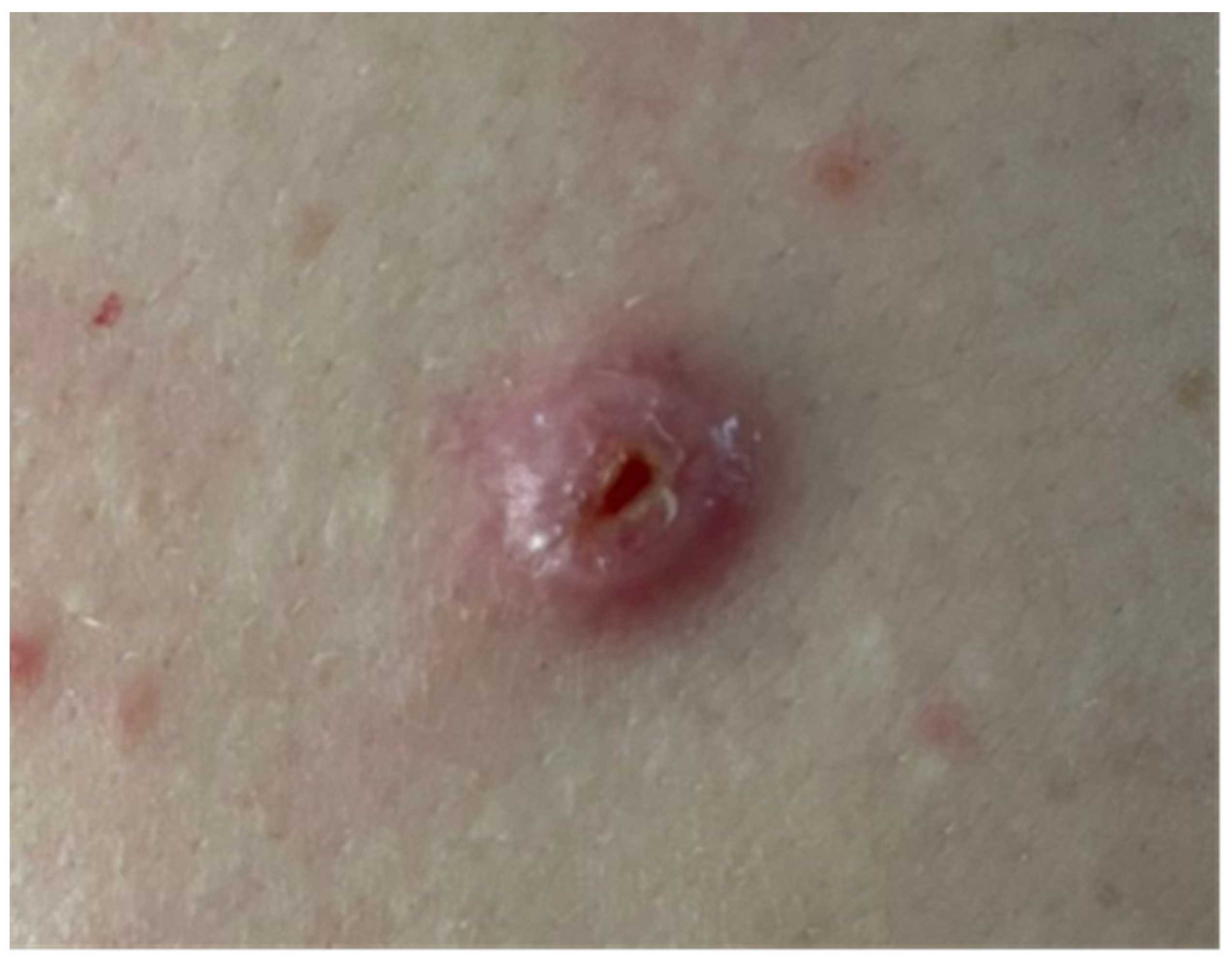
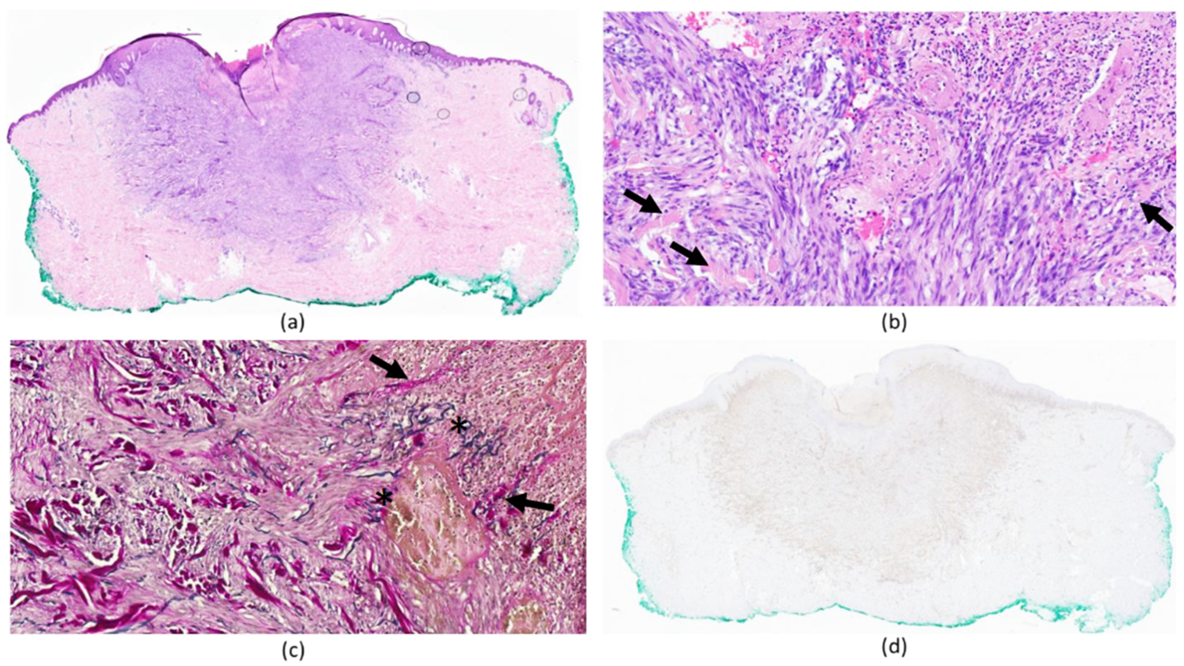
2. Case Report
3. Discussion
4. Conclusions
Author Contributions
Funding
Institutional Review Board Statement
Informed Consent Statement
Data Availability Statement
Conflicts of Interest
References
- Han, T.Y.; Chang, H.S.; Lee, J.H.; Lee, W.M.; Son, S.J. A clinical and histopathological study of 122 cases of dermatofibroma (benign fibrous histiocytoma). Ann. Dermatol. 2011, 23, 185–192. [Google Scholar] [CrossRef] [PubMed]
- Aloi, D.F.; Albertazzi, M.P. Dermatofibroma with Granular Cells: A Report of Two Cases. Dermatology 1999, 199, 54–56. [Google Scholar] [CrossRef] [PubMed]
- Lee, J. Epithelioid Cell Histiocytoma with Granular Cells. (Another Nonneural Granular Cell Neoplasm). Am. J. Dermatopathol. 2007, 29, 475–476. [Google Scholar] [CrossRef] [PubMed]
- del Carmen Gómez-Mateo, M.; Monteagudo, C. Nonepithelial skin tumors with multinucleated giant cells. Semin. Diagn. Pathol. 2013, 30, 58–72. [Google Scholar] [CrossRef] [PubMed]
- Alves, J.V.P.; Matos, D.M.; Barreiros, H.F.; Bártolo, E.A.F.L.F. Variants of dermatofibroma—A histopathological study. An. Bras. Dermatol. 2014, 89, 472–477. [Google Scholar] [CrossRef] [PubMed]
- Luzar, B.; Calonje, E. Cutaneous fibrohistiocytic tumours—An update. Histopathology 2010, 56, 148–165. [Google Scholar] [CrossRef] [PubMed]
- Kim, S.W.; Kim, M.S.; Lee, J.H.; Son, S.J.; Park, K.Y.; Li, K.; Seo, S.J.; Han, T.Y. A clinicopathologic study of thirty cases of acquired perforating dermatosis in Korea. Ann. Dermatol. 2014, 26, 162–171. [Google Scholar] [CrossRef] [PubMed]
- Zaccaria, E.; Rebora, A.; Rongioletti, F. Multiple eruptive dermatofibromas and im-munossupression: Report of two cases and review of the literature. Int. J. Dermatol. 2008, 47, 723–727. [Google Scholar] [CrossRef]
- Płaszczyca, A.; Nilsson, J.; Magnusson, L.; Brosjö, O.; Larsson, O.; von Steyern, F.V.; Domanski, H.; Lilljebjörn, H.; Fioretos, T.; Tayebwa, J.; et al. Fusions involving protein kinase C and membrane-associated proteins in benign fibrous histiocytoma. Int. J. Biochem. Cell Biol. 2014, 53, 475–481. [Google Scholar] [CrossRef]
- Walther, C.; Hofvander, J.; Nilsson, J.; Magnusson, L.; Domanski, H.A.; Gisselsson, D.; Tayebwa, J.; Doyle, L.A.; Fletcher, C.D.M.; Mertens, F. Gene fusion detection in formalin-fixed paraffin-embedded benign fibrous histiocytomas using fluorescence in situ hybridization and RNA sequencing. Lab. Investig. 2015, 95, 1071–1076. [Google Scholar] [CrossRef]
- Kim, E.J.; Park, H.S.; Yoon, H.S.; Cho, S. A case of perforating dermatofibroma with floret-like giant cells. Clin. Exp. Dermatol. 2014, 40, 305–308. [Google Scholar] [CrossRef] [PubMed]
- Aydin, E.; Vardareli, O.S.; Bilezikçi, B.; Ozgirgin, O.N. Dermatofibroma accompanied by perforating dermatosis in the auricle: A case report. Kulak Burun Bogaz Ihtis Derg. 2005, 15, 83–86. [Google Scholar] [PubMed]
- Samlaska, S.P.; Sandberg, G.D.; Maggio, K.L.; Sakas, E.L. Generalized perforating granuloma annulare. J. Am. Acad. Dermatol. 1992, 27, 319–322. [Google Scholar] [CrossRef] [PubMed]
- Kobayashi, H.; Oishi, K.; Miyake, M.; Nishijima, C.; Kawashima, A.; Kobayashi, H.; Inaoki, M. Spitz nevus on the sole of the foot presenting with transepidermal elimination. Dermatol. Pract. Concept. 2014, 4, 41–43. [Google Scholar] [CrossRef] [PubMed]
- Kang, H.Y.; Kang, W.H. Guess what! Perforating pilomatricoma resembling keratoacanthoma. Eur. J. Dermatol. 2000, 10, 63–64. [Google Scholar] [PubMed]
- Abbas, O.; Salem, Z.; Haddad, F.; Kibbi, A.-G. Perforating cutaneous metastasis from an ovarian adenocarcinoma. J. Cutan. Pathol. 2010, 37, 53–56. [Google Scholar] [CrossRef]
- Morton, C.A.; Henderson, I.S.; Jones, M.C.; Lowe, J.G. Acquired perforating dermatosis in a British dialysis population. Br. J. Dermatol. 1996, 135, 671–677. [Google Scholar] [CrossRef]
- Patterson, J.W. The perforating disorders. J. Am. Acad. Dermatol. 1984, 10, 561. [Google Scholar] [CrossRef]
- Pigariolo, C.B.; Maronese, C.A.; Giacalone, S.; Gianotti, R.; Marzano, A.V.; Veraldi, S. Persistent variant of acquired verrucous perforating collagenoma. Int. J. Dermatol. 2022, 61, 333–335. [Google Scholar]
- Bohdanowicz, M.; Bradshaw, S.H. Perforating Gout: Expanding the Differential for Transepidermal Elimination. Dermatopathology 2023, 10, 207–218. [Google Scholar] [CrossRef]
- Penas, P.F.; Jones-Caballero, M.; Fraga, J.; Sánchez-Pérez, J.; García-Díez, A. Perforating granuloma annulare. Int. J. Dermatol. 1997, 36, 340–348. [Google Scholar] [CrossRef]


| Features | Characteristics | Case 1 | Case 2 | Our Case |
|---|---|---|---|---|
| Clinical | Gender | Male | Female | Male |
| Age (years) | 12 | 82 | 31 | |
| Site | Back of the knee | Left auricle | Lower back | |
| Diameter (mm) of the lesion | 6 | 8 | 10 | |
| Histopathological | Epidermal perforation | Present | Present | Present |
| Extruded material | Giant cells Macrophages Collagen bundles | Collagen bundles | Collagen bundles Elastic fibers Spindle cells |
Disclaimer/Publisher’s Note: The statements, opinions and data contained in all publications are solely those of the individual author(s) and contributor(s) and not of MDPI and/or the editor(s). MDPI and/or the editor(s) disclaim responsibility for any injury to people or property resulting from any ideas, methods, instructions or products referred to in the content. |
© 2023 by the authors. Licensee MDPI, Basel, Switzerland. This article is an open access article distributed under the terms and conditions of the Creative Commons Attribution (CC BY) license (https://creativecommons.org/licenses/by/4.0/).
Share and Cite
Lungu, A.; Hsieh, A.; Kaya, G.; Menzinger, S. Perforating Fibrous Histiocytoma Mimicking Keratoacanthoma: A Case Report. Dermatopathology 2024, 11, 8-12. https://doi.org/10.3390/dermatopathology11010002
Lungu A, Hsieh A, Kaya G, Menzinger S. Perforating Fibrous Histiocytoma Mimicking Keratoacanthoma: A Case Report. Dermatopathology. 2024; 11(1):8-12. https://doi.org/10.3390/dermatopathology11010002
Chicago/Turabian StyleLungu, Alina, Aurélie Hsieh, Gürkan Kaya, and Sébastien Menzinger. 2024. "Perforating Fibrous Histiocytoma Mimicking Keratoacanthoma: A Case Report" Dermatopathology 11, no. 1: 8-12. https://doi.org/10.3390/dermatopathology11010002