


Cells 2022, 11(17), 2630; https://doi.org/10.3390/cells11172630 - 24 Aug 2022
Cited by 5 | Viewed by 4458
Abstract
►
Show Figures
Interferon regulatory factor 8 (IRF8) is a transcription factor of the IRF protein family. IRF8 was originally identified as an essentialfactor for myeloid cell lineage commitment and differentiation. Deletion of Irf8 leads to massive accumulation of CD11b+Gr1+ immature myeloid cells
[...] Read more.
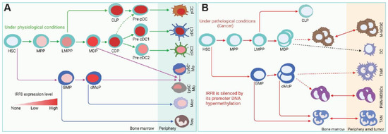
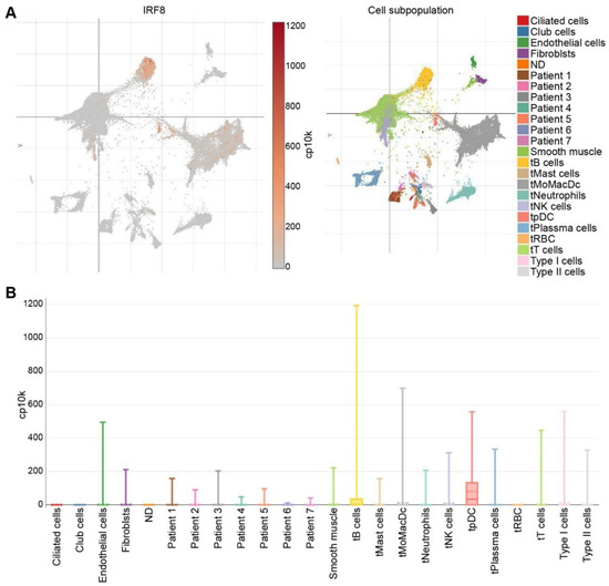
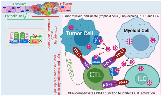
Interferon regulatory factor 8 (IRF8) is a transcription factor of the IRF protein family. IRF8 was originally identified as an essentialfactor for myeloid cell lineage commitment and differentiation. Deletion of Irf8 leads to massive accumulation of CD11b+Gr1+ immature myeloid cells (IMCs), particularly the CD11b+Ly6Chi/+Ly6G− polymorphonuclear myeloid-derived suppressor cell-like cells (PMN-MDSCs). Under pathological conditions such as cancer, Irf8 is silenced by its promoter DNA hypermethylation, resulting in accumulation of PMN-MDSCs and CD11b+ Ly6G+Ly6Clo monocytic MDSCs (M-MDSCs) in mice. IRF8 is often silenced in MDSCs in human cancer patients. MDSCs are heterogeneous populations of immune suppressive cells that suppress T and NK cell activity to promote tumor immune evasion and produce growth factors to exert direct tumor-promoting activity. Emerging experimental data reveals that IRF8 is also expressed in non-hematopoietic cells. Epithelial cell-expressed IRF8 regulates apoptosis and represses Osteopontin (OPN). Human tumor cells may use the IRF8 promoter DNA methylation as a mechanism to repress IRF8 expression to advance cancer through acquiring apoptosis resistance and OPN up-regulation. Elevated OPN engages CD44 to suppress T cell activation and promote tumor cell stemness to advance cancer. IRF8 thus is a transcription factor that regulates both the immune and non-immune components in human health and diseases.
Full article
Figure 1