


Cells 2022, 11(10), 1662; https://doi.org/10.3390/cells11101662 - 17 May 2022
Cited by 5 | Viewed by 3722
Abstract
►
Show Figures
Neurodegenerative diseases are deteriorating conditions of the nervous system that are rapidly increasing in the ageing population. Increasing evidence suggests that neuroinflammation, largely mediated by microglia, the resident immune cells of the brain, contributes to the onset and progression of neurodegenerative diseases. Hence,
[...] Read more.
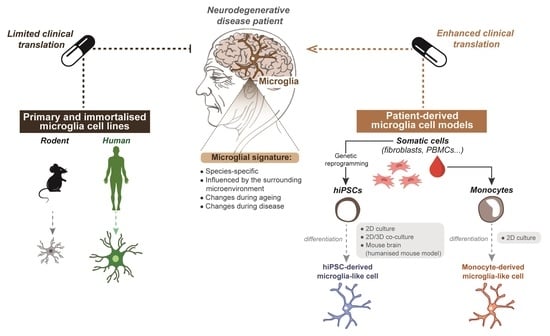
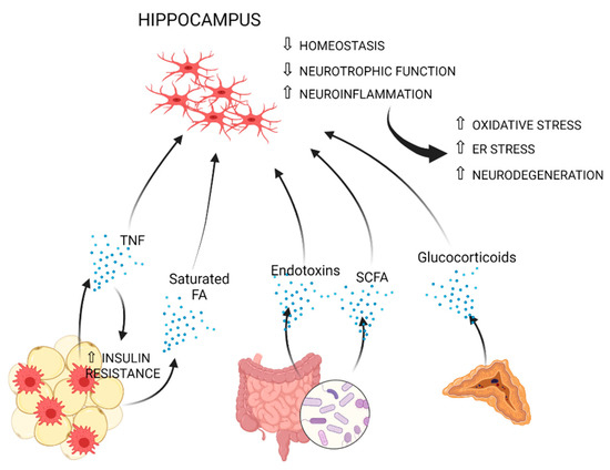
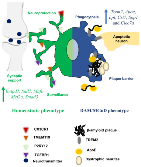
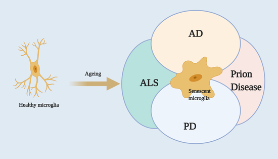
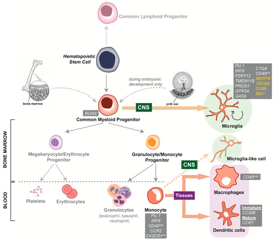
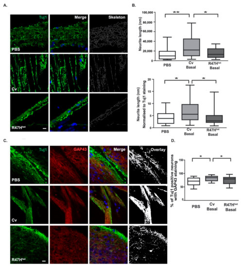
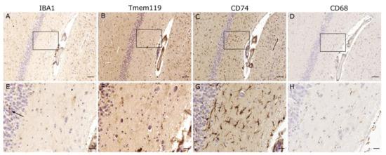
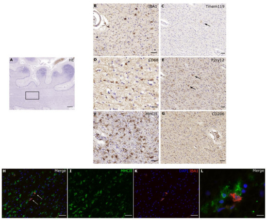
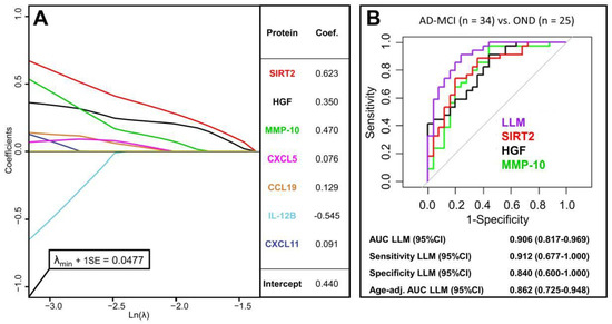
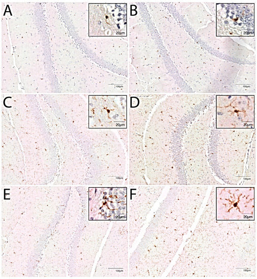
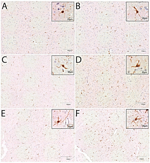
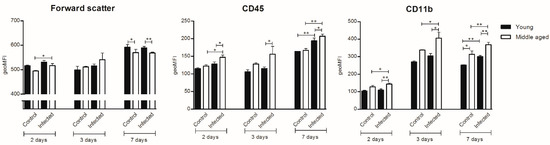
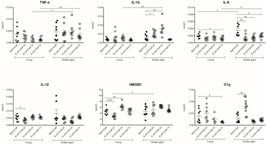
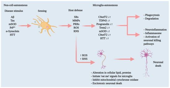
Neurodegenerative diseases are deteriorating conditions of the nervous system that are rapidly increasing in the ageing population. Increasing evidence suggests that neuroinflammation, largely mediated by microglia, the resident immune cells of the brain, contributes to the onset and progression of neurodegenerative diseases. Hence, microglia are considered a major therapeutic target that could potentially yield effective disease-modifying treatments for neurodegenerative diseases. Despite the interest in studying microglia as drug targets, the availability of cost-effective, flexible, and patient-specific microglia cellular models is limited. Importantly, the current model systems do not accurately recapitulate important pathological features or disease processes, leading to the failure of many therapeutic drugs. Here, we review the key roles of microglia in neurodegenerative diseases and provide an update on the current microglial plaforms utilised in neurodegenerative diseases, with a focus on human microglia-like cells derived from peripheral blood mononuclear cells as well as human-induced pluripotent stem cells. The described microglial platforms can serve as tools for investigating disease biomarkers and improving the clinical translatability of the drug development process in neurodegenerative diseases.
Full article
Graphical abstract