Feature Papers in 'Cell Proliferation and Division'
Share This Topical Collection
Editor
 Prof. Dr. Zhixiang Wang
Prof. Dr. Zhixiang Wang
E-Mail
Website
Collection Editor
Signal Transduction Research Group, Department of Medical Genetics, Faculty of Medicine and Dentistry, University of Alberta, Edmonton, AB T6G 2H7, Canada
Interests: receptor tyrosine kinases; EGFR; signal transduction; cancer therapy; breast cancer treatment; targeted therapy
Special Issues, Collections and Topics in MDPI journals
Topical Collection Information
Dear Colleagues,
The present Topical Collection entitled “Feature Papers in Cell Proliferation and Division” collates high-quality research articles, communications, and review articles addressing and investigating the cutting-edge fields of cell proliferation and division. The Topical Collection aims to illustrate, through the choice of the selected works, the frontier research in cell proliferation and division, and we therefore invite the Editorial Board Members of Cells or relevant experts and colleagues to contribute papers reflecting the latest progress in their particular field of research.
The topics of interest include, but are not limited to, the following:
- Cell proliferation;
- Cell division;
- Cell cycle;
- Cell reproduction;
- Cell growth;
- Stem cells;
- Cancer;
- Chromosoma;
- Mitosis;
- Meiosis;
- Cyclin;
- Cohesins;
- Centromeres;
- Kinetochore;
- Cytokinesis.
Prof. Dr. Zhixiang Wang
Collection Editor
Manuscript Submission Information
Manuscripts should be submitted online at www.mdpi.com by registering and logging in to this website. Once you are registered, click here to go to the submission form. Manuscripts can be submitted until the deadline. All submissions that pass pre-check are peer-reviewed. Accepted papers will be published continuously in the journal (as soon as accepted) and will be listed together on the collection website. Research articles, review articles as well as short communications are invited. For planned papers, a title and short abstract (about 100 words) can be sent to the Editorial Office for announcement on this website.
Submitted manuscripts should not have been published previously, nor be under consideration for publication elsewhere (except conference proceedings papers). All manuscripts are thoroughly refereed through a single-blind peer-review process. A guide for authors and other relevant information for submission of manuscripts is available on the Instructions for Authors page. Cells is an international peer-reviewed open access semimonthly journal published by MDPI.
Please visit the Instructions for Authors page before submitting a manuscript.
The Article Processing Charge (APC) for publication in this open access journal is 2700 CHF (Swiss Francs).
Submitted papers should be well formatted and use good English. Authors may use MDPI's
English editing service prior to publication or during author revisions.
Published Papers (3 papers)
2022
Open AccessArticle
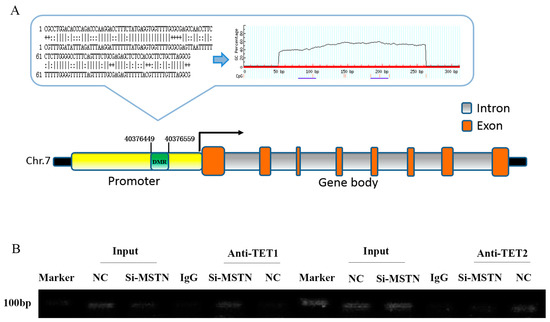
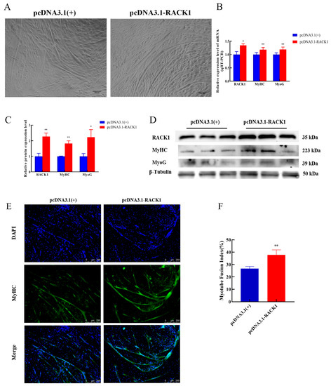
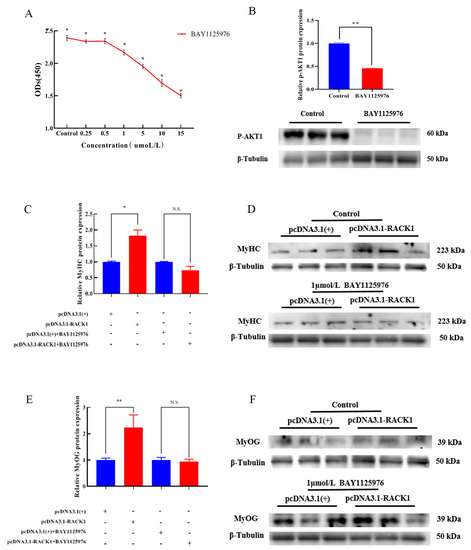
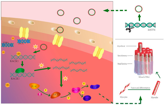
Myostatin Mutation Enhances Bovine Myogenic Differentiation through PI3K/AKT/mTOR Signalling via Removing DNA Methylation of RACK1
by
Yiping Zhao, Xiaoxia Xia, Qiaomeng Wang, Debao Hu, Linlin Zhang, Xin Li, Xiangbin Ding, Hong Guo and Yiwen Guo
Cited by 3 | Viewed by 1325
Abstract
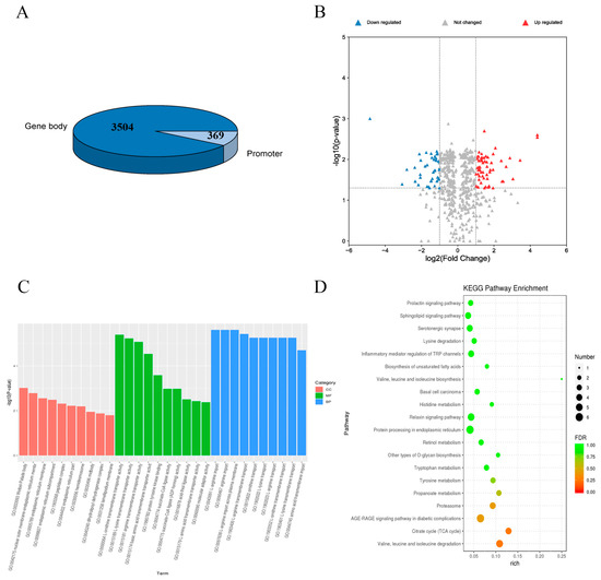
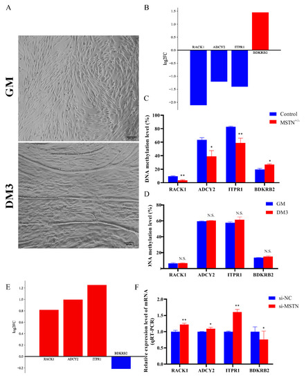
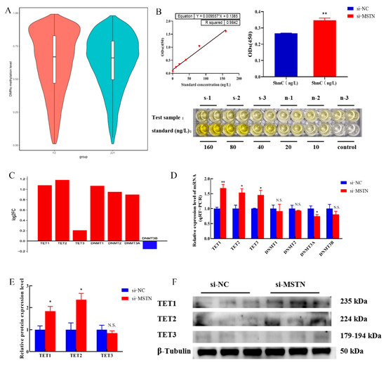
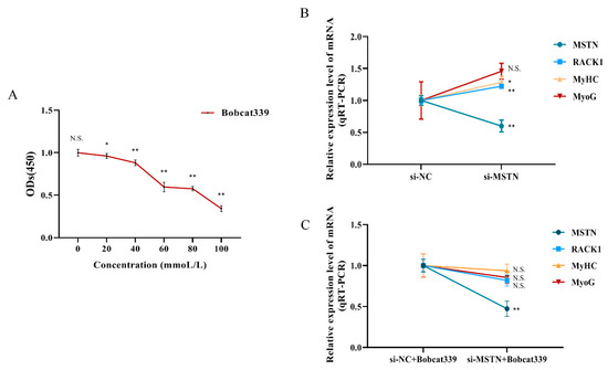
Myostatin (MSTN) is a negative regulator of skeletal muscle development and plays an important role in muscle development. Fluctuations in gene expression influenced by DNA methylation are critical for homeostatic responses in muscle. However, little is known about the mechanisms underlying this fluctuation
[...] Read more.
Myostatin (MSTN) is a negative regulator of skeletal muscle development and plays an important role in muscle development. Fluctuations in gene expression influenced by DNA methylation are critical for homeostatic responses in muscle. However, little is known about the mechanisms underlying this fluctuation regulation and myogenic differentiation of skeletal muscle. Here we report a genome-wide analysis of DNA methylation dynamics in bovine skeletal muscle myogenesis after myostatin editing. We show that, after myostatin editing, an increase in TETs (DNA demethylases) and a concomitant increase in the receptor for activated C kinase 1 (RACK1) control the myogenic development of skeletal muscle. Interestingly, enhancement of PI3K/AKT/mTOR signaling by RACK1 appears to be an essential driver of myogenic differentiation, as it was associated with an increase in myogenic differentiation marker factors (MyHC and MyoG) during muscle differentiation. Overall, our results suggest that loss of myostatin promotes the myogenic differentiation response in skeletal muscle by decreasing DNA methylation of RACK1.
Full article
►▼
Show Figures
Open AccessArticle
A Novel Mechanism Underlying the Inhibitory Effects of Trastuzumab on the Growth of HER2-Positive Breast Cancer Cells
by
Hamid Maadi and Zhixiang Wang
Cited by 3 | Viewed by 1751
Abstract
To improve the efficacy of trastuzumab, it is essential to understand its mechanism of action. One of the significant issues that makes it difficult to determine the precise mechanism of trastuzumab action is the formation of various HER receptor dimers in HER2-positive breast
[...] Read more.
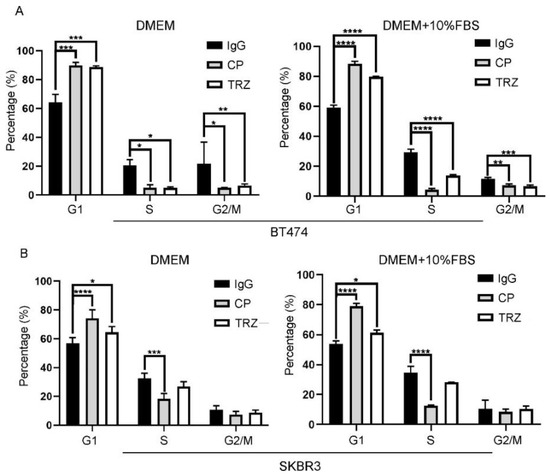
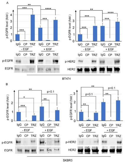
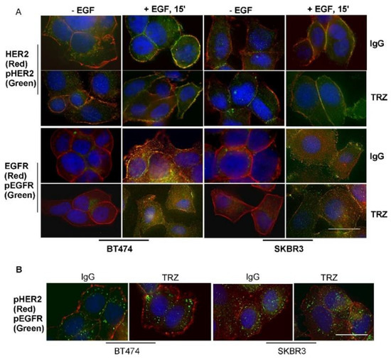
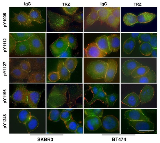
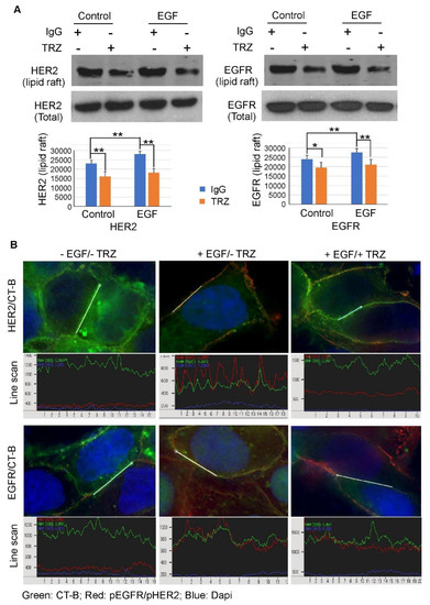
To improve the efficacy of trastuzumab, it is essential to understand its mechanism of action. One of the significant issues that makes it difficult to determine the precise mechanism of trastuzumab action is the formation of various HER receptor dimers in HER2-positive breast cancer cells. So far, studies have focused on the role of HER2–HER3 heterodimers, and little is known regarding EGFR–HER2 heterodimers. Here, we study the role of trastuzumab on the cell signaling and cell proliferation mediated by EGFR–HER2 heterodimers in BT474 and SRBR3 cells. EGF stimulates the formation of both EGFR homodimer and EGFR–HER2 heterodimer. Trastuzumab only binds to HER2, not EGFR. Therefore, any effects of trastuzumab on EGF-induced activation of EGFR, HER2, and downstream signaling proteins, as well as cell proliferation, are through its effects on EGFR–HER2 heterodimers. We show that trastuzumab inhibits EGF-induced cell proliferation and cell cycle progression in BT474 and SKBR3 cells. Interestingly trastuzumab strongly inhibits EGF-induced Akt phosphorylation and slightly inhibits EGF-induced Erk activation, in both BT474 and SKBR3 cells. These data suggest the presence of a novel mechanism that allows trastuzumab to inhibit EGR-induced Akt activation and cell proliferation, without blocking EGF-induced EGFR–HER2 heterodimerization and activation. We show that trastuzumab inhibits EGF-induced lipid raft localization of the EGFR–HER2 heterodimer. Disruption of the lipid raft with MβCD blocks HER2-mediated AKT activation in a similar way to trastuzumab. MβCD and trastuzumab synergically inhibit AKT activation. We conclude that trastuzumab inhibits EGF-induced lipid raft localization of EGFR–HER2 heterodimer, which leads to the inhibition of Akt phosphorylation and cell proliferation, without blocking the formation and phosphorylation of the EGFR–HER2 heterodimer.
Full article
►▼
Show Figures
Open AccessArticle
In Vitro Transfection of Up-Regulated Genes Identified in Favorable-Outcome Neuroblastoma into Cell Lines
by
Yoko Hiyama, Emi Yamaoka, Takahiro Fukazawa, Masato Kojima, Yusuke Sotomaru and Eiso Hiyama
Viewed by 2028
Abstract
We previously used microarrays to show that high expression of
DHRS3,
NROB1, and
CYP26A1 predicts favorable NB outcomes. Here, we investigated whether expression of these genes was associated with suppression of NB cell (SK-N-SH, NB12, and TGW) growth. We assessed morphology
[...] Read more.
We previously used microarrays to show that high expression of
DHRS3,
NROB1, and
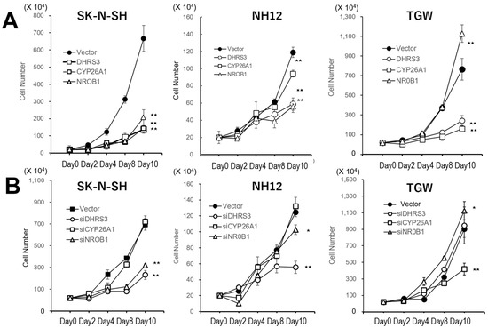
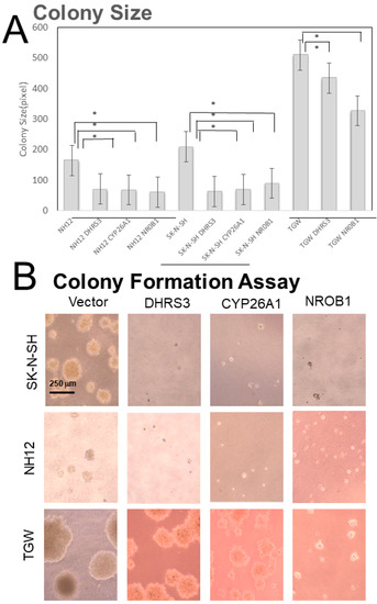
CYP26A1 predicts favorable NB outcomes. Here, we investigated whether expression of these genes was associated with suppression of NB cell (SK-N-SH, NB12, and TGW) growth. We assessed morphology and performed growth, colony-formation, and migration assays, as well as RNA sequencing. The effects of the transient expression of these genes were also assessed with a tetracycline-controlled expression (Tet-On) system. Gene overexpression reduced cell growth and induced morphological senescence. Gene-expression analysis identified pathways involving cellular senescence and cell adhesion. In these cells, transduced gene dropout occurred during passage, making long-term stable gene transfer difficult. Tet-On-induced gene expression caused more pronounced cell-morphology changes. Specifically,
DHRS3 and
NROB1 led to rapid inhibition and arrest of cell growth, though
CYP26A1 did not affect cell-growth rate or cell cycle.
DHRS3 arrested the cell cycle by interacting with the all-trans-retinol pathway and drove differentiation and senescence in tumors. Overexpression of these genes reduced the malignant grade of these cells. A new therapeutic strategy might be the induction of these genes, as they suppress the growth of high-risk neuroblastoma and lead to differentiation and senescence.
Full article
►▼
Show Figures


