


Cancers 2024, 16(1), 124; https://doi.org/10.3390/cancers16010124 - 26 Dec 2023
Viewed by 1472
Abstract
►
Show Figures
Breast cancer remains a significant health challenge, and novel treatment approaches are critically needed. This review presents an in-depth analysis of engineered adoptive T-cell therapies (E-ACTs), an innovative frontier in cancer immunotherapy, focusing on their application in breast cancer. We explore the evolving
[...] Read more.
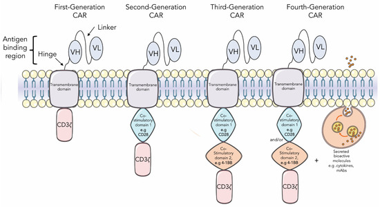
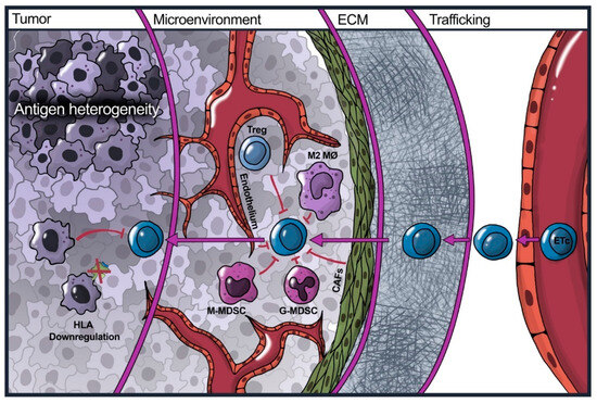
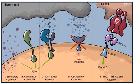
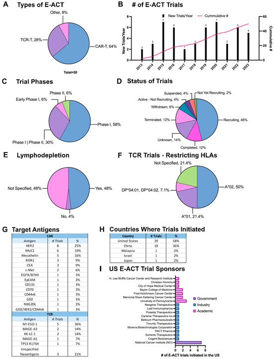
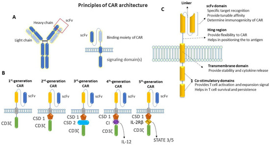
Breast cancer remains a significant health challenge, and novel treatment approaches are critically needed. This review presents an in-depth analysis of engineered adoptive T-cell therapies (E-ACTs), an innovative frontier in cancer immunotherapy, focusing on their application in breast cancer. We explore the evolving landscape of chimeric antigen receptor (CAR) and T-cell receptor (TCR) T-cell therapies, highlighting their potential and challenges in targeting breast cancer. The review addresses key obstacles such as target antigen selection, the complex breast cancer tumor microenvironment, and the persistence of engineered T-cells. We discuss the advances in overcoming these barriers, including strategies to enhance T-cell efficacy. Finally, our comprehensive analysis of the current clinical trials in this area provides insights into the future possibilities and directions of E-ACTs in breast cancer treatment.
Full article
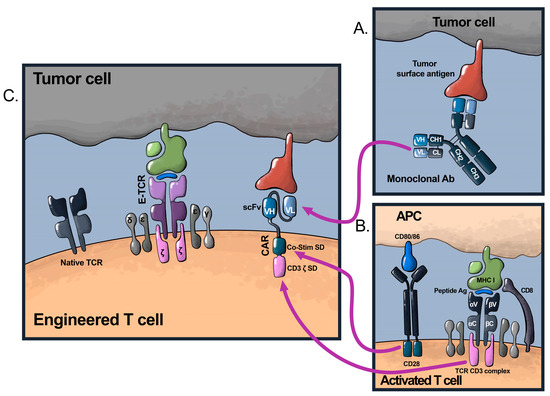
Figure 1


