


Cancers 2023, 15(3), 641; https://doi.org/10.3390/cancers15030641 - 19 Jan 2023
Cited by 2 | Viewed by 1627
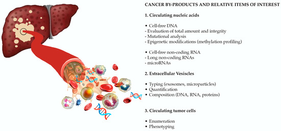
Abstract
►
Show Figures
Malignant pleural mesothelioma (MPM) is an asbestos-associated, highly aggressive cancer characterized by late-stage diagnosis and poor prognosis. Gold standards for diagnosis are pleural biopsy and cytology of pleural effusion (PE), both of which are limited by low sensitivity and markedly inter-observer variations. Therefore,
[...] Read more.
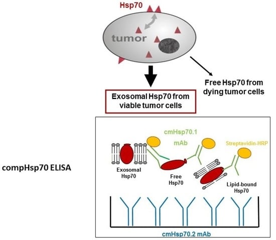
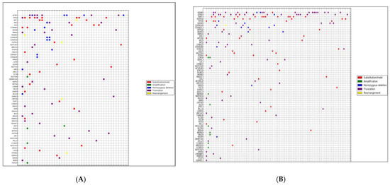
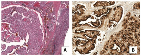
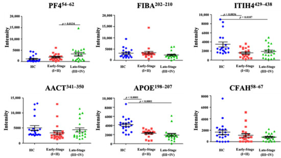
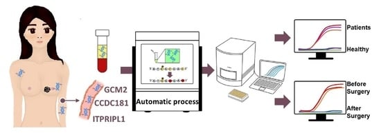
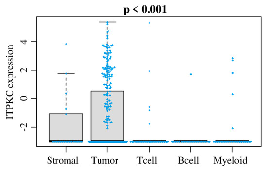
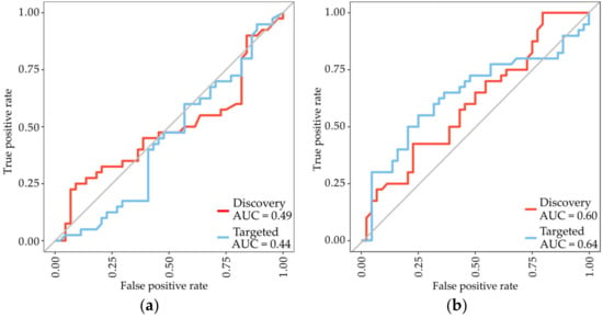
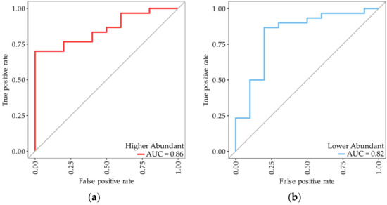
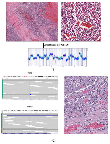
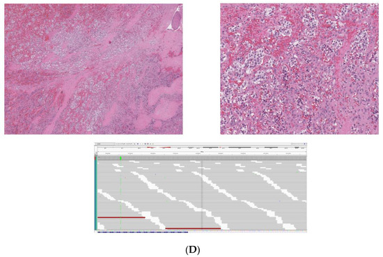
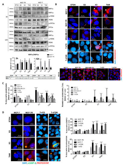
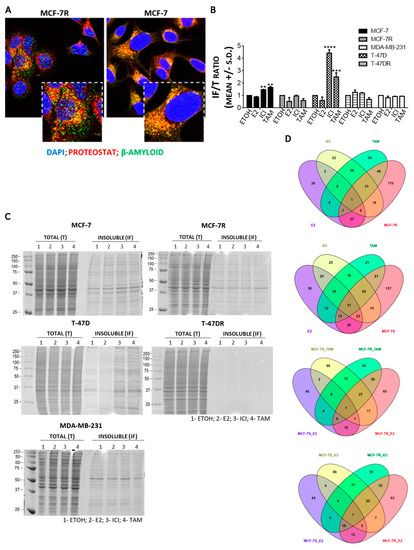
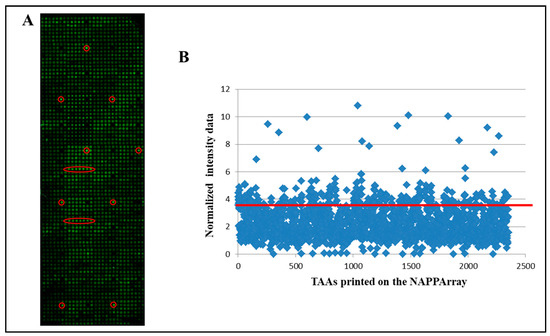
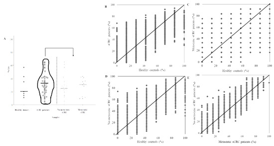
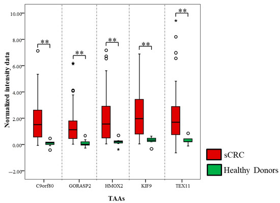
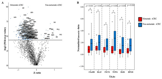
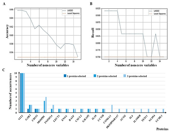
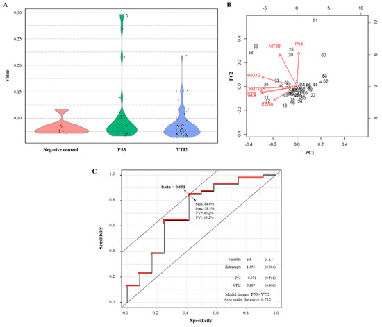
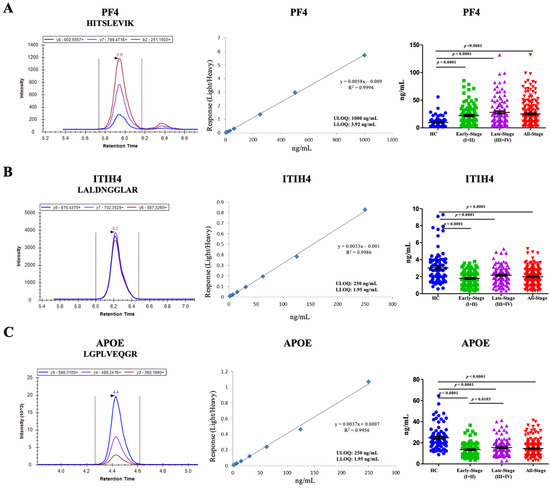
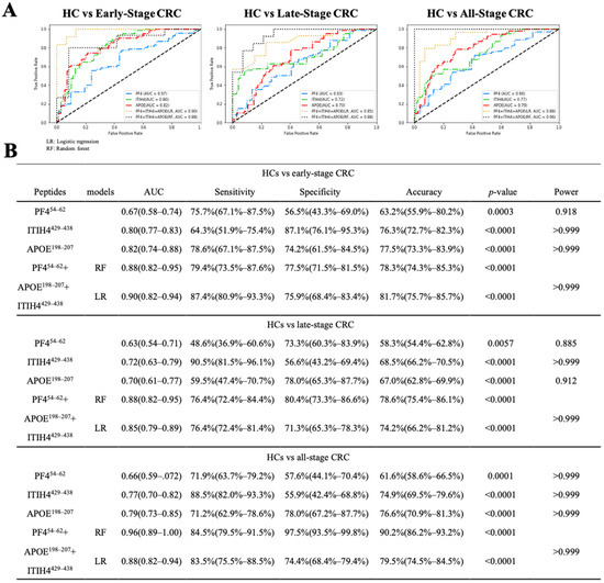
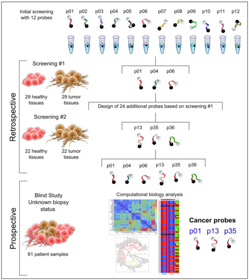
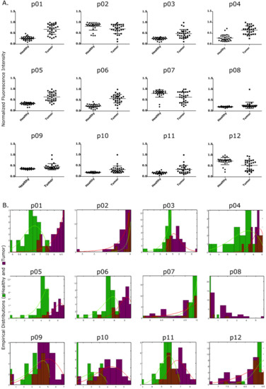
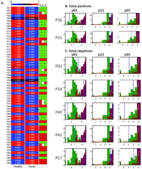
Malignant pleural mesothelioma (MPM) is an asbestos-associated, highly aggressive cancer characterized by late-stage diagnosis and poor prognosis. Gold standards for diagnosis are pleural biopsy and cytology of pleural effusion (PE), both of which are limited by low sensitivity and markedly inter-observer variations. Therefore, the assessment of PE biomarkers is considered a viable and objective diagnostic tool for MPM diagnosis. We applied a novel affinity-enrichment mass spectrometry-based proteomics method for explorative analysis of pleural effusions from a prospective cohort of 84 patients referred for thoracoscopy due to clinical suspicion of MPM. Protein biomarkers with a high capability to discriminate MPM from non-MPM patients were identified, and a Random Forest algorithm was applied for building classification models. Immunohistology of pleural biopsies confirmed MPM in 40 patients and ruled out MPM in 44 patients. Proteomic analysis of pleural effusions identified panels of proteins with excellent diagnostic properties (90–100% sensitivities, 89–98% specificities, and AUC 0.97–0.99) depending on the specific protein combination. Diagnostic proteins associated with cancer growth included galactin-3 binding protein, testican-2, haptoglobin, Beta ig-h3, and protein AMBP. Moreover, we also confirmed previously reported diagnostic accuracies of the MPM markers fibulin-3 and mesothelin measured by two complementary mass spectrometry-based methods. In conclusion, a novel affinity-enrichment mass spectrometry-based proteomics identified panels of proteins in pleural effusion with extraordinary diagnostic accuracies, which are described here for the first time as biomarkers for MPM.
Full article
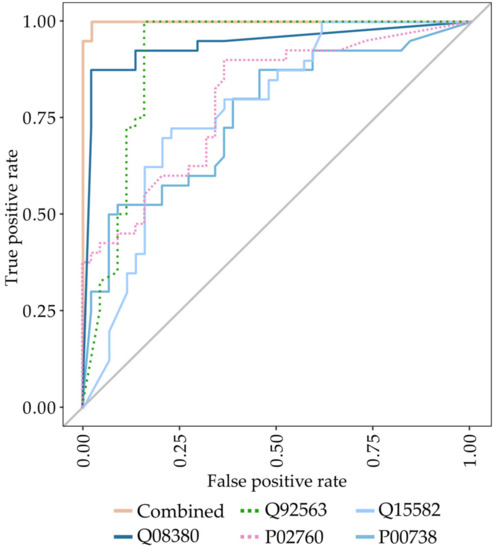
Figure 1


