Article

Integrative Molecular Analysis of DNA Methylation Dynamics Unveils Molecules with Prognostic Potential in Breast Cancer

1
Department of Life Sciences, College of Science and General Studies, Alfaisal University, Riyadh 11533, Saudi Arabia
2
Department of Immunology, Division of Translational Immuno-Oncology, Roswell Park Comprehensive Cancer Center, New York, NY 14203, USA
3
Department of Plant Breeding and Genetics, The University of Agriculture, Peshawar 25130, Pakistan
4
Institute of Basic Medical Sciences, Khyber Medical University, Peshawar 25120, Pakistan
5
Department of Biostatistics, School of Public Health, The University of Michigan, Ann Arbor, MI 48109, USA
6
Office of Research, Innovation, and Graduate Studies, Alfaisal University, Riyadh 11533, Saudi Arabia
7
Department of Life and Environmental Sciences, College of Health and Natural Sciences, Zayed University, Abu Dhabi 144534, United Arab Emirates
8
Department of Bioinformatics, Biocenter, University of Würzburg, Am Hubland, 97074 Wuerzburg, Germany
9
Graduate School of Integrated Basic Sciences, Nihon University, Setagaya-ku, Tokyo 156-8550, Japan
*
Author to whom correspondence should be addressed.
BioMedInformatics 2023, 3(2), 434-445; https://doi.org/10.3390/biomedinformatics3020029
Submission received: 10 April 2023 / Revised: 6 May 2023 / Accepted: 22 May 2023 / Published: 5 June 2023
(This article belongs to the Special Issue Feature Papers in Computational Biology and Medicine)

Abstract

:
DNA methylation acts as a major epigenetic modification in mammals, characterized by the transfer of a methyl group to a cytosine. DNA methylation plays a pivotal role in regulating normal development, and misregulation in cells leads to an abnormal phenotype as is seen in several cancers. Any mutations or expression anomalies of genes encoding regulators of DNA methylation may lead to abnormal expression of critical molecules. A comprehensive genomic study encompassing all the genes related to DNA methylation regulation in relation to breast cancer is lacking. We used genomic and transcriptomic datasets from the Cancer Genome Atlas (TGCA) Pan-Cancer Atlas, Genotype-Tissue Expression (GTEx) and microarray platforms and conducted in silico analysis of all the genes related to DNA methylation with respect to writing, reading and erasing this epigenetic mark. Analysis of mutations was conducted using cBioportal, while Xena and KMPlot were utilized for expression changes and patient survival, respectively. Our study identified multiple mutations in the genes encoding regulators of DNA methylation. The expression profiling of these showed significant differences between normal and disease tissues. Moreover, deregulated expression of some of the genes, namely DNMT3B, MBD1, MBD6, BAZ2B, ZBTB38, KLF4, TET2 and TDG, was correlated with patient prognosis. The current study, to our best knowledge, is the first to provide a comprehensive molecular and genetic profile of DNA methylation machinery genes in breast cancer and identifies DNA methylation machinery as an important determinant of the disease progression. The findings of this study will advance our understanding of the etiology of the disease and may serve to identify alternative targets for novel therapeutic strategies in cancer.

Graphical Abstract

1. Introduction

Among the various types of epigenetic modifications of the genome, DNA methylation is the first and highly characterized one that involves addition of a methyl group to the fifth carbon of the cytosine residue at CpG dinucleotides [1,2,3]. DNA methylation is carried out by the action of a group of enzymes called DNA methyltransferases (DNMTs), which are also termed as writers of this modification and are conserved in various species including bacteria, plants and mammals [4]. DNA methylation has a critical role in regulating the gene expression in various physiological processes; for example, in X-chromosome inactivation, genomic imprinting, repetitive elements repression and silencing of potentially active transposable elements [5,6]. Various techniques have been developed for detection of DNA methylation, including sodium bisulfite conversion, methylation-sensitive restriction enzymes and methylated DNA immunoprecipitation. The choice of DNA methylation detection technique depends on several factors such as the scale of analysis (gene-specific vs. genome-wide), the resolution required and the availability of resources [7]. While methylation at the CpG sites is the most frequent one, recent studies in the brain and embryonic stem cells have identified methylation beyond the CpG context [8,9]. Dynamic regulation of genome methylation is a general attribute of early mammalian development that is characterized by highly regulated methylation and demethylation of lineage specific genes [10,11]. The dynamics of DNA methylation during early development necessitates the high expression of DNA methyltransferases such as DNMT3A and DNMT3B, which are de novo methyltransferases, and DNMT1, which is maintenance methyltransferase [12,13].
DNA methylation in gene promoters affects gene expression by regulating the recruitment of methylated DNA binding proteins (MBPs), also called readers of DNA methylation, which influence binding of transcription factor and overall chromatin structure [14]. Methyl CpG binding protein 2 (MeCP2) was the first methylated DNA binding protein that was identified by its ability to bind DNA sequences when they are methylated at CpG [3]. MeCP2 contains a methyl-CpG-binding domain (MBD) that comprises 70–85 amino acids [15] that was used to identify other proteins with methylated DNA ability to bind [16]. To date, a number of methyl-CpG binding proteins have been identified that are classified based on methylated DNA binding domains [17]. After binding to the methylated DNA, MBPs relay the DNA methylation signal into various functional states through interactions with diverse partners that eventually regulate gene expression by specific mechanisms, such as chromatin organization and epigenome maintenance [18].
DNA methylation is considered relatively stable epigenetic modification compared with most histone modifications; however, active loss of DNA methylation was mechanistically described recently. The discovery of ten eleven translocation protein 1 (TET1) and its ability to modify methylcytosine and remove methyl group from DNA provided initial insights regarding active demethylation [19]. TET1 belongs to a family of three enzymes namely TET1, TET2 and TET3, that are involved in the stepwise oxidation of 5-methylcytosine (5mC) to 5-hydroxymethylcytosine (5hmC), 5-formylcytosine (5fC) and 5-carboxylcytosine (5caC) [20,21]. These enzymes are collectively called “erasers” of DNA methylation.
One of the hallmarks of cancer is the disruption of epigenetic profiles including DNA methylation patterns. Abnormal DNA methylation is one of the most critical epigenetic factors directly linked with tumorigenesis, because aberrant methylation can contribute to transcriptional changes of tumor suppressors and oncogenes [22,23,24]. Disruption in the activities of members of “readers”, “writers” and “erasers” of DNA methylation has been reported [25,26]. DNA methyltransferases coordinate mRNA expression in normal tissue and tumors [27]. Moreover, elevated expressions of DNMTs have been reported for hepatic, prostate and breast cancers [28,29,30,31].
Although multiple reports that have focused on the role of one or a few regulators of DNA methylation in cancer progression have significantly enhanced our understanding of the role of this important epigenetic modification in cancer [32,33,34], to our knowledge a comprehensive study that provides overarching molecular and genetic profile of DNA methylation machinery is lacking. This study provides a genomic and molecular landscape of writers, readers and erasers of DNA methylation in breast cancer. The majority of molecules show significant expression anomalies in breast cancer. Additionally, we identify that deregulated expression of some of the molecules have prognostic potential in breast cancer.

2. Materials and Methods

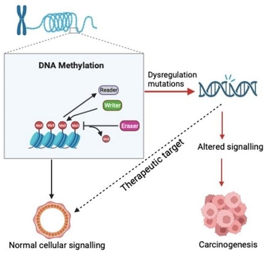
2.1. Writers, Readers and Erasers of DNA Methylation

The genes coding for writers, readers and erasers of DNA methylation are listed in Table 1. The list was compiled using data from earlier studies [17,35]. All the genes listed in the table were used for mutational analysis, expression changes and patient overall survival.

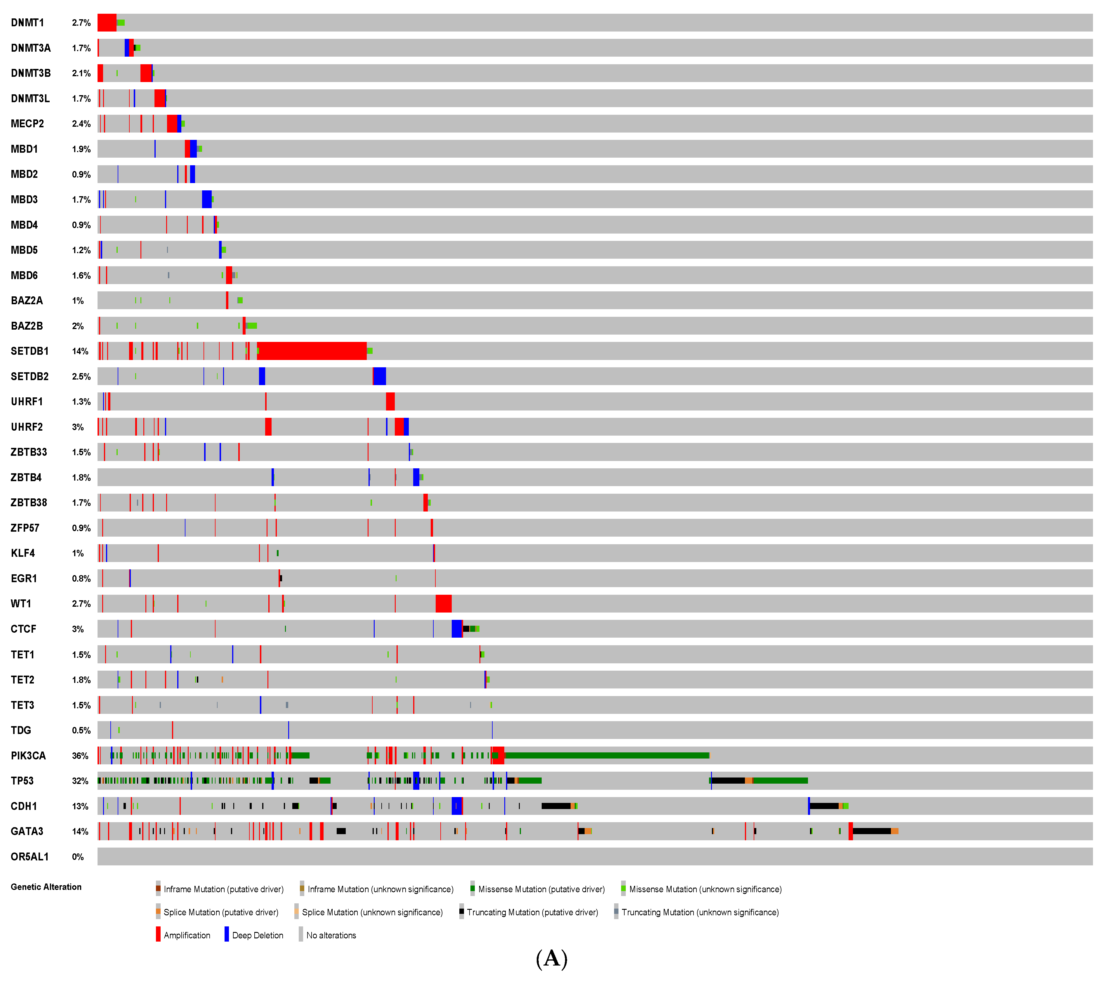
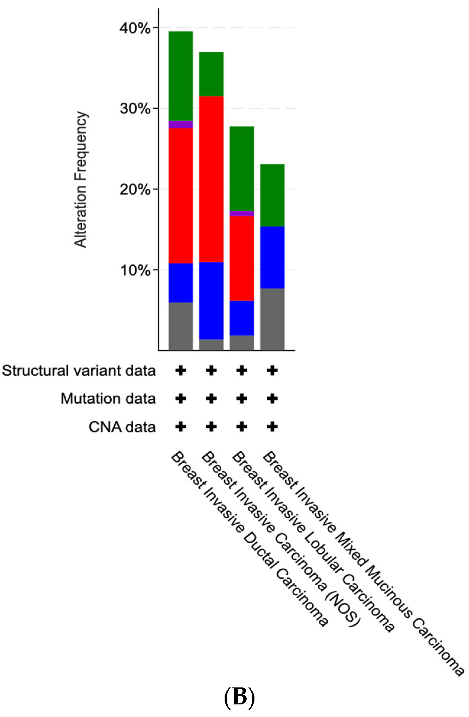
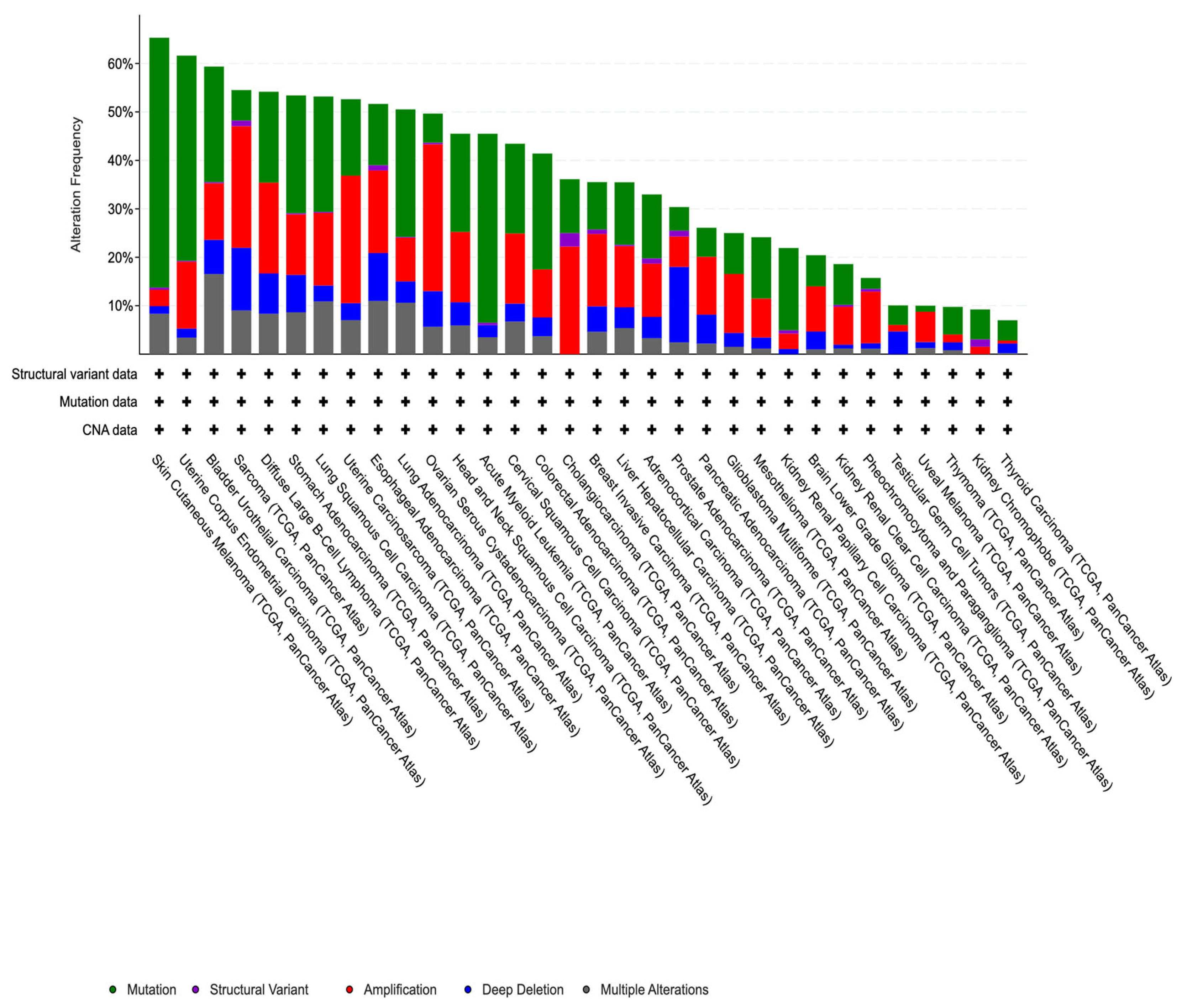
2.2. Genetic Alterations

To identify mutations in various genes encoding DNA methylation regulators, we utilized the Cancer Genome Atlas (TCGA) (https://www.cancer.gov/tcga, accessed on 10 April 2022) data using cBioportal [36,37]. Breast cancer datasets from multiple sources are accessible at cBioportal. We selected the latest “TCGA Breast Invasive Carcinoma (TCGA, Pan-Cancer Atlas)” dataset that consisted of 996 patient samples with copy-number alterations (CNAs) and mutation data. Therefore, following genetic alterations were identified in these 996 breast cancer patients’ datasets: (1) copy-number alterations (CNAs) and (2) mutations.

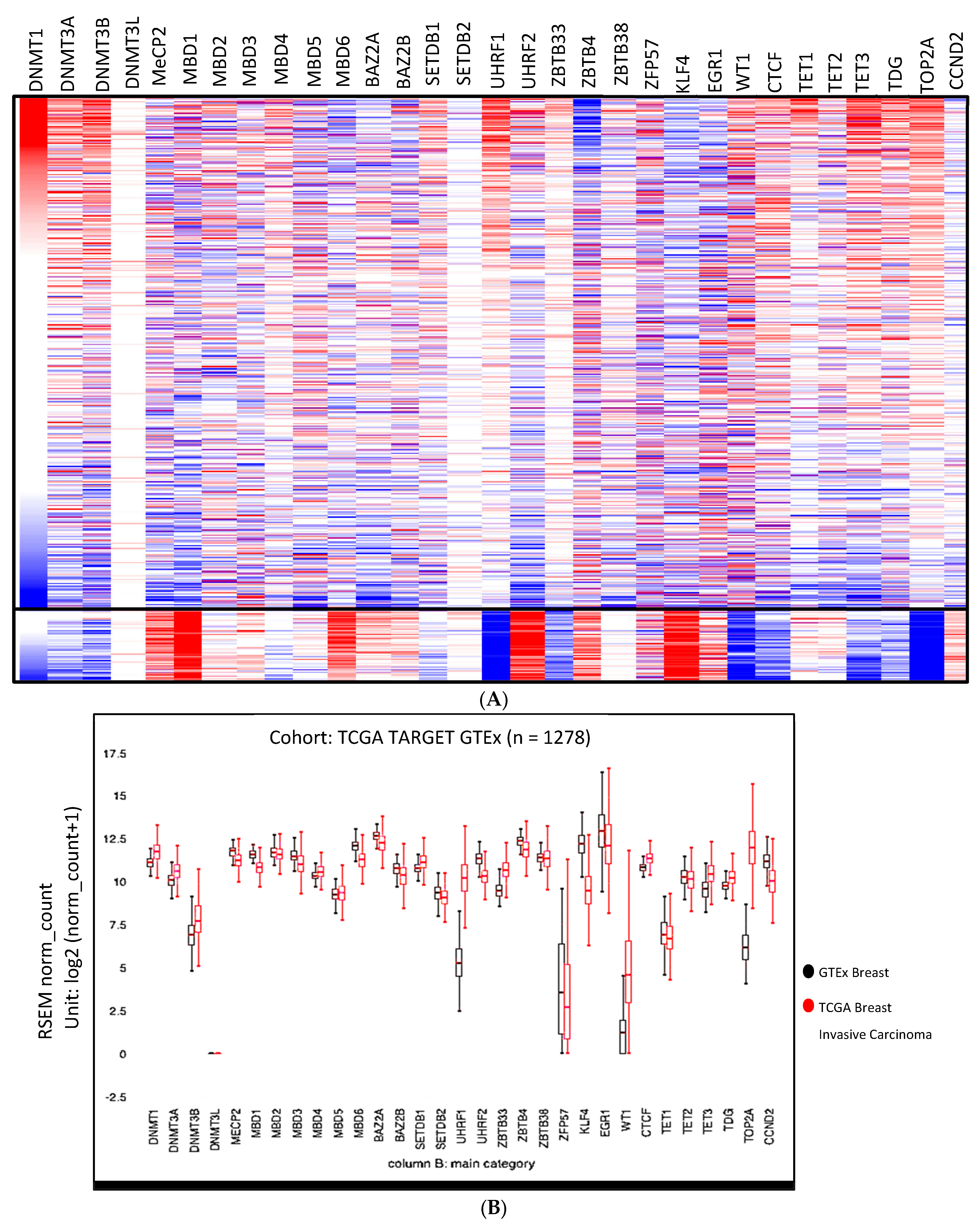
2.3. Changes in the Transcriptome

We employed Xena to compare transcriptome changes between normal tissue and breast cancer samples [38], which assimilates data from TCGA and GTEX to draw a comparison [39]. The heat map was generated using the gene list in Table 1. After launching Xena, all the genes in the list were incorporated. We selected the first variable phenotype “main category” and “gene expression” as the second genomic variable. All the genes encoding writers, readers and erasers of DNA methylation were incorporated to generate the heatmap. The transcription changes in the individual genes were obtained from the “view chart” tool to generate box plots that compare GTEX and TCGA breast cancer patients’ datasets. We used Xena specifically for generating heatmap and box plots because Xena employs the same pipeline for TCGA and GTEx samples to eradicate batch effects [38].

2.4. Survival Analysis

The Kaplan–Meier Plotter [40] was used to generate Kaplan–Meier plots utilizing multiple microarray expression datasets from 7462 breast cancer patients. For overall survival (OS), the analysis was run on 2879 breast cancer patients that were split by median. For the best probe selection, “Jetset” was selected, and patient survival was analyzed for the indicated time spans [40].

3. Results

3.1. Mutational Analysis of DNA Methylation Regulators in Breast Cancer

In our previous study on the genomic analysis of TCGA datasets, we reported that several genes encoding molecules related to nucleocytoplasmic shuttling were mutated [41]. In the current study, using cBioportal and selecting breast invasive carcinoma (TCGA, Firehose Legacy) datasets, we carried out mutational analysis of the genes encoding proteins corresponding to writers, readers and erasers of the DNA methylation. The characterization of TCGA datasets using cBioportal led to the identification of various mutations in breast cancer patients. The frequency genetic alteration for various genes differed markedly (ranging from 0.6% in EGR1 to 11% in SETDB1) (Figure 1A) amongst various genes under study. The most prevalent genetic alteration was found to be the gene amplification. Additional mutations detected were deep deletions, base substitutions and multiple alterations. Interestingly, alteration frequency in all the genes related to DNA methylation was found to be in 371 (37%) of queried patients, representing significance of DNA methylation dynamics (Figure 1), though with variable rates and mutation types, genetic alterations in these genes were detected in all breast cancer subtypes. We used PIK3CA, TP53, CDH1 and GATA3 genes as controls to validate our analysis. Consistent with the literature, higher mutation rates were found in these genes (Figure 1A). One of the olfactory receptor genes, OR5AL1, with no known connection with breast cancer, was used as a negative control.
We stratified breast cancer patients into subtypes and analyzed mutation frequency. Alteration frequency was variable depending on subtypes, and amplification was non-existent in breast invasive mixed mucinous carcinoma (Figure 1B). Interestingly, the genetic alterations in DNA methylation machinery were not specific to breast cancer. Querying 10,953 patients/10,967 samples in 32 studies from TCGA Pan-Cancer Atlas Studies identified comparable trends in mutations in the majority of cancers. This reveals that mutations in the genes coding for writers, readers and erasers of DNA methylation span a spectrum of cancers (Figure 2).

3.2. Expression Anomalies in Breast Cancer

Recent studies have shown that transcriptomic anomalies underlie tumor development. Aberrant gene expression may potentially serve as a biomarker in breast cancer. Therefore, we analyzed transcripts of all the genes under study in the TCGA and GTEx datasets. As can be seen in Figure 3A, several genes under study showed significant expression deregulation in breast cancer. There has been a mixed trend regarding over- or under-expression of DNA-methylation-related genes in cancer patients compared with normal tissue samples. MBD1 and MBD3 showed clear loss of expression, and this trend correlated with high rates of deletions as shown in Figure 1A. Similarly, overexpression of UHRF1 correlated with genetic amplification. Figure 3B shows expression changes in the individual genes between TCGA and GTEx datasets. Overall, our results show that the majority of genes encoding DNA methylation writers, readers and erasers go through massive expression deregulation in breast cancer patients.

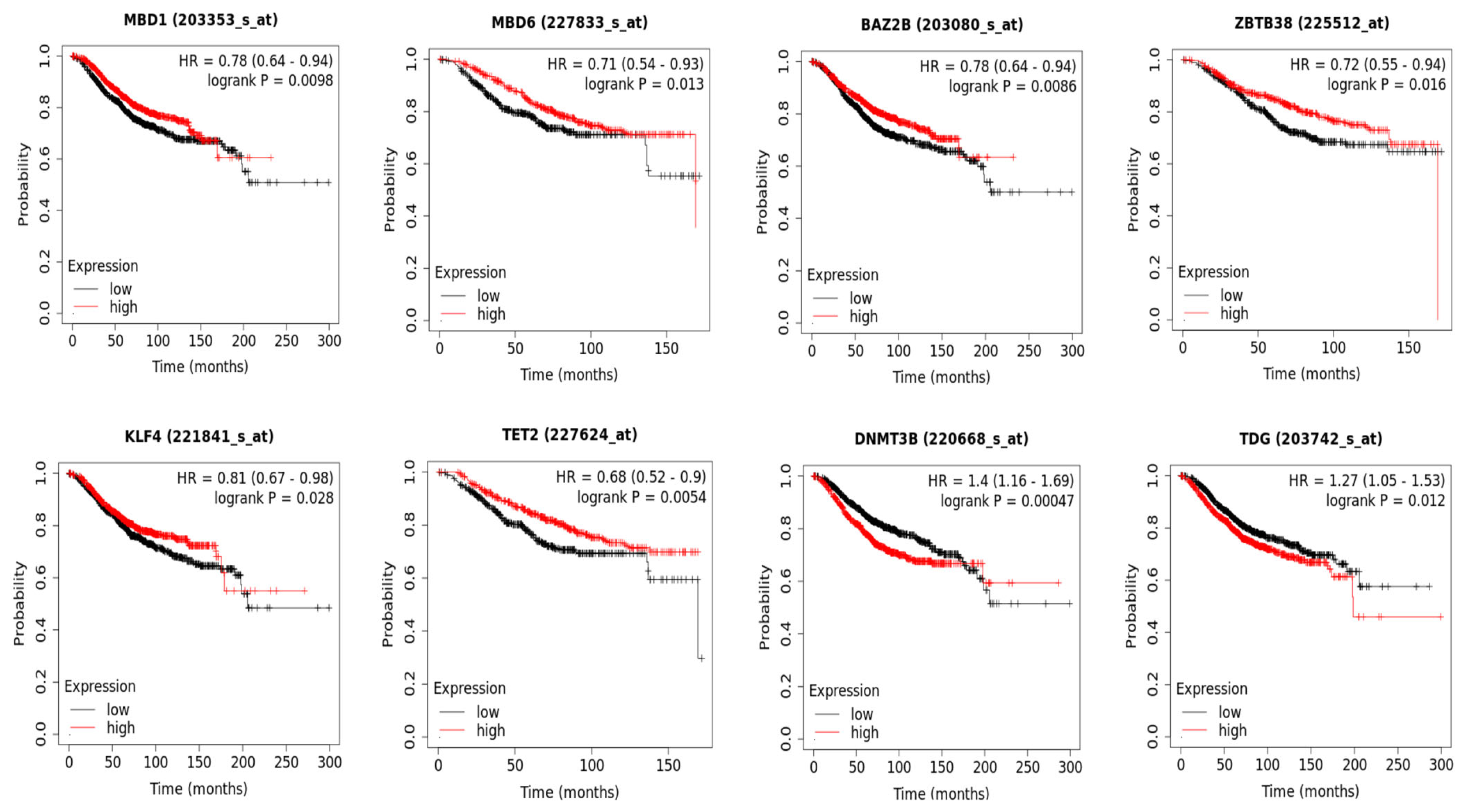
3.3. DNA Methylation and Patient Overall Survival

After establishing that genetic alterations and expression changes have significantly high prevalence in cancer patients, we next determined whether these changes in gene expression have prognostic potential. The Kaplan–Meier (KM) Plotter [40] has integrated microarray datasets that can be used to calculate overall survival (OS) of breast cancer patients according to expression levels of DNA methylated related genes. The current analysis was run on 2879 breast cancer patients. We tested all the genes and found that overexpression of MBD1, MBD6, BAZ2B, ZBTZ3B, KLF4 and TET2 correlated with better overall survival, while DNMT3B and TDG overexpression were correlated with poor patient overall survival (Figure 4). TOP2A was used as a control in our analysis, and, consistent with its published role as a marker for poor patient survival, the results were replicated in our analysis. The other genes under study were not significantly correlated with patient survival (Supplementary Figure S1). Collectively, our analysis identified several molecules regulating DNA methylation dynamics that might have potential for prognostication in breast cancer.

4. Discussion

In the current study, a comprehensive genomic and molecular landscape of genes encoding writers, readers and erasers of DNA methylation in breast cancer is provided. Our data show that these genes are not only genetically altered but show expression anomalies in breast cancer patients. Almost all the genes in the above categories are genetically altered to varying degrees. Mutations and expression deregulations in these genes may have a variety of effects on various signaling pathways. As myriads of genes are regulated by DNA methylation, alterations in DNA methylation regulators have far-reaching consequences.
The role of DNA methylation in cancer became apparent when the role of DNA methylation in the inactivation of tumor suppressor genes was revealed [42]. While earlier studies mainly focused on the genetic aspects of cancer, the epigenetic regulation in cancer became pivotal with our understanding of DNA methylation in cancer development [43] and its connection with gene regulation quantitatively. DNMT1, DNMT3A and DNMT3B are the major DNA methyltransferases, but, surprisingly, the mutation rates in these genes remained low (Figure 1). However, consistent with other studies, we found major expression elevations in all these methyltransferases that may contribute to significant alterations in DNA methylation [44,45,46,47].
In addition to changes in DNA methylation, equally important is the mechanisms that relay the DNA methylation information. The methylation marks are read and interpreted by the action of “reader” proteins that can recognize and bind to the methylated DNA regions. Out of all the readers under study, SETDB1 showed highest levels of genetic alterations with 11% of patients having gene amplifications. However, other readers had massive expression deregulations, mostly showing loss of expression, despite having relatively low representation in genetic alterations. Diversity in proteins that interpret methylation signals indicates the variation of the downstream signaling, e.g., MBD1 directs histone methylation to sites of DNA methylation, MeCP2 is involved in heterochromatin clustering, MBD2-3 dictate transcriptional silencing through NuRD/Mi-2 complexes, MBD4 maintains correct DNA methylation by suppressing CpG mutability, SETDB1 is involved in heterochromatin formation and BAZ2A is a component of the nucleolar remodeling complex NoRC [18]. Additional functions of MBPs have also been identified that are independent of methylated DNA binding. ChIP-seq studies for both MBD3 and MBD2 have shown that both proteins bind at transcriptionally active and unmethylated promoters [48,49].
The erasers of DNA methylation also showed dramatic expression differences. Consistent with earlier studies, TET1 and TET2 were modestly downregulated [50,51]. However, TET3 and TDG showed massive upregulation in the TCGA data. Differential regulation of TET and TDG also had opposing outcomes. While TET2 expression was correlated with better overall survival, TDG overexpression correlated with poor patient survival. TET2 has been reported to act as an anti-tumor factor, and its decreased expression has been associated with solid malignant tumors, such as colorectal, gastric, ovarian, prostate and breast cancers [52,53,54,55,56]. TET2 regulates the PD-L1 gene in breast cancer cells without altering the DNA methylation and hydroxymethylation (hmC) level at the promoter region pointing to functions that are independent of DNA demethylation [56]. TDG, on the other hand, has contradictory roles. Based on its ability to potentiate p53 signaling [57] and positively regulate p15 (ink4b) expression [58], it was suggested to act as a tumor suppressor. However, it is overexpressed in human colorectal cancer (CRC) patients, and knocking down TDG in several CRC cell lines inhibited cell growth [59]. Moreover, TDG knockdown suppressed tumor formation of melanoma cell lines in xenograft models, indicating that TDG activity is important for tumorigenesis [60]. Our data evince its potential oncogenic function in breast cancer.
DNMT3B was the only “writer” of DNA methylation that was not only overexpressed but also had poor prognostication. This is consistent with earlier studies wherein DNMT3b was proposed to be a prognostic biomarker for breast cancer [33]. Interestingly, the majority of the readers of DNA methylation showed better overall survival that included MBD1, MBD6, BAZ2B, ZBTB38 and KLF4. While the functions of many DNA methylation readers are now clear, more research is needed as some of these proteins are still less explored [17]. Future work on these candidates will unravel their anti-tumorigenic functions and underlying mechanisms. Molecular studies using cellular and xenograft models are required to understand the functions of these molecules.

5. Conclusions

In conclusion, the role of DNA methylation regulation in cancer therapeutics has gained attention in recent years after their role became apparent in various tumors. Loss of function studies have further substantiated their potential as therapeutic targets. Currently, FDA-approved DNMTis include 5-aza-2′-deoxycytidine (decitabine) and 5-azacytidine, which are cytidine analogs with a nitrogen atom replacing carbon in the fifth position of the ring. However, the lack of specificity of these therapeutic agents remains a major challenge. To overcome this challenge, the combination of traditional treatments with epigenetic drugs and the latest genomic tools is necessary to confer target specificity. Additionally, our study has identified several molecules such as DNMT3B, MBD1, MBD6, BAZ2B, ZBTB38, KLF4, TET2 and TDG in the DNA methylation pathway that have potential prognostic roles. This is the first study to unravel a comprehensive molecular and genetic profile of DNA methylation machinery genes in breast cancer and identifies DNA methylation machinery as an important determinant of disease progression. However, future research is needed to validate and target these molecules using cellular and animal models. These efforts will advance our understanding of the underlying mechanisms of DNA methylation in cancer and pave the way for the development of novel targeted therapeutic strategies.

Supplementary Materials

The following supporting information can be downloaded at: https://www.mdpi.com/article/10.3390/biomedinformatics3020029/s1, Figure S1.

Author Contributions

Conceptualization, R.M.; methodology, A.A. and R.M.; software, A.A. and R.M.; validation, R.M., N.Y., S.S., I.A., M.I., M.N. and F.A.A.; formal analysis, R.M.; investigation, R.M. and A.A.; resources, R.M.; data curation, R.M. and N.Y.; writing—original draft preparation, R.M., M.Y.W. and N.Y.; writing—review and editing, R.M. and M.Y.W.; visualization, R.M. and A.A.; supervision, R.M.; project administration, R.M.; funding acquisition, R.M. All authors have read and agreed to the published version of the manuscript.

Funding

This research was funded by a grant (IRG 22414) to RM from Alfaisal University, KSA.

Institutional Review Board Statement

Not applicable.

Informed Consent Statement

Not applicable.

Data Availability Statement

Data included in article/Supplementary Material is referenced in the article (https://www.cancer.gov/tcga, accessed on 10 April 2022).

Conflicts of Interest

The authors declare no conflict of interest. The funders had no role in the design of the study; in the collection, analyses, or interpretation of data; in the writing of the manuscript or in the decision to publish the results.

References

  1. Razin, A.; Cedar, H. DNA methylation and gene expression. Microbiol. Rev. 1991, 55, 451–458. [Google Scholar] [CrossRef] [PubMed]
  2. Jaenisch, R.; Bird, A. Epigenetic regulation of gene expression: How the genome integrates intrinsic and environmental signals. Nat. Genet. 2003, 33, 245–254. [Google Scholar] [CrossRef] [PubMed]
  3. Meehan, R.R.; Lewis, J.D.; McKay, S.; Kleiner, E.L.; Bird, A.P. Identification of a mammalian protein that binds specifically to DNA containing methylated CpGs. Cell 1989, 58, 499–507. [Google Scholar] [CrossRef] [PubMed]
  4. Bestor, T.H. The DNA methyltransferases of mammals. Hum. Mol. Genet. 2000, 9, 2395–2402. [Google Scholar] [CrossRef] [PubMed] [Green Version]
  5. Jones, P.A.; Takai, D. The role of DNA methylation in mammalian epigenetics. Science 2001, 293, 1068–1070. [Google Scholar] [CrossRef] [PubMed]
  6. Bird, A. The essentials of DNA methylation. Cell 1992, 70, 5–8. [Google Scholar] [CrossRef]
  7. Martisova, A.; Holcakova, J.; Izadi, N.; Sebuyoya, R.; Hrstka, R.; Bartosik, M. DNA Methylation in Solid Tumors: Functions and Methods of Detection. Int. J. Mol. Sci. 2021, 22, 4247. [Google Scholar] [CrossRef]
  8. Guo, J.U.; Su, Y.; Shin, J.H.; Shin, J.; Li, H.; Xie, B.; Zhong, C.; Hu, S.; Le, T.; Fan, G.; et al. Distribution, recognition and regulation of non-CpG methylation in the adult mammalian brain. Nat. Neurosci. 2014, 17, 215–222. [Google Scholar] [CrossRef] [Green Version]
  9. Lister, R.; Pelizzola, M.; Dowen, R.H.; Hawkins, R.D.; Hon, G.; Tonti-Filippini, J.; Nery, J.R.; Lee, L.; Ye, Z.; Ngo, Q.M.; et al. Human DNA methylomes at base resolution show widespread epigenomic differences. Nature 2009, 462, 315–322. [Google Scholar] [CrossRef] [Green Version]
  10. Smith, Z.D.; Chan, M.M.; Humm, K.C.; Karnik, R.; Mekhoubad, S.; Regev, A.; Eggan, K.; Meissner, A. DNA methylation dynamics of the human preimplantation embryo. Nature 2014, 511, 611–615. [Google Scholar] [CrossRef] [Green Version]
  11. Okae, H.; Chiba, H.; Hiura, H.; Hamada, H.; Sato, A.; Utsunomiya, T.; Kikuchi, H.; Yoshida, H.; Tanaka, A.; Suyama, M.; et al. Genome-wide analysis of DNA methylation dynamics during early human development. PLoS Genet. 2014, 10, e1004868. [Google Scholar] [CrossRef] [PubMed] [Green Version]
  12. Egger, G.; Jeong, S.; Escobar, S.G.; Cortez, C.C.; Li, T.W.; Saito, Y.; Yoo, C.B.; Jones, P.A.; Liang, G. Identification of DNMT1 (DNA methyltransferase 1) hypomorphs in somatic knockouts suggests an essential role for DNMT1 in cell survival. Proc. Natl. Acad. Sci. USA 2006, 103, 14080–14085. [Google Scholar] [CrossRef] [PubMed] [Green Version]
  13. Fatemi, M.; Hermann, A.; Gowher, H.; Jeltsch, A. Dnmt3a and Dnmt1 functionally cooperate during de novo methylation of DNA. Eur. J. Biochem. 2002, 269, 4981–4984. [Google Scholar] [CrossRef] [PubMed]
  14. Parry, L.; Clarke, A.R. The Roles of the Methyl-CpG Binding Proteins in Cancer. Genes. Cancer 2011, 2, 618–630. [Google Scholar] [CrossRef] [Green Version]
  15. Nan, X.; Meehan, R.R.; Bird, A. Dissection of the methyl-CpG binding domain from the chromosomal protein MeCP2. Nucleic Acids Res. 1993, 21, 4886–4892. [Google Scholar] [CrossRef] [Green Version]
  16. Hendrich, B.; Bird, A. Identification and characterization of a family of mammalian methyl-CpG binding proteins. Mol. Cell. Biol. 1998, 18, 6538–6547. [Google Scholar] [CrossRef] [Green Version]
  17. Mahmood, N.; Rabbani, S.A. DNA Methylation Readers and Cancer: Mechanistic and Therapeutic Applications. Front. Oncol. 2019, 9, 489. [Google Scholar] [CrossRef] [Green Version]
  18. Du, Q.; Luu, P.L.; Stirzaker, C.; Clark, S.J. Methyl-CpG-binding domain proteins: Readers of the epigenome. Epigenomics 2015, 7, 1051–1073. [Google Scholar] [CrossRef]
  19. Tahiliani, M.; Koh, K.P.; Shen, Y.; Pastor, W.A.; Bandukwala, H.; Brudno, Y.; Agarwal, S.; Iyer, L.M.; Liu, D.R.; Aravind, L.; et al. Conversion of 5-methylcytosine to 5-hydroxymethylcytosine in mammalian DNA by MLL partner TET1. Science 2009, 324, 930–935. [Google Scholar] [CrossRef] [Green Version]
  20. He, Y.F.; Li, B.Z.; Li, Z.; Liu, P.; Wang, Y.; Tang, Q.; Ding, J.; Jia, Y.; Chen, Z.; Li, L.; et al. Tet-mediated formation of 5-carboxylcytosine and its excision by TDG in mammalian DNA. Science 2011, 333, 1303–1307. [Google Scholar] [CrossRef] [Green Version]
  21. Ito, S.; Shen, L.; Dai, Q.; Wu, S.C.; Collins, L.B.; Swenberg, J.A.; He, C.; Zhang, Y. Tet proteins can convert 5-methylcytosine to 5-formylcytosine and 5-carboxylcytosine. Science 2011, 333, 1300–1303. [Google Scholar] [CrossRef] [Green Version]
  22. Brinkman, A.B.; Nik-Zainal, S.; Simmer, F.; Rodriguez-Gonzalez, F.G.; Smid, M.; Alexandrov, L.B.; Butler, A.; Martin, S.; Davies, H.; Glodzik, D.; et al. Partially methylated domains are hypervariable in breast cancer and fuel widespread CpG island hypermethylation. Nat. Commun. 2019, 10, 1749. [Google Scholar] [CrossRef] [PubMed] [Green Version]
  23. Batra, R.N.; Lifshitz, A.; Vidakovic, A.T.; Chin, S.F.; Sati-Batra, A.; Sammut, S.J.; Provenzano, E.; Ali, H.R.; Dariush, A.; Bruna, A.; et al. DNA methylation landscapes of 1538 breast cancers reveal a replication-linked clock, epigenomic instability and cis-regulation. Nat. Commun. 2021, 12, 5406. [Google Scholar] [CrossRef] [PubMed]
  24. Umeh-Garcia, M.; O’Geen, H.; Simion, C.; Gephart, M.H.; Segal, D.J.; Sweeney, C.A. Aberrant promoter methylation contributes to LRIG1 silencing in basal/triple-negative breast cancer. Br. J. Cancer 2022, 127, 436–448. [Google Scholar] [CrossRef]
  25. Zhang, W.; Xu, J. DNA methyltransferases and their roles in tumorigenesis. Biomark. Res. 2017, 5, 1. [Google Scholar] [CrossRef] [PubMed] [Green Version]
  26. Rasmussen, K.D.; Helin, K. Role of TET enzymes in DNA methylation, development, and cancer. Genes Dev. 2016, 30, 733–750. [Google Scholar] [CrossRef] [Green Version]
  27. Robertson, K.D.; Uzvolgyi, E.; Liang, G.; Talmadge, C.; Sumegi, J.; Gonzales, F.A.; Jones, P.A. The human DNA methyltransferases (DNMTs) 1, 3a and 3b: Coordinate mRNA expression in normal tissues and overexpression in tumors. Nucleic Acids Res. 1999, 27, 2291–2298. [Google Scholar] [CrossRef] [Green Version]
  28. Saito, Y.; Kanai, Y.; Nakagawa, T.; Sakamoto, M.; Saito, H.; Ishii, H.; Hirohashi, S. Increased protein expression of DNA methyltransferase (DNMT) 1 is significantly correlated with the malignant potential and poor prognosis of human hepatocellular carcinomas. Int. J. Cancer 2003, 105, 527–532. [Google Scholar] [CrossRef]
  29. Zhao, Z.; Wu, Q.; Cheng, J.; Qiu, X.; Zhang, J.; Fan, H. Depletion of DNMT3A suppressed cell proliferation and restored PTEN in hepatocellular carcinoma cell. J. Biomed. Biotechnol. 2010, 2010, 737535. [Google Scholar] [CrossRef] [Green Version]
  30. Patra, S.K.; Patra, A.; Zhao, H.; Dahiya, R. DNA methyltransferase and demethylase in human prostate cancer. Mol. Carcinog. 2002, 33, 163–171. [Google Scholar] [CrossRef]
  31. Butcher, D.T.; Rodenhiser, D.I. Epigenetic inactivation of BRCA1 is associated with aberrant expression of CTCF and DNA methyltransferase (DNMT3B) in some sporadic breast tumours. Eur. J. Cancer 2007, 43, 210–219. [Google Scholar] [CrossRef] [PubMed]
  32. Billard, L.M.; Magdinier, F.; Lenoir, G.M.; Frappart, L.; Dante, R. MeCP2 and MBD2 expression during normal and pathological growth of the human mammary gland. Oncogene 2002, 21, 2704–2712. [Google Scholar] [CrossRef] [PubMed]
  33. Man, X.; Li, Q.; Wang, B.; Zhang, H.; Zhang, S.; Li, Z. DNMT3A and DNMT3B in Breast Tumorigenesis and Potential Therapy. Front. Cell Dev. Biol. 2022, 10, 916725. [Google Scholar] [CrossRef] [PubMed]
  34. Xu, B.; Wang, H.; Tan, L. Dysregulated TET Family Genes and Aberrant 5mC Oxidation in Breast Cancer: Causes and Consequences. Cancers 2021, 13, 6039. [Google Scholar] [CrossRef]
  35. Lan, J.; Hua, S.; He, X.; Zhang, Y. DNA methyltransferases and methyl-binding proteins of mammals. Acta Biochim. Biophys. Sin. 2010, 42, 243–252. [Google Scholar] [CrossRef] [Green Version]
  36. Cerami, E.; Gao, J.; Dogrusoz, U.; Gross, B.E.; Sumer, S.O.; Aksoy, B.A.; Jacobsen, A.; Byrne, C.J.; Heuer, M.L.; Larsson, E.; et al. The cBio cancer genomics portal: An open platform for exploring multidimensional cancer genomics data. Cancer Discov. 2012, 2, 401–404. [Google Scholar] [CrossRef] [Green Version]
  37. Gao, J.; Aksoy, B.A.; Dogrusoz, U.; Dresdner, G.; Gross, B.; Sumer, S.O.; Sun, Y.; Jacobsen, A.; Sinha, R.; Larsson, E.; et al. Integrative analysis of complex cancer genomics and clinical profiles using the cBioPortal. Sci. Signal. 2013, 6, pl1. [Google Scholar] [CrossRef] [Green Version]
  38. Goldman, M.J.; Craft, B.; Hastie, M.; Repecka, K.; McDade, F.; Kamath, A.; Banerjee, A.; Luo, Y.; Rogers, D.; Brooks, A.N.; et al. Visualizing and interpreting cancer genomics data via the Xena platform. Nat. Biotechnol. 2020, 38, 675–678. [Google Scholar] [CrossRef]
  39. Consortium, G.T. The Genotype-Tissue Expression (GTEx) project. Nat. Genet. 2013, 45, 580–585. [Google Scholar] [CrossRef]
  40. Gyorffy, B. Survival analysis across the entire transcriptome identifies biomarkers with the highest prognostic power in breast cancer. Comput. Struct. Biotechnol. J. 2021, 19, 4101–4109. [Google Scholar] [CrossRef]
  41. Mehmood, R.; Jibiki, K.; Shibazaki, N.; Yasuhara, N. Molecular profiling of nucleocytoplasmic transport factor genes in breast cancer. Heliyon 2021, 7, e06039. [Google Scholar] [CrossRef]
  42. Jones, P.A.; Laird, P.W. Cancer epigenetics comes of age. Nat. Genet. 1999, 21, 163–167. [Google Scholar] [CrossRef] [PubMed]
  43. Ballestar, E.; Esteller, M. Methyl-CpG-binding proteins in cancer: Blaming the DNA methylation messenger. Biochem. Cell. Biol. 2005, 83, 374–384. [Google Scholar] [CrossRef]
  44. Girault, I.; Tozlu, S.; Lidereau, R.; Bieche, I. Expression analysis of DNA methyltransferases 1, 3A, and 3B in sporadic breast carcinomas. Clin. Cancer Res. 2003, 9, 4415–4422. [Google Scholar]
  45. Oh, B.K.; Kim, H.; Park, H.J.; Shim, Y.H.; Choi, J.; Park, C.; Park, Y.N. DNA methyltransferase expression and DNA methylation in human hepatocellular carcinoma and their clinicopathological correlation. Int. J. Mol. Med. 2007, 20, 65–73. [Google Scholar] [CrossRef] [Green Version]
  46. Melki, J.R.; Warnecke, P.; Vincent, P.C.; Clark, S.J. Increased DNA methyltransferase expression in leukaemia. Leukemia 1998, 12, 311–316. [Google Scholar] [CrossRef] [PubMed] [Green Version]
  47. Ahluwalia, A.; Hurteau, J.A.; Bigsby, R.M.; Nephew, K.P. DNA methylation in ovarian cancer. II. Expression of DNA methyltransferases in ovarian cancer cell lines and normal ovarian epithelial cells. Gynecol. Oncol. 2001, 82, 299–304. [Google Scholar] [CrossRef]
  48. Shimbo, T.; Du, Y.; Grimm, S.A.; Dhasarathy, A.; Mav, D.; Shah, R.R.; Shi, H.; Wade, P.A. MBD3 localizes at promoters, gene bodies and enhancers of active genes. PLoS Genet. 2013, 9, e1004028. [Google Scholar] [CrossRef] [PubMed] [Green Version]
  49. Baubec, T.; Ivanek, R.; Lienert, F.; Schubeler, D. Methylation-dependent and -independent genomic targeting principles of the MBD protein family. Cell 2013, 153, 480–492. [Google Scholar] [CrossRef] [PubMed] [Green Version]
  50. Yang, H.; Liu, Y.; Bai, F.; Zhang, J.-Y.; Ma, S.-H.; Liu, J.; Xu, Z.-D.; Zhu, H.-G.; Ling, Z.-Q.; Ye, D.; et al. Tumor development is associated with decrease of TET gene expression and 5-methylcytosine hydroxylation. Oncogene 2013, 32, 663–669. [Google Scholar] [CrossRef] [Green Version]
  51. Chen, J.Y.; Luo, C.W.; Lai, Y.S.; Wu, C.C.; Hung, W.C. Lysine demethylase KDM2A inhibits TET2 to promote DNA methylation and silencing of tumor suppressor genes in breast cancer. Oncogenesis 2017, 6, e369. [Google Scholar] [CrossRef] [Green Version]
  52. Huang, Y.; Wang, G.; Liang, Z.; Yang, Y.; Cui, L.; Liu, C.Y. Loss of nuclear localization of TET2 in colorectal cancer. Clin. Epigenetics 2016, 8, 9. [Google Scholar] [CrossRef] [Green Version]
  53. He, Z.; Wang, X.; Huang, C.; Gao, Y.; Yang, C.; Zeng, P.; Chen, Z. The FENDRR/miR-214-3P/TET2 axis affects cell malignant activity via RASSF1A methylation in gastric cancer. Am. J. Transl. Res. 2018, 10, 3211–3223. [Google Scholar]
  54. Zhang, L.Y.; Li, P.L.; Wang, T.Z.; Zhang, X.C. Prognostic values of 5-hmC, 5-mC and TET2 in epithelial ovarian cancer. Arch. Gynecol. Obstet. 2015, 292, 891–897. [Google Scholar] [CrossRef] [PubMed]
  55. Nickerson, M.L.; Das, S.; Im, K.M.; Turan, S.; Berndt, S.I.; Li, H.; Lou, H.; Brodie, S.A.; Billaud, J.N.; Zhang, T.; et al. TET2 binds the androgen receptor and loss is associated with prostate cancer. Oncogene 2017, 36, 2172–2183. [Google Scholar] [CrossRef] [PubMed] [Green Version]
  56. Shen, Y.; Liu, L.; Wang, M.; Xu, B.; Lyu, R.; Shi, Y.G.; Tan, L. TET2 Inhibits PD-L1 Gene Expression in Breast Cancer Cells through Histone Deacetylation. Cancers 2021, 13, 2207. [Google Scholar] [CrossRef] [PubMed]
  57. Kim, E.J.; Um, S.J. Thymine-DNA glycosylase interacts with and functions as a coactivator of p53 family proteins. Biochem. Biophys. Res. Commun. 2008, 377, 838–842. [Google Scholar] [CrossRef]
  58. Thillainadesan, G.; Chitilian, J.M.; Isovic, M.; Ablack, J.N.; Mymryk, J.S.; Tini, M.; Torchia, J. TGF-beta-dependent active demethylation and expression of the p15ink4b tumor suppressor are impaired by the ZNF217/CoREST complex. Mol. Cell. 2012, 46, 636–649. [Google Scholar] [CrossRef] [Green Version]
  59. Xu, X.; Yu, T.; Shi, J.; Chen, X.; Zhang, W.; Lin, T.; Liu, Z.; Wang, Y.; Zeng, Z.; Wang, C.; et al. Thymine DNA glycosylase is a positive regulator of Wnt signaling in colorectal cancer. J. Biol. Chem. 2014, 289, 8881–8890. [Google Scholar] [CrossRef] [Green Version]
  60. Mancuso, P.; Tricarico, R.; Bhattacharjee, V.; Cosentino, L.; Kadariya, Y.; Jelinek, J.; Nicolas, E.; Einarson, M.; Beeharry, N.; Devarajan, K.; et al. Thymine DNA glycosylase as a novel target for melanoma. Oncogene 2019, 38, 3710–3728. [Google Scholar] [CrossRef] [Green Version]
Figure 1. Genes encoding writers, readers and erasers of DNA methylation show genetic alterations in breast cancer. (A) Frequency of genetic alterations in these genes are in breast cancer patients. Individual lines in the horizontal bars are 996 breast cancer patients. Color schemes in the figure correspond to various genetic alterations including missense mutation, deep deletion, amplification, in frame mutation and truncating mutations. PIK3CA, TP53, CDH1 and GATA3 are used as positive controls, while OR5AL1 is used as a negative control with 0% mutation rate. (B) Subtypes of breast cancer showing various genetic alterations in DNA methylation regulators genes.
Biomedinformatics 03 00029 g001aBiomedinformatics 03 00029 g001b
Figure 2. Frequency of genetic alterations in genes encoding DNA methylation regulators in a wide variety of cancers. TCGA Pan-Cancer Atlas datasets employing 10,953 patients/10,967 samples in 32 studies. Genes encoding DNA methylation regulators are mutated in 4213 (38%) of samples.
Biomedinformatics 03 00029 g002
Figure 3. Expression level of genes that encode writers, readers and erasers of DNA methylation. (A) Red/blue bars represent over/under-expression of the genes under study. Expression levels are compared between GTEx and TCGA datasets. As control, TOP2A and CCND2 are employed. (B) Expression comparison of individual gene transcripts in normal vs. breast cancer patients. Welch’s t-test was used to calculate the p-values for individual genes. DNMT1 p = 5.781 × 10−74 (t = −22.11), DNMT3A p = 1.981 × 10−39 (t = −15.12), DNMT3B p = 1.076 × 10⁻³³ (t = −13.78), DNMT3L p = 0.07770 (t = −1.770), MECP2 p = 0.000 (t = 17.54), MBD1 p = 0.000 (t = 34.01), MBD2 p = 0.00002861 (t = 4.248), MBD3p = 0.000 (t = 12.02), MBD4 p = 4.561 × 10⁻³⁰ (t = −12.52), MBD5 p = 0.1683 (t = −1.381), MBD6 p = 0.000 (t = 23.56), BAZ2A p = 0.000 (t = 13.08), BAZ2B p = 0.000 (t = 10.07), SETDB1 p = 1.064 × 10⁻³¹ (t = −12.98), SETDB2 p = 2.190 × 10⁻⁸ (t = 5.781), UHRF1 p = 6.499 × 10−120 (t = −47.29), UHRF2 p = 0.000 (t = 29.62), ZBTB33 p = 4.126 × 10−94 (t = −30.02), ZBTB4 p = 0.000 (t = 17.21), ZBTB38 p = 0.005314 (t = 2.800), ZFP57 p = 0.05441 (t = 1.933), KLF4 p = 0.000 (t = 37.18), EGR1 p = 7.124 × 10⁻¹¹ (t = 6.803), WT1 p = 1.442 × 10−106 (t = −28.61), CTCF p = 4.217 × 10−65 (t = −21.70), TET1 p = 0.01331 (t = 2.491), TET2 p = 0.06953 (t = 1.821), TET3 p = 5.981 × 10−48 (t = −17.60), TDG p = 5.148 × 10−49 (t = −17.12), TOP2A p = 6.588 × 10−156 (t = −58.62), CCND2 p = 0.000 (t = 20.99).
Biomedinformatics 03 00029 g003
Figure 4. Expression of genes encoding DNA methylation machinery correlate with breast cancer patients’ overall survival. Kaplan–Meier plots indicate patient overall survival. The X axis denotes patient overall survival percentage, while the Y axis represents months after the diagnosis. Black color represents the low expression group, while the high expression group is indicated by the red color. The patients were split by median. The log-rank test is used to determine p-values.
Biomedinformatics 03 00029 g004
Table 1. This is a table showing list of genes encoding for writers, readers and erasers used in the study.
CategoryGene SymbolProtein Function
WriterDNMT1Maintenance methylation
DNMT3ADe novo methylation
DNMT3BDe novo methylation
DNMT3LAssists the de novo methyltransferases
ReaderMeCP2Methylated DNA binding
MBD1Methylated DNA binding
MBD2Methylated DNA binding
MBD3Methylated DNA binding
MBD4Methylated DNA binding
MBD5Methylated DNA binding
MBD6Methylated DNA binding
BAZ2AMethylated DNA binding
BAZ2BMethylated DNA binding
SETDB1Methylated DNA binding
SETDB2Methylated DNA binding
UHRF1Methylated DNA binding
UHRF2Methylated DNA binding
ZBTB33Methylated DNA binding
ZBTB4Methylated DNA binding
ZBTB38Methylated DNA binding
ZFP57Methylated DNA binding
KLF4Methylated DNA binding
EGR1Methylated DNA binding
WT1Methylated DNA binding
CTCFMethylated DNA binding
EraserTET1DNA demethylation
TET2DNA demethylation
TET3DNA demethylation
TDGDNA demethylation
Disclaimer/Publisher’s Note: The statements, opinions and data contained in all publications are solely those of the individual author(s) and contributor(s) and not of MDPI and/or the editor(s). MDPI and/or the editor(s) disclaim responsibility for any injury to people or property resulting from any ideas, methods, instructions or products referred to in the content.

