
Exploitation of Quercetin’s Antioxidative Properties in Potential Alternative Therapeutic Options for Neurodegenerative Diseases


Abstract
:1. Introduction
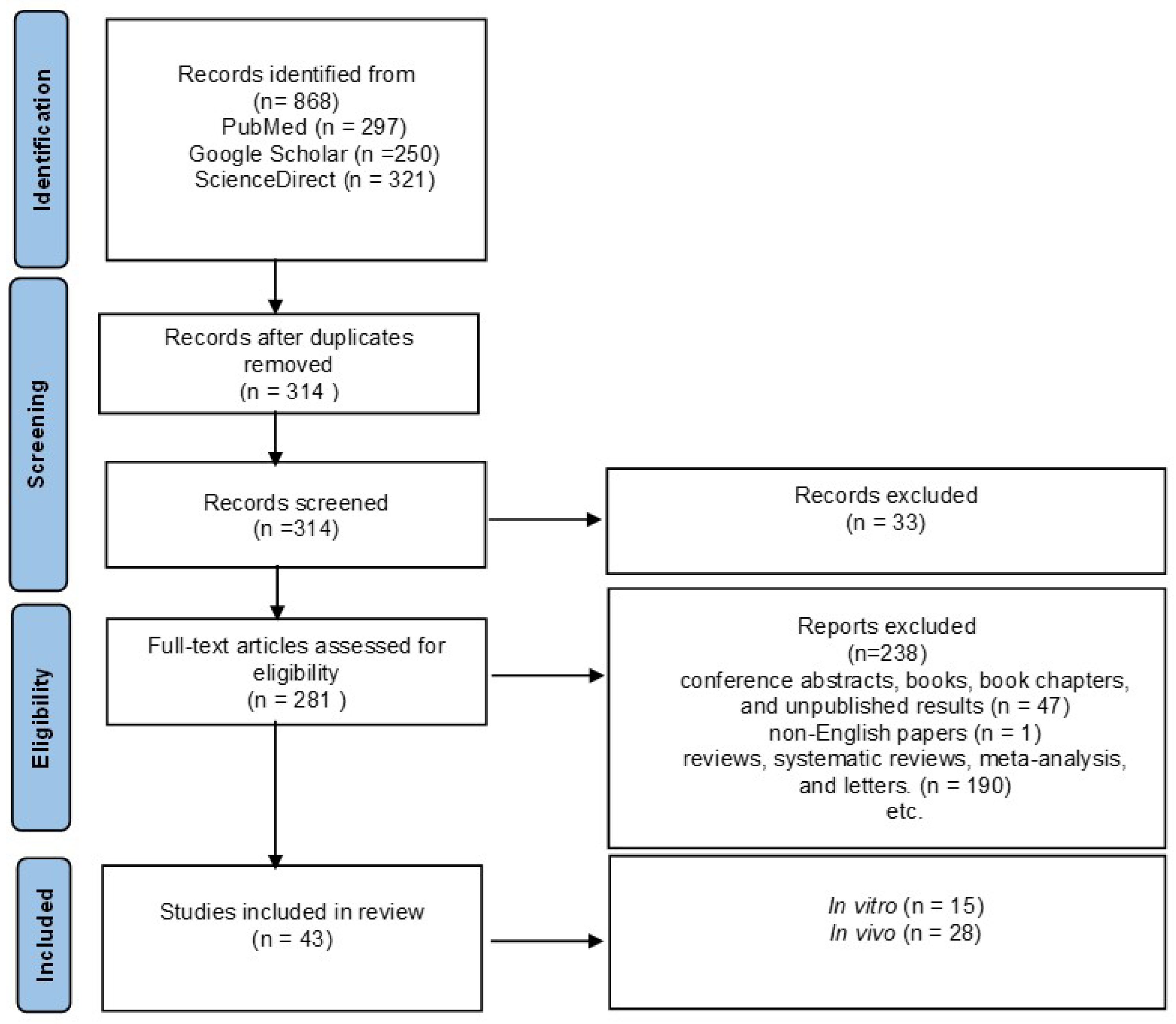
2. Methodology
3. Oxidative Stress
3.1. Alzheimer’s Disease

| Types of Que | Concentration of Que | Model | Exposure | Effects | Ref. |
|---|---|---|---|---|---|
| Que | Dosage: 2.2 μM; Duration: 24 h; | HT-22 mouse hippocampal cell | H2O2 | lipid peroxidation, ↑ intracellular GSH, ROS | [69] |
| Dosage: 10–100 µmol L−1; Duration: 10 min; | PC12 cells | H2O2 | ↓ lipid peroxidation, ↓ GSH, mitochondrial protection mechanisms | [70] | |
| Dosage: 50 mg; kg−1 b.w.; Duration: 2 times a week for 4 weeks; | homozygotic transgenic mouse line B6.129S7-Sod2tm1Leb/J | H2O2 and Aβ | ↓ ROS levels, improved the typical morphology of mitochondria, prevented mitochondrial dysfunction | [71] | |
| Dosage: 10 μM; | APP695-transfected SH-SY5Y cells | Aβ25–35 | ↓ ROS, ↓ BACE, ↓ Aβ, ↓ GSH, ↓ lipid peroxidation | [73] | |
| Dosage: 10 and 50 μM; Duration: 7 days; | hek cells | Aβ1–42 or Aβ1–40 | ↓ Aβ peptides, ↓ the performed mature fibrils | [74] | |
| Dosage: 5 or 10 mg kg−1 b.w.; Ad: p.o.; Duration: once daily; | hBMECs | fAβ1–40 | ↓ SOD, ↓ LDH | [75] | |
| Dosage: 2.4 µg mL−1; | HT-22 murine neuroblastoma cells | Aβ25–35 | ↓ amyloidogenic Aβ peptides, inhibited Aβ fibril formation. | [76] | |
| Dosage: 10, 20, 40, and 80 μmol L−1; Duration: 24 h, 48 h, and 72 h; | PC12 cells | Aβ25–35 | ↑ the survival rate of PC12 injured by Aβ25-35, promoted cell proliferation, and antagonized the toxicity of Aβ, ↓ ROS | [78] | |
| Q3G | Dosage: 25 μmol L−1; | Tg2576 AD primary neuron cultures | Aβ1–40, Aβ1–42 | ↑ neuronal survival, ↑ c-Jun N-terminal kinases, ↓ stress-induced impairments | [82] |
| Que/Ginkgo biloba | Dosage: 1.5–6 μg mL−1; | SHSY5Y human neuroblastoma cells | Aβ1–42 | ↓ Akt signaling pathways, ↓ Aβ toxicity, ↓ platelet-activating factor | [83] |
| Que/ Acanthopanax henryi | Dosage: 2.5, 5, 10, 20, and 40 μg mL−1; | cell-free system | ↓ AchE activity, ↑ antioxidant activity | [84] |
| Types of Que | Concentration | Model | Exposure | Effects | Ref. |
|---|---|---|---|---|---|
| Que | Dosage: 25 mg kg−1 b.w.; Ad: i.p.; Duration: every 2 days for 3 months; | 3xTg-AD mice | ↓ tauopathy, ↓ β-amyloidosis, ↑ memory, ↑ learning, ↓ microgliosis, ↓ astrogliosis | [86] | |
| Dosage: 100 mg kg−1 b.w.; Ad: gavage; Duration: every 48 h for 12 months; | 3xTg-AD mice | ↓ neurodegeneration, ↓ β-amyloidosis | [87] | ||
| Dosage: 20 and 40 mg kg−1 b.w.; Ad: p.o.; Duration: 16 weeks; | adult male C57BL mice | ↑ MMP, ↑ ATP levels, ↓ ROS | [88] | ||
| Dosage: 20 mg; Ad: p.o.; Duration: 5 weeks; | APP23 AD mice model | Aβ | ↓ eIF2α, ↓ ATF4, ↓ GADD34, ↑ memory in aged mice, ↓ memory deterioration in the early stage of AD, ↓ memory dysfunction, ↓ OS | [89] | |
| Dosage: 1% in mouse chow; Ad: p.o.; Duration: from 3 to 13 months; | double transgenic female mice | ↓ neuroinflammation, ↓ neurodegeneration, ↓ IL-1β | [90] | ||
| Dosage: 25 mg kg−1; Ad: p.o.; Duration: 2 times a week for 2 months; | SAMP8 mice | ↑ the cognition and memory impairments, ↓ astrogliosis | [92] | ||
| Dosage: 100 mg kg−1 b.w.; Ad: p.o.; Duration: 22 days; | adult male Sprague Dawley rats | Aβ1–42 | ↑ expression of Nrf2/HO-1 in rat brains, ↓ Aβ1-42 level, ↓ antioxidant activity | [93] | |
| Dosage: 12.5 and 25 mg kg−1; | mice | Scopolamine | ↓ OS, ↓ AchE activity | [94] | |
| Dosage: 30 mg kg−1 b.w.; Ad: i.p.; Duration: every day for 8 days; | male albino Wistar rats | Scopolamine | abridged transfer latency, ↓ avoidance response, ↓ 3,4-methylenedioxyamphetamine, acetylcholinesterase levels, ↑ CAT, ↑ GSH levels | [95] | |
| Dosage: 10 mg kg −1 b.w.; Ad: p.o.; Duration: every day for 12 weeks; | male albino Wistar rats | aluminum | ↓ ROS production, ↑ mitochondrial superoxide dismutase activity | [96] | |
| Que/ginkgo flavonols | Dosage: 4.8% in extract, all based on weight; | double transgenic (TgAPP/PS1) mice | - | reversed the spatial learning deficit | [98] |
3.2. Parkinson’s Disease
3.3. Huntington’s Disease
3.4. Epilepsy
4. Conclusions
Author Contributions
Funding
Institutional Review Board Statement
Informed Consent Statement
Data Availability Statement
Conflicts of Interest
References
- Zhang, Y.M.; Zhang, Z.Y.; Wang, R.X. Protective Mechanisms of Quercetin Against Myocardial Ischemia Reperfusion Injury. Front. Physiol. 2020, 11, 956. [Google Scholar] [CrossRef] [PubMed]
- Singh, A.; Kukreti, R.; Saso, L.; Kukreti, S. Oxidative Stress: A Key Modulator in Neurodegenerative Diseases. Molecules 2019, 24, 1583. [Google Scholar] [CrossRef] [PubMed] [Green Version]
- Shukla, V.; Mishra, S.K.; Pant, H.C. Oxidative Stress in Neurodegeneration. Adv. Pharmacol. Sci. 2011, 2011, 572634. [Google Scholar] [CrossRef] [PubMed] [Green Version]
- Gkekas, I.; Gioran, A.; Boziki, M.K.; Grigoriadis, N.; Chondrogianni, N.; Petrakis, S. Oxidative Stress and Neurodegeneration: Interconnected Processes in PolyQ Diseases. Antioxidants 2021, 10, 1450. [Google Scholar] [CrossRef]
- Bandiwadekar, A.; Jose, J.; Khayatkashani, M.; Habtemariam, S.; Kashani, H.R.K.; Nabavi, S.M. Emerging Novel Approaches for the Enhanced Delivery of Natural Products for the Management of Neurodegenerative Diseases. J. Mol. Neurosci. 2021, 72, 653–676. [Google Scholar] [CrossRef] [PubMed]
- Chapman, J.M.; Muday, G.K. Flavonols modulate lateral root emergence by scavenging reactive oxygen species in Arabidopsis thaliana. J. Biol. Chem. 2021, 296, 100222. [Google Scholar] [CrossRef]
- Ozcan, A.; Ogun, M. Biochemistry of Reactive Oxygen and Nitrogen Species. In Basic Principles and Clinical Significance of Oxidative Stress; Intechopen: London, UK, 2015. [Google Scholar]
- Klebanoff, S.J. Oxygen Metabolism and the Toxic Properties of Phagocytes. Ann. Intern. Med. 1980, 93, 391–398. [Google Scholar] [CrossRef]
- Sheikh, S.; Safia, U.; Haque, E.; Mir, S.S. Neurodegenerative Diseases: Multifactorial Conformational Diseases and Their Therapeutic Interventions. J. Neurodegener. Dis. 2013, 2013, 1–8. [Google Scholar] [CrossRef] [Green Version]
- Erden Inal, M.; Kahraman, A.; Köken, T. Beneficial effects of quercetin on oxidative stress induced by ultraviolet A. Clin. Exp. Dermatol. 2001, 26, 536–539. [Google Scholar] [CrossRef]
- Elumalai, P.; Lakshmi, S. Role of Quercetin Benefits in Neurodegeneration. Benefits Nat. Prod. Neurodegener. Dis. 2016, 12, 229–245. [Google Scholar] [CrossRef]
- Halliwell, B. Oxidants and human disease: Some new concepts 1. FASEB J. 1987, 1, 358–364. [Google Scholar] [CrossRef]
- Ansari, M.A.; Abdul, H.M.; Joshi, G.; Opii, W.O.; Butterfield, D.A. Protective effect of quercetin in primary neurons against Aβ(1–42): Relevance to Alzheimer’s disease. J. Nutr. Biochem. 2009, 20, 269–275. [Google Scholar] [CrossRef] [PubMed] [Green Version]
- Islam, S.; Quispe, C.; Hossain, R.; Islam, M.T.; Al-Harrasi, A.; Al-Rawahi, A.; Martorell, M.; Mamurova, A.; Seilkhan, A.; Altybaeva, N.; et al. Neuropharmacological Effects of Quercetin: A Literature-Based Review. Front. Pharmacol. 2021, 12, 665031. [Google Scholar] [CrossRef] [PubMed]
- Ames, B.N.; Shigenaga, M.K.; Hagen, T.M. Oxidants, antioxidants, and the degenerative diseases of aging. Proc. Natl. Acad. Sci. USA 1993, 90, 7915–7922. [Google Scholar] [CrossRef] [PubMed]
- Zalpoor, H.; Nabi-Afjadi, M.; Forghaniesfidvajani, R.; Tavakol, C.; Farahighasreaboonasr, F.; Pakizeh, F.; Dana, V.G.; Seif, F. Quercetin as a JAK–STAT inhibitor: A potential role in solid tumors and neurodegenerative diseases. Cell. Mol. Biol. Lett. 2022, 27, 60. [Google Scholar] [CrossRef]
- Shen, P.; Lin, W.; Deng, X.; Ba, X.; Han, L.; Chen, Z.; Qin, K.; Huang, Y.; Tu, S. Potential Implications of Quercetin in Autoimmune Diseases. Front. Immunol. 2021, 12, 1991. [Google Scholar] [CrossRef]
- Haytowitz, D.; Wu, X.; Bhagwat, S. USDA Database for the Flavonoid Content of Selected Foods, Release 3.3; USDA Agricultureal Research Service: Washington, DC, USA, 2018.
- Harwood, M.; Danielewska-Nikiel, B.; Borzelleca, J.F.; Flamm, G.W.; Williams, G.M.; Lines, T.C. A critical review of the data related to the safety of quercetin and lack of evidence of in vivo toxicity, including lack of genotoxic/carcinogenic properties. Food Chem. Toxicol. 2007, 45, 2179–2205. [Google Scholar] [CrossRef]
- Hoffmann-Ribani, R.; Huber, L.S.; Rodriguez-Amaya, D.B. Flavonols in fresh and processed Brazilian fruits. J. Food Compos. Anal. 2009, 22, 263–268. [Google Scholar] [CrossRef]
- Tsao, R.; Yang, R.; Young, J.C.; Zhu, H. Polyphenolic Profiles in Eight Apple Cultivars Using High-Performance Liquid Chromatography (HPLC). J. Agric. Food Chem. 2003, 51, 6347–6353. [Google Scholar] [CrossRef]
- Sakakibara, H.; Honda, Y.; Nakagawa, S.; Ashida, H.; Kanazawa, K. Simultaneous Determination of All Polyphenols in Vegetables, Fruits, and Teas. J. Agric. Food Chem. 2003, 51, 571–581. [Google Scholar] [CrossRef]
- Dragovic-Uzelac, V.; Levaj, B.; Mrkic, V.; Bursac, D.; Boras, M. The content of polyphenols and carotenoids in three apricot cultivars depending on stage of maturity and geographical region. Food Chem. 2007, 102, 966–975. [Google Scholar] [CrossRef]
- Bilyk, A.; Sapers, G.M. Varietal differences in the quercetin, kaempferol, and myricetin contents of highbush blueberry, cranberry, and thornless blackberry fruits. J. Agric. Food Chem. 1986, 34, 585–588. [Google Scholar] [CrossRef]
- Kirakosyan, A.; Seymour, E.M.; Urcuyo Llanes, D.E.; Kaufman, P.B.; Bolling, S.F. Chemical profile and antioxidant capacities of tart cherry products. Food Chem. 2009, 115, 20–25. [Google Scholar] [CrossRef]
- Justesen, U.; Knuthsen, P.; Leth, T. Quantitative analysis of flavonols, flavones, and flavanones in fruits, vegetables and beverages by high-performance liquid chromatography with photo-diode array and mass spectrometric detection. J. Chromatogr. A 1998, 799, 101–110. [Google Scholar] [CrossRef]
- Giuffrida, D.; Salvo, F.; Ziino, M.; Toscano, G.; Dugo, G. Initial Investigation on Some Chemical Constituents of Capers (Capparis Spinosa L.) from the Island of Salina. Ital. J. Food Sci. 2002, 14, 25. [Google Scholar]
- Lugast, A.; Hóvári, J. Flavonoid aglycons in foods of plant origin I. vegetables. Acta Aliment. 2000, 29, 345–352. [Google Scholar] [CrossRef]
- Lin, L.-Z.; Mukhopadhyay, S.; Robbins, R.J.; Harnly, J.M. Identification and quantification of flavonoids of Mexican oregano (Lippia graveolens) by LC-DAD-ESI/MS analysis. J. Food Compos. Anal. 2007, 20, 361–369. [Google Scholar] [CrossRef] [Green Version]
- Arabbi, P.R.; Genovese, M.I.; Lajolo, F.M. Flavonoids in Vegetable Foods Commonly Consumed in Brazil and Estimated Ingestion by the Brazilian Population. J. Agric. Food Chem. 2004, 52, 1124–1131. [Google Scholar] [CrossRef]
- Innocenti, M.; Gallori, S.; Giaccherini, C.; Ieri, F.; Vincieri, F.F.; Mulinacci, N. Evaluation of the Phenolic Content in the Aerial Parts of Different Varieties of Cichorium intybus L. J. Agric. Food Chem. 2005, 53, 6497–6502. [Google Scholar] [CrossRef]
- Price, K.R.; Rhodes, M.J.C.; Barnes, K.A. Flavonol Glycoside Content and Composition of Tea Infusions Made from Commercially Available Teas and Tea Products. J. Agric. Food Chem. 1998, 46, 2517–2522. [Google Scholar] [CrossRef]
- Mishra, D.; Flora, S.J.S. Quercetin Administration During Chelation Therapy Protects Arsenic-Induced Oxidative Stress in Mice. Biol. Trace Element Res. 2008, 122, 137–147. [Google Scholar] [CrossRef] [PubMed]
- Lesjak, M.; Beara, I.; Simin, N.; Pintać, D.; Majkić, T.; Bekvalac, K.; Orčić, D.; Mimica-Dukić, N. Antioxidant and anti-inflammatory activities of quercetin and its derivatives. J. Funct. Foods 2018, 40, 68–75. [Google Scholar] [CrossRef]
- Dhaouadi, Z.; Nsangou, M.; Garrab, N.; Anouar, E.; Marakchi, K.; Lahmar, S. DFT study of the reaction of quercetin with O2 and OH radicals. J. Mol. Struct. THEOCHEM 2009, 904, 35–42. [Google Scholar] [CrossRef]
- Reyes-Farias, M.; Carrasco-Pozo, C. The Anti-Cancer Effect of Quercetin: Molecular Implications in Cancer Metabolism. Int. J. Mol. Sci. 2019, 20, 3177. [Google Scholar] [CrossRef] [Green Version]
- Vafadar, A.; Shabaninejad, Z.; Movahedpour, A.; Fallahi, F.; Taghavipour, M.; Ghasemi, Y.; Akbari, M.; Shafiee, A.; Hajighadimi, S.; Moradizarmehri, S.; et al. Quercetin and cancer: New insights into its therapeutic effects on ovarian cancer cells. Cell Biosci. 2020, 10, 1–17. [Google Scholar] [CrossRef] [PubMed] [Green Version]
- Shafabakhsh, R.; Asemi, Z. Quercetin: A natural compound for ovarian cancer treatment. J. Ovarian Res. 2019, 12, 55. [Google Scholar] [CrossRef] [Green Version]
- Tang, S.-M.; Deng, X.-T.; Zhou, J.; Li, Q.-P.; Ge, X.-X.; Miao, L. Pharmacological basis and new insights of quercetin action in respect to its anti-cancer effects. Biomed. Pharmacother. 2020, 121, 109604. [Google Scholar] [CrossRef] [PubMed]
- Ezzati, M.; Yousefi, B.; Velaei, K.; Safa, A. A review on anti-cancer properties of Quercetin in breast cancer. Life Sci. 2020, 248, 117463. [Google Scholar] [CrossRef]
- Hou, D.-D.; Zhang, W.; Gao, Y.-L.; Sun, Y.-Z.; Wang, H.-X.; Qi, R.-Q.; Chen, H.-D.; Gao, X.-H. Anti-inflammatory effects of quercetin in a mouse model of MC903-induced atopic dermatitis. Int. Immunopharmacol. 2019, 74, 105676. [Google Scholar] [CrossRef]
- Kawabata, K.; Baba, N.; Sakano, T.; Hamano, Y.; Taira, S.; Tamura, A.; Baba, S.; Natsume, M.; Ishii, T.; Murakami, S.; et al. Functional properties of anti-inflammatory substances from quercetin-treated Bifidobacterium adolescentis. Biosci. Biotechnol. Biochem. 2018, 82, 689–697. [Google Scholar] [CrossRef] [Green Version]
- Saeedi-Boroujeni, A.; Mahmoudian-Sani, M.-R. Anti-inflammatory potential of Quercetin in COVID-19 treatment. J. Inflamm. 2021, 18, 1–9. [Google Scholar] [CrossRef] [PubMed]
- Liu, M.; Yu, Q.; Xiao, H.; Li, M.; Huang, Y.; Zhang, Q.; Li, P. The Inhibitory Activities and Antiviral Mechanism of Medicinal Plant Ingredient Quercetin Against Grouper Iridovirus Infection. Front. Microbiol. 2020, 11, 586331. [Google Scholar] [CrossRef] [PubMed]
- Di Petrillo, A.; Orrù, G.; Fais, A.; Fantini, M.C. Quercetin and its derivates as antiviral potentials: A comprehensive review. Phytotherapy Res. 2022, 36, 266–278. [Google Scholar] [CrossRef] [PubMed]
- Kim, C.H.; Kim, J.E.; Song, Y.J. Antiviral Activities of Quercetin and Isoquercitrin Against Human Herpesviruses. Molecules 2020, 25, 2379. [Google Scholar] [CrossRef]
- Wang, S.; Yao, J.; Zhou, B.; Yang, J.; Chaudry, M.T.; Wang, M.; Xiao, F.; Li, Y.; Yin, W. Bacteriostatic Effect of Quercetin as an Antibiotic Alternative In Vivo and Its Antibacterial Mechanism In Vitro. J. Food Prot. 2018, 81, 69–78. [Google Scholar] [CrossRef] [PubMed]
- Olewnik-Kruszkowska, E.; Gierszewska, M.; Richert, A.; Grabska-Zielińska, S.; Rudawska, A.; Bouaziz, M. Antibacterial Films Based on Polylactide with the Addition of Quercetin and Poly(Ethylene Glycol). Materials 2021, 14, 1643. [Google Scholar] [CrossRef]
- Júnior, S.D.D.C.; Santos, J.V.D.O.; Campos, L.A.D.A.; Pereira, M.A.; Santos-Magalhaes, N.; Cavalcanti, I.M.F. Antibacterial and antibiofilm activities of quercetin against clinical isolates of Staphyloccocus aureus and Staphylococcus saprophyticus with resistance profile. Int. J. Environ. Agric. Biotechnol. 2018, 3, 1948–1958. [Google Scholar] [CrossRef] [Green Version]
- Jaisinghani, R.N. Antibacterial properties of quercetin. Microbiol. Res. 2017, 8, 1. [Google Scholar] [CrossRef] [Green Version]
- Aljadaan, S.A.N.; Elias, R.S.; Al-Anssari, R.A. Investigation of the Antioxidant and Antibacterial Activity of Novel Quercetin Derivatives. Biointerface Res. Appl. Chem. 2020, 10, 7329–7336. [Google Scholar] [CrossRef]
- Patel, R.V.; Mistry, B.M.; Shinde, S.K.; Syed, R.; Singh, V.; Shin, H.-S. Therapeutic potential of quercetin as a cardiovascular agent. Eur. J. Med. Chem. 2018, 115, 889–904. [Google Scholar] [CrossRef]
- Zhang, L.; Ma, J.; Yang, F.; Li, S.; Ma, W.; Chang, X.; Yang, L. Neuroprotective Effects of Quercetin on Ischemic Stroke: A Literature Review. Front. Pharmacol. 2022, 13, 854249. [Google Scholar] [CrossRef] [PubMed]
- Yang, R.; Shen, Y.J.; Chen, M.; Zhao, J.Y.; Chen, S.H.; Zhang, W.; Song, J.K.; Li, L.; Du, G.H. Quercetin attenuates ischemia reperfusion injury by protecting the blood-brain barrier through Sirt1 in MCAO rats. J. Asian Nat. Prod. Res. 2022, 24, 278–289. [Google Scholar] [CrossRef]
- Andres, S.; Pevny, S.; Ziegenhagen, R.; Bakhiya, N.; Schäfer, B.; Hirsch-Ernst, K.I.; Lampen, A. Safety Aspects of the Use of Quercetin as a Dietary Supplement. Mol. Nutr. Food Res. 2018, 62. [Google Scholar] [CrossRef]
- Harishkumar, R.; Reddy, L.P.K.; Karadkar, S.H.; Al Murad, M.; Karthik, S.S.; Manigandan, S.; Selvaraj, C.I.; Christopher, J.G. Toxicity and Selective Biochemical Assessment of Quercetin, Gallic Acid, and Curcumin in Zebrafish. Biol. Pharm. Bull. 2019, 42, 1969–1976. [Google Scholar] [CrossRef] [PubMed] [Green Version]
- Simunkova, M.; Alwasel, S.H.; Alhazza, I.M.; Jomova, K.; Kollar, V.; Rusko, M.; Valko, M. Management of oxidative stress and other pathologies in Alzheimer’s disease. Arch. Toxicol. 2019, 93, 2491–2513. [Google Scholar] [CrossRef] [PubMed] [Green Version]
- Page, M.J.; McKenzie, J.E.; Bossuyt, P.M.; Boutron, I.; Hoffmann, T.C.; Mulrow, C.D.; Shamseer, L.; Tetzlaff, J.M.; Akl, E.A.; Brennan, S.E.; et al. The PRISMA 2020 Statement: An Updated Guideline for Reporting Systematic Reviews. BMJ 2021, 372, n71. [Google Scholar] [CrossRef] [PubMed]
- Kook, D.; Wolf, A.H.; Yu, A.L.; Neubauer, A.S.; Priglinger, S.G.; Kampik, A.; Welge-Lu¨ssen, U.C. The Protective Effect of Quercetin against Oxidative Stress in the Human RPE In Vitro. Investig. Opthalmology Vis. Sci. 2008, 49, 1712–1720. [Google Scholar] [CrossRef] [Green Version]
- Xu, D.; Hu, M.-J.; Wang, Y.-Q.; Cui, Y.-L. Antioxidant Activities of Quercetin and Its Complexes for Medicinal Application. Molecules 2019, 24, 1123. [Google Scholar] [CrossRef] [Green Version]
- Valério, D.A.; Georgetti, S.R.; Magro, D.A.; Casagrande, R.; Cunha, T.M.; Vicentini, F.T.M.C.; Vieira, S.M.; Fonseca, M.J.V.; Ferreira, S.H.; Cunha, F.Q.; et al. Quercetin Reduces Inflammatory Pain: Inhibition of Oxidative Stress and Cytokine Production. J. Nat. Prod. 2009, 72, 1975–1979. [Google Scholar] [CrossRef]
- Grewal, A.K.; Singh, T.G.; Sharma, D.; Sharma, V.; Singh, M.; Rahman, H.; Najda, A.; Walasek-Janusz, M.; Kamel, M.; Albadrani, G.M.; et al. Mechanistic insights and perspectives involved in neuroprotective action of quercetin. Biomed. Pharmacother. 2021, 140, 111729. [Google Scholar] [CrossRef]
- De Ture, M.A.; Dickson, D.W. The neuropathological diagnosis of Alzheimer’s disease. Mol. Neurodegener. 2019, 14, 32. [Google Scholar] [CrossRef] [PubMed] [Green Version]
- Kommaddi, R.P.; Das, D.; Karunakaran, S.; Nanguneri, S.; Bapat, D.; Ray, A.; Shaw, E.; Bennett, D.A.; Nair, D.; Ravindranath, V. Aβ mediates F-actin disassembly in dendritic spines leading to cognitive deficits in Alzheimer’s disease. J. Neurosci. 2018, 38, 1085–1099. [Google Scholar] [CrossRef] [PubMed] [Green Version]
- Parent, M.J.; Zimmer, E.R.; Shin, M.; Kang, M.S.; Fonov, V.S.; Mathieu, A.; Aliaga, A.; Kostikov, A.; Do Carmo, S.; Dea, D.; et al. Multimodal Imaging in Rat Model Recapitulates Alzheimer’s Disease Biomarkers Abnormalities. J. Neurosci. 2017, 37, 12263–12271. [Google Scholar] [CrossRef] [PubMed] [Green Version]
- Wallace, R.A.; Dalton, A.J. What can we learn from study of Alzheimer’s disease in patients with Down syndrome for early-onset Alzheimer’s disease in the general population? Alzheimer’s Res. Ther. 2011, 3, 13. [Google Scholar] [CrossRef] [PubMed] [Green Version]
- Hollingworth, P.; Harold, D.; Jones, L.; Owen, M.J.; Williams, J. Alzheimer’s disease genetics: Current knowledge and future challenges. Int. J. Geriatr. Psychiatry 2011, 26, 793–802. [Google Scholar] [CrossRef]
- Mayeux, R.; Stern, Y. Epidemiology of Alzheimer Disease. Cold Spring Harb. Perspect. Med. 2012, 2, a006239. [Google Scholar] [CrossRef] [Green Version]
- Ishige, K.; Schubert, D.; Sagara, Y. Flavonoids protect neuronal cells from oxidative stress by three distinct mechanisms. Free. Radic. Biol. Med. 2001, 30, 433–446. [Google Scholar] [CrossRef]
- Heo, H.J.; Lee, C.Y. Protective Effects of Quercetin and Vitamin C against Oxidative Stress-Induced Neurodegeneration. J. Agric. Food Chem. 2004, 52, 7514–7517. [Google Scholar] [CrossRef]
- Godoy, J.A.; Lindsay, C.B.; Quintanilla, R.A.; Carvajal, F.J.; Cerpa, W.; Inestrosa, N.C. Quercetin Exerts Differential Neuroprotective Effects Against H2O2 and Aβ Aggregates in Hippocampal Neurons: The Role of Mitochondria. Mol. Neurobiol. 2017, 54, 7116–7128. [Google Scholar] [CrossRef]
- Porat, Y.; Abramowitz, A.; Gazit, E. Inhibition of Amyloid Fibril Formation by Polyphenols: Structural Similarity and Aromatic Interactions as a Common Inhibition Mechanism. Chem. Biol. Drug Des. 2006, 67, 27–37. [Google Scholar] [CrossRef]
- Jiménez-Aliaga, K.; Bermejo-Bescós, P.; Benedí, J.; Martín-Aragón, S. Quercetin and rutin exhibit antiamyloidogenic and fibril-disaggregating effects in vitro and potent antioxidant activity in APPswe cells. Life Sci. 2011, 89, 939–945. [Google Scholar] [CrossRef] [PubMed]
- Ono, K.; Yoshiike, Y.; Takashima, A.; Hasegawa, K.; Naiki, H.; Yamada, M. Potent anti-amyloidogenic and fibril-destabilizing effects of polyphenols in vitro: Implications for the prevention and therapeutics of Alzheimer’s disease. J. Neurochem. 2003, 87, 172–181. [Google Scholar] [CrossRef] [PubMed]
- Li, Y.; Zhou, S.; Li, J.; Sun, Y.; Hasimu, H.; Liu, R.; Zhang, T. Quercetin protects human brain microvascular endothelial cells from fibrillar β-amyloid1–40-induced toxicity. Acta Pharm. Sin. B 2015, 5, 47–54. [Google Scholar] [CrossRef] [PubMed] [Green Version]
- Kim, H.; Park, B.-S.; Lee, K.-G.; Choi, C.Y.; Jang, S.S.; Kim, Y.-H.; Lee, S.-E. Effects of Naturally Occurring Compounds on Fibril Formation and Oxidative Stress of β-Amyloid. J. Agric. Food Chem. 2005, 53, 8537–8541. [Google Scholar] [CrossRef]
- Rattanajarasroj, S.; Unchern, S. Comparable Attenuation of Aβ25–35-Induced Neurotoxicity by Quercitrin and 17β-Estradiol in Cultured Rat Hippocampal Neurons. Neurochem. Res. 2010, 35, 1196–1205. [Google Scholar] [CrossRef]
- Yu, X.; Li, Y.; Mu, X. Effect of Quercetin on PC12 Alzheimer’s Disease Cell Model Induced by Aβ25-35 and Its Mechanism Based on Sirtuin1/Nrf2/HO-1 Pathway. BioMed Res. Int. 2020, 2020, 8210578. [Google Scholar] [CrossRef]
- Ishisaka, A.; Mukai, R.; Terao, J.; Shibata, N.; Kawai, Y. Specific localization of quercetin-3-O-glucuronide in human brain. Arch. Biochem. Biophys. 2014, 557, 11–17. [Google Scholar] [CrossRef]
- Khan, H.; Ullah, H.; Aschner, M.; Cheang, W.S.; Akkol, E.K. Neuroprotective Effects of Quercetin in Alzheimer’s Disease. Biomolecules 2020, 10, 59. [Google Scholar] [CrossRef] [Green Version]
- Ren, S.-C.; Suo, Q.-F.; Du, W.-T.; Pan, H.; Yang, M.-M.; Wang, R.-H.; Liu, J. Quercetin permeability across blood-brain barrier and its effect on the viability of U251 cells. Sichuan da xue xue bao. Yi xue ban = J. Sichuan Univ. Med. Sci. Ed. 2010, 41, 751–754. [Google Scholar]
- Ho, L.; Ferruzzi, M.G.; Janle, E.M.; Wang, J.; Gong, B.; Lobo, J.; Cooper, B.; Wu, Q.L.; Talcott, S.T.; Percival, S.S.; et al. Identification of brain-targeted bioactive dietary quercetin-3-O-glucuronide as a novel intervention for Alzheimer’s disease. FASEB J. 2013, 27, 769–781. [Google Scholar] [CrossRef] [Green Version]
- Shi, C.; Zhao, L.; Zhu, B.; Li, Q.; Yew, D.T.; Yao, Z.; Xu, J. Protective effects of Ginkgo biloba extract (EGb761) and its constituents quercetin and ginkgolide B against β-amyloid peptide-induced toxicity in SH-SY5Y cells. Chem. Interact. 2009, 181, 115–123. [Google Scholar] [CrossRef] [PubMed]
- Zhang, X.D.; Liu, X.Q.; Kim, Y.H.; Whang, W.K. Chemical constituents and their acetyl cholinesterase inhibitory and antioxidant activities from leaves of Acanthopanax henryi: Potential complementary source against Alzheimer’s disease. Arch. Pharmacal Res. 2014, 37, 606–616. [Google Scholar] [CrossRef]
- Miriyala, S.; Holley, A.K.; Clair, D.K.S. Mitochondrial superoxide dismutase—Signals of distinction. Anti-Cancer Agents Med. Chem. 2012, 11, 181–190. [Google Scholar] [CrossRef] [Green Version]
- Sabogal-Guáqueta, A.M.; Muñoz-Manco, J.I.; Ramírez-Pineda, J.R.; Lamprea-Rodriguez, M.; Osorio, E.; Cardona-Gómez, G.P. The flavonoid quercetin ameliorates Alzheimer’s disease pathology and protects cognitive and emotional function in aged triple transgenic Alzheimer’s disease model mice. Neuropharmacology 2015, 93, 134–145. [Google Scholar] [CrossRef] [PubMed] [Green Version]
- Paula, P.-C.; Maria, S.-G.A.; Luis, C.-H.; Patricia, C.-G.G. Preventive Effect of Quercetin in a Triple Transgenic Alzheimer’s Disease Mice Model. Molecules 2019, 24, 2287. [Google Scholar] [CrossRef] [PubMed] [Green Version]
- Wang, D.-M.; Li, S.-Q.; Wu, W.-L.; Zhu, X.-Y.; Wang, Y.; Yuan, H.-Y. Effects of Long-Term Treatment with Quercetin on Cognition and Mitochondrial Function in a Mouse Model of Alzheimer’s Disease. Neurochem. Res. 2014, 39, 1533–1543. [Google Scholar] [CrossRef]
- Hayakawa, M.; Itoh, M.; Ohta, K.; Li, S.; Ueda, M.; Wang, M.-X.; Nishida, E.; Islam, S.; Suzuki, C.; Ohzawa, K.; et al. Quercetin reduces eIF2α phosphorylation by GADD34 induction. Neurobiol. Aging 2015, 36, 2509–2518. [Google Scholar] [CrossRef]
- Jung, S.H.; Murphy, E.A.; McClellan, J.L.; Carmichael, M.D.; Davis, J.M. The dietary flavonoid quercetin decreases neuroinflammation in a mouse model of Alzheimer’s disease. FASEB J. 2010, 24, 604–617. [Google Scholar] [CrossRef]
- Zhang, X.; Hu, J.; Zhong, L.; Wang, N.; Yang, L.; Liu, C.-C.; Li, H.; Wang, X.; Zhou, Y.; Zhang, Y.; et al. Quercetin stabilizes apolipoprotein E and reduces brain Aβ levels in amyloid model mice. Neuropharmacology 2016, 108, 179–192. [Google Scholar] [CrossRef]
- Moreno, L.C.G.E.I.; Puerta, E.; Suárez-Santiago, J.E.; Santos-Magalhães, N.S.; Ramirez, M.J.; Irache, J.M. Effect of the oral administration of nanoencapsulated quercetin on a mouse model of Alzheimer’s disease. Int. J. Pharm. 2017, 14, 927–937. [Google Scholar] [CrossRef]
- Li, Y.; Tian, Q.; Li, Z.; Dang, M.; Lin, Y.; Hou, X. Activation of Nrf2 signaling by sitagliptin and quercetin combination against β-amyloid induced Alzheimer’s disease in rats. Drug Dev. Res. 2019, 80, 837–845. [Google Scholar] [CrossRef] [PubMed]
- Olayinka, J.N.; Eduviere, A.; Adeoluwa, O.; Akinluyi, E.; Obisesan, A.; Akawa, O.; Adebanjo, A. Quercetin mitigates scopolamine-induced memory dysfunction: Impact on oxidative stress and cholinergic mechanisms. Metab. Brain Dis. 2022, 37, 265–277. [Google Scholar] [CrossRef] [PubMed]
- Palle, S.; Neerati, P. Quercetin nanoparticles attenuates scopolamine induced spatial memory deficits and pathological damages in rats. Bull. Fac. Pharmacy, Cairo Univ. 2017, 55, 101–106. [Google Scholar] [CrossRef] [Green Version]
- Sharma, D.; Wani, W.; Sunkaria, A.; Kandimalla, R.; Sharma, R.; Verma, D.; Bal, A.; Gill, K. Quercetin attenuates neuronal death against aluminum-induced neurodegeneration in the rat hippocampus. Neuroscience 2016, 324, 163–176. [Google Scholar] [CrossRef]
- Wani, W.Y.; Gudup, S.; Sunkaria, A.; Bal, A.; Singh, P.P.; Kandimalla, R.J.; Sharma, D.R.; Gill, K.D. Protective efficacy of mitochondrial targeted antioxidant MitoQ against dichlorvos induced oxidative stress and cell death in rat brain. Neuropharmacology 2011, 61, 1193–1201. [Google Scholar] [CrossRef] [PubMed]
- Hou, Y.; Aboukhatwa, M.A.; Lei, D.-L.; Manaye, K.; Khan, I.; Luo, Y. Anti-depressant natural flavonols modulate BDNF and beta amyloid in neurons and hippocampus of double TgAD mice. Neuropharmacology 2010, 58, 911–920. [Google Scholar] [CrossRef] [PubMed] [Green Version]
- Halli-Tierney, A.D.; Luker, J.; Carroll, D.G. Parkinson Disease. Am. Fam. Physician 2020, 102, 679–691. [Google Scholar] [CrossRef] [PubMed]
- Vargas-Restrepo, F.; Guaqueta, A.M.S.; Cardona-Gómez, G.P. Quercetin ameliorates inflammation in CA1 hippocampal region in aged triple transgenic Alzheimer’s disease mice model. Biomedica 2018, 38, 69–76. [Google Scholar] [CrossRef] [PubMed]
- Benameur, T.; Soleti, R.; Porro, C. The Potential Neuroprotective Role of Free and Encapsulated Quercetin Mediated by miRNA against Neurological Diseases. Nutrients 2021, 13, 1318. [Google Scholar] [CrossRef] [PubMed]
- Bournival, J.; Plouffe, M.; Renaud, J.; Provencher, C.; Martinoli, M.-G. Quercetin and Sesamin Protect Dopaminergic Cells from MPP+-Induced Neuroinflammation in a Microglial (N9)-Neuronal (PC12) Coculture System. Oxidative Med. Cell. Longev. 2012, 2012, 1–11. [Google Scholar] [CrossRef] [Green Version]
- Bureau, G.; Longpré, F.; Martinoli, M.-G. Resveratrol and quercetin, two natural polyphenols, reduce apoptotic neuronal cell death induced by neuroinflammation. J. Neurosci. Res. 2008, 86, 403–410. [Google Scholar] [CrossRef]
- Amanzadeh, E.; Esmaeili, A.; Rahgozar, S.; Nourbakhshnia, M. Application of quercetin in neurological disorders: From nutrition to nanomedicine. Rev. Neurosci. 2019, 30, 555–572. [Google Scholar] [CrossRef]
- Zhu, M.; Han, S.; Fink, A.L. Oxidized quercetin inhibits α-synuclein fibrillization. Biochim. et Biophys. Acta (BBA)—Gen. Subj. 2013, 1830, 2872–2881. [Google Scholar] [CrossRef] [PubMed]
- Magalingam, K.B.; Radhakrishnan, A.; Haleagrahara, N. Protective effects of flavonol isoquercitrin, against 6-hydroxy dopamine (6-OHDA)—Induced toxicity in PC12 cells. BMC Res. Notes 2014, 7, 49. [Google Scholar] [CrossRef] [Green Version]
- Magalingam, K.B.; Radhakrishnan, A.; Haleagrahara, N. Protective effects of quercetin glycosides, rutin, and isoquercetrin against 6-hydroxydopamine (6-OHDA)-induced neurotoxicity in rat pheochromocytoma (PC-12) cells. Int. J. Immunopathol. Pharmacol. 2016, 29, 30–39. [Google Scholar] [CrossRef] [Green Version]
- Pany, S.; Pal, A.; Sahu, P.K. Neuroprotective Effect of Quercetin in Neurotoxicity Induced Rats: Role of Neuroinflammation in Neurodegeneration. Asian J. Pharm. Clin. Res. 2014, 7, 152–156. [Google Scholar]
- Kumar, P.; Singh, S.; Jamwal, S. Neuroprotective potential of quercetin in combination with piperine against 1-methyl-4-phenyl-1,2,3,6-tetrahydropyridine-induced neurotoxicity. Neural Regen. Res. 2017, 12, 1137–1144. [Google Scholar] [CrossRef]
- Lv, C.; Hong, T.; Yang, Z.; Zhang, Y.; Wang, L.; Dong, M.; Zhao, J.; Mu, J.; Meng, Y. Effect of Quercetin in the 1-Methyl-4-phenyl-1, 2, 3, 6-tetrahydropyridine-Induced Mouse Model of Parkinson’s Disease. Evid.-Based Complement. Altern. Med. 2012, 2012, 928643. [Google Scholar] [CrossRef] [Green Version]
- Sriraksa, N.; Wattanathorn, J.; Muchimapura, S.; Tiamkao, S.; Brown, K.; Chaisiwamongkol, K. Cognitive-Enhancing Effect of Quercetin in a Rat Model of Parkinson’s Disease Induced by 6-Hydroxydopamine. Evid.-Based Complement. Altern. Med. 2011, 2012, 823206. [Google Scholar] [CrossRef] [Green Version]
- Mehdizadeh, M.; Joghataei, M.T.; Nobakht, M.; Aryanpour, R. Neuroprotective Effect of Quercetin in a Model of Parkinson’s Disease in Rat: A Histochemical Analysis. Basic Clin. Neurosci. 2009, 1, 3–6. [Google Scholar]
- Karuppagounder, S.; Madathil, S.; Pandey, M.; Haobam, R.; Rajamma, U.; Mohanakumar, K. Quercetin up-regulates mitochondrial complex-I activity to protect against programmed cell death in rotenone model of Parkinson’s disease in rats. Neuroscience 2013, 236, 136–148. [Google Scholar] [CrossRef] [PubMed]
- Madiha, S.; Batool, Z.; Tabassum, S.; Liaquat, L.; Sadir, S.; Shahzad, S.; Naqvi, F.; Saleem, S.; Yousuf, S.; Nawaz, A.; et al. Quercetin exhibits potent antioxidant activity, restores motor and non-motor deficits induced by rotenone toxicity. PLoS ONE 2021, 16, e0258928. [Google Scholar] [CrossRef] [PubMed]
- Denny Joseph, K.M.; Muralidhara. Combined Oral Supplementation of Fish Oil and Quercetin Enhances Neuroprotection in a Chronic Rotenone Rat Model: Relevance to Parkinson’s Disease. Neurochem. Res. 2015, 40, 894–905. [Google Scholar] [CrossRef]
- Medina, A.; Mahjoub, Y.; Shaver, L.; Pringsheim, T. Prevalence and Incidence of Huntington’s Disease: An Updated Systematic Review and Meta-Analysis. Mov. Disord. 2022, 37, 2327–2335. [Google Scholar] [CrossRef] [PubMed]
- Sandhir, R.; Mehrotra, A. Quercetin supplementation is effective in improving mitochondrial dysfunctions induced by 3-nitropropionic acid: Implications in Huntington’s disease. Biochim. et Biophys. Acta (BBA)—Mol. Basis Dis. 2013, 1832, 421–430. [Google Scholar] [CrossRef] [Green Version]
- Chakraborty, J.; Singh, R.; Dutta, D.; Naskar, A.; Rajamma, U.; Mohanakumar, K.P. Quercetin Improves Behavioral Deficiencies, Restores Astrocytes and Microglia, and Reduces Serotonin Metabolism in 3-Nitropropionic Acid-Induced Rat Model of Huntington’s Disease. CNS Neurosci. Ther. 2014, 20, 10–19. [Google Scholar] [CrossRef]
- Jain, D.; Gangshettiwar, A. Combination of lycopene, quercetin and poloxamer188 alleviates anxiety and depression in 3-nitropropionic acid-induced Huntingtons disease in rats. J. Intercult. Ethnopharmacol. 2014, 3, 186–191. [Google Scholar] [CrossRef]
- Joseph, K.D. Muralidhara Enhanced neuroprotective effect of fish oil in combination with quercetin against 3-nitropropionic acid induced oxidative stress in rat brain. Prog. Neuro-Psychopharmacol. Biol. Psychiatry 2013, 40, 83–92. [Google Scholar] [CrossRef]
- Kuhad, A.; Singla, S.; Arora, V.; Chopra, K. Neuroprotective effect of sesamol and quercetin against QA induced neurotoxicity: An experimental paradigm of Huntington’s disease. J. Neurol. Sci. 2013, 333, e149–e150. [Google Scholar] [CrossRef]
- Patel, D.C.; Tewari, B.P.; Chaunsali, L.; Sontheimer, H. Neuron–glia interactions in the pathophysiology of epilepsy. Nat. Rev. Neurosci. 2019, 20, 282–297. [Google Scholar] [CrossRef]
- Sefil, F.; Kahraman, I.; Dokuyucu, R.; Gokce, H.; Ozturk, A.; Tutuk, O.; Aydin, M.; Ozkan, U.; Pinar, N. Ameliorating effect of quercetin on acute pentylenetetrazole induced seizures in rats. Int. J. Clin. Exp. Med. 2014, 7, 2471–2477. [Google Scholar] [PubMed]
- Kızılaslan, N.; Aydın, D.; Sumbul, O.; Koroglu, R.; Aygun, H. The effect of quercetin on absence epilepsy in WAG/Rij rats. Neurol. Res. 2023, 1–7. [Google Scholar] [CrossRef] [PubMed]
- Nassiri-Asl, M.; Moghbelinejad, S.; Abbasi, E.; Yonesi, F.; Haghighi, M.-R.; Lotfizadeh, M.; Bazahang, P. Effects of quercetin on oxidative stress and memory retrieval in kindled rats. Epilepsy Behav. 2013, 28, 151–155. [Google Scholar] [CrossRef] [PubMed]
- Singh, T.; Kaur, T.; Goel, R.K. Adjuvant quercetin therapy for combined treatment of epilepsy and comorbid depression. Neurochem. Int. 2017, 104, 27–33. [Google Scholar] [CrossRef]
- Choudhary, N.; Bijjem, K.R.V.; Kalia, A.N. Antiepileptic potential of flavonoids fraction from the leaves of Anisomeles malabarica. J. Ethnopharmacol. 2011, 135, 238–242. [Google Scholar] [CrossRef]


| Source | Que | References | |||
|---|---|---|---|---|---|
| Food | Common Name | Scientific Name | Active Portions | mg 100 g−1 Weight | |
| Fruits | Acerola | Malpighia emarginata | Fruits | 4.74 | [20] |
| Apple | Malus domestica | Fruits | 19.36 | [21] | |
| Cranberry | Vaccinium oxycoccus | Fruits | 25.0 | [22] | |
| Apricots | Prunus armeniaca | Fruits | 1.63 | [23] | |
| Blackberries | Rubus spp. | Fruits | 3.58 | [24] | |
| Blueberries | Vaccinium spp. | Fruits | 7.67 | [24] | |
| Cherries | Prunus avium | Fruits | 17.44 | [25] | |
| Cranberries | Vaccinium macrocarpon | Fruits | 14.84 | [24] | |
| Grapefruit | Citrus paradisi | Fruits | 0.50 | [26] | |
| Grapes | Vitis vinifera | Fruits | 3.7 | [26] | |
| Vegetables | Capers, raw | Capparis spinosa | Flower buds | 233.84 | [27] |
| Onions, raw | Allium cepa | Bulbs | 34.8 | [26] | |
| Dill weed, fresh | Anethum graveolens | Leaves | 74.5 | [28] | |
| Oregano | Origanum vulgare | Leaves | 42.00 | [29] | |
| Tarragon, fresh | Artemisia dracunculus | Leaves | 10.00 | [26] | |
| Chicory | Cichorium intybus | Leaves | 25.2 | [30] | |
| Beverages | mg 100 mL−1 | ||||
| Black tea | 2.50 | [31] | |||
| Red wine | 3.16 | [32] |
| Types of Que | Concentration | Model | Exposure | Effects | Ref. |
|---|---|---|---|---|---|
| Que | Dosage: 0.1 μM | Microglial (N9)-neuronal (PC12) cells | MPP | ↓ iNOS gene expression, ↓ ROS, ↓ cellular death, ↓ DNA fragmentation, ↓apoptosis, ↓ nuclear translocation of apoptosis-inducing factor, ↓ caspase-3 activation | [102] |
| Dosage: 10 mM | PC12 cells | α-Synuclein | ↓ Aβ fibrillation | [105] | |
| Isoquercetin | Dosage: 10, 50, and 100 μM | PC12 cells | 6-OHDA | ↓ ROS, ↑ SOD, ↑ GSH, ↑ CAT, ↑ GPx | [106] |
| Quercetin glycoside | Dosage: 10, 50, and 100 μM | PC12 cells | 6-OHDA | ↑ antioxidant activity, ↑ GSH, ↑ GPx | [107] |
| Types of Que | Concentration | Model | Exposure | Effects | Ref. |
|---|---|---|---|---|---|
| Que | Dosage: 25 mg kg−1 Ad: p.o. | Wistar rats | Haloperidol MPTP | ↓ cataleptic score, ↑ actophotometer activity score, ↑ GSH, ↓ lipid peroxidation, ↓ ROS | [108] |
| Dosage: 25 and 50 mg kg−1 Ad: intragastrically Duration: 14 days | Wistar rats | MPTP | ↓ TNF-α, ↓ IL-1β and ↓ IL-6, ↓ glutamate level, | [109] | |
| Dosage: 50, 100, and 200 mg kg−1 Ad: p.o. Duration: 14 days | adult male C57BL/6 mice | MPTP | ↓ striatal dopamine depletion, ↓ level of acetylcholine, ↑ AchE activity, ↑ motor deficits, ↑ GPx, ↑ SOD | [110] | |
| Dosage: 100, 200, and 300 mg kg −1 Duration: 14 days | Wistar rats | 6-OHDA | ↑ spatial memory, ↓ OS, ↓ AchE activity, ↑ antioxidant activity, ↓ neuronal damage | [111] | |
| Dosage: 20 mg kg−1 Ad: i.p. Duration: 1 month | Wistar rats | 6-OHDA | ↓ neuroplastic changes in neural circuits, ↓ excitability in neurons involved in epilepsy, ↓ NMDA receptor functionality | [112] | |
| Dosage: 25–75 mg kg−1 Duration: 12 h intervals for 4 days | Wistar rats | Rotenone | ↓ nigral GSH depletion, ↓ ROS, ↓ striatal DA loss, ↑ mitochondrial complex, ↓ neuronal death | [113] | |
| Dosage: 50 mg kg−1 Ad: p.o. Duration: 14 days | Wistar rats | Rotenone | ↑ AchE activity, ↑ SOD, ↓ GPx, ↓ CAT | [114] | |
| Que + fish oil | Dosage: 25 mg kg−1 Ad: p.o. Duration: 28 days | Wistar rats | Rotenone | ↑ mitochondrial functions, ↑ GSH, ↑ antioxidant defenses | [115] |
| Types of Que | Concentration | Model | Exposure | Effects | Ref. |
|---|---|---|---|---|---|
| Que | Dosage: 25 mg kg−1 Ad: p.o. Duration: 21 days | Wistar rats | 3-NPA | ↑ ATP, ↑ activity of complex II and V enzyme of respiratory chain complex, ↓ ROS, ↑ SOD, ↑ CAT, ↓ lipid peroxidation, | [117] |
| Dosage: 25–50 mg kg−1 Ap: i.p. Duration: 4 days | Sprague Dawley rats | 3-NPA | ↓ gait despair, ↓ microglial proliferation, ↓ anxiety, ↑ astrocyte numbers in the lesion core, ↓ motor coordination deficits, ↓ serotonin metabolism | [118] | |
| Que + lycopene | Dosage: 50 mg kg−1 Duration: 14 days | Wistar rats | 3-NPA | ↓ anxiety, ↓ depression | [119] |
| Que + fish oil | Dosage: 25 mg kg−1 | Wistar rats | 3-NPA | ↓ OS, ↑ motor function | [120] |
| Que + sesamol | Dosage: 25, 50, and 100 mg kg−1 Ad: i.p. Duration: 14 days before and 14 days after QA administration | Wistar rats | QA | ↓ behavioral, biochemical, and neurochemical alterations in the rat brain, ↑ antioxidant effects, ↑ anti-inflammatory activity | [121] |
| Types of Que | Concentration | Model | Type of Test | Exposure | Effects | Ref. |
|---|---|---|---|---|---|---|
| Que | Dosage: 5, 10, 20, and 40 mg kg−1 | Albino rats | in vivo | PTZ | ↑ antiseizure effect, ↑ anticonvulsant effect | [123] |
| Dosage: 25, 50, and 100 mg kg−1 Ad: i.p. | Wistar rats | in vivo | PTZ | ↑ anticonvulsant effects, ↓ seizure severity, ↓ lipid peroxidation, ↑ antioxidant effect, ↑ memory retrieval in the passive avoidance task | [125] | |
| Dosage: 10, 20, and 40 mg kg−1 Ad.: p.o. Duration: 15 days | Swiss albino mice | in vivo | PTZ | ↑ immobility time, ↓ seizure severity | [126] | |
| Que/ Anisomelesma labarica | Dosage: 25 and 50 mg kg−1 Ad: i.p. | Wistar rats | in vivo | PTZ | ↓ locomotor activity and motor activity performance | [127] |
| Dosage: 6.25 and 12.5 mg kg−1 Ad: i.p. Duration: 1 week | Wistar rats | in vivo | PTZ | potentiating the GABAergic system, inhibition of the NMDA receptor and Na+ channels. |
Disclaimer/Publisher’s Note: The statements, opinions and data contained in all publications are solely those of the individual author(s) and contributor(s) and not of MDPI and/or the editor(s). MDPI and/or the editor(s) disclaim responsibility for any injury to people or property resulting from any ideas, methods, instructions or products referred to in the content. |
© 2023 by the authors. Licensee MDPI, Basel, Switzerland. This article is an open access article distributed under the terms and conditions of the Creative Commons Attribution (CC BY) license (https://creativecommons.org/licenses/by/4.0/).
Share and Cite
Rarinca, V.; Nicoara, M.N.; Ureche, D.; Ciobica, A. Exploitation of Quercetin’s Antioxidative Properties in Potential Alternative Therapeutic Options for Neurodegenerative Diseases. Antioxidants 2023, 12, 1418. https://doi.org/10.3390/antiox12071418
Rarinca V, Nicoara MN, Ureche D, Ciobica A. Exploitation of Quercetin’s Antioxidative Properties in Potential Alternative Therapeutic Options for Neurodegenerative Diseases. Antioxidants. 2023; 12(7):1418. https://doi.org/10.3390/antiox12071418
Chicago/Turabian StyleRarinca, Viorica, Mircea Nicusor Nicoara, Dorel Ureche, and Alin Ciobica. 2023. "Exploitation of Quercetin’s Antioxidative Properties in Potential Alternative Therapeutic Options for Neurodegenerative Diseases" Antioxidants 12, no. 7: 1418. https://doi.org/10.3390/antiox12071418