Article

Role of Chemokine Cxcl12a in Mediating the Stimulatory Effects of Ethanol on Embryonic Development of Subpopulations of Hypocretin/Orexin Neurons and Their Projections

Laboratory of Behavioral Neurobiology, The Rockefeller University, New York, NY 10065, USA
*
Author to whom correspondence should be addressed.
These authors contributed equally to this work.
Cells 2023, 12(10), 1399; https://doi.org/10.3390/cells12101399
Submission received: 23 March 2023 / Revised: 11 May 2023 / Accepted: 11 May 2023 / Published: 16 May 2023
(This article belongs to the Special Issue Alcohol and Neuroimmunology)

Abstract

:
Studies in zebrafish and rats show that embryonic ethanol exposure at low-moderate concentrations stimulates hypothalamic neurons expressing hypocretin/orexin (Hcrt) that promote alcohol consumption, effects possibly involving the chemokine Cxcl12 and its receptor Cxcr4. Our recent studies in zebrafish of Hcrt neurons in the anterior hypothalamus (AH) demonstrate that ethanol exposure has anatomically specific effects on Hcrt subpopulations, increasing their number in the anterior AH (aAH) but not posterior AH (pAH), and causes the most anterior aAH neurons to become ectopically expressed further anterior in the preoptic area (POA). Using tools of genetic overexpression and knockdown, our goal here was to determine whether Cxcl12a has an important function in mediating the specific effects of ethanol on these Hcrt subpopulations and their projections. The results demonstrate that the overexpression of Cxcl12a has stimulatory effects similar to ethanol on the number of aAH and ectopic POA Hcrt neurons and the long anterior projections from ectopic POA neurons and posterior projections from pAH neurons. They also demonstrate that knockdown of Cxcl12a blocks these effects of ethanol on the Hcrt subpopulations and projections, providing evidence supporting a direct role of this specific chemokine in mediating ethanol’s stimulatory effects on embryonic development of the Hcrt system.

1. Introduction

Exposure to ethanol in utero causes long-lasting neuronal changes in the offspring [1,2]. Studies in rodent models show that prenatal exposure to ethanol at low-moderate concentrations stimulates neuronal development, increasing the proliferation, neurogenesis and migration of neurons expressing the hypothalamic peptide, hypocretin/orexin (Hcrt) [3,4,5,6]. This stimulatory effect on peptidergic neurons is shown in rats to be anatomically specific, with ethanol increasing the number of enkephalin-expressing neurons in the core but not shell of the nucleus accumbens (NAc) [5] and also causing Hcrt neurons to be ectopically expressed outside the hypothalamus, further anterior in the NAc core and ventromedial caudate putamen [7].
These findings in rodents have been confirmed and further extended in zebrafish, an especially genetically tractable model that is useful for neurodevelopmental studies due to their transparency and small size, permitting observance of the embryonic brain in vivo and enabling experiments to be performed that are difficult or impossible to achieve using rodent models [8]. In zebrafish embryos and larvae, embryonic ethanol exposure at a low-moderate dose (0.5% v/v) in the water increases the number of Hcrt neurons in the anterior hypothalamus (AH) in an area-dependent manner, specifically in the most anterior (aAH) but not posterior (pAH) region of the AH [9,10]. Ethanol also causes some of these aAH neurons in zebrafish to migrate further anterior and to be ectopically expressed in the preoptic area (POA) [7,11], consistent with other zebrafish studies showing ethanol inducing ectopically located oxytocin neurons and facial branchial motor neurons [12,13]. The induction of these ectopic neurons is likely due to ethanol’s effects on the timing and directionality of neuronal migration [4,12,14], resulting in their clustering at inappropriate sites that leads to the formation of heterotopias [15], a phenomenon observed in the brains of children with fetal alcohol spectrum disorders [16].
In addition to affecting the number and location of neurons, embryonic exposure to ethanol also alters axonal pathfinding and the morphology of neuronal projections [17,18]. These effects are also brain-area dependent, with ethanol increasing the number, length and complexity of projections from neurons in the hippocampus [19] while decreasing these measures from neurons in the prefrontal cortex [20,21]. We recently found in both rats and zebrafish that ethanol increases the number of processes emanating from the soma of normally located Hcrt neurons [7]. Further, while having little effect on the short projections from Hcrt neurons to local regions, ethanol in zebrafish stimulates the long descending projections from pAH Hcrt neurons that innervate the locus coeruleus (LC) and the long ascending projections from aAH Hcrt neurons that become ectopically expressed in the POA and innervate the subpallium (SP) and dorsal pallium (DP) [9].
Studies suggest that the neuroimmune system, including neuronal chemokines, may be involved in these ethanol-induced changes in the development of Hcrt neurons and their projections [10,22,23]. The chemokine ligand, Cxcl12, and its receptor, Cxcr4, are shown to stimulate the proliferation and migration of neurons [24,25] and have a role in axonal guidance and dendritic development [26,27,28,29]. Additionally, the homologues Cxcl12a and Cxcr4b in zebrafish are found to colocalize with Hcrt neurons under control conditions, with ethanol increasing their expression in the hypothalamus and their colocalization with Hcrt neurons in embryos, and these effects are blocked by pretreatment with a Cxcr4 receptor antagonist prior to ethanol exposure [10,22].
Here we sought to determine the underlying neuroimmune mechanisms of ethanol’s anatomically specific effects on the Hcrt system. Specifically, using genetic overexpression and knockdown of the cxcl12a gene followed by live imaging of the Hcrt system in larval Hcrt:EGFP transgenic zebrafish, we investigated the direct role of the chemokine Cxcl12a in mediating the stimulatory effects identified in zebrafish of embryonic ethanol exposure on the development of anatomically localized and ectopic Hcrt neuronal subpopulations and their projections.

2. Materials and Methods

2.1. Animals and Housing

Transgenic Hcrt:EGFP [30] zebrafish (Danio rerio) of the AB strain background were used in this study. Adult zebrafish were group-housed in 3 L tanks (Aquatic Habitat, Apopka, FL, USA), with recirculating water flow at a temperature between 28 and 29 °C and a pH between 6.9 and 7.4 as previously described [11]. All breeding and raising of embryos occurred within an AAALAC-accredited facility using protocols approved by the Rockefeller University Institutional Animal Care and Use Committee and guidance of the NIH Guide for the Care and Use of Laboratory Animals. This study was carried out in compliance with the ARRIVE guidelines.

2.2. Embryonic Ethanol Treatment

Embryonic exposure of zebrafish to ethanol was performed, as described in our previous reports [11,22] and briefly summarized here. At 22 h post-fertilization (hpf), embryos were removed from an incubator and placed in a solution of either 0.0% (control) or 0.5% (vol/vol%) ethanol for 2 h, washed in fresh embryo medium, and then returned to the incubator. This timing of ethanol exposure from 22 to 24 hpf was selected because it coincides with the beginning of the development of the zebrafish hypothalamus and is when Hcrt neurons begin migrating [10].

2.3. cxcl12a mRNA Synthesis and Injection

Cxcl12a plasmid [31] with a complete coding sequence subcloned into a pGEM-T vector (provided by Dr. Yoshihiro Yoshihara lab, Riken Center for Brain Science, Japan) was linearized with NcoI restriction enzyme (ThermoFisher Scientific, Waltham, MA, USA). Then, a mMESSAGE mMACHINE T7 Transcription kit (ThermoFisher Scientific, MA, USA) was used to generate capped cxcl12a mRNA transcripts, according to the manufacturer’s protocol. A mixture of MOsp (850 ng/µL) and cxcl12a mRNA (50 ng/µL) diluted in a 0.1 M KCl vehicle solution was used for rescue experiments. This dose of cxcl12a mRNA was chosen based on initial tests showing it to have no effect on morphology or survival, in contrast to higher concentrations (100 ng/µL, 150 ng/µL and 200 ng/µL) which were found to reduce embryo survival. For negative control experiments, both control and ethanol-exposed fish were injected with the vehicle solution. Injections were performed using a Pneumatic PicoPump Micro-injector (World Precision Instruments, Sarasota, FL, USA). The groups tested in experiments involving cxcl12a mRNA injection compared to ethanol consisted of (1) control zebrafish injected with vehicle (“Control + Vehicle”); (2) ethanol-exposed zebrafish injected with vehicle (“Ethanol + Vehicle”); and (3) control zebrafish injected with cxcl12a mRNA (“Control + cxcl12a mRNA”).

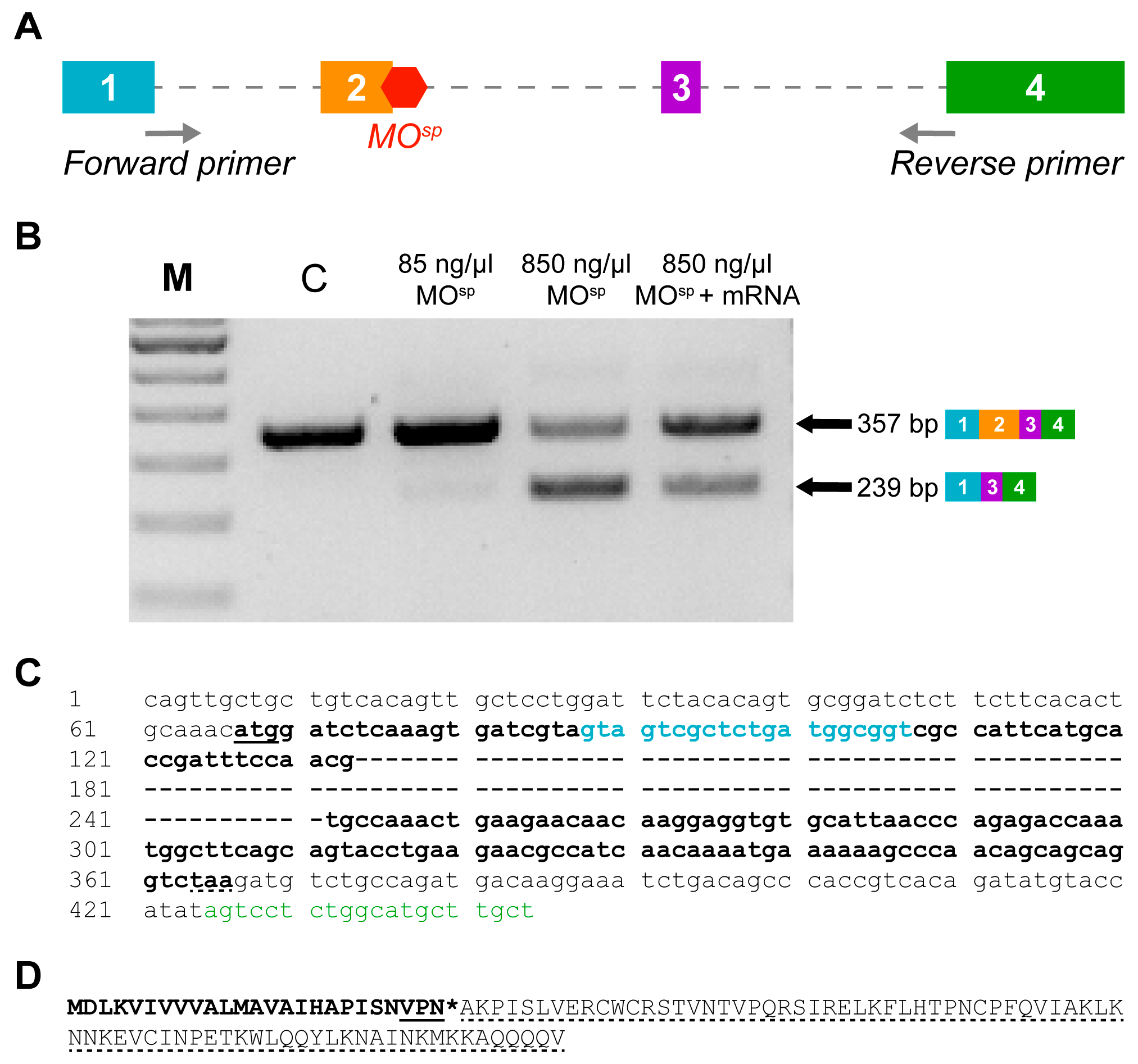
2.4. cxcl12a Morpholino Design and Injection

To achieve knockdown of Cxcl12a protein expression, we designed a splice morpholino (MO) (MOsp: 5′-AAGTGCAGATACTCACATGACTTGG-3′) which was synthesized by Gene Tools (Philomath, OR, USA) and analyzed with the NCBI BLAST tool (https://blast.ncbi.nlm.nih.gov/Blast.cgi, accessed on 15 December 2022) to assess any potential off-target effects. The MOsp was designed to target cxcl12a pre-mRNA splicing at exon 2/intron 2 (E2I2) and bind to the last 9 bases of E2 and first 12 bases of I2. This effectively skips exon 2 entirely with its 118 bp. Moreover, we additionally used a Cxcl12a translation blocking MO (MOaug: 5′-TTGAGATCCATGTTTGCAGTGTGAA-3′) [28,32] to validate that, unlike the MOsp, it does not create alternate transcripts to decrease Cxcl12a mRNA levels. Two doses of MOsp, including a low dose of 85 ng/µL and a high dose of 850 ng/µL, were tested to determine the highest and most effective concentration that does not induce a phenotype in wild-type embryos. An amount of 1 nl of both the MOsp and a standard control MO (5’-CCTCTTACCTCAGTTACAATTTATA-3’) at 1 mM [33] were injected into single-cell stage Hcrt:EGFP embryos. We determined that the 850 ng/µL of MOsp effectively induced alternate splicing and then used a translation tool from www.ExPASy.com to determine the amino acids produced by alternately spliced cxcl12a mRNA. No morphological abnormalities were observed in the injected zebrafish at both high and low doses. The groups tested in experiments involving the cxcl12a MOsp consisted of (1) control zebrafish injected with the control MO (“Control + Control MO”); (2) ethanol-exposed zebrafish injected with the control MO (“Ethanol + Control MO”); (3) ethanol-exposed zebrafish injected with the cxcl12a MOsp (“Ethanol + cxcl12a MOsp”); and (4) control zebrafish injected with the cxcl12a MOsp (“Control + cxcl12a MOsp”).

2.5. RNA Extraction, cDNA Synthesis and RT-PCR

To confirm the splicing efficiency of the cxcl12a MOsp, total RNA was extracted from the whole embryo at 28 hpf using TRIzol reagent (Invitrogen, Carlsbad, CA, USA) [34]. An amount of 1 μg of total RNA was used to synthesize cDNA, according to the manufacturer’s procedure in the TaqMan Reverse Transcription kit (ThermoFisher Scientific, Waltham, MA, USA). Using the Phire Tissue Direct RT-PCR Kit (ThermoFisher Scientific, Waltham, MA, USA) and the primers (F: 5′-GTAGTCGCTCTGATGGCGGT-3′ and R: 5′-AGTCCTCTGGCATGCTTGCT-3′), we amplified a region that partially spans exons 1 and 4 and the full length of exons 2 and 3 of the mature cxcl12a transcript. The RT-PCR reaction was run at a 65.5 °C annealing temperature in 30 cycles.

2.6. Microscopy and Image Analysis

To acquire images of Hcrt neurons and their projections throughout the brain, transgenic 6 dpf Hcrt:EGFP zebrafish were live-imaged using confocal microscopy, with a 25× objective lens on a Zeiss LSM 780 laser scanning confocal microscope and a z step of 1.0 μm. All images were analyzed using Imaris 9.9.1 software. The AH of the zebrafish brain was first divided into equal halves along the AP axes, into regions defined as anterior AH (aAH) and posterior AH (pAH). The number of Hcrt neurons in the aAH and pAH and also further anterior in the POA was quantified manually using the “Spots” function in Imaris. We then created 3D reconstructions of neuronal projections from Hcrt neurons located in the aAH, POA and pAH using the “Filaments” function of Imaris [35]. Spot sizes for the start point and seeding points of the filament were 7 µm and 0.9 µm with a threshold of 1350 and 350, respectively, followed by manual correction. To quantify the density of terminal points and branch points within certain brain areas of interest, we first used the “Surface” function of Imaris to mask out functionally relevant brain regions that are known to be innervated by Hcrt neurons, as described [36,37]. These included the subpallium (SP), dorsal pallium (DP), locus coeruleus (LC) and lateral forebrain bundle (LFB). For this analysis, we excluded the Hcrt projections to the furthest anterior regions including the olfactory bulb, pineal gland and optic tectum and those to the most posterior regions beyond the LC including the spinal cord. The anatomy for each brain region was derived from the Zebrafish Brain Browser atlas, following previously published guidelines [38]. The density of branch points and terminal points was calculated within each of these brain areas by quantifying their number and then dividing these values by the volume (exported from Imaris “Surface” function) of each respective region.

2.7. Statistical Analyses

All number data for Hcrt neurons in the aAH and pAH were analyzed using two-way ANOVA, which tested the main effects of the condition and brain area and their interaction, followed by post-hoc Holm–Sidak’s multiple comparisons test. The projection data of Hcrt neurons derived from the brain regions innervated by these neurons was analyzed by a one-way ANOVA for each individual brain area. With the ectopic POA neurons and their projections absent in the control conditions, the POA data from the experimental groups are presented in the text of Section 3 but could not be statistically compared to a control group as there were no POA neurons present in the control zebrafish. All tests were two-tailed, and significance was determined at p < 0.05. All data were analyzed using Prism (version 9, GraphPad, San Diego, CA, USA) and are presented as mean ± SEM in the figures and tables.

3. Results

3.1. Confirmation of cxcl12a mRNA and MOsp

To assess the function of Cxcl12a in mediating the ethanol-induced effects on Hcrt neurons and their projections, we used in vitro-transcribed cxcl12a mRNA and an antisense splice-altering MO (MOsp) injected at the single-cell stage. Injection of the cxcl12a mRNA was to overexpress Cxcl12a protein, whereas the MOsp was used to knock down the cxcl12a gene by inhibiting the splicing of exon 2 into the mature mRNA product. The cxcl12a gene, located on chromosome 13, generates a 4-exon transcript that encodes a protein composed of 99 amino acids. The schematic in Figure 1A shows the sites on the cxcl12a pre-mRNA to which the MOsp binds. Whereas the low dose (85 ng/µL) MOsp has no splice-altering effect, targeted binding of the high dose (850 ng/µL) MOsp causes alternate splicing that fully excludes exon 2 from the mature cxcl12a mRNA (Figure 1B). The removal of exon 2 creates a smaller PCR product (239 bp), which can cause translation failure and therefore effectively knock down Cxcl12a protein expression [39,40].
This decreased expression is detected by RT-PCR, which also shows that rescue by co-injecting cxcl12a MOsp and cxcl12a mRNA increases the expression of the wild-type band intensity compared to the mutant band intensity (Figure 1B). Moreover, sequence analysis shows complete deletion of the 118 bp of exon 2 (Figure 1C), and from this analysis, the resulting transcript encoding a 25 amino acid protein with a premature stop codon is expected (Figure 1D). Taken together, the MOsp effectively truncates the mature cxcl12a mRNA, which causes a frameshift in the downstream sequence to insert a premature termination codon and delete 77 amino acids that ultimately decreases wild-type Cxcl12a expression.

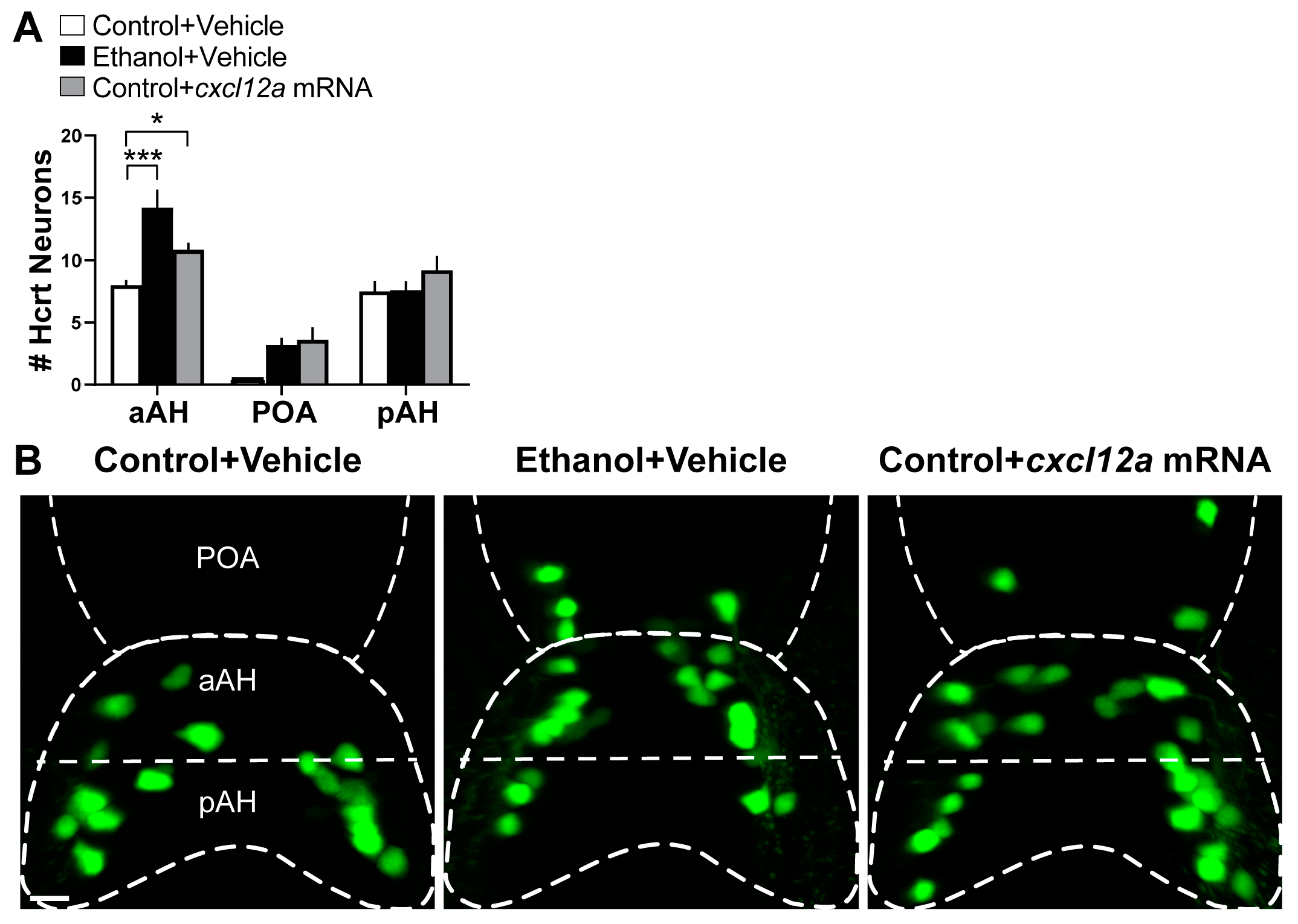
3.2. Cxcl12a Overexpression Mimics the Ethanol-Induced Increase in Number of Hcrt Neurons in the aAH and POA with No Effect in the pAH

We first tested in 6 dpf zebrafish if the overexpression of Cxcl12a has effects similar to those produced by embryonic ethanol exposure (0.5% v/v, 22–24 hpf), specifically on the number of Hcrt neurons in the aAH, POA and pAH subpopulations. Analysis of Hcrt:EGFP zebrafish showed that the Hcrt neurons, while similar in number in the aAH and pAH and not evident in the POA of the Control + Vehicle-injected zebrafish, were significantly altered in the Ethanol + Vehicle-injected zebrafish (Figure 2A), as illustrated in the photomicrographs (Figure 2B). Ethanol increased the number of Hcrt neurons specifically in the aAH (p = 0.0001), and it additionally caused the most anterior Hcrt neurons of the aAH (3.2 ± 0.583) in Control + Vehicle zebrafish to become ectopically expressed further anterior in the POA, while having no effect on the number of Hcrt neurons in the pAH (p = 0.939). Notably, injection of cxcl12a mRNA in the Control + cxcl12a mRNA zebrafish had very similar effects to those of embryonic ethanol exposure (Figure 2A,B). It produced a significant increase in the number of Hcrt neurons in the aAH (p = 0.019) and again caused some (3.6 ± 1.03) of the most anterior aAH Hcrt neurons to become ectopically expressed in the POA. It also had no effect on the number of Hcrt neurons in the pAH (p = 0.497). These results demonstrate that cxcl12a overexpression has the same effects as embryonic ethanol exposure on the development of these Hcrt subpopulations.

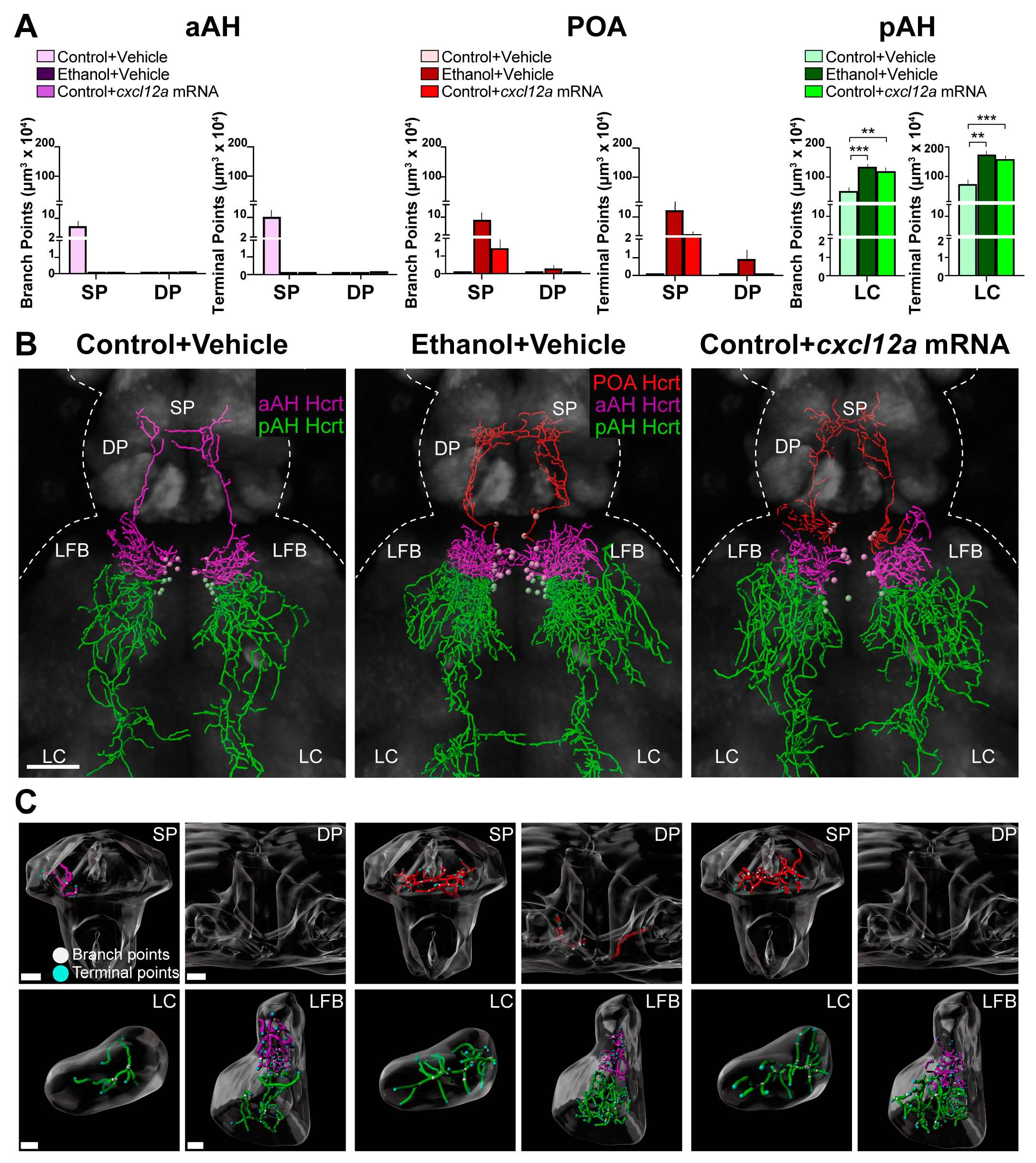
3.3. Cxcl12a Overexpression Mimics the Ethanol-Induced Stimulation of Hcrt Projections to Distant Areas in the Anterior and Posterior Brain

Here we tested whether the overexpression of Cxcl12a has similar effects to those produced by embryonic ethanol exposure (0.5% v/v, 22–24 hpf) on the projections of the aAH, POA and pAH subpopulations. As previously described [9], we examined the long projections from the aAH Hcrt neurons that project anteriorly to the SP and DP and are not seen in any posterior structures, the ectopic POA Hcrt neurons that, like aAH Hcrt neurons, project anteriorly to the SP and DP, and the pAH Hcrt neurons that project posteriorly to the LC and are not seen in any anterior structures. We also examined the short projections from the aAH and pAH neurons that project laterally to the LFB. While the branch points (6.6 ± 2.08) and terminal points (10.20 ± 2.54) of the long projections from aAH Hcrt neurons in the Control + Vehicle zebrafish were evident in the SP but not the DP, these projections were not detected in either of these areas from aAH Hcrt neurons in the Ethanol + Vehicle or the Control + cxcl12a mRNA zebrafish (Figure 3A), as illustrated in the photomicrographs (Figure 3B) and brain region enlargements (Figure 3C). This is due to the loss in these two experimental groups of the specific Hcrt neurons which, while originally in the most anterior region of the aAH with long projections ascending to the SP, were induced by ethanol and Cxcl12a mRNA to migrate further anterior to become ectopically expressed in the POA. The branch points and terminal points of the projections from these ectopic POA neurons in the Ethanol + Vehicle zebrafish were detected not only in the SP (9.16 ± 2.76 and 12.80 ± 3.05, respectively) but also more dorsally in the DP (0.30 ± 0.19 and 0.88 ± 0.46), an area not reached by the projections from the aAH neurons in Control + Vehicle zebrafish. In the Control + cxcl12a mRNA zebrafish, they were similarly detected in the SP (1.44 ± 0.52 and 2.64 ± 0.87, respectively), although they were not evident in the DP (Figure 3A–C). Like these long anterior projections from the POA Hcrt neurons, ethanol increased the branch points (p = 0.0004) and terminal points (p = 0.00046) of the long projections from the pAH Hcrt neurons in the LC, and cxcl12a mRNA injection had a similar effect on these measures of the pAH projections to the LC (p = 0.003 and p = 0.002, respectively). In contrast to these long projections, both ethanol and cxcl12a mRNA had no effect on the branch points and terminal points of the short, lateral projections to the LFB from the aAH and pAH subpopulations of Hcrt neurons (Table 1). These results demonstrate that overexpression of Cxcl12a produces the same effects as embryonic ethanol exposure on Hcrt neuronal projections, indicating that Cxcl12a is an important mediator of ethanol’s effects on Hcrt development.

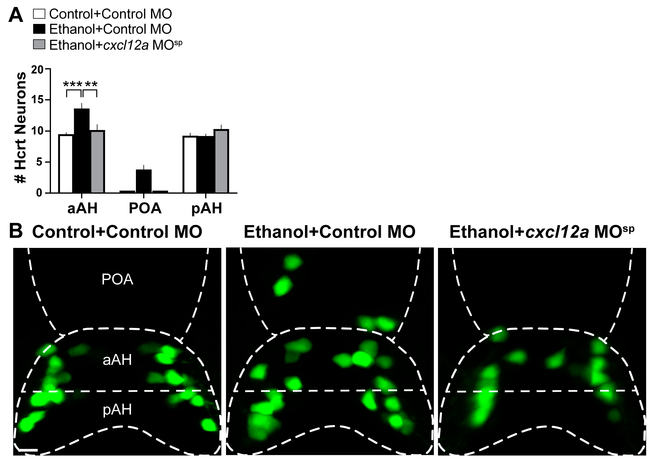
3.4. Cxcl12a Knockdown Blocks the Ethanol-Induced increase in Number of Hcrt Neurons in the aAH and POA

To further test the involvement of Cxcl12a in mediating the effects of embryonic ethanol exposure, we next tested if knockdown of Cxcl12a by injecting a cxcl12a MOsp at the single-cell stage blocks the stimulatory effects of ethanol on the number of Hcrt neurons in the aAH and POA. We found as in Experiment 3.2 that the Hcrt neuron number was stimulated in the Ethanol + Control MO zebrafish (Figure 4A), as illustrated in the photomicrographs (Figure 4B). While the Hcrt neurons of the Control + Control MO fish were similar in number in the aAH and pAH and were not evident in the POA, the number of Hcrt neurons in the Ethanol + Control MO fish was increased in the aAH (p = 0.0005) but not the pAH (p = 0.999), and some (3.2 ± 0.58) ectopically located Hcrt neurons originating in the aAH were observed in the POA (Figure 4A,B). Injection of the cxcl12a MOsp in Ethanol + cxcl12a MOsp zebrafish blocked the ethanol-induced increase in the number of Hcrt neurons in the aAH (p = 0.0012), while having no effect on Hcrt neurons in the pAH (p = 0.416). The cxcl12a MOsp also completely prevented the formation of ectopic Hcrt neurons further anterior in the POA that were observed in the Ethanol + Control MO zebrafish (Figure 4A,B). Compared to the Control + Control MO zebrafish, injection of the cxcl12a MOsp in Control + cxcl12a MOsp at the dose used had no effects of its own on the number of Hcrt neurons in the aAH, POA and pAH subpopulations (Table 2). These results provide additional evidence that Cxcl12a is involved in the anatomically specific stimulatory effect of embryonic ethanol exposure on the Hcrt neurons specifically in the aAH and POA subpopulations that is not evident in the pAH.

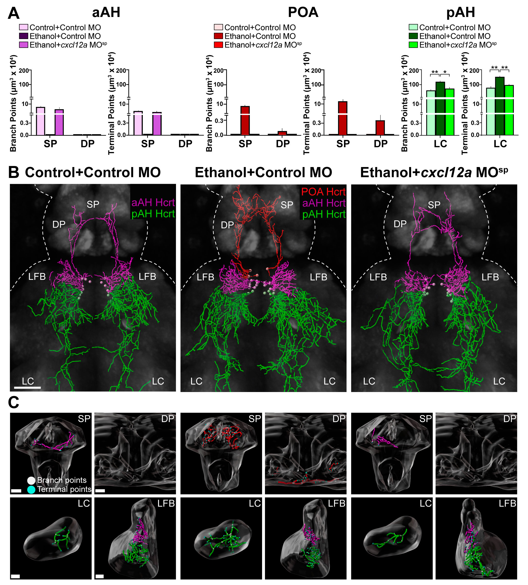
3.5. Cxcl12a Knockdown Blocks the Ethanol-Induced Stimulation of Hcrt Projections to Distant Areas in the Anterior and Posterior Brain

Here we tested whether Cxcl12a knockdown also blocks the stimulatory effects of ethanol on the projections from the Hcrt subpopulations to specific anterior and posterior brain areas. While branch points (3.3 ± 0.45) and terminal points (7.14 ± 1.04) of the long projections from normally located aAH Hcrt neurons in the Control + Control MO zebrafish were evident in the SP but not DP as shown in Experiment 3.3, they were not detected in either area of the Ethanol + Control MO zebrafish (Figure 5A), as illustrated in the photomicrographs (Figure 5B) and brain region enlargements (Figure 5C), again due to the ethanol-induced loss in this group of the most anterior aAH Hcrt neurons with long ascending projections to the SP. Injection of cxcl12a MOsp in the ethanol-exposed zebrafish, while preventing the formation of the ectopic POA neurons, restored the branch points (2.56 ± 0.86) and terminal points (5.19 ± 1.48) of the long projections from normally located aAH neurons in the SP, as seen in the Control + Control MO zebrafish (Figure 5A–C). Additionally, in the Ethanol + Control MO zebrafish, the branch points and terminal points of the projections from ectopic POA neurons clearly seen in both the SP (8.64 ± 1.7 and 12.72 ± 2.14, respectively) and DP (0.08 ± 0.05 and 0.37 ± 0.18, respectively) were not detected in the Ethanol + cxcl12a MOsp zebrafish similar to the Control + Control MO zebrafish (Figure 5A–C). Like these long anterior projections from POA Hcrt neurons, the Ethanol + Control MO compared to Control + Control MO zebrafish exhibited a significant increase in branch points (p = 0.0097) and terminal points (p = 0.0014) of the long posterior projections from the pAH Hcrt neurons in the LC. Further, the Ethanol + cxcl12a MOsp zebrafish exhibited a blockade of this ethanol effect, showing a significant decrease in the branch points (p = 0.025) and terminal points (p = 0.0037) of these pAH projections to the LC. Once again, ethanol had no effect on the branch points and terminal points of the short, lateral projections from the aAH and pAH neurons to the LFB (Table 2). In the Control + cxcl12a MOsp-injected zebrafish compared to the Control + Control MO zebrafish, the cxcl12a MOsp had little effect of its own under control conditions, except for reducing the branch points and terminal points of projections in the SP from aAH Hcrt neurons (Table 3). These results, showing that knockdown of Cxcl12a inhibits the ethanol-induced effects on Hcrt projections, further demonstrates that Cxcl12a is directly involved in mediating how ethanol alters the development of the Hcrt neuronal network.

4. Discussion

This report presents the novel finding that Cxcl12a is directly involved in the effects of embryonic exposure to ethanol on the development of anatomically distinct and ectopically located subpopulations of Hcrt neurons and their projections. As we have recently described [10], this study confirms that embryonic ethanol exposure increases the number of Hcrt neurons specifically in the aAH but not the pAH and causes the most anterior aAH neurons to become ectopically located further anterior in the POA. Notably, overexpression of Cxcl12a closely mimics these stimulatory effects of ethanol on the number of Hcrt neurons normally located in the aAH and abnormally located in the POA, and the knockdown of Cxcl12a via injection of a cxcl12a MOsp prevents these specific effects of ethanol. Building on studies in rats showing prenatal Cxcl12 administration to stimulate the overall density of peptide-expressing neurons in the hypothalamus [25] similar to the effects of ventricular injection of Cxcl12 shown in adult rats [41], here we provide the first evidence that embryonic exposure to Cxcl12 can affect specific neuronal subpopulations in a highly localized manner. This agrees with evidence that Cxcr4, the receptor for Cxcl12, is also involved in these anatomically specific effects, with embryonic pretreatment of the Cxcr4 antagonist AMD3100 found to block the ethanol-induced increase in the number of Hcrt neurons in the aAH and induction of ectopic Hcrt neurons in the POA [10].
The increase in number of Hcrt neurons in the aAH and POA induced similarly by ethanol and cxcl12a mRNA injection may be due, in part, to an increase in their proliferation and migration, which is stimulated by low-moderate concentrations of embryonic ethanol [6,11,42] and by Cxcl12 itself [24,43,44]. With embryonic ethanol exposure shown to increase the expression of Cxcl12 in the hypothalamus [22,23], Cxcl12a overexpression in turn likely increases the proliferation and migratory speed of the aAH Hcrt neurons, specifically those located in the most anterior region of the aAH, causing them to become ectopically expressed in the POA. Importantly, this induction of ectopic neurons in the POA by both ethanol and Cxcl12a overexpression is not simply due to a redistribution of Hcrt neurons from the aAH but occurs in conjunction with an increase in Hcrt number in the aAH. Further evidence linking the ethanol-induced increase in proliferation to Cxcl12 is suggested by our recent studies [9,10], showing that the more anterior areas of the aAH and POA have naturally higher levels of cxcl12a transcripts along with greater proliferative activity compared to the posterior area of the pAH. While it is possible that the increased number of Hcrt neurons produced by both ethanol and Cxcl12a overexpression results from decreased programmed cell death of these neurons, prior studies of embryonic exposure at comparably low concentrations of ethanol in both rats and zebrafish have reported no changes in apoptosis [5,45].
Cxcl12 has a well-established role in controlling migration, as shown in rodent studies of neurons expressing gonadotropin-releasing hormone (GnRH) [28,46] and of trigeminal sensory and motor neurons [47,48], consistent with an in vitro study showing the velocity and distance migrated by hypothalamic peptide-expressing neurons to be increased by the chemokine CCL2 [49]. With both Cxcl12a overexpression and ethanol exposure increasing Hcrt neuron number, future studies will be needed to determine if the combination of these two treatments produces an additive effect and an even greater increase in Hcrt number, or if there is a ceiling to the increase in Hcrt neuron number. Further, with Cxcl12a MOsp administration blocking the ethanol-induced increase in Hcrt neurons, future work will be needed to evaluate Cxcl12 as a potential target to reverse the increased recruitment of Hcrt neurons produced by a number of substances of misuse, including cocaine [50] and alcohol [23].
The induction of ectopic Hcrt neurons in the POA from the anterior aAH subpopulation may also involve an endogenous Cxcl12 concentration gradient, where neurons that express Cxcr4 migrate towards the higher concentrations of this chemokine. While the role of a Cxcl12a gradient in cellular migration has been clearly demonstrated in peripheral systems such as the lateral line in zebrafish [51] and the in vitro retinal ganglion cells from mice [52], we recently found in the embryonic zebrafish brain a natural posterior-to-anterior concentration gradient of cxcl12a transcripts, with the lowest levels in the pAH, higher levels in the aAH and POA and the highest levels in the telencephalon [10]. We also discovered that ethanol stimulates cxcl12a transcript expression along this natural gradient, while having an anatomically specific stimulatory effect on the co-expression of Cxcr4b in Hcrt neurons in the aAH and POA subpopulations but not in the pAH [10]. Similar to ethanol, the injection of cxcl12a mRNA likely increases endogenous Cxcl12a expression along this gradient causing Cxcr4b-expressing Hcrt neurons to migrate towards the higher Cxcl12a levels in the anterior brain, while injection of the cxcl12a MOsp likely has the opposite effect on Cxcl12a expression, thereby blocking the ethanol-induced abnormal migration of aAH Hcrt neurons further anterior into the POA. This idea is further supported by evidence showing that the transplantation of Cxcl12a in the anterior zebrafish brain causes trigeminal Cxcr4b-expressing sensory neurons to migrate anteriorly towards the transplanted Cxcl12a [53] and by in vitro findings showing dorsal root ganglion neurons to be attracted to the local source of Cxcl12 [54]. Another possible mechanism in the anatomically specific effects of ethanol and Cxcl12a on Hcrt neurons may involve radial glia progenitor cells. These neuroprogenitor cells, which in zebrafish and rats have an important role in cell proliferation and migration [55,56,57], are densely concentrated in the hypothalamus where they co-express chemokines [23,58] and are strongly stimulated by embryonic ethanol exposure, consequently contributing to alterations in the proliferation and migration of Hcrt neurons.
In addition to controlling neuronal proliferation and migration, Cxcl12 is also known to mediate the development of neuronal projections, including projection pathfinding and positioning, axonal elongation, branch formation and dendrite generation and density [26,27,28,29]. Here we provide the first direct evidence that Cxcl12a mediates the effects of embryonic ethanol exposure on the development of Hcrt neuronal projections, with Cxcl12a overexpression producing stimulatory effects similar to ethanol. It is worth noting, however, that the effects of Cxcl12a overexpression on Hcrt projections are not as strong as those produced by ethanol, including a lack of an effect of Cxcl12a overexpression on DP projections from POA Hcrt neurons that was seen in ethanol-exposed fish. This may be due to Cxcl12a overexpression producing less of an increase in Cxcl12a protein levels, possibly via some mRNA degradation, or due to the involvement of other neuroimmune mediators, such as the CCL2/CCR2 chemokine system, shown to be stimulated by embryonic ethanol and to alter neuronal development [59]. While having no effect on the short, lateral projections of Hcrt neurons to the LFB, Cxcl12a overexpression like ethanol increases the branch points and terminal points of the long projections from the aAH, POA and pAH subpopulations. Although the mechanisms have yet to be determined, this finding that ethanol has differential effects on Hcrt neuronal projections depending on their length is similar to a prior report in mice [60], showing that early exposure to ethanol differentially affects the long and short projections from pyramidal neurons in the medial prefrontal cortex.
It is notable that the Hcrt neurons, which become ectopically expressed in the POA after ethanol exposure and Cxcl12a overexpression, share the same projection characteristics as the most anterior aAH neurons normally located in control zebrafish, with both having long ascending projections to the SP. This supports the idea that these ethanol-induced ectopic neurons in the POA do, in fact, originate from the anterior aAH subpopulation. Our results, showing that knockdown of Cxcl12a completely blocks the formation of these ectopic POA neurons with long ascending projections and restores their position in the anterior aAH region, provides evidence that Cxcl12a is an important mediator of these ethanol effects on the location of Hcrt neurons. Our findings also suggest that Cxcl12a plays a critical role in the anterior pathfinding of the Hcrt projections, with injection of the cxcl12a MOsp decreasing the branch points and terminal points of aAH Hcrt neurons in the SP of control zebrafish and completely blocking the ethanol-induced branch points and terminal points in the DP, a more dorsal region not typically innervated by Hcrt neurons at this young age [36].
With our recent study showing ethanol to increase the concentration gradient of Cxcl12a in the brain with highest levels in the telencephalon where the SP and DP are located [10], it is likely that this gradient also contributes to the ethanol-induced guidance of the Hcrt neuron projections in the anterior direction. With Hcrt soma found to co-express Cxcr4b receptors in both rats and zebrafish [10,23], the Hcrt neuronal projections also likely co-express Cxcr4b receptors that contribute to the guidance of these projections towards higher Cxcl12a concentrations. This idea is consistent with findings in mice, showing that Cxcr4 receptors are expressed along the axons of embryonic GnRH neurons [46] and Cxcl12 is expressed at high levels where motor neuron projections terminate [61], and also in zebrafish, where both Cxcl12a and Cxcr4b expression are positively correlated with the axonal pathway of GnRH projections [28]. In addition to these effects on the anterior Hcrt projections, Cxcl12a overexpression completely reproduces the effects of ethanol on the posterior projections from pAH neurons, increasing their branch points and terminal points in the LC, an effect that is totally blocked by Cxcl12a knockdown in ethanol-exposed fish. Although a chemokine concentration gradient has yet to be described in the area of the LC, there are previous reports showing that Cxcr4 is expressed in the LC of both rats and humans [62,63], indicating that this chemokine system may be associated with the pathfinding of neuronal projections to this region.
The effects described here of ethanol and Cxcl12a overexpression on the development of different subpopulations of Hcrt neurons and their projections are likely to have significant behavioral consequences. In both zebrafish and rats, Hcrt has a positive relationship with behaviors that are stimulated by embryonic ethanol exposure [36,42], including alcohol consumption [64,65] as well as locomotor activity and anxiety-like behavior [42,66,67]. Similar to ethanol, chemokines are also shown to stimulate these behaviors [25,68], and chemokine receptor antagonists are found to block the behaviors induced by ethanol in larval zebrafish [10] as well as rats [68]. Studies in rodents show that Hcrt neuron subpopulations mediate different behaviors related to substances of misuse [50,69], possibly due to the known neurochemical and genetic heterogeneity of Hcrt neurons [70]. For example, Hcrt neurons co-express other neurochemicals such as dynorphin and glutamate [36,71], and it is possible that embryonic ethanol exposure or Cxcl12a overexpression alters their level of co-expression within specific subpopulations of Hcrt neurons that in turn affect neuronal functioning and contribute to behavioral disturbances. Our laboratory recently demonstrated the functional importance of specific Hcrt subpopulations in mediating ethanol-induced behaviors [7], showing that ablation of the ectopic POA Hcrt neurons or the Hcrt neurons in the most anterior part of the aAH blocks the ethanol-induced increase in anxiety-like behavior, and ablation of the ectopic Hcrt neurons blocks the ethanol-induced increase in locomotor behavior. In this report, we take the next step and reveal that Cxcl12a is involved in mediating the ethanol-induced effects on the projections of different Hcrt neuron subpopulations to functionally different brain areas. Ethanol and Cxcl12a both induce Hcrt neurons in the most anterior region in the aAH that become ectopically expressed in the POA and send long projections to the SP, a brain area homologous to the mammalian basal ganglia that contains Hcrt receptors and is rich in cells expressing neurotransmitters associated with ethanol-related behaviors [72,73,74]. Notably, ethanol also causes ectopic Hcrt neurons in the POA to send projections further anterior to the DP, an area shown to participate in drug responses of zebrafish [75,76]. In contrast, the Hcrt subpopulation in the pAH innervates the LC, a region that is rich in norepinephrine and important for locomotor and sleep–wake behaviors [77,78] and has a functional relationship with Hcrt neurons in terms of these behaviors [79]. These alterations in Hcrt projections within the LC may act as a potential mechanism underlying sleep disorders that are often comorbid with substance use disorders [80], consistent with evidence showing Hcrt receptor antagonists to have clinical efficacy in improving sleep and reducing withdrawal symptoms [81]. Together, our finding here, that Cxcl12a knockdown blocks the ethanol-induced effects on these Hcrt projections from the Hcrt subpopulations to distant brain areas, suggests that this knockdown will also reduce or block the behaviors induced by ethanol exposure.
In summary, this study demonstrates that the chemokine Cxcl12a directly mediates the stimulatory effects of embryonic ethanol exposure on the number of Hcrt neurons in anatomically specific subpopulations and the induction of ectopic Hcrt neurons, effects mimicked by injection of cxcl12a mRNA and blocked by knockdown using a cxcl12a MOsp. This chemokine is also involved in ethanol’s effects on the pathfinding of projections from Hcrt neurons to distant brain areas known to have important behavioral functions. These neurodevelopmental changes likely alter the functioning of Hcrt neurons, contributing to disturbances in alcohol-related behaviors shown to be produced by embryonic ethanol exposure.

Author Contributions

Conceptualization, N.Y., A.D.C., O.K. and S.F.L.; Data curation, S.F.L.; Formal analysis, N.Y., A.D.C., B.Y. and M.F.; Funding acquisition, S.F.L.; Investigation, N.Y., A.D.C., A.R.A. and S.F.L.; Methodology, N.Y., A.D.C., A.R.A. and M.F.; Project administration, S.F.L.; Resources, S.F.L.; Software, N.Y., A.D.C., A.R.A., B.Y. and M.F.; Supervision, A.D.C., O.K. and S.F.L.; Validation, N.Y., A.D.C., A.R.A. and O.K.; Visualization, N.Y., A.D.C., A.R.A., O.K. and B.Y.; Writing—original draft, A.D.C., O.K. and S.F.L.; Writing—review and editing, N.Y., A.D.C., A.R.A., O.K., B.Y., M.F. and S.F.L. All authors have read and agreed to the published version of the manuscript.

Funding

This research was funded by the National Institute on Alcohol Abuse and Alcoholism of the National Institutes of Health under award numbers R01AA027653 (S.F.L.) and F32AA027702 (A.D.C.). The content is solely the responsibility of the authors and does not necessarily represent the official views of the National Institutes of Health.

Institutional Review Board Statement

The animal study protocol was approved by the Institutional Animal Care and Use Committee (IACUC) of The Rockefeller University (protocol code 21067-H (PRV 18045-H) approved on 29 June 2021).

Informed Consent Statement

Not applicable.

Data Availability Statement

The datasets used and/or analyzed during the current study are available from the corresponding author on reasonable request.

Acknowledgments

We thank Lior Appelbaum (Bar-Ilan University) and Nobuhiko Miyasaka (RIKEN Brain Science Institute) who kindly shared the transgenic Hcrt:EGFP zebrafish and pGEM sdf-1a plasmid with us, respectively. We also thank The Rockefeller University’s BioImaging Resource Center for the use of their equipment and for providing guidance.

Conflicts of Interest

The authors declare no conflict of interest.

References

  1. Gupta, K.K.; Gupta, V.K.; Shirasaka, T. An Update on Fetal Alcohol Syndrome-Pathogenesis, Risks, and Treatment. Alcohol. Clin. Exp. Res. 2016, 40, 1594–1602. [Google Scholar] [CrossRef]
  2. Petrelli, B.; Weinberg, J.; Hicks, G.G. Effects of prenatal alcohol exposure (PAE): Insights into FASD using mouse models of PAE. Biochem. Cell Biol. 2018, 96, 131–147. [Google Scholar] [CrossRef]
  3. Kleiber, M.L.; Mantha, K.; Stringer, R.L.; Singh, S.M. Neurodevelopmental alcohol exposure elicits long-term changes to gene expression that alter distinct molecular pathways dependent on timing of exposure. J. Neurodev. Disord. 2013, 5, 6. [Google Scholar] [CrossRef] [PubMed]
  4. Cuzon, V.C.; Yeh, P.W.L.; Yanagawa, Y.; Obata, K.; Yeh, H.H. Ethanol Consumption during Early Pregnancy Alters the Disposition of Tangentially Migrating GABAergic Interneurons in the Fetal Cortex. J. Neurosci. 2008, 28, 1854–1864. [Google Scholar] [CrossRef] [PubMed]
  5. Chang, G.-Q.; Karatayev, O.; Liang, S.C.; Barson, J.R.; Leibowitz, S.F. Prenatal ethanol exposure stimulates neurogenesis in hypothalamic and limbic peptide systems: Possible mechanism for offspring ethanol overconsumption. Neuroscience 2012, 222, 417–428. [Google Scholar] [CrossRef]
  6. Chang, G.-Q.; Karatayev, O.; Leibowitz, S.F. Prenatal exposure to ethanol stimulates hypothalamic CCR2 chemokine receptor system: Possible relation to increased density of orexigenic peptide neurons and ethanol drinking in adolescent offspring. Neuroscience 2015, 310, 163–175. [Google Scholar] [CrossRef] [PubMed]
  7. Collier, A.D.; Yasmin, N.; Chang, G.Q.; Karatayev, O.; Khalizova, N.; Fam, M.; Abdulai, A.R.; Yu, B.; Leibowitz, S.F. Embryonic ethanol exposure induces ectopic Hcrt and MCH neurons outside hypothalamus in rats and zebrafish: Role in ethanol-induced behavioural disturbances. Addict. Biol. 2022, 27, e13238. [Google Scholar] [CrossRef]
  8. Lovely, C.B.; Fernandes, Y.; Eberhart, J.K. Fishing for Fetal Alcohol Spectrum Disorders: Zebrafish as a Model for Ethanol Teratogenesis. Zebrafish 2016, 13, 391–398. [Google Scholar] [CrossRef] [PubMed]
  9. Yasmin, N.; Collier, A.D.; Karatayev, O.; Abdulai, A.R.; Yu, B.; Fam, M.; Khalizova, N.; Leibowitz, S.F. Subpopulations of hypocretin/orexin neurons differ in measures of their cell proliferation, dynorphin co-expression, projections, and response to embryonic ethanol exposure. Sci. Rep. 2023. in review. [Google Scholar]
  10. Collier, A.D.; Yasmin, N.; Karatayev, O.; Abdulai, A.R.; Yu, B.; Khalizova, N.; Fam, M.; Leibowitz, S.F. Neuronal chemokine concentration gradients mediate effects of embryonic ethanol exposure on ectopic hypocretin/orexin neurons and behavior in zebrafish. Sci. Rep. 2023, 13, 1447. [Google Scholar] [CrossRef]
  11. Collier, A.D.; Halkina, V.; Min, S.S.; Roberts, M.Y.; Campbell, S.D.; Camidge, K.; Leibowitz, S.F. Embryonic Ethanol Exposure Affects the Early Development, Migration, and Location of Hypocretin/Orexin Neurons in Zebrafish. Alcohol. Clin. Exp. Res. 2019, 43, 1702–1713. [Google Scholar] [CrossRef] [PubMed]
  12. Boric, K.; Orio, P.; Viéville, T.; Whitlock, K. Quantitative Analysis of Cell Migration Using Optical Flow. PLoS ONE 2013, 8, e69574. [Google Scholar] [CrossRef]
  13. Coffey, C.M.; Solleveld, P.A.; Fang, J.; Roberts, A.K.; Hong, S.-K.; Dawid, I.B.; Laverriere, C.E.; Glasgow, E. Novel Oxytocin Gene Expression in the Hindbrain Is Induced by Alcohol Exposure: Transgenic Zebrafish Enable Visualization of Sensitive Neurons. PLoS ONE 2013, 8, e53991. [Google Scholar] [CrossRef] [PubMed]
  14. Cole, G.J.; Zhang, C.; Ojiaku, P.; Bell, V.; Devkota, S.; Mukhopadhyay, S. Effects of Ethanol Exposure on Nervous System Development in Zebrafish. Int. Rev. Cell Mol. Biol. 2012, 299, 255–315. [Google Scholar] [CrossRef] [PubMed]
  15. Chevassus-Au-Louis, N.; Represa, A. The right neuron at the wrong place: Biology of heterotopic neurons in cortical neuronal migration disorders, with special reference to associated pathologies. Cell. Mol. Life Sci. 1999, 55, 1206–1215. [Google Scholar] [CrossRef]
  16. Wozniak, J.R.; Riley, E.P.; Charness, M.E. Clinical presentation, diagnosis, and management of fetal alcohol spectrum disorder. Lancet Neurol. 2019, 18, 760–770. [Google Scholar] [CrossRef]
  17. Dunty, W.C., Jr.; Zucker, R.M.; Sulik, K.K. Hindbrain and cranial nerve dysmorphogenesis result from acute maternal ethanol administration. Dev. Neurosci. 2002, 24, 328–342. [Google Scholar] [CrossRef]
  18. Abbott, C.W.; Rohac, D.J.; Bottom, R.T.; Patadia, S.; Huffman, K.J. Prenatal Ethanol Exposure and Neocortical Development: A Transgenerational Model of FASD. Cereb. Cortex 2018, 28, 2908–2921. [Google Scholar] [CrossRef] [PubMed]
  19. Goeke, C.M.; Roberts, M.L.; Hashimoto, J.G.; Finn, D.A.; Guizzetti, M. Neonatal Ethanol and Choline Treatments Alter the Morphology of Developing Rat Hippocampal Pyramidal Neurons in Opposite Directions. Neuroscience 2018, 374, 13–24. [Google Scholar] [CrossRef] [PubMed]
  20. Hamilton, G.F.; Whitcher, L.T.; Klintsova, A. Postnatal binge-like alcohol exposure decreases dendritic complexity while increasing the density of mature spines in mPFC Layer II/III pyramidal neurons. Synapse 2010, 64, 127–135. [Google Scholar] [CrossRef] [PubMed]
  21. Hamilton, G.F.; Criss, K.J.; Klintsova, A.Y. Voluntary exercise partially reverses neonatal alcohol-induced deficits in mPFC layer II/III dendritic morphology of male adolescent rats. Synapse 2015, 69, 405–415. [Google Scholar] [CrossRef]
  22. Collier, A.D.; Khalizova, N.; Chang, G.Q.; Min, S.; Campbell, S.; Gulati, G.; Leibowitz, S.F. Involvement of Cxcl12a/Cxcr4b Chemokine System in Mediating the Stimulatory Effect of Embryonic Ethanol Exposure on Neuronal Density in Zebrafish Hypothalamus. Alcohol. Clin. Exp. Res. 2020, 44, 2519–2535. [Google Scholar] [CrossRef] [PubMed]
  23. Chang, G.Q.; Collier, A.D.; Karatayev, O.; Gulati, G.; Boorgu, D.; Leibowitz, S.F. Moderate Prenatal Ethanol Exposure Stimulates CXCL12/CXCR4 Chemokine System in Radial Glia Progenitor Cells in Hypothalamic Neuroepithelium and Peptide Neurons in Lateral Hypothalamus of the Embryo and Postnatal Offspring. Alcohol. Clin. Exp. Research. 2020, 44, 866–879. [Google Scholar] [CrossRef]
  24. Watson, A.E.S.; Goodkey, K.; Footz, T.; Voronova, A. Regulation of CNS precursor function by neuronal chemokines. Neurosci. Lett. 2020, 715, 134533. [Google Scholar] [CrossRef]
  25. Poon, K.; Barson, J.R.; Shi, H.; Chang, G.Q.; Leibowitz, S.F. Involvement of the CXCL12 System in the Stimulatory Effects of Prenatal Exposure to High-Fat Diet on Hypothalamic Orexigenic Peptides and Behavior in Offspring. Front. Behav. Neurosci. 2017, 11, 91. [Google Scholar] [CrossRef] [PubMed]
  26. Chédotal, A. Roles of axon guidance molecules in neuronal wiring in the developing spinal cord. Nat. Rev. Neurosci. 2019, 20, 380–396. [Google Scholar] [CrossRef]
  27. Festa, L.K.; Irollo, E.; Platt, B.J.; Tian, Y.; Floresco, S.; Meucci, O. CXCL12-induced rescue of cortical dendritic spines and cognitive flexibility. Elife 2020, 9, e49717. [Google Scholar] [CrossRef]
  28. Palevitch, O.; Abraham, E.; Borodovsky, N.; Levkowitz, G.; Zohar, Y.; Gothilf, Y. Cxcl12a–Cxcr4b signaling is important for proper development of the forebrain GnRH system in zebrafish. Gen. Comp. Endocrinol. 2010, 165, 262–268. [Google Scholar] [CrossRef]
  29. Li, J.; Wu, Y.; Chen, P.; Huang, X.; Liu, Y.; Peng, M.; Wu, R.-Q. CXCL12 promotes spinal nerve regeneration and functional recovery after spinal cord injury. Neuroreport 2021, 32, 450–457. [Google Scholar] [CrossRef]
  30. Appelbaum, L.; Wang, G.X.; Maro, G.S.; Mori, R.; Tovin, A.; Marin, W.; Yokogawa, T.; Kawakami, K.; Smith, S.J.; Gothilf, Y.; et al. Sleep-wake regulation and hypocretin-melatonin interaction in zebrafish. Proc. Natl. Acad. Sci. USA 2009, 106, 21942–21947. [Google Scholar] [CrossRef] [PubMed]
  31. Miyasaka, N.; Knaut, H.; Yoshihara, Y. Cxcl12/Cxcr4 chemokine signaling is required for placode assembly and sensory axon pathfinding in the zebrafish olfactory system. Development 2007, 134, 2459–2468. [Google Scholar] [CrossRef]
  32. Marsay, K.S.; Greaves, S.; Mahabaleshwar, H.; Ho, C.M.; Roehl, H.; Monk, P.N.; Carney, T.J.; Partridge, L.J. Tetraspanin Cd9b and Cxcl12a/Cxcr4b have a synergistic effect on the control of collective cell migration. PLoS ONE 2021, 16, e0260372. [Google Scholar] [CrossRef]
  33. Otten, A.B.C.; Kamps, R.; Lindsey, P.; Gerards, M.; Pendeville-Samain, H.; Muller, M.; van Tienen, F.H.J.; Smeets, H.J.M. Tfam Knockdown Results in Reduction of mtDNA Copy Number, OXPHOS Deficiency and Abnormalities in Zebrafish Embryos. Front. Cell Dev. Biol. 2020, 8, 381. [Google Scholar] [CrossRef]
  34. Green, M.R.; Sambrook, J. Isolation of Total RNA from Zebrafish Embryos and Adults. Cold Spring Harb. Protoc. 2020, 2020, 101667. [Google Scholar] [CrossRef] [PubMed]
  35. Dudink, I.; White, T.A.; Ardalan, M.; Mallard, C.; Ballerin, G.; Creed, S.J.; Pham, Y.; Sutherland, A.E.; Castillo-Melendez, M.; Allison, B.J.; et al. An Optimized and Detailed Step-by-Step Protocol for the Analysis of Neuronal Morphology in Golgi-Stained Fetal Sheep Brain. Dev. Neurosci. 2022, 44, 344–362. [Google Scholar] [CrossRef] [PubMed]
  36. Elbaz, I.; Levitas-Djerbi, T.; Appelbaum, L. The Hypocretin/Orexin Neuronal Networks in Zebrafish. Behav. Neurosci. Orexin/Hypocretin 2016, 33, 75–92. [Google Scholar] [CrossRef]
  37. Kaslin, J.; Nystedt, J.M.; Östergård, M.; Peitsaro, N.; Panula, P. The Orexin/Hypocretin System in Zebrafish Is Connected to the Aminergic and Cholinergic Systems. J. Neurosci. 2004, 24, 2678–2689. [Google Scholar] [CrossRef]
  38. Marquart, G.D.; Tabor, K.M.; Brown, M.; Strykowski, J.L.; Varshney, G.K.; LaFave, M.C.; Mueller, T.; Burgess, S.M.; Higashijima, S.-I.; Burgess, H.A. A 3D Searchable Database of Transgenic Zebrafish Gal4 and Cre Lines for Functional Neuroanatomy Studies. Front. Neural Circuits 2015, 9, 78. [Google Scholar] [CrossRef] [PubMed]
  39. Palasin, K.; Uechi, T.; Yoshihama, M.; Srisowanna, N.; Choijookhuu, N.; Hishikawa, Y.; Kenmochi, N.; Chotigeat, W. Abnormal development of zebrafish after knockout and knockdown of ribosomal protein L10a. Sci. Rep. 2019, 9, 18130. [Google Scholar] [CrossRef]
  40. Guo, R.; Li, F.; Lu, M.; Ge, K.; Gan, L.; Sheng, D. LIM Homeobox 9 knockdown by morpholino does not affect zebrafish retinal development. Biol. Open 2021, 10, bio056382. [Google Scholar] [CrossRef]
  41. Poon, K.; Barson, J.R.; Ho, H.T.; Leibowitz, S.F. Relationship of the Chemokine, CXCL12, to Effects of Dietary Fat on Feeding-Related Behaviors and Hypothalamic Neuropeptide Systems. Front. Behav. Neurosci. 2016, 10, 51. [Google Scholar] [CrossRef] [PubMed]
  42. Collier, A.D.; Yasmin, N.; Khalizova, N.; Campbell, S.; Onoichenco, A.; Fam, M.; Albeg, A.S.; Leibowitz, S.F. Sexually dimorphic and asymmetric effects of embryonic ethanol exposure on hypocretin/orexin neurons as related to behavioral changes in zebrafish. Sci. Rep. 2021, 11, 16078. [Google Scholar] [CrossRef]
  43. Wu, Y.; Peng, H.; Cui, M.; Whitney, N.P.; Huang, Y.; Zheng, J.C. CXCL12 increases human neural progenitor cell proliferation through Akt-1/FOXO3a signaling pathway. J. Neurochem. 2009, 109, 1157–1167. [Google Scholar] [CrossRef]
  44. Teicher, B.A.; Fricker, S.P. CXCL12 (SDF-1)/CXCR4 pathway in cancer. Clin. Cancer Res. 2010, 16, 2927–2931. [Google Scholar] [CrossRef] [PubMed]
  45. Pinheiro-da-Silva, J.; Luchiari, A.C. Embryonic ethanol exposure on zebrafish early development. Brain Behav. 2021, 11, e02062. [Google Scholar] [CrossRef] [PubMed]
  46. Memi, F.; Abe, P.; Cariboni, A.; MacKay, F.; Parnavelas, J.G.; Stumm, R. CXC Chemokine Receptor 7 (CXCR7) Affects the Migration of GnRH Neurons by Regulating CXCL12 Availability. J. Neurosci. 2013, 33, 17527–17537. [Google Scholar] [CrossRef] [PubMed]
  47. Lewellis, S.W.; Nagelberg, D.; Subedi, A.; Staton, A.; LeBlanc, M.; Giraldez, A.; Knaut, H. Precise SDF1-mediated cell guidance is achieved through ligand clearance and microRNA-mediated decay. J. Cell Biol. 2013, 200, 337–355. [Google Scholar] [CrossRef]
  48. Sapède, D.; Rossel, M.; Dambly-Chaudière, C.; Ghysen, A. Role of SDF1 chemokine in the development of lateral line efferent and facial motor neurons. Proc. Natl. Acad. Sci. USA 2005, 102, 1714–1718. [Google Scholar] [CrossRef]
  49. Poon, K.; Ho, H.T.; Barson, J.R.; Leibowitz, S.F. Stimulatory role of the chemokine CCL2 in the migration and peptide expression of embryonic hypothalamic neurons. J. Neurochem. 2014, 131, 509–520. [Google Scholar] [CrossRef] [PubMed]
  50. James, M.H.; Aston-Jones, G. Orexin Reserve: A Mechanistic Framework for the Role of Orexins (Hypocretins) in Addiction. Biol. Psychiatry 2022, 92, 836–844. [Google Scholar] [CrossRef]
  51. Lau, S.; Feitzinger, A.; Venkiteswaran, G.; Wang, J.; Lewellis, S.W.; Koplinski, C.A.; Peterson, F.C.; Volkman, B.F.; Meier-Schellersheim, M.; Knaut, H. A negative-feedback loop maintains optimal chemokine concentrations for directional cell migration. Nat. Cell Biol. 2020, 22, 266–273. [Google Scholar] [CrossRef] [PubMed]
  52. Hilla, A.M.; Baehr, A.; Leibinger, M.; Andreadaki, A.; Fischer, D. CXCR4/CXCL12-mediated entrapment of axons at the injury site compromises optic nerve regeneration. Proc. Natl. Acad. Sci. USA 2021, 118, e2016409118. [Google Scholar] [CrossRef]
  53. Knaut, H.; Blader, P.; Strähle, U.; Schier, A.F. Assembly of Trigeminal Sensory Ganglia by Chemokine Signaling. Neuron 2005, 47, 653–666. [Google Scholar] [CrossRef]
  54. Belmadani, A.; Tran, P.B.; Ren, D.; Assimacopoulos, S.; Grove, E.A.; Miller, R.J. The Chemokine Stromal Cell-Derived Factor-1 Regulates the Migration of Sensory Neuron Progenitors. J. Neurosci. 2005, 25, 3995–4003. [Google Scholar] [CrossRef] [PubMed]
  55. Lindsey, B.W.; Hall, Z.J.; Heuzé, A.; Joly, J.-S.; Tropepe, V.; Kaslin, J. The role of neuro-epithelial-like and radial-glial stem and progenitor cells in development, plasticity, and repair. Prog. Neurobiol. 2018, 170, 99–114. [Google Scholar] [CrossRef] [PubMed]
  56. Turrero García, M.; Harwell, C.C. Radial glia in the ventral telencephalon. FEBS Lett. 2017, 591, 3942–3959. [Google Scholar] [CrossRef]
  57. Bertipaglia, C.; Gonçalves, J.C.; Vallee, R.B. Nuclear migration in mammalian brain development. Semin. Cell Dev. Biol. 2018, 82, 57–66. [Google Scholar] [CrossRef]
  58. Diotel, N.; Vaillant, C.; Gueguen, M.-M.; Mironov, S.; Anglade, I.; Servili, A.; Pellegrini, E.; Kah, O. Cxcr4 and Cxcl12 expression in radial glial cells of the brain of adult zebrafish. J. Comp. Neurol. 2010, 518, 4855–4876. [Google Scholar] [CrossRef] [PubMed]
  59. Chang, G.-Q.; Karatayev, O.; Kavya, B.D.S.S.; Leibowitz, S.F. CCL2/CCR2 Chemokine System in Embryonic Hypothalamus: Involvement in Sexually Dimorphic Stimulatory Effects of Prenatal Ethanol Exposure on Peptide-Expressing Neurons. Neuroscience 2020, 424, 155–171. [Google Scholar] [CrossRef] [PubMed]
  60. Louth, E.L.; Luctkar, H.D.; Heney, K.A.; Bailey, C.D.C. Developmental ethanol exposure alters the morphology of mouse prefrontal neurons in a layer-specific manner. Brain Res. 2018, 1678, 94–105. [Google Scholar] [CrossRef] [PubMed]
  61. Lieberam, I.; Agalliu, D.; Nagasawa, T.; Ericson, J.; Jessell, T.M. A Cxcl12-Cxcr4 Chemokine Signaling Pathway Defines the Initial Trajectory of Mammalian Motor Axons. Neuron 2005, 47, 667–679. [Google Scholar] [CrossRef]
  62. Mithal, D.S.; Banisadr, G.; Miller, R.J. CXCL12 Signaling in the Development of the Nervous System. J. Neuroimmune Pharmacol. 2012, 7, 820–834. [Google Scholar] [CrossRef] [PubMed]
  63. Meyrath, M.; Szpakowska, M.; Zeiner, J.; Massotte, L.; Merz, M.P.; Benkel, T.; Simon, K.; Ohnmacht, J.; Turner, J.D.; Krüger, R.; et al. The atypical chemokine receptor ACKR3/CXCR7 is a broad-spectrum scavenger for opioid peptides. Nat. Commun. 2020, 11, 3033. [Google Scholar] [CrossRef]
  64. Hayton, S.J.; Mahoney, M.K.; Olmstead, M.C. Behavioral Traits Predicting Alcohol Drinking in Outbred Rats: An Investigation of Anxiety, Novelty Seeking, and Cognitive Flexibility. Alcohol. Clin. Exp. Res. 2012, 36, 594–603. [Google Scholar] [CrossRef]
  65. Karatayev, O.; Barson, J.R.; Carr, A.J.; Baylan, J.; Chen, Y.W.; Leibowitz, S.F. Predictors of ethanol consumption in adult Sprague-Dawley rats: Relation to hypothalamic peptides that stimulate ethanol intake. Alcohol 2010, 44, 323–334. [Google Scholar] [CrossRef] [PubMed]
  66. Sterling, M.E.; Chang, G.-Q.; Karatayev, O.; Chang, S.Y.; Leibowitz, S.F. Effects of embryonic ethanol exposure at low doses on neuronal development, voluntary ethanol consumption and related behaviors in larval and adult zebrafish: Role of hypothalamic orexigenic peptides. Behav. Brain Res. 2016, 304, 125–138. [Google Scholar] [CrossRef]
  67. Pinheiro-Da-Silva, J.; Agues-Barbosa, T.; Luchiari, A.C. Embryonic Exposure to Ethanol Increases Anxiety-Like Behavior in Fry Zebrafish. Alcohol 2020, 55, 581–590. [Google Scholar] [CrossRef]
  68. Chang, G.Q.; Karatayev, O.; Halkina, V.; Edelstien, J.; Ramirez, E.; Leibowitz, S.F. Hypothalamic CCL2/CCR2 Chemokine System: Role in Sexually Dimorphic Effects of Maternal Ethanol Exposure on Melanin-Concentrating Hormone and Behavior in Adolescent Offspring. J. Neurosci. 2018, 38, 9072–9090. [Google Scholar] [CrossRef] [PubMed]
  69. James, M.H.; Mahler, S.V.; Moorman, D.E.; Aston-Jones, G. A decade of orexin/hypocretin and addiction: Where are we now? In Behavioral Neuroscience of Orexin/Hypocretin; Lawrence, A.J., De Lucea, L., Eds.; Springer: New York, NY, USA, 2016; Volume 33, pp. 247–281. [Google Scholar]
  70. Sagi, D.; de Lecea, L.; Appelbaum, L. Heterogeneity of Hypocretin/Orexin Neurons. Front Neurol. Neurosci. 2021, 45, 61–74. [Google Scholar] [CrossRef]
  71. Muschamp, J.W.; Hollander, J.A.; Thompson, J.L.; Voren, G.; Hassinger, L.C.; Onvani, S.; Kamenecka, T.M.; Borgland, S.L.; Kenny, P.J.; Carlezon, W.A., Jr. Hypocretin (orexin) facilitates reward by attenuating the antireward effects of its cotransmitter dynorphin in ventral tegmental area. Proc. Natl. Acad. Sci. USA 2014, 111, E1648–E1655. [Google Scholar] [CrossRef]
  72. Wee, C.L.; Song, E.; Nikitchenko, M.; Herrera, K.J.; Wong, S.; Engert, F.; Kunes, S. Social isolation modulates appetite and avoidance behavior via a common oxytocinergic circuit in larval zebrafish. Nat. Commun. 2022, 13, 2573. [Google Scholar] [CrossRef]
  73. Paredes-Zúñiga, S.; Ormeño, F.; Allende, M.L. Triadimefon triggers circling behavior and conditioned place preference/aversion in zebrafish in a dose dependent manner. Neurotoxicol. Teratol. 2021, 86, 106979. [Google Scholar] [CrossRef] [PubMed]
  74. Carpenter, R.E.; Watt, M.J.; Forster, G.L.; Øverli; Bockholt, C.; Renner, K.J.; Summers, C.H. Corticotropin releasing factor induces anxiogenic locomotion in trout and alters serotonergic and dopaminergic activity. Horm. Behav. 2007, 52, 600–611. [Google Scholar] [CrossRef] [PubMed]
  75. Von Trotha, J.W.; Vernier, P.; Bally-Cuif, L. Emotions and motivated behavior converge on an amygdala-like structure in the zebrafish. Eur. J. Neurosci. 2014, 40, 3302–3315. [Google Scholar] [CrossRef]
  76. Mathuru, A.S.; Kibat, C.; Cheong, W.F.; Shui, G.; Wenk, M.R.; Friedrich, R.W.; Jesuthasan, S. Chondroitin Fragments Are Odorants that Trigger Fear Behavior in Fish. Curr. Biol. 2012, 22, 538–544. [Google Scholar] [CrossRef]
  77. Singh, C.; Rihel, J.; Prober, D.A. Neuropeptide Y Regulates Sleep by Modulating Noradrenergic Signaling. Curr. Biol. 2017, 27, 3796–3811.e3795. [Google Scholar] [CrossRef] [PubMed]
  78. Oikonomou, G.; Altermatt, M.; Zhang, R.-W.; Coughlin, G.M.; Montz, C.; Gradinaru, V.; Prober, D.A. The Serotonergic Raphe Promote Sleep in Zebrafish and Mice. Neuron 2019, 103, 686–701.e688. [Google Scholar] [CrossRef]
  79. Singh, C.; Oikonomou, G.; Prober, D.A. Norepinephrine is required to promote wakefulness and for hypocretin-induced arousal in zebrafish. Elife 2015, 4, e07000. [Google Scholar] [CrossRef]
  80. López-Muciño, L.A.; García-García, F.; Cueto-Escobedo, J.; Acosta-Hernández, M.; Venebra-Muñoz, A.; Rodríguez-Alba, J.C. Sleep loss and addiction. Neurosci. Biobehav. Rev. 2022, 141, 104832. [Google Scholar] [CrossRef]
  81. Gyawali, U.; James, M.H. Sleep disturbance in substance use disorders: The orexin (hypocretin) system as an emerging pharmacological target. Neuropsychopharmacology 2023, 48, 228–229. [Google Scholar] [CrossRef]
Figure 1. Verification of the efficiency of injection of a splice-altering cxcl12a MO (MOsp) and of the rescue with cxcl12a mRNA in zebrafish embryo. (A) A schematic representation of cxcl12a pre-mRNA showing its 4 exons, introns (dotted line) and the binding position of MOsp on exon 2 to cause alternate splicing. The positions of the MOsp detection primers (designed to obtain product smaller than full-length gene of 1521 bp), which partially spans exons 1 and 4 and the full length of exons 2 and 3, are shown. (B) RT-PCR analysis of Cxcl12a transcript confirming the splice-altering effect of MOsp (850 ng/µL) compared to embryos injected with control MO. A low dose (85 ng/µL) MOsp has no marked splice-altering effect, whereas MOsp + mRNA shows rescue of wild-type (WT) product and increases its expression. M is a 100 bp ladder. A smaller PCR product (239 bp) was observed from MOsp-injected embryos because exon 2 (118 bp) was completely skipped; the WT product was 357 bp. (C) A segment of the cxcl12a gene is shown. The underline and dotted underline show the start and stop codons, respectively. The nucleotides in bold show the coding sequence (cds), whereas the hyphens show the 118 nucleotides of exon 2 that were excised after MOsp injection. Finally, the colored nucleotides show the positions of the forward primer (blue) on exon 1 and the reverse primer (green) on exon 4. (D) The resulting truncated transcript, which has 77 amino acids removed (dotted underline), is expected to be only 25 amino acids long (bold) and has 3 new insertions (underline). The asterisk shows the premature stop codon.
Cells 12 01399 g001
Figure 2. Effects of exposure to embryonic ethanol (0.5% v/v, 22–24 hpf) and injection of cxcl12a mRNA (50 ng/µL) on the number of Hcrt neurons in the aAH, POA and pAH of 6 dpf Hcrt:EGFP zebrafish. (A) Bar graphs (n = 4–5/group) show the number of Hcrt neurons in the aAH, POA and pAH (Two-way ANOVA, Condition main effect: (F (2, 33) = 8.577, p = 0.0009), Brain area main effect: (F (2, 33) = 68.52, p < 0.0001), Condition ×Brain area interaction: (F (4, 33) = 3.431, p = 0.019), followed by Holm–Sidak post-hoc analysis). (B) Photomicrographs (25×, dorsal view) illustrate Hcrt neurons (green) that are located in the aAH, POA and pAH of Control + Vehicle (left), Ethanol + Vehicle (middle) and Control + cxcl12a mRNA (right) zebrafish. Scale bar: 10 µm. All results are shown as means ± standard errors. * p < 0.05, *** p < 0.001. Abbreviations: aAH: anterior part of the anterior hypothalamus, POA: preoptic area, pAH: posterior part of the anterior hypothalamus, Hcrt: hypocretin, hpf: hours post fertilization, dpf: days post fertilization.
Cells 12 01399 g002
Figure 3. Effects of exposure to embryonic ethanol (0.5% v/v, 22–24 hpf) and injection of cxcl12a mRNA (50 ng/µL) on the density of branch points and terminal points in different brain areas of the projections from Hcrt neurons in the aAH, POA and pAH subpopulations of 6 dpf Hcrt:EGFP zebrafish. (A) Bar graphs (n = 4–5/group) with purple-colored bars show projection data of aAH Hcrt neurons and their branch points (left) and terminal points (right) in the SP and DP. Bar graphs (n = 4–5/group) with red-colored bars show projection data of ectopic POA Hcrt neurons and their branch points (left) and terminal points (right) in the SP and DP. Bar graphs (n = 4–5/group) with green-colored bars show projection data of pAH Hcrt neurons and their branch points (left, F (2, 10) = 17.14, p = 0.0006) and terminal points (right, F (2, 10) = 15.91, p = 0.008) in the LC. (B) Images show digital representations of Hcrt neuronal projections, created from photomicrographs (25×, dorsal view) of the Control + Vehicle-injected (left), Ethanol + Vehicle-injected (middle) and Control + cxcl12a mRNA-injected (right) zebrafish, obtained using confocal microscopy with the “Filaments” function of Imaris software. As with the bar graph data, the primarily ascending aAH Hcrt projections are shown in purple, the ascending POA Hcrt projections are shown in red and the primarily descending pAH Hcrt projections are shown in green. (C) Digitally constructed enlargements of the projections in the SP, DP, LC and LFB from the aAH (purple), POA (red) and pAH (green) Hcrt neurons are shown below, with branch points indicated by white dots and terminal points indicated by blue dots. Scale bars: low magnification 50 µm; SP: 15 µm; DP: 15 µm; LC: 10 µm; LFB: 10 µm. All results are shown as means ± standard errors. ** p < 0.01, *** p < 0.001. Abbreviations: aAH: anterior part of the anterior hypothalamus, POA: preoptic area, pAH: posterior part of the anterior hypothalamus, LFB: lateral forebrain bundle, SP: subpallium, DP: dorsal pallium, LC: locus coeruleus, Hcrt: hypocretin, hpf: hours post fertilization, dpf: days post fertilization.
Cells 12 01399 g003
Figure 4. Effects of exposure to embryonic ethanol (0.5% v/v, 22–24 hpf) and injection of cxcl12a MOsp (850 ng/µL) on the number of Hcrt neurons in the aAH, POA and pAH subpopulations of 6 dpf transgenic Hcrt:EGFP zebrafish brains. (A) Bar graphs (n = 4–6/group) show the number of Hcrt neurons in the aAH, POA and pAH. (Two-way ANOVA, Condition main effect: (F (3, 45) = 7.765, p = 0.0003), Brain area main effect: (F (2, 45) = 209.4, p < 0.0001), Condition ×Brain area interaction: (F (6, 45) = 4.381, p = 0.014), followed by Holm–Sidak post-hoc analysis). (B) Photomicrographs (25×, dorsal view) illustrate Hcrt neurons (green) that are located in the aAH and pAH of Control + Control MO (left), Ethanol + Control MO (middle) and Ethanol + cxcl12a MOsp (right). Scale bar: 10 µm. All results are shown as means ± standard errors. ** p < 0.01, *** p < 0.001. Abbreviations: MO: morpholino, aAH: anterior part of the anterior hypothalamus, POA: preoptic area, pAH: posterior part of the anterior hypothalamus, Hcrt: hypocretin, hpf: hours post fertilization, dpf: days post fertilization.
Cells 12 01399 g004
Figure 5. Effects of exposure to embryonic ethanol (0.5% v/v, 22–24 hpf) and injection of cxcl12a MOsp (850 ng/µL) with ethanol on the density of branch points and terminal points of the projections in different brain areas from Hcrt neurons in the aAH, POA and pAH of 6 dpf transgenic Hcrt:EGFP zebrafish. (A) Bar graphs (n = 4–6/group) with purple-colored bars show projection data of Hcrt neurons located in the aAH and their branch points (left) and terminal points (right) in the SP and DP. Bar graphs (n = 4–6/group) with red-colored bars show projection data of Hcrt neurons located in the POA and their branch points (left) and terminal points (right) in the SP and DP. Bar graphs (n = 4–6/group) with green-colored bars show projection data of Hcrt neurons located in the pAH and their branch points (left, F (3, 15) = 4.89, p = 0.014) and terminal points (right, F (3, 15) = 8.34, p = 0.002) in the LC. (B) Images show digital representations of Hcrt neuronal projections created from photomicrographs (25×, dorsal view) of Control + Control MO-injected zebrafish (left), Ethanol + Control MO-injected zebrafish (middle) and Control + cxcl12a MOsp-injected zebrafish (right) obtained using confocal microscopy using the “Filaments” function of Imaris software. The primarily ascending aAH Hcrt projections are shown in purple, the ascending POA Hcrt projections in red and the primarily descending pAH Hcrt projections are shown in green. (C) Digitally constructed enlargements of the SP, DP, LC and LFB containing Hcrt projections from the aAH (purple), POA (red) and pAH (green) are shown below, with branch points indicated by white dots and terminal points indicated by blue dots. Scale bars: low magnification 50 µm; SP: 15 µm; DP: 15 µm; LC: 10 µm; LFB: 10 µm. All results are shown as means ± standard errors. * p < 0.05, ** p < 0.01Abbreviations: aAH: anterior part of the anterior hypothalamus, POA: preoptic area, pAH: posterior part of the anterior hypothalamus, LFB: lateral forebrain bundle, SP: subpallium, DP: dorsal pallium, LC: locus coeruleus, Hcrt: hypocretin, hpf: hours post fertilization, dpf: days post fertilization.
Cells 12 01399 g005
Table 1. Effects of ethanol and cxcl12a mRNA injection compared to control on short projections to the lateral forebrain bundle (LFB). One-way ANOVA analysis and post-hoc tests were performed on the density of projection branch points and terminal points of the aAH and pAH neurons innervating the LFB in Ethanol + Vehicle (0.5% v/v, 22–24 hpf) and Control + cxcl12a mRNA (50 ng/µL) zebrafish compared to Control + Vehicle Hcrt:EGFP zebrafish (6 dpf).
Hcrt Projection Branch and Terminal Points in the LFB
One-way ANOVA
Brain AreasMeasuresdfFpValue
aAHBranch Points2, 103.880.05
Terminal Points2, 102.911.00
pAHBranch Points2, 100.470.63
Terminal Points2, 100.290.75
Post-Hoc Tests
Brain AreasMeasuresControl +
Vehicle
Ethanol +
Vehicle
pValue
aAHBranch Points8.23 ± 1.7911.08 ± 0.740.35
Terminal Points11.70 ± 1.6716.02 ± 1.490.19
pAHBranch Points15.16 ± 2.0011.88 ± 2.040.59
Terminal Points21.93 ± 3.7818.26 ± 2.510.71
Brain AreasMeasuresControl +
Vehicle
Control +
cxcl12amRNA
pValue
aAHBranch Points8.23 ± 1.795.85 ± 1.170.38
Terminal Points11.70 ± 1.6710.87 ± 0.630.67
pAHBranch Points15.16 ± 2.0013.93 ± 3.260.93
Terminal Points21.93 ± 3.7819.32 ± 4.350.71
Post-hoc data are represented as mean ± SEM. Branch points and terminal points are measured as density (µm3 × 104). Abbreviations: Hcrt: Hypocretin, aAH: anterior part of the anterior hypothalamus, POA: preoptic area, pAH: posterior part of the anterior hypothalamus; LFB: lateral forebrain bundle.
Table 2. Effects of ethanol and cxcl12a MOsp injection compared to control on short projections to the lateral forebrain bundle (LFB). One-way ANOVA analysis and post-hoc tests were performed on the density of branch points and terminal points of the projections from aAH and pAH neurons innervating the LFB in Ethanol + Control MO (0.5% v/v, 22–24 hpf) zebrafish compared to Control + Control MO and Ethanol + cxcl12a MOsp (850 ng/µL) Hcrt:EGFP zebrafish (6 dpf).
Hcrt Projection Branch and Terminal Points in the LFB
One-way ANOVA
Brain AreasMeasuresdfFpValue
aAHBranch Points2, 103.880.05
Terminal Points2, 102.911.00
pAHBranch Points2, 100.470.63
Terminal Points2, 100.290.75
Post-Hoc Tests
Brain AreasMeasuresControl +
Control MO
Ethanol +
Control MO
pValue
aAHBranch Points10.44 ± 2.998.11 ± 1.440.97
Terminal Points14.59 ± 3.1211.81 ± 1.830.52
pAHBranch Points12.21 ± 1.5110.60 ± 1.830.69
Terminal Points20.84 ± 1.5919.03 ± 3.020.64
Brain AreasMeasuresEthanol +
Control MO
Ethanol +
cxcl12aMOsp
pValue
aAHBranch Points8.11 ± 1.442.99 ± 0.770.15
Terminal Points11.81 ± 1.835.15 ± 0.1.510.20
pAHBranch Points10.60 ± 1.837.68 ± 1.970.57
Terminal Points19.03 ± 3.0211.96 ± 2.590.16
Post-hoc data are represented as mean ± SEM. Branch points and terminal points are measured as density (µm3 × 104). Abbreviations: Hcrt: Hypocretin, aAH: anterior part of the anterior hypothalamus, POA: preoptic area, pAH: posterior part of the anterior hypothalamus; LFB: lateral forebrain bundle.
Table 3. Effects of cxcl12a MOsp injection in control zebrafish on Hcrt neuron number and projections in brain areas of interest. Two-way ANOVA analysis and post-hoc tests were performed on the number of Hcrt neurons in the aAH, POA and pAH, and t test analysis was performed on the density of projection branch points and terminal points of aAH and pAH neurons, with all comparisons made between Control + Control MO and Control + cxcl12a MOsp (850 ng/µL) Hcrt:EGFP zebrafish (6 dpf).
Hcrt Neuron Number Two-Way ANOVA Post-Hoc Tests
Brain AreasMeasuresControl +
Control MO
Control +
cxcl12aMOsp
pValue
aAH# Hcrt neurons9.50 ± 0.2810.00 ± 0.410.85
POA# Hcrt neurons00NA
pAH# Hcrt neurons9.25 ± 0.479.25 ± 0.750.99
Hcrt Projection Branch and Terminal Pointsttest results
Brain AreasProjection AreasMeasuresControl +
Control MO
Control +
cxcl12aMOsp
pValue
aAHSPBranch Points3.30 ± 0.450.54 ± 0.340.003 **
Terminal Points7.14 ± 1.033.00 ± 1.300.047 *
DPBranch Points00NA
Terminal Points00NA
LFBBranch Points10.44 ± 2.996.05 ± 1.660.24
Terminal Points14.59 ± 3.1216.01 ± 1.520.19
POASPBranch Points00NA
Terminal Points00NA
DPBranch Points00NA
Terminal Points00NA
pAHLCBranch Points60.52 ± 1.5192.43 ± 23.180.32
Terminal Points80.98 ± 8.95111.24 ± 24.730.16
LFBBranch Points12.21 ± 1.518.50 ± 1.500.13
Terminal Points20.84 ± 1.5914.77 ± 2.510.08
Significant effects are boldface. * p < 0.05; ** p < 0.01. Post-hoc data are represented as mean ± SEM. Branch points and terminal points are measured as density (µm3 × 104). Abbreviations: Hcrt: Hypocretin, MO: morpholino; MOsp: splice morpholino, #: number; aAH: anterior part of the anterior hypothalamus, POA: preoptic area, pAH: posterior part of the anterior hypothalamus, SP: subpallium; DP: dorsal pallium; LFB: lateral forebrain bundle; LC locus coeruleus.
Disclaimer/Publisher’s Note: The statements, opinions and data contained in all publications are solely those of the individual author(s) and contributor(s) and not of MDPI and/or the editor(s). MDPI and/or the editor(s) disclaim responsibility for any injury to people or property resulting from any ideas, methods, instructions or products referred to in the content.

