
Identification of Putative Novel Rotavirus H VP7, VP4, VP6 and NSP4 Genotypes in Pigs

 , , , and
Abstract
:1. Introduction
2. Materials and Methods
2.1. Samples
2.2. RNA Extraction and Amplicon Generation
2.3. Sequencing
2.4. Genotyping and Phylogenetic Analyses
2.5. Recombination Analyses
3. Results
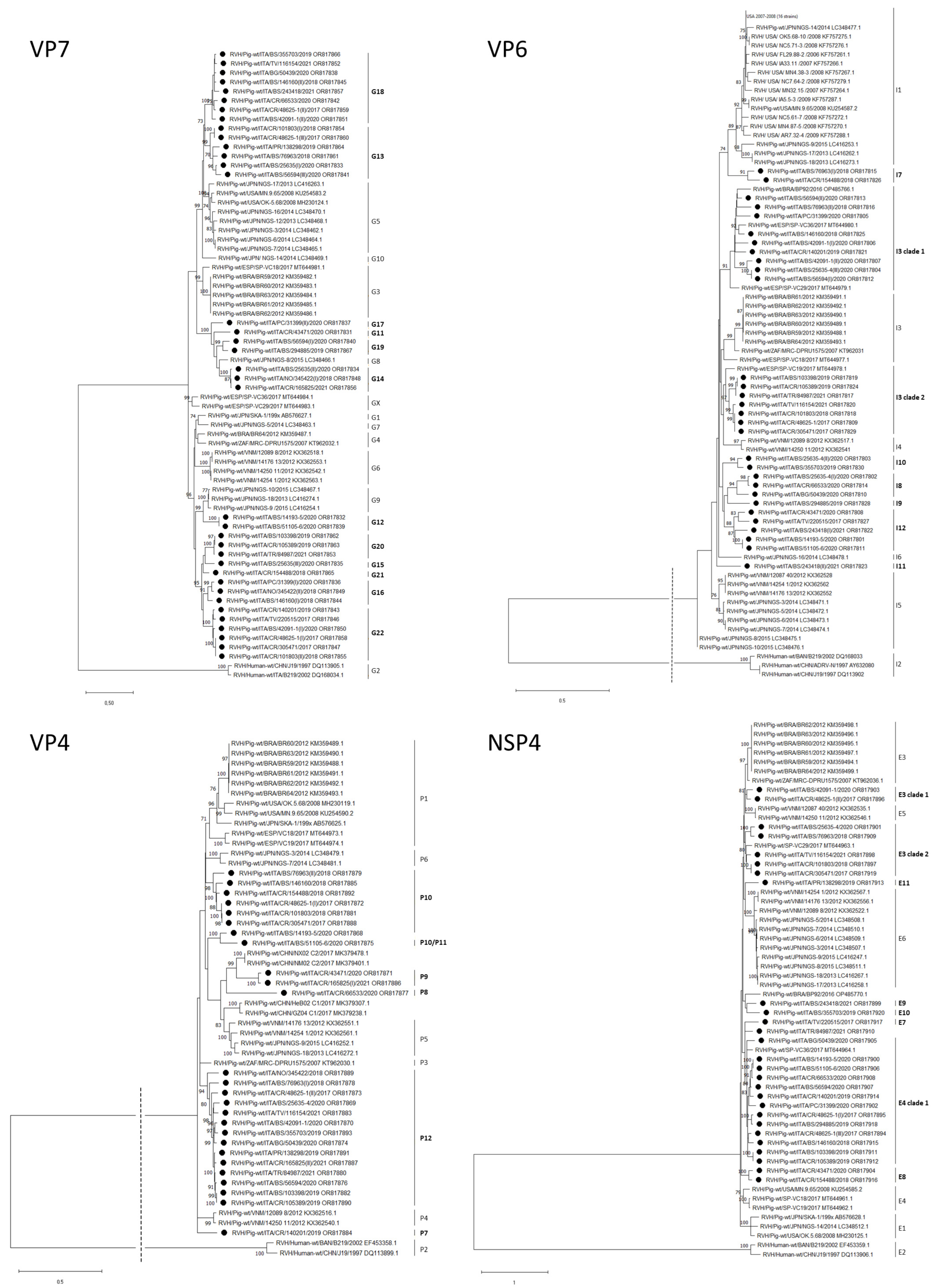
3.1. VP7
3.2. VP4
3.3. VP6
3.4. NSP4
4. Discussion
5. Conclusions
Supplementary Materials
Author Contributions
Funding
Institutional Review Board Statement
Informed Consent Statement
Data Availability Statement
Acknowledgments
Conflicts of Interest
References
- Pesavento, J.B.; Crawford, S.E.; Estes, M.K.; Prasad, B.V.V. Rotavirus Proteins: Structure and Assembly. Curr. Top. Microbiol. Immunol. 2006, 309, 189–219. [Google Scholar] [CrossRef] [PubMed]
- Desselberger, U. Rotaviruses. Virus Res. 2014, 190, 75–96. [Google Scholar] [CrossRef] [PubMed]
- Martella, V.; Ciarlet, M.; Camarda, A.; Pratelli, A.; Tempesta, M.; Greco, G.; Cavalli, A.; Elia, G.; Decaro, N.; Terio, V.; et al. Molecular Characterization of the VP4, VP6, VP7, and NSP4 Genes of Lapine Rotaviruses Identified in Italy: Emergence of a Novel VP4 Genotype. Virology 2003, 314, 358–370. [Google Scholar] [CrossRef] [PubMed]
- Lahon, A.; Chitambar, S.D. Molecular Characterization of VP4, VP6, VP7 and NSP4 Genes of Group B Rotavirus Strains from Outbreaks of Gastroenteritis. Asian Pac. J. Trop. Med. 2011, 4, 846–849. [Google Scholar] [CrossRef]
- Almeida, P.R.; Lorenzetti, E.; Cruz, R.S.; Watanabe, T.T.; Zlotowski, P.; Alfieri, A.A.; Driemeier, D. Diarrhea Caused by Rotavirus A, B, and C in Suckling Piglets from Southern Brazil: Molecular Detection and Histologic and Immunohistochemical Characterization. J. Vet. Diagn. Investig. 2018, 30, 370–376. [Google Scholar] [CrossRef]
- Marthaler, D.; Homwong, N.; Rossow, K.; Culhane, M.; Goyal, S.; Collins, J.; Matthijnssens, J.; Ciarlet, M. Rapid Detection and High Occurrence of Porcine Rotavirus A, B, and C by RT-QPCR in Diagnostic Samples. J. Virol. Methods 2014, 209, 30–34. [Google Scholar] [CrossRef]
- Ferrari, E.; Salogni, C.; Martella, V.; Alborali, G.L.; Scaburri, A.; Boniotti, M.B. Assessing the Epidemiology of Rotavirus A, B, C and H in Diarrheic Pigs of Different Ages in Northern Italy. Pathogens 2022, 11, 467. [Google Scholar] [CrossRef]
- Jiang, S.; Ji, S.; Tang, Q.; Cui, X.; Yang, H.; Kan, B.; Gao, S. Molecular Characterization of a Novel Adult Diarrhoea Rotavirus Strain J19 Isolated in China and Its Significance for the Evolution and Origin of Group B Rotaviruses. J. Gen. Virol. 2008, 89, 2622–2629. [Google Scholar] [CrossRef]
- Yang, H.; Chen, S.; Ji, S. A novel rotavirus causing large scale of adult diarrhea in Shi Jiazhuang. Zhonghua Liu Xing Bing Xue Za Zhi 1998, 19, 336–338. [Google Scholar]
- Matthijnssens, J.; Otto, P.H.; Ciarlet, M.; Desselberger, U.; Van Ranst, M.; Johne, R. VP6-Sequence-Based Cutoff Values as a Criterion for Rotavirus Species Demarcation. Arch. Virol. 2012, 157, 1177–1182. [Google Scholar] [CrossRef]
- Yang, H.; Makeyev, E.V.; Kang, Z.; Ji, S.; Bamford, D.H.; van Dijk, A.A. Cloning and Sequence Analysis of DsRNA Segments 5, 6 and 7 of a Novel Non-Group A, B, C Adult Rotavirus That Caused an Outbreak of Gastroenteritis in China. Virus Res. 2004, 106, 15–26. [Google Scholar] [CrossRef] [PubMed]
- Wakuda, M. Porcine Rotavirus Closely Related to Novel Group of Human Rotaviruses. Emerg. Infect. Dis. 2011, 17, 1491. [Google Scholar] [CrossRef] [PubMed]
- Marthaler, D.; Rossow, K.; Culhane, M.; Goyal, S.; Collins, J.; Matthijnssens, J.; Nelson, M.; Ciarlet, M. Widespread Rotavirus H in Commercially Raised Pigs, United States. Emerg. Infect. Dis. 2014, 20, 1203–1206. [Google Scholar] [CrossRef] [PubMed]
- Molinari, B.L.D.; Lorenzetti, E.; Otonel, R.A.A.; Alfieri, A.F.; Alfieri, A.A. Species H Rotavirus Detected in Piglets with Diarrhea, Brazil, 2012. Emerg. Infect. Dis. 2014, 20, 1019–1022. [Google Scholar] [CrossRef]
- Nyaga, M.M.; Peenze, I.; Potgieter, C.A.; Seheri, L.M.; Page, N.A.; Yinda, C.K.; Steele, A.D.; Matthijnssens, J.; Mphahlele, M.J. Complete Genome Analyses of the First Porcine Rotavirus Group H Identified from a South African Pig Does Not Provide Evidence for Recent Interspecies Transmission Events. Infect. Genet. Evol. 2016, 38, 1–7. [Google Scholar] [CrossRef] [PubMed]
- Phan, M.V.T.; Anh, P.H.; Cuong, N.V.; Munnink, B.B.O.; van der Hoek, L.; My, P.T.; Tri, T.N.; Bryant, J.E.; Baker, S.; Thwaites, G.; et al. Unbiased Whole-Genome Deep Sequencing of Human and Porcine Stool Samples Reveals Circulation of Multiple Groups of Rotaviruses and a Putative Zoonotic Infection. Virus Evol. 2016, 2, vew027. [Google Scholar] [CrossRef]
- Shi, K.; Zhou, H.; Feng, S.; He, J.; Li, B.; Long, F.; Shi, Y.; Yin, Y.; Li, Z. Development of a Quadruplex RT-QPCR for the Detection of Porcine Rotaviruses and the Phylogenetic Analysis of Porcine RVH in China. Pathogens 2023, 12, 1091. [Google Scholar] [CrossRef]
- Puente, H.; Cortey, M.; de Nova, P.J.G.; Mencía-Ares, Ó.; Gómez-García, M.; Díaz, I.; Arguello, H.; Martín, M.; Rubio, P.; Carvajal, A. First Identification and Characterization of Rotavirus H in Swine in Spain. Transbound. Emerg. Dis. 2021, 68, 3055–3069. [Google Scholar] [CrossRef]
- Krasnikov, N.; Yuzhakov, A. Interspecies Recombination in NSP3 Gene in the First Porcine Rotavirus H in Russia Identified Using Nanopore-Based Metagenomic Sequencing. Front. Vet. Sci. 2023, 10, 1302531. [Google Scholar] [CrossRef]
- Flores, P.S.; Costa, F.B.; Amorim, A.R.; Mendes, G.S.; Rojas, M.; Santos, N. Rotavirus A, C, and H in Brazilian Pigs: Potential for Zoonotic Transmission of RVA. J. Vet. Diagn. Investig. 2021, 33, 129–135. [Google Scholar] [CrossRef]
- Suzuki, T.; Inoue, D. Full Genome-Based Genotyping System for Rotavirus H and Detection of Potential Gene Recombination in Nonstructural Protein 3 between Porcine Rotavirus H and Rotavirus C. J. Gen. Virol. 2018, 99, 1582–1589. [Google Scholar] [CrossRef] [PubMed]
- Hull, J.J.A.; Qi, M.; Montmayeur, A.M.; Kumar, D.; Velasquez, D.E.; Moon, S.-S.; Magaña, L.C.; Betrapally, N.; Ng, T.F.F.; Jiang, B.; et al. Metagenomic Sequencing Generates the Whole Genomes of Porcine Rotavirus A, C, and H from the United States. PLoS ONE 2020, 15, e0244498. [Google Scholar] [CrossRef] [PubMed]
- Nyaga, M.M.; Jere, K.C.; Esona, M.D.; Seheri, M.L.; Stucker, K.M.; Halpin, R.A.; Akopov, A.; Stockwell, T.B.; Peenze, I.; Diop, A.; et al. Whole Genome Detection of Rotavirus Mixed Infections in Human, Porcine and Bovine Samples Co-Infected with Various Rotavirus Strains Collected from Sub-Saharan Africa. Infect. Genet. Evol. 2015, 31, 321–334. [Google Scholar] [CrossRef] [PubMed]
- Martella, V.; Bányai, K.; Lorusso, E.; Bellacicco, A.L.; Decaro, N.; Camero, M.; Bozzo, G.; Moschidou, P.; Arista, S.; Pezzotti, G.; et al. Prevalence of Group C Rotaviruses in Weaning and Post-Weaning Pigs with Enteritis. Vet. Microbiol. 2007, 123, 26–33. [Google Scholar] [CrossRef] [PubMed]
- Kim, Y.; Chang, K.O.; Straw, B.; Saif, L.J. Characterization of Group C Rotaviruses Associated with Diarrhea Outbreaks in Feeder Pigs. J. Clin. Microbiol. 1999, 37, 1484–1488. [Google Scholar] [CrossRef] [PubMed]
- Matthijnssens, J.; Ciarlet, M.; Heiman, E.; Arijs, I.; Delbeke, T.; McDonald, S.M.; Palombo, E.A.; Iturriza-Gómara, M.; Maes, P.; Patton, J.T.; et al. Full Genome-Based Classification of Rotaviruses Reveals a Common Origin between Human Wa-Like and Porcine Rotavirus Strains and Human DS-1-like and Bovine Rotavirus Strains. J. Virol. 2008, 82, 3204–3219. [Google Scholar] [CrossRef]
- Matthijnssens, J.; Ciarlet, M.; Rahman, M.; Attoui, H.; Bányai, K.; Estes, M.K.; Gentsch, J.R.; Iturriza-Gómara, M.; Kirkwood, C.D.; Martella, V.; et al. Recommendations for the Classification of Group A Rotaviruses Using All 11 Genomic RNA Segments. Arch. Virol. 2008, 153, 1621–1629. [Google Scholar] [CrossRef]
- Bankevich, A.; Nurk, S.; Antipov, D.; Gurevich, A.A.; Dvorkin, M.; Kulikov, A.S.; Lesin, V.M.; Nikolenko, S.I.; Pham, S.; Prjibelski, A.D.; et al. SPAdes: A New Genome Assembly Algorithm and Its Applications to Single-Cell Sequencing. J. Comput. Biol. 2012, 19, 455–477. [Google Scholar] [CrossRef]
- Hall, T.A. BioEdit: A User-Friendly Biological Sequence Alignment Editor and Analysis Program for Windows 95/98/NT. Nucleic Acids Symp. Ser. 1999, 41, 95–98. [Google Scholar]
- Tamura, K.; Stecher, G.; Kumar, S. MEGA11: Molecular Evolutionary Genetics Analysis Version 11. Mol. Biol. Evol. 2021, 38, 3022–3027. [Google Scholar] [CrossRef]
- Martin, D.P.; Murrell, B.; Golden, M.; Khoosal, A.; Muhire, B. RDP4: Detection and Analysis of Recombination Patterns in Virus Genomes. Virus Evol. 2015, 1, vev003. [Google Scholar] [CrossRef] [PubMed]


| Strain | Management of Herd | Age | RVA | RVB | RVC | RVH |
|---|---|---|---|---|---|---|
| RVH/Pig-wt/ITA/CR/48625-1/2017 | Farrow to weaning | weaning | + | + | + | + |
| RVH/Pig-wt/ITA/TV/220515/2017 | Farrow to weaning | post-weaning | + | + | + | + |
| RVH/Pig-wt/ITA/CR/305471/2017 | Farrow to weaning | fattening | + | + | + | + |
| RVH/Pig-wt/ITA/BS/76963/2018 | Farrow to weaning | fattening | + | + | + | + |
| RVH/Pig-wt/ITA/CR/101803/2018 | Farrow to weaning | weaning | + | + | + | + |
| RVH/Pig-wt/ITA/BS/146160/2018 | Farrow to weaning | weaning | + | + | + | + |
| RVH/Pig-wt/ITA/CR/154488/2018 | Farrow to weaning | fattening | + | + | + | + |
| RVH/Pig-wt/ITA/NO/345422/2018 | Farrow to weaning | fattening | + | + | + | + |
| RVH/Pig-wt/ITA/BS/103398/2019 | Farrow to weaning | weaning | + | + | + | + |
| RVH/Pig-wt/ITA/CR/105389/2019 | Farrow to weaning | suckling | + | + | + | + |
| RVH/Pig-wt/ITA/PR/138298/2019 | Weaning | post-weaning | + | + | + | + |
| RVH/Pig-wt/ITA/CR/140201/2019 | Farrow to weaning | fattening | + | + | + | + |
| RVH/Pig-wt/ITA/BS/294885/2019 | Weaning | fattening | + | + | + | + |
| RVH/Pig-wt/ITA/BS/355703/2019 | Farrow to weaning | fattening | + | + | + | |
| RVH/Pig-wt/ITA/BS/14193-5/2020 | Farrow to weaning | post-weaning | + | + | ||
| RVH/Pig-wt/ITA/BS/25635-4/2020 | Weaning | weaning | + | + | + | + |
| RVH/Pig-wt/ITA/PC/31399/2020 | Farrow to weaning | post-weaning | + | + | + | + |
| RVH/Pig-wt/ITA/BS/42091-1/2020 | Weaning | fattening | + | + | + | + |
| RVH/Pig-wt/ITA/CR/43471/2020 | Weaning | fattening | + | + | + | + |
| RVH/Pig-wt/ITA/BG/50439/2020 | Weaning | fattening | + | + | + | + |
| RVH/Pig-wt/ITA/BS/51105-6/2020 | Farrow to weaning | weaning | + | + | + | + |
| RVH/Pig-wt/ITA/BS/56594/2020 | Weaning | weaning | + | + | + | + |
| RVH/Pig-wt/ITA/CR/66533/2020 | Farrow to weaning | fattening | + | + | ||
| RVH/Pig-wt/ITA/TR/84987/2021 | Weaning | weaning | + | + | + | + |
| RVH/Pig-wt/ITA/TV/116154/2021 | Farrow to weaning | fattening | + | + | + | |
| RVH/Pig-wt/ITA/CR/165825/2021 | Farrow to weaning | post-weaning | + | + | ||
| RVH/Pig-wt/ITA/BS/243418/2021 | Farrow to weaning | weaning | + | + |
| Gene Target | Primer | Sequence 5′-3′ | Position |
|---|---|---|---|
| VP7 | PoRVH_VP7 F1 | GCC ATG TTG TTC CTA CTA AYC | 12–32 |
| PoRVH_VP7 R2 | GAT GTA AYG GAT TTC TCR ACG TT | 781–803 | |
| VP4 | PoRVH_VP4 F1 | CAA GAR AAA TTR GAT CGY GAG | 106–126 |
| PoRVH_VP4 R1 | TGA CCA CAY TTY CTT GTT GG | 1051–1070 | |
| PoRVH_VP4 F2 | TGG ATG ATT GAY TCA GGA TTT AA | 955–977 | |
| PoRVH_VP4 R3 | GAC ATT ATG CCT GAA GTY AGA TC | 1711–1733 | |
| PoRVH_VP4 F4 | GCA ATT GTT CCA GCT GAT GC | 2089–2108 | |
| PoRVH_VP4 R4 | CTA CAG CAT ATT CTG CAA GAT G | 2227–2248 | |
| PoRVH_VP4 R2 | ACT AAT GYC ACT ACR GTC TAT G | 2498–2519 | |
| VP6 | PoRVH_VP6_F1 | TAC AAG TGA CCC ACA AGG ATG | 14–34 |
| PoRVH_VP6_R1 | ACA GGT ATR TTA TTT GGA GGC T | 732–753 | |
| PoRVH_VP6_F2 | AGT ACC ATG TTC AGG AGT AAT G | 658–679 | |
| PoRVH_VP6_R3 | GTT GCA ACA ATY CTT CCA CC | 821–837 | |
| NSP4 | PoRVH_NSP4 F1 | ATC AAA GTM ACG ATG GAG CAC | 1–21 |
| PoRVH_NSP4 R2 | TTG CGC AAG GGT GRA CAC T | 704–722 |
| Strains | Region | Farm | Genotype (N) | ||||||||||||||
|---|---|---|---|---|---|---|---|---|---|---|---|---|---|---|---|---|---|
| VP4 | VP7 | VP6 | NSP4 | VP4 (N = 6) | VP7 (N = 12) | VP6 (N = 8) | NSP4 (N = 8) | ||||||||||
| RVH/Pig-wt/ITA/TV/220515/2017 | Veneto | 1 | - | 1 | 1 | 1 | - | G22 | I12 | E7 | |||||||
| RVH/Pig-wt/ITA/CR/305471/2017 | Lombardia | 2 | 1 | 1 | 1 | 1 | P10 | G22 | I3 clade 2 | E3 clade 2 | |||||||
| RVH/Pig-wt/ITA/CR/48625-1/2017 | Lombardia | 2 | 2 | 3 | 1 | 3 | P12 | P10 | G13 | G18 | G22 | I3 clade 2 | E3 clade 1 | E4 clade 1 | E4 clade 1 | ||
| RVH/Pig-wt/ITA/BS/76963/2018 | Lombardia | 3 | 2 | 1 | 2 | 1 | P12 | P10 | G13 | I7 | I3 clade 1 | E3 clade 2 | |||||
| RVH/Pig-wt/ITA/CR/101803/2018 | Lombardia | 4 | 1 | 2 | 1 | 1 | P10 | G13 | G22 | I3 clade 2 | E3 clade 2 | ||||||
| RVH/Pig-wt/ITA/CR/105389/2019 | Lombardia | 4 | 1 | 1 | 1 | 1 | P12 | G20 | I3 clade 2 | E4 clade 1 | |||||||
| RVH/Pig-wt/ITA/PR/138298/2019 | Emilia Romagna | 5 | 1 | 1 | - | 1 | P12 | G13 | - | E11 | |||||||
| RVH/Pig-wt/ITA/BS/146160/2018 | Lombardia | 6 | 1 | 2 | 1 | 1 | P10 | G16 | G18 | I3 clade 1 | E4 clade 1 | ||||||
| RVH/Pig-wt/ITA/CR/154488/2018 | Lombardia | 7 | 1 | 1 | 1 | 1 | P10 | G21 | I7 | E8 | |||||||
| RVH/Pig-wt/ITA/NO/345422/2018 | Piemonte | 8 | 1 | 2 | - | - | P12 | G14 | G16 | - | - | ||||||
| RVH/Pig-wt/ITA/BS/103398/2019 | Lombardia | 9 | 1 | 1 | 1 | 1 | P12 | G20 | I3 clade 2 | E4 clade 1 | |||||||
| RVH/Pig-wt/ITA/CR/140201/2019 | Lombardia | 10 | 1 | 1 | 1 | 1 | P7 | G22 | I3 clade 1 | E4 clade 1 | |||||||
| RVH/Pig-wt/ITA/BS/294885/2019 | Lombardia | 11 | - | 1 | 1 | 1 | - | G19 | I9 | E4 clade 1 | |||||||
| RVH/Pig-wt/ITA/BS/14193-5/2020 | Lombardia | 12 | 1 | 1 | 1 | 1 | P10 | G12 | I12 | E4 clade 1 | |||||||
| RVH/Pig-wt/ITA/BS/51105-6/2020 | Lombardia | 12 | 1 | 1 | 1 | 1 | P10/11 | G12 | I12 | E4 clade 1 | |||||||
| RVH/Pig-wt/ITA/BS/25635-4/2020 | Lombardia | 13 | 1 | 3 | 3 | 1 | P12 | G13 | G14 | G15 | I8 | I10 | I3 clade 1 | E3 clade 2 | |||
| RVH/Pig-wt/ITA/BS/56594/2020 | Lombardia | 13 | 1 | 2 | 2 | 1 | P12 | G13 | G19 | I3 clade 1 | I3 clade 1 | E4 clade 1 | |||||
| RVH/Pig-wt/ITA/PC/31399/2020 | Emilia Romagna | 14 | - | 2 | 1 | 1 | - | G16 | G17 | I3 clade 1 | E4 clade 1 | ||||||
| RVH/Pig-wt/ITA/CR/43471/2020 | Lombardia | 15 | 1 | 1 | 1 | 1 | P9 | G11 | I12 | E8 | |||||||
| RVH/Pig-wt/ITA/BG/50439/2020 | Lombardia | 16 | 1 | 1 | 1 | 1 | P12 | G18 | I8 | E4 clade 1 | |||||||
| RVH/Pig-wt/ITA/CR/66533/2020 | Lombardia | 17 | 1 | 1 | 1 | 1 | P8 | G18 | I8 | E4 clade 1 | |||||||
| RVH/Pig-wt/ITA/BS/355703/2019 | Lombardia | 18 | 1 | 1 | 1 | 1 | P12 | G18 | I10 | E10 | |||||||
| RVH/Pig-wt/ITA/BS/42091-1/2020 | Lombardia | 19 | 1 | 2 | 2 | 1 | P12 | G18 | G22 | I3 clade 1 | I3 clade 1 | E3 clade 1 | |||||
| RVH/Pig-wt/ITA/TR/84987/2021 | Umbria | 20 | 1 | 1 | 1 | 1 | P12 | G20 | I3 clade 2 | ND | |||||||
| RVH/Pig-wt/ITA/TV/116154/2021 | Veneto | 21 | 1 | 1 | 1 | 1 | P12 | G18 | I3 clade 2 | E3 clade 2 | |||||||
| RVH/Pig-wt/ITA/CR/165825/2021 | Lombardia | 22 | 2 | 1 | - | - | P12 | P9 | G14 | - | - | ||||||
| RVH/Pig-wt/ITA/BS/243418/2021 | Lombardia | 23 | - | 1 | 2 | 1 | - | G18 | I12 | I11 | E9 | ||||||
| Strain | Gene | Recombination Breakpoints (bp) | Major Parent | Minor Parent | Detection Method | p-Value |
|---|---|---|---|---|---|---|
| ITA/BS/14193-5/2020 | VP4 | 945 | ITA/BS/51105-6/2020 | ITA154488/2018 | RDP | 8.48 × 10³ |
| ITA/48625-1(I)/2017 | GENECONV | 1.88 × 10⁻⁵ | ||||
| ITA/101803/2018 | Bootscan | 1.32 × 10⁻⁴ | ||||
| ITA/305471/2017 | Maxchi | 1.51 × 10⁻⁶ | ||||
| Chimaera | 3.46 × 10⁻⁹ | |||||
| SiSscan | 6.18 × 10⁻¹¹ | |||||
| 3 Seq | 1.21 × 10−36 | |||||
| ITA/BS/51105-6/2020 | VP4 | 945 | ITA/BS/14193-5/2020 | unknown | RDP | 8.48 × 10³ |
| GENECONV | 1.88 × 10⁻⁵ | |||||
| Bootscan | 4.20 × 10⁻⁴ | |||||
| Maxchi | 3.51 × 10⁻² | |||||
| Chimaera | 4.18 × 10⁻⁴ | |||||
| SiSscan | 6.18 × 10⁻¹¹ | |||||
| 3 Seq | 1.21 × 10−36 |
Disclaimer/Publisher’s Note: The statements, opinions and data contained in all publications are solely those of the individual author(s) and contributor(s) and not of MDPI and/or the editor(s). MDPI and/or the editor(s) disclaim responsibility for any injury to people or property resulting from any ideas, methods, instructions or products referred to in the content. |
© 2023 by the authors. Licensee MDPI, Basel, Switzerland. This article is an open access article distributed under the terms and conditions of the Creative Commons Attribution (CC BY) license (https://creativecommons.org/licenses/by/4.0/).
Share and Cite
Ferrari, E.; Vignola, G.; Bertasio, C.; Chiapponi, C.; Alborali, G.L.; Martella, V.; Boniotti, M.B. Identification of Putative Novel Rotavirus H VP7, VP4, VP6 and NSP4 Genotypes in Pigs. Viruses 2024, 16, 68. https://doi.org/10.3390/v16010068
Ferrari E, Vignola G, Bertasio C, Chiapponi C, Alborali GL, Martella V, Boniotti MB. Identification of Putative Novel Rotavirus H VP7, VP4, VP6 and NSP4 Genotypes in Pigs. Viruses. 2024; 16(1):68. https://doi.org/10.3390/v16010068
Chicago/Turabian StyleFerrari, Elena, Greta Vignola, Cristina Bertasio, Chiara Chiapponi, Giovanni Loris Alborali, Vito Martella, and Maria Beatrice Boniotti. 2024. "Identification of Putative Novel Rotavirus H VP7, VP4, VP6 and NSP4 Genotypes in Pigs" Viruses 16, no. 1: 68. https://doi.org/10.3390/v16010068