Article

Treatability Changes of Radiata Pine Heartwood Induced by White-Rot Fungus Trametes versicolor

Research Institute of Wood Industry, Chinese Academy of Forestry, Beijing 100091, China
*
Author to whom correspondence should be addressed.
Forests 2023, 14(5), 1040; https://doi.org/10.3390/f14051040
Submission received: 21 April 2023 / Revised: 16 May 2023 / Accepted: 17 May 2023 / Published: 18 May 2023
(This article belongs to the Special Issue Wood Treatments and Modification Technologies)

Abstract

:
Desired retention and depth into wood are necessary for wood preservatives to provide long-term durability. In general, heartwood of wood is difficult to treat, and bioincising was investigated as a potential technique to improve the treatability of refractory wood and heartwood. In order to study the effects of bioincising treatment with white-rot fungus Trametes versicolor on the pore structure and treatability of radiata pine heartwood, this research conducted tests of mass loss, microscopic structures, pore structure parameters, uptake, and penetration of preservative of radiata pine heartwood specimens incubated by T. versicolor for 4, 8, and 12 weeks. The results showed that the optimal inoculation time of T. versicolor bioincising on radiata pine heartwood was 4 to 8 weeks. At this time, the retention of injected preservatives increased by 5.01%–17.73%, the penetration depth of preservatives increased significantly, and the corresponding mass loss was 3.04%–6.45%. The results of microstructure and pore structure showed that T. versicolor entered the adjacent tracheids via apertures, with less impact on the cell wall, mainly degrading pit membranes and ray parenchyma cells early in the inoculation of radiata pine heartwood. As the structures impeding fluid flow were connected, the porosity of the wood and the range of the main pore size distribution increased significantly, thus increasing the treatability of radiata pine heartwood.

1. Introduction

As a natural and renewable material, wood is widely used in building and construction due to its soft touch, beautiful texture, and high strength-to-weight ratio [1,2,3]. However, wood is susceptible to attack by fungi and insects, especially when used in outdoor conditions [4]. Chemical treatment is one of the most common protections to reduce wood deterioration. Studies have shown that wood has good resistance to biological decay after proper preservative treatment [5,6,7]. Generally, it is desirable to achieve a significant degree of penetration in order to provide long-term durability [8,9]. Although wood is porous, many wood species are not easy to treat [10]. Many commercial wood species are difficult to treat with wood preservatives due to a thin sapwood band and refractory heartwood. The difficulty for these species to meet international standards limits their use in an environment with a high risk of a biological attack, such as ground-contact applications and areas with severe termite hazards [11,12]. Therefore, improving the treatability of wood is important for the efficient use of wood.
The treatability depends on the interconnectivity of the cavity system in the wood [13,14]. Pit membranes are interconnecting pathways between adjacent tracheids which are considered to be the most important influence on liquid permeability [15]. The flow of chemicals is enabled and controlled by the size, structure, and amount of pits [14]. Related studies have shown that resistance to flow in coniferous wood exists entirely in the pit membrane [16]. In coniferous woods, bordered pits in the tracheid become aspirated during wood drying [17,18]. The torus is pressed against the porus due to the high capillary tension forces, blocking the pit aperture and preventing the flow of fluids, which even makes the sapwood of some wood species untreatable. Hence, aspiration of the bordered pits significantly reduces the treatability of the wood for fluids. This same aspiration process occurs during heartwood formation [13], so the heartwood of most wood species is usually more difficult to treat than sapwood [19,20]. Additionally, less treatability of coniferous heartwood may also be due to the deposition of extractives in the pit membrane [20,21,22].
To overcome the problem related to the low treatability of refractory wood, many methods have been attempted over the past 100 years, and some of these methods, such as mechanical incising and steaming, have been used in the industry [17]. However, the use of them is not always desired due to operating costs and qualities of treated wood [17,23]. The biological method is a potential technique to improve wood treatability by the application of enzymes [24,25,26], bacteria [27,28,29], and fungi [30,31,32,33,34,35,36,37,38,39,40,41,42,43]. An increase in wood permeability is supposedly induced by the selective degradation of pit membranes in the bordered and half-bordered pits, entailing only negligible changes in the tracheid cell wall [33,38]. Bacteria, enzymes, and fungi have all been studied for their ability to improve permeability without having excessively negative effects on other wood properties. Of these, the bioincised treatment by fungi may be the most successful, as fungi can quickly colonize wood and transport their specific enzymes deep into the wood substrate in a short time [11,44]. Earlier work using fungal pre-treatment has shown promising results. Picea abies inoculated with a white-rot fungus Physisporinus vitreus for six weeks showed a significant improvement in permeability and only a slight decrease in bending strength [32]. Bioincising with P. vitreus has also shown promise in the heartwood of Norway spruce [35], Douglas fir compression wood [40], and Oriental spruce heartwood [43]. Dichomitus squalens, a white-rot basidiomycete, was used to successfully improve permeability and penetration in Norway spruce heartwood [30] and spruce lumber [11]. The white-rot fungus Hypoxylon fragiforme was used to successfully improve the permeability of Norway spruce with no negative effect on compression strength after 14-day incubation [38]. Populus davidiana bioincised by white-rot Trametes versicolor for 17 days had significantly higher durability by increasing the uptake and penetration of wood preservatives with little effect on bending strength [41]. Thus, fungal pre-treatment is a promising approach to improve the treatability of wood without excessive effects on other properties of the wood.
The application of fungi to improve the treatability of refractory wood in bioincising is still on a laboratory scale [17]. The fungus type, incubation time, and wood species appear as important factors for bioincising and need to be further explored and studied. Radiata pine is one of the most imported wood species in China in recent years and is widely used in construction and home decoration. However, radiata pine heartwood has low decay resistance and is difficult to treat, which will largely limit its efficient utilization. Hernandez has used albino strains of Ophiostoma fungi to successfully increase the permeability of radiata pine [37]. The white-rot fungus, T. versicolor grows rapidly and has also shown promising results in biological treatment [41]. In order to investigate and study the effect of bioincising treatment with white-rot fungus T. versicolor on the pore structure and treatability of radiata pine heartwood, this study used T. versicolor to inoculate radiata pine heartwood at 25 °C and 85% relative humidity for 4, 8, and 12 weeks. Afterward, mass loss, microscopic structure, pore structure parameters, uptake, and penetration of preservatives were determined to investigate the effects of bioincising treatment with T. versicolor on the heartwood of radiata pine.

2. Materials and Methods

2.1. Materials

The radiata pine was native to New Zealand and was purchased from the timber market in Shanghai. Radial pine heartwood free of bugs, knots, discoloration, and other defects was selected and sawn into specimens with dimensions of 10 mm (L) × 20 mm (T) × 20 mm (R) for inoculation with white-rot fungus. Referring to GB/T 27654-2011 standard [45], a mass fraction of 0.2% copper azole wood preservative (CuAz-4) was prepared for the impregnation treatment of the specimens. The white-rot fungus T. versicolor was selected to inoculate the heartwood of radiata pine, and the strain was obtained from the Research Institute of Wood Industry, Chinese Academy of Forestry. The white-rot fungus T. versicolor was grown and maintained on Potato Dextrose Agar Medium (PDA) at 28 °C until inoculation.

2.2. Fungal Pre-Treatment

Naturally air-dried specimens of radiata pine heartwood were equilibrated in a constant temperature and humidity chamber (25 °C, 85% relative humidity) for 7 days, and all specimens were then placed in an oven at 60 °C for 8 h until a constant weight was reached prior to incubation. Afterward, steam-sterilized (30 min, 121 °C, 1.5 bar) heartwood of radiata pine specimens was exposed to white-rot fungus T. versicolor according to GB/T 13942.1-2009 standard [46]. The specimens were placed on a mixed medium with river sand and sawdust previously inoculated with T. versicolor in 250 mL experimental jars. Incubation of the specimens to T. versicolor took place in the growth chamber at 25 °C and 85% relative humidity for 4, 8, and 12 weeks. After incubation, the specimens were removed, cleaned of mycelium with a scalpel, and placed in an oven at 60 °C for 8 h until they reached a constant weight. The mass loss was calculated using the constant weight at 60 °C loss of each specimen before and after incubation with white-rot fungus T. versicolor. The control specimens were treated the same way as the inoculated specimens except for the fungal exposure. The average mass loss of radiata pine heartwood specimens at different incubation times was calculated with six replicates per group.
The mass loss ratio is calculated as follows:
L = (M0 − M1)/M0 × 100%
where L is the mass loss rate of the specimen (%); M0 is the constant mass of the specimen before incubation with T. versicolor (g); M1 is the constant mass of the specimen after incubation (g).

2.3. Uptake and Penetration of Preservative

The retention and cross-sectional penetration of wood preservatives were used as indicators for evaluating wood treatability. The specimens treated with different incubation times were weighed after air drying for a week. The weighed specimens were then placed in a large 2-L beaker; about 1 L of wood preservative was poured into the beaker, and then the specimens were pressed into the agent with an iron block. Subsequently, the beaker was placed in a vacuum-drying oven and subjected to a vacuum impregnation process at −0.09 MPa for 10 min, followed by a return to atmospheric pressure for another 10 min. After vacuum impregnation treatment, the specimens were removed, the preservative on the surface of the specimens with filter paper was wiped off, and then the specimens were weighed again to calculate the retention of the preservative injected into the specimens. Each group had six replicates, and the results were averaged.
The retention of injected preservative is calculated as follows:
R = (M3 − M2) × C/V × 103
where R is the retention of injected preservative of the specimen (kg/m3); M2 is the mass of the specimen before vacuum impregnation treatment (g); M3 is the mass of the specimen after vacuum impregnation treatment (g); C is the active ingredient concentration in preservatives (%); V is the volume of the specimen before vacuum impregnation treatment (cm3).
The vacuum-impregnated specimens were naturally air-dried for a week and then cut along the axial direction from the middle. Then, copper ion chromogenic agent was sprayed on the cross-section of the specimens to observe the penetration depth and area of the wood preservative.

2.4. Scanning Electron Microscopy (SEM) Observation

Specimens from different inoculation times were sliced to obtain approximately 40 µm thick tangential and cross sections. The microstructures of the radiata pine heartwood were then observed using scanning electron microscopy (S-4800, Hitachi, Tokyo, Japan) mainly for pits, ray parenchyma cells, and longitudinal tracheids.

2.5. Pore Structure

Specimens of radiata pine heartwood at 0, 4, and 8 weeks of fungal inoculation were selected and then made into small specimen blocks of 10 mm (L) × 5 mm (T) × 5 mm (R). The pore structure parameters of the small specimen blocks were quantified using a Micromeritics AutoPore IV9500 mercury piezometer. According to the measuring principle of the mercury piezometer, the mercury is pressed into the wood by a certain pressure, which increases faster at the beginning and at a decreasing rate as the pressure increases. The volume of mercury pressed into the wood is equal to the volume of pores in the wood. The distribution of pore size is obtained according to the Washburn equation [47].
The Washburn equation is as follows:
r = −γcosθ/P
where r is the pore diameter (μm); P is the pressurized pressure (MPa); γ is the surface of mercury tension (0.485 N/m, ambient temperature 20 °C); θ is the angle of wetting of mercury with wood (141°)

3. Results and Discussion

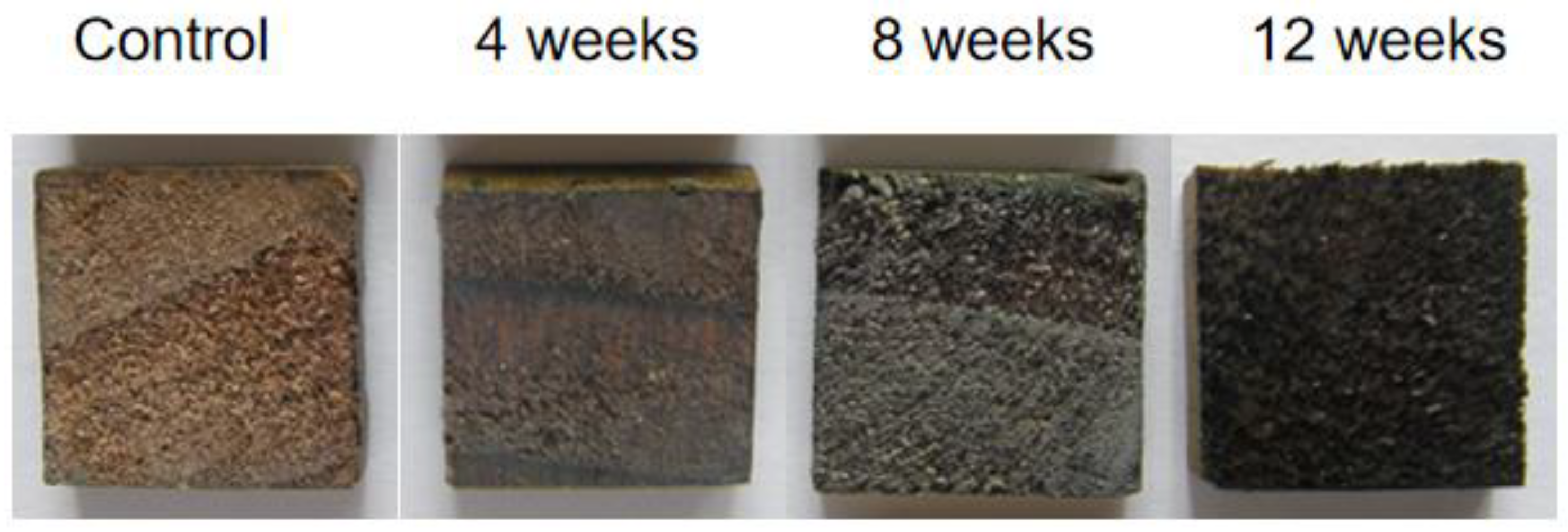
3.1. Mass Loss and Treatability

The mass loss and preservative retention of radiation pine heartwood at different incubation time periods are shown in Table 1, and the penetration area and depth of the preservative in cross-section are shown in Figure 1.
As can be seen from Table 1 and Figure 1, the bioincising treatment with T. versicolor achieved higher uptake and deeper penetration depth of wood preservatives. Moreover, the treatability of radiata pine heartwood increased with increasing inoculation time. The results of the preservative retention showed that the retention of radiata pine heartwood increased by 5.01%, 12.72%, and 2.82% during the 0–4, 4–8, and 8–12 week inoculation time periods, respectively. The greatest increase rate occurred between 4 and 8 weeks, followed by 0–4 weeks, and only a small increase in the 8–12 weeks. The mass loss results showed that the mass loss of radiata pine heartwood was 3.04%, 3.41%, and 12.3% in the 0 to 4, 4 to 8, and 8 to 12 week inoculation time periods, respectively. Of these, a small increase in mass loss was observed at 0–4 and 4–8 weeks and a large increase at 8–12 weeks. Studies have shown that there was a strong correlation between the degree of degradation of the cell structure and mass loss when using P. vitreus to inoculate oriental spruce wood. Extensive degradation of bordered pits was observed at a mass loss of 5%–10% in oriental spruce wood. At a mass loss of 10%–15%, the cell walls were strongly degraded [42]. It was assumed that the first eight weeks was the period when the white-rot fungus T. versicolor invaded and colonized the heartwood of radiata pine, with a gradual increase in mass loss as the incubation time increased. The mass loss of radiata pine heartwood reached 6.45% after 8-week inoculation, at which point structures such as bordered pits that blocked fluid flow were extensively degraded, with a consequent increase in treatability, and the greatest increase occurred between 4 and 8 weeks. During the 8–12 week incubation time period, the mass loss rose rapidly with increasing incubation time. Mass loss reached 18.75% after 12 weeks of incubation time, at which point the cell walls were severely degraded. When treated by vacuum impregnation, radiata pine heartwood cell walls possibly collapsed due to overfilling with wood preservatives [48]. As a result, the retention increase rate of injected preservatives showed a decrease during the 8–12 week incubation time period.

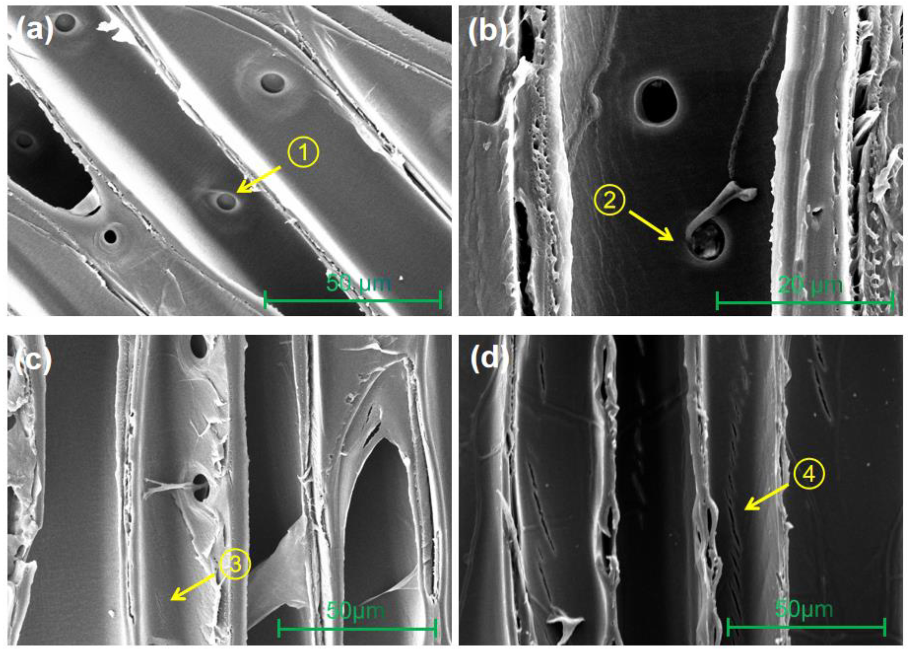
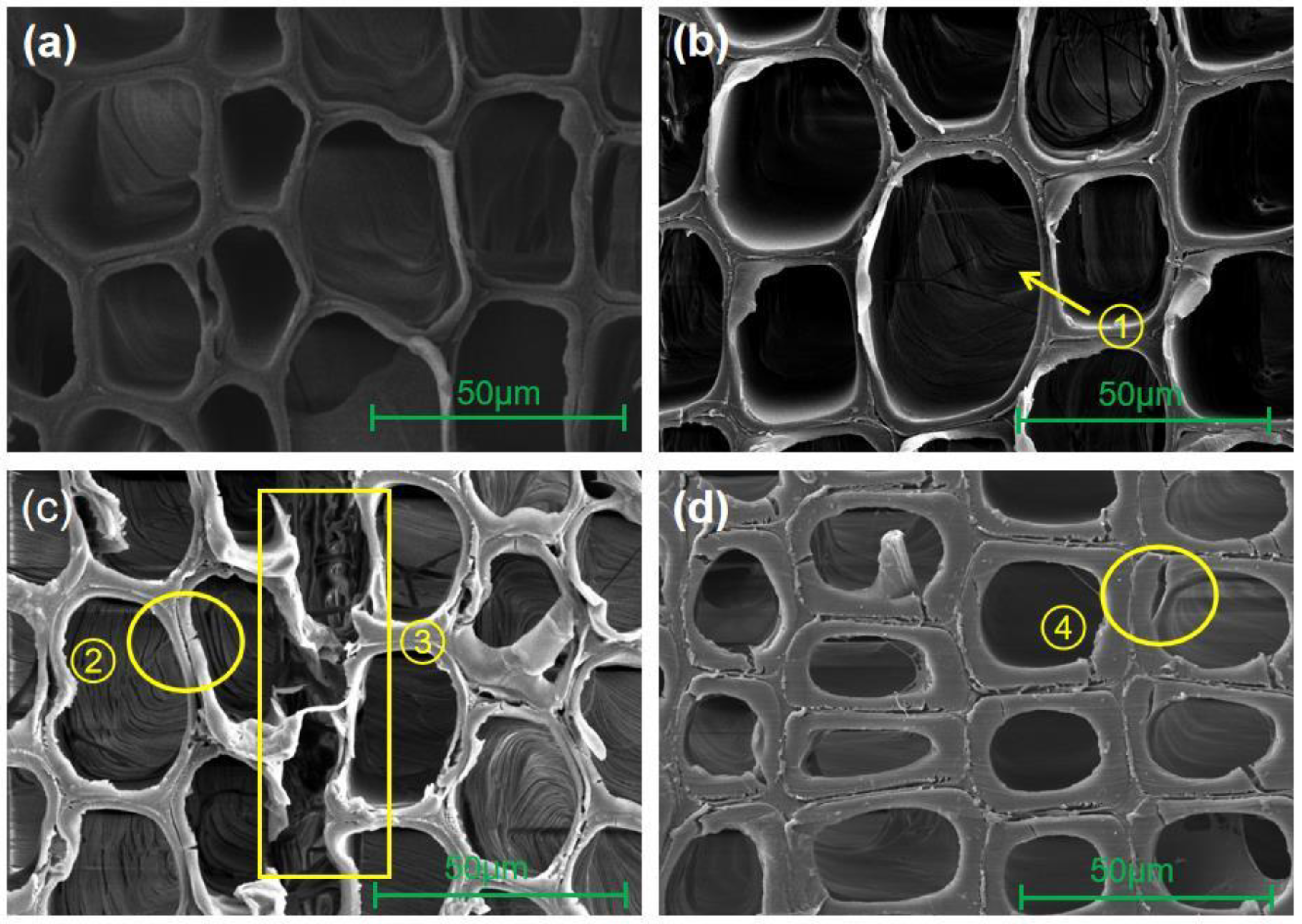
3.2. Microscopic Structure
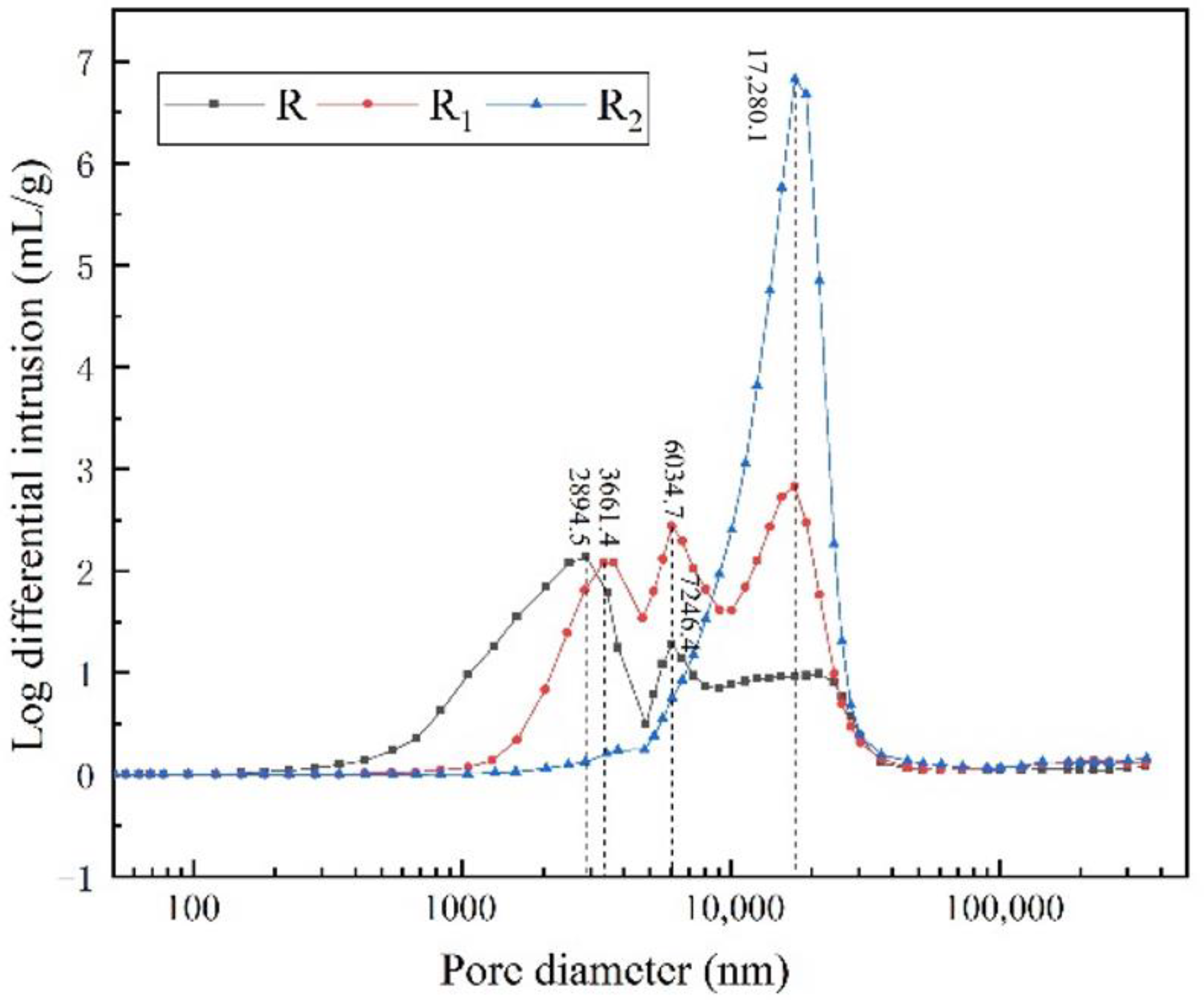
SEM images of the tangential section and cross section at different incubation times are shown in Figure 2 and Figure 3.
As shown in Figure 2a,b, the tracheid cell walls of the control sample were neatly arranged and well preserved at the cross-section, and bordered pits between tracheids became aspirated at the tangential section. Bordered pits are only channels for the flow of liquid between adjacent tracheids, so “pit aspiration” greatly reduced the treatability of the wood. After inoculation with white-rot fungus T. versicolor, the degradation of the cellular structure of radiata pine heartwood became more and more severe as the inoculation time increased (Figure 2 and Figure 3). When exposed to T. versicolor for four weeks, the tracheid cell walls remained intact, with hyphae entering the adjacent tracheids via apertures (Figure 2b and Figure 3b). At this point, the mass loss of radiata pine heartwood was only 3.04%, indicating that T. versicolor was in the early stage of cultivation and that damage to the wood was limited, mainly degrading bordered pits. When exposed to T. versicolor for eight weeks, ray parenchyma cells were severely degraded, and tiny cracks appeared in the tracheid cell walls (Figure 2c and Figure 3c). At this point, the mass loss of radiata pine heartwood reached 6.45%, indicating that T. versicolor had gradually completed initial colonization and affected the cell wall structure to some extent. When exposed to T. versicolor for 12 weeks, a significant change in cell morphology was observed in the cross-section, with many visible cracks in tracheid cell walls, and in the tangential section, the cracks were observed throughout the tracheid cell wall axially (Figure 2d and Figure 3d). During this time period, the mass loss rose rapidly to 18.75%, and the cell walls were strongly degraded. Thus, the white-rot fungus T. versicolor degraded mainly bordered pits and ray parenchyma cells of radiata pine heartwood in the early stages of inoculation, with relatively little effect on tracheid cell walls. However, as the inoculation time increased, the mass loss rose rapidly, and tracheid cell walls were strongly degraded.
The cell wall is the actual load-bearing structure of the wood and has an extremely important influence on the macro-mechanical properties of the material. As inoculation time increased, although the treatability of the wood increased, the tracheid cell walls were subjected to increasingly severe degradation due to its rising mass loss, resulting in loss of mechanical properties. Therefore, the fungal inoculation time (controlled mass loss) is critical for bioincising. The results of microstructure, mass loss, and retention of injected preservatives showed that the maximum increase in retention was between four and eight weeks. After a four-week inoculation time, T. versicolor had no significant effect on the cell wall. After an eight-week inoculation time, tiny cracks appeared in the tracheid cell wall. The mass loss of radiata pine heartwood ranged from 3.04% to 6.45% after four to eight weeks of inoculation. The relevant literature shows that the mechanical property of wood is not significantly decreased within 10% mass loss in bioincising [42]. Therefore, the optimum time for inoculation of radiata pine heartwood by the white-rot fungus T. versicolor was between 4 and 8 weeks, corresponding to a mass loss of between 3.04% and 6.45%.

3.3. Pore Structure

The pore structure parameters of control and inoculated wood by T. versicolor for four and eight weeks are shown in Table 2. The cumulative mercury intrusion into the heartwood of radiata pine increased with increasing inoculation time. The cumulative mercury intrusion was 0.339 mL/g and 0.472 mL/g higher than the control after four and eight-week inoculation time, respectively. This was reflected in the porosity, which increased by 8.2% and 11.5% in radiata pine heartwood after four and eight-week inoculation, respectively. In combination with microstructural analysis, the degradative enzymes secreted by T. versicolor destroyed the structures of radiata pine heartwood, such as bordered pits and rays, increasing the internal porosity of the wood and causing an increase in cumulative mercury intrusion. Meanwhile, due to the invasion of the fungus, some of the small pore size pores inside the wood were connected, resulting in a decrease in the specific surface area of the wood (3.585 m2/g > 1.690 m2/g > 0.772 m2/g) and an increase in median pore diameter (16,041.8 nm > 7793.9 nm > 3162.9 nm). The median pore diameter of the inoculated wood was much higher than that of the control sample, indicating that most of the pores in the inoculated wood were concentrated in the relatively large pore diameter area compared to the control.
Figure 4 shows the pore diameter versus log-differential intrusion for the control and inoculated radiata pine heartwood, reflecting the rate of change in pore volume for the material at the corresponding pore diameter. For the control sample, the maximum increase in pore volume corresponds to a pore diameter of 2894.5 nm, i.e., the highest number of micropores at this pore diameter. In addition, the number of pores around the pore diameter of 6034.7 nm was also relatively high. A significant increase in the range of major pore diameter distribution occurred in radiata pine heartwood after inoculation with white-rot fungi T. versicolor. After specimens were inoculated for four weeks, the highest number of pores was found around pore diameter 17,280.1 nm, followed by pores around 6034.7 nm and 3661.4 nm. After specimens were inoculated for eight weeks, the pores were mainly concentrated around the pore diameter of 17,280.1 nm. The inoculated wood showed a significant increase in the number of pores of relatively large pore diameter and a decrease in the number of pores of relatively small pore diameter. In the pore diameter range > 3661.4 nm, the number of pores of the four-week inoculated wood was higher than that of the control, and 0.906 mL/g more mercury was injected than the control, accounting for 38.6% of the total pore intake; in the pore diameter range > 7246.4 nm, the number of pores of the eight-week inoculated wood was higher than that of the control, and 1.676 mL/g more mercury was injected than the control, accounting for 67.6% of the total intake. In general, the pore diameter is inversely proportional to the capillary pressure that needs to be overcome during fluid penetration. When radiata pine heartwood was inoculated with T. versicolor, structures that blocked fluid flow were connected, and the range of distribution of the main pore diameters was significantly increased, thus increasing the efficiency of fluid penetration.

4. Conclusions

Radial pine heartwood could be treated with white-rot fungus T. versicolor to improve treatability, and the optimum time for inoculation was between four and eight weeks. At this time, the retention of injected preservatives increased by 5.01%–17.73%, the penetration depth of preservatives increased significantly, and the corresponding mass loss was 3.04%–6.45%. Microstructural and pore structure results revealed that early in the inoculation of radiata pine heartwood, T. versicolor entered the adjacent tracheids via apertures, with less impact on the cell wall, mainly degrading pit membranes, and ray parenchyma cells. As the structures blocking fluid flow were connected, the porosity of the wood and the range of the main pore size distribution increased significantly, thus increasing the treatability of radiata pine heartwood.
From the findings of this study, the following aspect can be considered for further research: the mechanical properties and feasibility of biological incising on an industrial scale or on full-size lumber needs further study.

Author Contributions

Conceptualization, X.F., J.L. and X.M.; methodology, X.F., J.L. and X.M.; investigation, X.F. and X.M.; data curation, X.F. and X.M.; writing—original draft preparation, X.F.; writing—review and editing, Y.X. and X.M.; supervision, Y.X. and X.M.; funding acquisition, X.M. All authors have read and agreed to the published version of the manuscript.

Funding

This research was supported financially by the National Natural Science Foundation of China (31971588) and the Fundamental Research Funds of the Chinese Academy of Forestry (CAFYBB2019ZB008).

Data Availability Statement

Data are available upon request to the corresponding author.

Acknowledgments

The authors thank the Research Institute of Wood Industry, Chinese Academy of Forestry for the equipment support.

Conflicts of Interest

The authors declare no conflict of interest.

References

  1. Mattos, B.D.; Lourençon, T.V.; Serrano, L.; Labidi, J.; Gatto, D.A. Chemical modification of fast-growing eucalyptus wood. Wood Sci. Technol. 2015, 49, 273–288. [Google Scholar] [CrossRef]
  2. Ramage, M.H.; Burridge, H.; Busse-Wicher, M.; Fereday, G.; Reynolds, T.; Shah, D.U.; Wu, G.; Yu, L.; Fleming, P.; Danielle, D.; et al. The wood from the trees: The use of timber in construction. Renew. Sust. Energy Rev. 2017, 68, 333–359. [Google Scholar] [CrossRef]
  3. Mai, C.; Schmitt, U.; Niemz, P. A brief overview on the development of wood research. Holzforschung 2022, 76, 102–119. [Google Scholar] [CrossRef]
  4. Zabel, R.A.; Morrell, J.J. Wood Microbiology: Decay and Its Prevention; Academic Press Inc.: San Diego, CA, USA, 1992. [Google Scholar]
  5. Tascioglu, C.; Tsunoda, K. Biological performance of copper azole-treated wood and wood-based composites. Holzforschung 2010, 64, 399–406. [Google Scholar] [CrossRef]
  6. Liu, M.; Zhong, H.; Ma, E.; Liu, R. Resistance to fungal decay of paraffin wax emulsion/copper azole compound system treated wood. Int. Biodeter. Biodegr. 2018, 129, 61–66. [Google Scholar] [CrossRef]
  7. Bao, M.; Tang, R.; Bao, Y.; He, S.; Chen, Y.; Li, N. Changes in Chemical Composition, Crystallizability, and Microstructure of Decayed Wood-Fiber-Mat-Reinforced Composite Treated with Copper Triazole Preservative. Forests 2022, 13, 1387. [Google Scholar] [CrossRef]
  8. Ahmed, S.A.; Sehlstedt-Persson, M.; Karlsson, O.; Morén, T. Uneven distribution of preservative in kiln-dried sapwood lumber of Scots pine: Impact of wood structure and resin allocation. Holzforschung 2012, 66, 251–258. [Google Scholar] [CrossRef]
  9. Messaoudi, D.; Ruel, K.; Joseleau, J.P.; Messaoudi, D.; Ruel, K.; Joseleau, J.P. Uptake of insecticides and fungicides by impregnable and refractory coniferous wood species treated with commercial bio-based emulsion gel formulations. Maderas-Cienc. Tecnol. 2020, 22, 505–516. [Google Scholar] [CrossRef]
  10. Sujatha, M.; Venmalar, D. Anatomical approach to evaluating the treatability class of five species of plantation timbers. J. Indian. Acad. Wood. S 2009, 16, 15–21. [Google Scholar] [CrossRef]
  11. Dale, A.; Morris, P.I.; Uzunovic, A.; Symons, P.; Stirling, R. Biological incising of lodgepole pine and white spruce lumber with Dichomitus squalens. Eur. J. Wood Wood Prod. 2019, 77, 1161–1176. [Google Scholar] [CrossRef]
  12. Winandy, J.E.; Hassan, B.; Morrell, J.J. Review of the effects of incising on treatability and strength of wood. Wood Mater. Sci. Eng. 2023, 2, 751–762. [Google Scholar] [CrossRef]
  13. Siau, J.F. Transport Processes in Wood; Springer: New York, NY, USA, 1984. [Google Scholar]
  14. Zimmer, K.P.; Høibø, O.A.; Vestøl, G.I.; Larnøy, E. Variation in treatability of Scots pine sapwood: A survey of 25 different northern European locations. Wood Sci. Technol. 2014, 48, 1049–1068. [Google Scholar] [CrossRef]
  15. Banks, W.B. Some factors affecting the permeability of Scots Pine and Norwaty Spruce. J. Inst. Wood Sci. 1970, 5, 10–17. [Google Scholar]
  16. Petty, J.A.; Preston, R.D. The dimensions and number of pit membrane pores in conifer wood. Proc. R. Soc. Lond. B. Biol. Sci. 1969, 172, 137–151. [Google Scholar] [CrossRef]
  17. Tarmian, A.; Zahedi Tajrishi, I.; Oladi, R.; Efhamisisi, D. Treatability of wood for pressure treatment processes: A literature review. Eur. J. Wood Wood Prod. 2020, 78, 635–660. [Google Scholar] [CrossRef]
  18. Schwarzkopf, M. Densified wood impregnated with phenol resin for reduced set-recovery. Wood Mater. Sci. Eng. 2021, 16, 35–41. [Google Scholar] [CrossRef]
  19. Erickson, H.D. Permeability of Southern Pine wood—A review. Wood Sci. 1970, 2, 149–158. [Google Scholar]
  20. Flynn, K.A. A review of the permeability, fluid flow, and anatomy of spruce (Picea spp.). Wood Fiber Sci. 1995, 27, 278–284. [Google Scholar]
  21. Liese, W.; Bauch, J. On the closure of bordered pits in conifers. Wood Sci. Technol. 1967, 1, 1–13. [Google Scholar] [CrossRef]
  22. Watanabe, U.; Imamura, Y.; Iida, I. Liquid penetration of precompressed wood VI: Anatomical characterization of pit fractures. J. Wood. Sci. 1998, 44, 158–162. [Google Scholar] [CrossRef]
  23. Evans, P.D. The effects of incising on the checking of wood: A review. Int. Wood Prod. J. 2016, 7, 12–25. [Google Scholar] [CrossRef]
  24. Bauch, J.; Liese, W.; Berndt, H. Biological investigations for the improvement of the permeability of softwoods. Holzforschung 1970, 24, 199–205. [Google Scholar] [CrossRef]
  25. Meyer, R.W. Effect of enzyme treatment on bordered-pit ultrastructure, permeability, and toughness of the sapwood of three western conifers. Wood Sci. 1974, 6, 220–230. [Google Scholar]
  26. Durmaz, S.; Yildiz, U.C.; Yildiz, S. Alkaline enzyme treatment of Spruce wood to increase permeability. BioRes 2015, 10, 4403–4410. [Google Scholar] [CrossRef]
  27. Kobayashi, Y.; Iida, I.; Imamura, Y.; Watanabe, U. Improvement of penetrability of sugi wood by impregnation of bacteria using sap-flow method. J. Wood Sci. 1998, 44, 482–485. [Google Scholar] [CrossRef]
  28. Hansmann, C.; Gindl, W.; Wimmer, R.; Teischinger, A. Permeability of wood—A review. Wood Res.–Drevársky Výskum. 2002, 47, 1–16. [Google Scholar]
  29. Yildiz, S.; Canakci, S.; Yildiz, U.C.; Ozgenc, O.; Tomak, E.D. Improving of the impregnability of refractory spruce wood by Bacillus licheniformis pretreatment. BioResources 2012, 7, 565–577. [Google Scholar] [CrossRef]
  30. Messner, K.; Bruce, A.; Bongers, H.P.M. Treatability of refractory wood species after fungal pre-treatment. In Proceedings of the First European Conference on Wood Modification, Ghent, Belgium, 3–4 April 2003; pp. 389–401. [Google Scholar]
  31. Wan, H.; Yang, D.Q.; Zhang, C. Impact of biological incising to improve phenolic resin retention and hardness of various wood species. Forest Prod. J. 2006, 56, 61–67. [Google Scholar]
  32. Schwarze, F.W.; Landmesser, H.; Zgraggen, B.; Heeb, M. Permeability changes in heartwood of Picea abies and Abies alba induced by incubation with Physisporinus vitreus. Holzforsch 2006, 60, 450–454. [Google Scholar] [CrossRef]
  33. Lehringer, C.; Hillebrand, K.; Richter, K.; Arnold, M.; Schwarze, F.W.M.R.; Militz, H. Anatomy of bioincised Norway spruce wood. Int. Biodeter. Biodegr. 2010, 64, 346–355. [Google Scholar] [CrossRef]
  34. Volkmer, T.; Landmesser, H.; Genoud, A.; Schwarze, F.W.M.R. Penetration of 3-iodo-2-propynyl butylcarbamate (IPBC) in coniferous wood pre-treated with Physisporinus vitreus. J. Coat. Technol. Res. 2010, 7, 721–726. [Google Scholar] [CrossRef]
  35. Schubert, M.; Volkmer, T.; Lehringer, C.; Schwarze, F.W.M.R. Resistance of bioincised wood treated with wood preservatives to blue-stain and wood-decay fungi. Int. Biodeter. Biodegr. 2011, 65, 108–115. [Google Scholar] [CrossRef]
  36. Fuhr, M.J.; Stührk, C.; Münch, B.; Schwarze, F.W.M.R.; Schubert, M. Automated quantification of the impact of the wood decay fungus Physisporinus vitreus on the cell wall structure of Norway spruce by tomographic microscopy. Wood Sci. Technol. 2012, 46, 769–779. [Google Scholar] [CrossRef]
  37. Hernandez, V.; Avramidis, S.; Navarrete, J. Albino strains of Ophiostoma spp. fungi effect on radiata pine permeability. Eur. J. Wood Wood Prod. 2012, 70, 551–556. [Google Scholar] [CrossRef]
  38. Thaler, N.; Lesar, B.; Kariž, M.; Humar, M. Bioincising of Norway spruce wood using wood inhabiting fungi. Int. Biodeter. Biodegr. 2012, 68, 51–55. [Google Scholar] [CrossRef]
  39. Gilani, M.S.; Schwarze, F.W.M.R. Hygric properties of Norway spruce and Sycamore after incubation with two white rot fungi. Holzforschung 2014, 69, 77–86. [Google Scholar] [CrossRef]
  40. Emaminasab, M.; Tarmian, A.; Pourtahmasi, K. Permeability of poplar normal wood and tension wood bioincised by Physisporinus vitreus and Xylaria longipes. Int. Biodeter. Biodegr. 2015, 105, 178–184. [Google Scholar] [CrossRef]
  41. Chang, L.; Rong, B.; Xu, G.; Meng, Q.; Wang, L. Mechanical properties, components and decay resistance of Populus davidiana bioincised by Coriolus versicolor. J. For. Res. 2020, 31, 2023–2029. [Google Scholar] [CrossRef]
  42. Bakir, D.; Dogu, D.; Nami kartal, S. Anatomical structure and degradation characteristics of bioincised oriental spruce wood by Physisporinus vitreus. Wood Mater. Sci. Eng. 2022, 17, 834–845. [Google Scholar] [CrossRef]
  43. Bakir, D.; Dogu, D.; Kartal, S.N.; Terzi, E. Evaluation of pit dimensions and uptake of preservative solutions in wood after permeability improvement by bioincising. Wood Mater. Sci. Eng. 2023, 1, 233–243. [Google Scholar] [CrossRef]
  44. Lehringer, C.; Richter, K.; Schwarze, F.W.M.R.; Militz, H. A review on promising approaches for liquid permeability improvement in softwoods. Wood Fiber Sci. 2009, 41, 373–385. [Google Scholar] [CrossRef]
  45. GB/T 27654-2011; Wood Preservatives. China Quality and Standards Publishing: Beijing, China, 2011.
  46. GB/T 13942.1-2009; Durability of Wood Part I: Laboratory Test Methods for Natural Decay Resistance. China Quality and Standards Publishing: Beijing, China, 2009.
  47. Lowell, S.; Shields, J.E.; Thomas, M.A.; Thommes, M. Pore Size and Surface Characteristics of Porous Solids by Mercury Porosimetry. Charact. Porous Solids Powders Surf. Area Pore Size Density 2004, 16, 189–212. [Google Scholar] [CrossRef]
  48. Macchioni, N.; Pizzo, B.; Capretti, C.; Giachi, G. How an integrated diagnostic approach can help in a correct evaluation of the state of preservation of waterlogged archaeological wooden artefacts. J. Archaeol. Sci. 2012, 39, 3255–3263. [Google Scholar] [CrossRef]
Figure 1. Preservative penetration at the cross-section of specimens.
Forests 14 01040 g001
Figure 2. SEM images of tangential section at different incubation times. (a) the control sample, ① pit aspiration; (b) 4-week inoculated sample with T. versicolor, ② the hyphae entered the adjacent tracheid via apertures; (c) 8-week inoculated sample with T. versicolor, ③ tiny cracks appeared in the tracheid cell walls; (d) 12-week inoculated sample of T. versicolor, ④ cracks run the axial direction through the entire cell wall.
Forests 14 01040 g002
Figure 3. SEM images of cross-sections at different incubation times. (a) the control sample; (b) 4-week inoculated sample with T. versicolor, ① hyphae were visible in tracheid lumina; (c) 8-week inoculated sample of T. versicolor, ② tiny cracks appeared in the tracheid cell walls, ③ ray parenchyma cells were strongly degraded; (d) 12-week inoculated sample of T. versicolor, ④ many visible cracks in tracheid cell walls.
Forests 14 01040 g003
Figure 4. Log differential intrusion versus pore diameter of control and inoculated samples.
Forests 14 01040 g004
Table 1. Mass loss and preservative retention of radiata pine heartwood at different incubation times.
Incubation Time
(Week)
Average Mass Loss (%)Average Retention (kg/m3)Retention Increase Rate (%)
0-1.416 (0.044)-
43.04 (0.67)1.487 (0.067)5.01
86.45 (2.44)1.667 (0.016)17.73
1218.75 (6.81)1.707 (0.032)20.55
Note: Standard deviations are in brackets.
Table 2. Mercury intrusion porosimetry test results of radiata pine heartwood sample.
SampleTotal Intrusion Volume (mL/g)Total Pore Area (m2/g)Median Pore Diameter (nm)Porosity (%)
R2.0063.5853162.964.5
R12.3451.6907793.972.7
R22.4780.77216,041.876.0
Note: R represents the control; R1 represents the inoculated wood by T. versicolor for four weeks; R2 represents the inoculated wood by T. versicolor for eight weeks.
Disclaimer/Publisher’s Note: The statements, opinions and data contained in all publications are solely those of the individual author(s) and contributor(s) and not of MDPI and/or the editor(s). MDPI and/or the editor(s) disclaim responsibility for any injury to people or property resulting from any ideas, methods, instructions or products referred to in the content.

