Next Issue
Volume 20, November-1
Previous Issue
Volume 20, October-1
 
 
ijms-logo

Journal Browser

Journal Browser

Int. J. Mol. Sci., Volume 20, Issue 20 (October-2 2019) – 274 articles

Cover Story (view full-size image): 
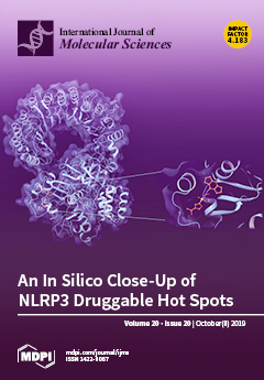
The present study was focused on NLRP3 inflammasome, a cytosolic complex that coordinates innate immunity responses. When over-activated, NLRP3 seems to be linked to several chronic pathologies, thus appears as an appealing target for the development of new therapeutic drugs. For this purpose, NLRP3 homology model was built and the putative protein binding region of a non-covalent inhibitor MCC950 (used as a probe) was investigated using a combination of in silico techniques. The main aim was to detect putative protein hot spots involved in the stabilization of protein-ligand complex. View this paper.
  • Issues are regarded as officially published after their release is announced to the table of contents alert mailing list.
  • You may sign up for e-mail alerts to receive table of contents of newly released issues.
  • PDF is the official format for papers published in both, html and pdf forms. To view the papers in pdf format, click on the "PDF Full-text" link, and use the free Adobe Reader to open them.
Order results
Result details
Section
Select all
Export citation of selected articles as:
21 pages, 7621 KiB  
Article
Identification of 15 T Cell Restricted Genes Evaluates T Cell Infiltration of Human Healthy Tissues and Cancers and Shows Prognostic and Predictive Potential
by Luigi Cari, Francesca De Rosa, Maria Grazia Petrillo, Graziella Migliorati, Giuseppe Nocentini and Carlo Riccardi
Int. J. Mol. Sci. 2019, 20(20), 5242; https://doi.org/10.3390/ijms20205242 - 22 Oct 2019
Cited by 4 | Viewed by 3685
Abstract
T cell gene signatures are used to evaluate T cell infiltration of non-lymphoid tissues and cancers in both experimental and clinical settings. However, some genes included in the available T cell signatures are not T cell-restricted. Herein, we propose a new human T [...] Read more.
T cell gene signatures are used to evaluate T cell infiltration of non-lymphoid tissues and cancers in both experimental and clinical settings. However, some genes included in the available T cell signatures are not T cell-restricted. Herein, we propose a new human T cell signature that has been developed via a six-step procedure and comprises 15 T cell restricted genes. We demonstrate the new T cell signature, named signature-H, that differs from other gene signatures since it shows higher sensitivity and better predictivity in the evaluation of T cell infiltration in healthy tissues as well as 32 cancers. Further, results from signature-H are highly concordant with the immunohistochemistry methods currently used for assessing the prognosis of neuroblastoma, as demonstrated by the Kaplan–Meier curves of patients ranked by tumor T cell infiltration. Moreover, T cell infiltration levels calculated using signature-H correlate with the risk groups determined by the staging of the neuroblastoma. Finally, multiparametric analysis of tumor-infiltrating T cells based on signature-H let us favorably predict the response of melanoma to the anti-PD-1 antibody nivolumab. These findings suggest that signature-H evaluates T cell infiltration levels of tissues and may be used as a prognostic tool in the precision medicine perspective after appropriate clinical validation. Full article
(This article belongs to the Special Issue Cancer Immunoediting and Beyond)
Show Figures

Graphical abstract

16 pages, 2131 KiB  
Article
Mutation of ONAC096 Enhances Grain Yield by Increasing Panicle Number and Delaying Leaf Senescence during Grain Filling in Rice
by Kiyoon Kang, Yejin Shim, Eunji Gi, Gynheung An and Nam-Chon Paek
Int. J. Mol. Sci. 2019, 20(20), 5241; https://doi.org/10.3390/ijms20205241 - 22 Oct 2019
Cited by 30 | Viewed by 4617
Abstract
Exploring genetic methods to improve yield in grain crops such as rice (Oryza sativa) is essential to help meet the needs of the increasing population. Here, we report that rice ONAC096 affects grain yield by regulating leaf senescence and panicle number. [...] Read more.
Exploring genetic methods to improve yield in grain crops such as rice (Oryza sativa) is essential to help meet the needs of the increasing population. Here, we report that rice ONAC096 affects grain yield by regulating leaf senescence and panicle number. ONAC096 expression increased rapidly in rice leaves upon the initiation of aging- and dark-induced senescence. Two independent T-DNA insertion mutants (onac096-1 and onac096-2) with downregulated ONAC096 expression retained their green leaf color during natural senescence in the field, thus extending their photosynthetic capacity. Reverse-transcription quantitative PCR analysis showed that ONAC096 upregulated genes controlling chlorophyll degradation and leaf senescence. Repressed OsCKX2 (encoding cytokinin oxidase/dehydrogenase) expression in the onac096 mutants led to a 15% increase in panicle number without affecting grain weight or fertility. ONAC096 mediates abscisic acid (ABA)-induced leaf senescence by upregulating the ABA signaling genes ABA INSENSITIVE5 and ENHANCED EM LEVEL. The onac096 mutants showed a 16% increase in grain yield, highlighting the potential for using this gene to increase grain production. Full article
(This article belongs to the Section Molecular Plant Sciences)
Show Figures

Graphical abstract

19 pages, 3971 KiB  
Article
Fusaricidin Produced by Paenibacillus polymyxa WLY78 Induces Systemic Resistance against Fusarium Wilt of Cucumber
by Yunlong Li and Sanfeng Chen
Int. J. Mol. Sci. 2019, 20(20), 5240; https://doi.org/10.3390/ijms20205240 - 22 Oct 2019
Cited by 41 | Viewed by 5176
Abstract
Cucumber is an important vegetable crop in China. Fusarium wilt is a soil-borne disease that can significantly reduce cucumber yields. Paenibacillus polymyxa WLY78 can strongly inhibit Fusarium oxysporum f. sp. Cucumerium, which causes Fusarium wilt disease. In this study, we screened the [...] Read more.
Cucumber is an important vegetable crop in China. Fusarium wilt is a soil-borne disease that can significantly reduce cucumber yields. Paenibacillus polymyxa WLY78 can strongly inhibit Fusarium oxysporum f. sp. Cucumerium, which causes Fusarium wilt disease. In this study, we screened the genome of WLY78 and found eight potential antibiotic biosynthesis gene clusters. Mutation analysis showed that among the eight clusters, the fusaricidin synthesis (fus) gene cluster is involved in inhibiting the Fusarium genus, Verticillium albo-atrum, Monilia persoon, Alternaria mali, Botrytis cinereal, and Aspergillus niger. Further mutation analysis revealed that with the exception of fusTE, the seven genes fusG, fusF, fusE, fusD, fusC, fusB, and fusA within the fus cluster were all involved in inhibiting fungi. This is the first time that demonstrated that fusTE was not essential. We first report the inhibitory mode of fusaricidin to inhibit spore germination and disrupt hyphal membranes. A biocontrol assay demonstrated that fusaricidin played a major role in controlling Fusarium wilt disease. Additionally, qRT-PCR demonstrated that fusaricidin could induce systemic resistance via salicylic acid (SA) signal against Fusarium wilt of cucumber. WLY78 is the first reported strain to both produce fusaricidin and fix nitrogen. Therefore, our results demonstrate that WLY78 will have great potential as a biocontrol agent in agriculture. Full article
(This article belongs to the Special Issue Plant Elicitors of Resistance and the Future of Plant Protection)
Show Figures

Graphical abstract

23 pages, 4229 KiB  
Article
Hyperacetylation of Cardiac Mitochondrial Proteins Is Associated with Metabolic Impairment and Sirtuin Downregulation after Chronic Total Body Irradiation of ApoE -/- Mice
by Zarko Barjaktarovic, Juliane Merl-Pham, Ignacia Braga-Tanaka, Satoshi Tanaka, Stefanie M. Hauck, Anna Saran, Mariateresa Mancuso, Michael J. Atkinson, Soile Tapio and Omid Azimzadeh
Int. J. Mol. Sci. 2019, 20(20), 5239; https://doi.org/10.3390/ijms20205239 - 22 Oct 2019
Cited by 24 | Viewed by 4185
Abstract
Chronic exposure to low-dose ionizing radiation is associated with an increased risk of cardiovascular disease. Alteration in energy metabolism has been suggested to contribute to radiation-induced heart pathology, mitochondrial dysfunction being a hallmark of this disease. The goal of this study was to [...] Read more.
Chronic exposure to low-dose ionizing radiation is associated with an increased risk of cardiovascular disease. Alteration in energy metabolism has been suggested to contribute to radiation-induced heart pathology, mitochondrial dysfunction being a hallmark of this disease. The goal of this study was to investigate the regulatory role of acetylation in heart mitochondria in the long-term response to chronic radiation. ApoE-deficient C57Bl/6J mice were exposed to low-dose-rate (20 mGy/day) gamma radiation for 300 days, resulting in a cumulative total body dose of 6.0 Gy. Heart mitochondria were isolated and analyzed using quantitative proteomics. Radiation-induced proteome and acetylome alterations were further validated using immunoblotting, enzyme activity assays, and ELISA. In total, 71 proteins showed peptides with a changed acetylation status following irradiation. The great majority (94%) of the hyperacetylated proteins were involved in the TCA cycle, fatty acid oxidation, oxidative stress response and sirtuin pathway. The elevated acetylation patterns coincided with reduced activity of mitochondrial sirtuins, increased the level of Acetyl-CoA, and were accompanied by inactivation of major cardiac metabolic regulators PGC-1 alpha and PPAR alpha. These observations suggest that the changes in mitochondrial acetylation after irradiation is associated with impairment of heart metabolism. We propose a novel mechanism involved in the development of late cardiac damage following chronic irradiation. Full article
(This article belongs to the Section Molecular Biology)
Show Figures

Figure 1

20 pages, 1262 KiB  
Review
The Predictive Role of the Biomarker Kidney Molecule-1 (KIM-1) in Acute Kidney Injury (AKI) Cisplatin-Induced Nephrotoxicity
by Daniela Maria Tanase, Evelina Maria Gosav, Smaranda Radu, Claudia Florida Costea, Manuela Ciocoiu, Alexandru Carauleanu, Cristina Mihaela Lacatusu, Minela Aida Maranduca, Mariana Floria and Ciprian Rezus
Int. J. Mol. Sci. 2019, 20(20), 5238; https://doi.org/10.3390/ijms20205238 - 22 Oct 2019
Cited by 91 | Viewed by 11917
Abstract
Acute kidney injury (AKI) following platinum-based chemotherapeutics is a frequently reported serious side-effect. However, there are no approved biomarkers that can properly identify proximal tubular injury while routine assessments such as serum creatinine lack sensitivity. Kidney-injury-molecule 1 (KIM-1) is showing promise in identifying [...] Read more.
Acute kidney injury (AKI) following platinum-based chemotherapeutics is a frequently reported serious side-effect. However, there are no approved biomarkers that can properly identify proximal tubular injury while routine assessments such as serum creatinine lack sensitivity. Kidney-injury-molecule 1 (KIM-1) is showing promise in identifying cisplatin-induced renal injury both in vitro and in vivo studies. In this review, we focus on describing the mechanisms of renal tubular cells cisplatin-induced apoptosis, the associated inflammatory response and oxidative stress and the role of KIM-1 as a possible biomarker used to predict cisplatin associated AKI. Full article
(This article belongs to the Special Issue Nephrotoxicity 2019)
Show Figures

Graphical abstract

16 pages, 4258 KiB  
Article
The SGLT2 Inhibitor Canagliflozin Prevents Carcinogenesis in a Mouse Model of Diabetes and Non-Alcoholic Steatohepatitis-Related Hepatocarcinogenesis: Association with SGLT2 Expression in Hepatocellular Carcinoma
by Teruo Jojima, Sho Wakamatsu, Masato Kase, Toshie Iijima, Yuko Maejima, Kenju Shimomura, Takahiko Kogai, Takuya Tomaru, Isao Usui and Yoshimasa Aso
Int. J. Mol. Sci. 2019, 20(20), 5237; https://doi.org/10.3390/ijms20205237 - 22 Oct 2019
Cited by 68 | Viewed by 7757
Abstract
The aim of the present study is to investigate the effects of canagliflozin, a selective sodium-glucose co-transporter 2 (SGLT2) inhibitor, on non-alcoholic steatohepatitis (NASH) and NASH-related hepatocellular carcinoma (HCC) in a mouse model of diabetes and NASH-HCC. First, mice aged five weeks were [...] Read more.
The aim of the present study is to investigate the effects of canagliflozin, a selective sodium-glucose co-transporter 2 (SGLT2) inhibitor, on non-alcoholic steatohepatitis (NASH) and NASH-related hepatocellular carcinoma (HCC) in a mouse model of diabetes and NASH-HCC. First, mice aged five weeks were divided into two groups (vehicle group and canagliflozin group) and were treated for three weeks. Then, mice aged five weeks were divided into three groups of nine animals each: the vehicle group, early canagliflozin group (treated from five to nine weeks), and continuous canagliflozin group (treated from five to 16 weeks). Canagliflozin was administered at a dose of 30 mg/kg in these experiments. In addition, the in vitro effects of canagliflozin were investigated using HepG2 cells, a human HCC cell line. At the age of eight or 16 weeks, the histological non-alcoholic fatty liver disease activity score was lower in the canagliflozin-treated mice than in vehicle-treated mice. There were significantly fewer hepatic tumors in the continuous canagliflozin group than in the vehicle group. Immunohistochemistry showed significantly fewer glutamine synthetase-positive nodules in the continuous canagliflozin group than in the vehicle group. Expression of α-fetoprotein mRNA, a marker of HCC, was downregulated in the continuous canagliflozin group when compared with the vehicle group. At 16 weeks, there was diffuse SGLT1 expression in the hepatic lobules and strong expression by hepatocytes in the vehicle group, while SGLT2 expression was stronger in liver tumors than in the lobules. In the in vitro study, canagliflozin (10 μM) suppressed the proliferation of HepG2 cells. Flow cytometry showed that canagliflozin reduced the percentage of HepG2 cells in the G2/M phase due to arrest in the G1 phase along with decreased expression of cyclin D and Cdk4 proteins, while it increased the percentage of cells in the G0/1 phase. Canagliflozin also induced apoptosis of HepG2 cells via activation of caspase 3. In this mouse model of diabetes and NASH/HCC, canagliflozin showed anti-steatotic and anti-inflammatory effects that attenuated the development of NASH and prevented the progression of NASH to HCC, partly due to the induction of cell cycle arrest and/or apoptosis as well as the reduction of tumor growth through the direct inhibition of SGLT2 in tumor cells. Full article
(This article belongs to the Section Molecular Pathology, Diagnostics, and Therapeutics)
Show Figures

Figure 1

12 pages, 436 KiB  
Review
Insights into the Pathophysiology of Infertility in Females with Classical Galactosaemia
by Zaza Abidin and Eileen P. Treacy
Int. J. Mol. Sci. 2019, 20(20), 5236; https://doi.org/10.3390/ijms20205236 - 22 Oct 2019
Cited by 11 | Viewed by 4978
Abstract
Classical galactosaemia (CG) (OMIM 230400) is a rare inborn error of galactose metabolism caused by the deficiency of the enzyme galactose-1-phosphate uridylyltransferase (GALT, EC 2.7.7.12). Primary ovarian insufficiency (POI) is the most common long-term complication experienced by females with CG, presenting with hypergonadotrophic [...] Read more.
Classical galactosaemia (CG) (OMIM 230400) is a rare inborn error of galactose metabolism caused by the deficiency of the enzyme galactose-1-phosphate uridylyltransferase (GALT, EC 2.7.7.12). Primary ovarian insufficiency (POI) is the most common long-term complication experienced by females with CG, presenting with hypergonadotrophic hypoestrogenic infertility affecting at least 80% of females despite new-born screening and lifelong galactose dietary restriction. In this review, we describe the hypothesized pathophysiology of POI from CG, implications of timing of the ovarian dysfunction, and the new horizons and future prospects for treatments and fertility preservation. Full article
(This article belongs to the Special Issue Molecular Basis of Fertility Preservation and Restoration)
Show Figures

Figure 1

15 pages, 2604 KiB  
Article
WIN55,212-2-Induced Expression of Mir-29b1 Favours the Suppression of Osteosarcoma Cell Migration in a SPARC-Independent Manner
by Antonietta Notaro, Sonia Emanuele, Fabiana Geraci, Antonella D’Anneo, Marianna Lauricella, Giuseppe Calvaruso and Michela Giuliano
Int. J. Mol. Sci. 2019, 20(20), 5235; https://doi.org/10.3390/ijms20205235 - 22 Oct 2019
Cited by 18 | Viewed by 2878
Abstract
WIN55,212-2 (WIN) is a synthetic agonist of cannabinoid receptors that displays promising antitumour properties. The aim of this study is to demonstrate that WIN is able to block the migratory ability of osteosarcoma cells and characterize the mechanisms involved. Using wound healing assay [...] Read more.
WIN55,212-2 (WIN) is a synthetic agonist of cannabinoid receptors that displays promising antitumour properties. The aim of this study is to demonstrate that WIN is able to block the migratory ability of osteosarcoma cells and characterize the mechanisms involved. Using wound healing assay and zymography, we showed that WIN affects cell migration and reduces the activity of the metalloproteases MMP2 and MMP9. This effect seemed to be independent of secreted protein acidic and rich in cysteine (SPARC), a matricellular protein involved in tissue remodeling and extracellular matrix deposition. SPARC release was indeed prevented by WIN, and SPARC silencing by RNA interference did not influence the effect of the cannabinoid on cell migration. WIN also increased the release of extracellular vesicles and dramatically upregulated miR-29b1, a key miRNA that modulates cell proliferation and migration. Interestingly, reduced cell migration was observed in stably miR-29b1-transfected cells, similarly to WIN-treated cells. Finally, we show the absence of SPARC in the extracellular vesicles released by osteosarcoma cells and no changes in SPARC level in miR-29b1 overexpressing cells. Overall, these findings suggest that WIN markedly affects cell migration, dependently on miR-29b1 and independently of SPARC, and can thus be considered as a potential innovative therapeutic agent in the treatment of osteosarcoma. Full article
(This article belongs to the Section Molecular Pathology, Diagnostics, and Therapeutics)
Show Figures

Graphical abstract

14 pages, 2127 KiB  
Article
Matrix Metalloproteinase-3 is Key Effector of TNF-α-Induced Collagen Degradation in Skin
by Ursula Mirastschijski, Blaž Lupše, Kathrin Maedler, Bhavishya Sarma, Arlo Radtke, Gazanfer Belge, Martina Dorsch, Dirk Wedekind, Lisa J. McCawley, Gabriele Boehm, Ulrich Zier, Kazuhiro Yamamoto, Sørge Kelm and Magnus S. Ågren
Int. J. Mol. Sci. 2019, 20(20), 5234; https://doi.org/10.3390/ijms20205234 - 22 Oct 2019
Cited by 30 | Viewed by 4510
Abstract
Inflammatory processes in the skin augment collagen degradation due to the up-regulation of matrix metalloproteinases (MMPs). The aim of the present project was to study the specific impact of MMP-3 on collagen loss in skin and its interplay with the collagenase MMP-13 under [...] Read more.
Inflammatory processes in the skin augment collagen degradation due to the up-regulation of matrix metalloproteinases (MMPs). The aim of the present project was to study the specific impact of MMP-3 on collagen loss in skin and its interplay with the collagenase MMP-13 under inflammatory conditions mimicked by the addition of the pro-inflammatory cytokine tumor necrosis factor-α (TNF-α). Skin explants from MMP-3 knock-out (KO) mice or from transgenic (TG) mice overexpressing MMP-3 in the skin and their respective wild-type counterparts (WT and WTT) were incubated ex vivo for eight days. The rate of collagen degradation, measured by released hydroxyproline, was reduced (p < 0.001) in KO skin explants compared to WT control skin but did not differ (p = 0.47) between TG and WTT skin. Treatment with the MMP inhibitor GM6001 reduced hydroxyproline media levels from WT, WTT and TG but not from KO skin explants. TNF-α increased collagen degradation in the WT group (p = 0.0001) only. More of the active form of MMP-13 was observed in the three MMP-3 expressing groups (co-incubation with receptor-associated protein stabilized MMP-13 subforms and enhanced detection in the media). In summary, the innate level of MMP-3 seems responsible for the accelerated loss of cutaneous collagen under inflammatory conditions, possibly via MMP-13 in mice. Full article
(This article belongs to the Special Issue Matrix Metalloproteinase)
Show Figures

Graphical abstract

18 pages, 1958 KiB  
Article
Genome-Wide Identification, Classification, and Expression Analysis of the Hsf Gene Family in Carnation (Dianthus caryophyllus)
by Wei Li, Xue-Li Wan, Jia-Yu Yu, Kui-Ling Wang and Jin Zhang
Int. J. Mol. Sci. 2019, 20(20), 5233; https://doi.org/10.3390/ijms20205233 - 22 Oct 2019
Cited by 32 | Viewed by 4003
Abstract
Heat shock transcription factors (Hsfs) are a class of important transcription factors (TFs) which play crucial roles in the protection of plants from damages caused by various abiotic stresses. The present study aimed to characterize the Hsf genes in carnation (Dianthus caryophyllus [...] Read more.
Heat shock transcription factors (Hsfs) are a class of important transcription factors (TFs) which play crucial roles in the protection of plants from damages caused by various abiotic stresses. The present study aimed to characterize the Hsf genes in carnation (Dianthus caryophyllus), which is one of the four largest cut flowers worldwide. In this study, a total of 17 non-redundant Hsf genes were identified from the D. caryophyllus genome. Specifically, the gene structure and motifs of each DcaHsf were comprehensively analyzed. Phylogenetic analysis of the DcaHsf family distinctly separated nine class A, seven class B, and one class C Hsf genes. Additionally, promoter analysis indicated that the DcaHsf promoters included various cis-acting elements that were related to stress, hormones, as well as development processes. In addition, cis-elements, such as STRE, MYB, and ABRE binding sites, were identified in the promoters of most DcaHsf genes. According to qRT-PCR data, the expression of DcaHsfs varied in eight tissues and six flowering stages and among different DcaHsfs, even in the same class. Moreover, DcaHsf-A1, A2a, A9a, B2a, B3a revealed their putative involvement in the early flowering stages. The time-course expression profile of DcaHsf during stress responses illustrated that all the DcaHsfs were heat- and drought-responsive, and almost all DcaHsfs were down-regulated by cold, salt, and abscisic acid (ABA) stress. Meanwhile, DcaHsf-A3, A7, A9a, A9b, B3a were primarily up-regulated at an early stage in response to salicylic acid (SA). This study provides an overview of the Hsf gene family in D. caryophyllus and a basis for the breeding of stress-resistant carnation. Full article
(This article belongs to the Special Issue Mapping Abiotic Stress-Tolerance Genes in Plants)
Show Figures

Figure 1

11 pages, 241 KiB  
Review
The Role of Glutamine in the Complex Interaction between Gut Microbiota and Health: A Narrative Review
by Simone Perna, Tariq A. Alalwan, Zahraa Alaali, Tahera Alnashaba, Clara Gasparri, Vittoria Infantino, Layla Hammad, Antonella Riva, Giovanna Petrangolini, Pietro Allegrini and Mariangela Rondanelli
Int. J. Mol. Sci. 2019, 20(20), 5232; https://doi.org/10.3390/ijms20205232 - 22 Oct 2019
Cited by 46 | Viewed by 11581
Abstract
The scientific literature has demonstrated that glutamine is one of the main beneficial amino acids. It plays an important role in gut microbiota and immunity. This paper provides a critical overview of experimental studies (in vitro, in vivo, and clinical) investigating the efficacy [...] Read more.
The scientific literature has demonstrated that glutamine is one of the main beneficial amino acids. It plays an important role in gut microbiota and immunity. This paper provides a critical overview of experimental studies (in vitro, in vivo, and clinical) investigating the efficacy of glutamine and its effect on gut microbiota. As a result of this review, we have summarized that glutamine could affect gut microbiota via different mechanisms including the reduction in the ratio of Firmicutes to Bacteroidetes, with the activation of NF-κB and PI3K-Akt pathways, reducing the intestinal colonization (Eimeria lesions) and bacterial overgrowth or bacterial translocation, increasing the production of secretory immunoglobulin A (SIgA) and immunoglobulin A+ (IgA+) cells in the intestinal lumen, and decreasing asparagine levels. The potential applications of glutamine on gut microbiota include, but are not limited to, the management of obesity, bacterial translocation and community, cytokines profiles, and the management of side effects during post-chemotherapy and constipation periods. Further studies and reviews are needed regarding the effects of glutamine supplementation on other conditions in humans. Full article
(This article belongs to the Special Issue Glutamine: An Essential Non-Essential Amino Acid)
19 pages, 2321 KiB  
Review
Hydrogen Sulfide in Bone Tissue Regeneration and Repair: State of the Art and New Perspectives
by Laura Gambari, Brunella Grigolo and Francesco Grassi
Int. J. Mol. Sci. 2019, 20(20), 5231; https://doi.org/10.3390/ijms20205231 - 22 Oct 2019
Cited by 26 | Viewed by 7699
Abstract
The importance of hydrogen sulfide (H2S) in the regulation of multiple physiological functions has been clearly recognized in the over 20 years since it was first identified as a novel gasotransmitter. In bone tissue H2S exerts a cytoprotective effect [...] Read more.
The importance of hydrogen sulfide (H2S) in the regulation of multiple physiological functions has been clearly recognized in the over 20 years since it was first identified as a novel gasotransmitter. In bone tissue H2S exerts a cytoprotective effect and promotes bone formation. Just recently, the scientific community has begun to appreciate its role as a therapeutic agent in bone pathologies. Pharmacological administration of H2S achieved encouraging results in preclinical studies in the treatment of systemic bone diseases, such as osteoporosis; however, a local delivery of H2S at sites of bone damage may provide additional opportunities of treatment. Here, we highlight how H2S stimulates multiple signaling pathways involved in various stages of the processes of bone repair. Moreover, we discuss how material science and chemistry have recently developed biomaterials and H2S-donors with improved features, laying the ground for the development of H2S-releasing devices for bone regenerative medicine. This review is intended to give a state-of-the-art description of the pro-regenerative properties of H2S, with a focus on bone tissue, and to discuss the potential of H2S-releasing scaffolds as a support for bone repair. Full article
(This article belongs to the Special Issue Hydrogen Sulfide in the Diseases and Tissue Regeneration)
Show Figures

Figure 1

9 pages, 957 KiB  
Article
The Detection of CMV in Saliva Can Mark a Systemic Infection with CMV in Renal Transplant Recipients
by Shelley Waters, Silvia Lee, Megan Lloyd, Ashley Irish and Patricia Price
Int. J. Mol. Sci. 2019, 20(20), 5230; https://doi.org/10.3390/ijms20205230 - 22 Oct 2019
Cited by 12 | Viewed by 4923
Abstract
Human cytomegalovirus (CMV) is often transmitted through saliva. The salivary gland is a site of CMV replication and saliva can be used to diagnose congenital CMV infections. CMV replication is monitored in whole blood or plasma in renal transplant recipients (RTR) and associates [...] Read more.
Human cytomegalovirus (CMV) is often transmitted through saliva. The salivary gland is a site of CMV replication and saliva can be used to diagnose congenital CMV infections. CMV replication is monitored in whole blood or plasma in renal transplant recipients (RTR) and associates with clinical disease. However, these assays may not detect replication in the salivary gland and there is little data linking detection in saliva with systemic infection and clinical sequelae. RTR (n = 82) were recruited > 2 years after transplantation. An in-house quantitative PCR assay was used to detect CMV UL54 in saliva samples. CMV DNA was sought in plasma using a commercial assay. Vascular health was predicted using flow mediated dilatation (FMD) and plasma biomarkers. CMV-reactive antibodies were quantified by ELISA and circulating CMV-specific T-cells by an interferon-γ ELISpot assay. Vδ2 γδ T-cells were detected using multicolor flow cytometry reflecting population expansion after CMV infection. The presence of CMV DNA in saliva and plasma associated with plasma levels of antibodies reactive with CMV gB and with populations of circulating Vδ2 γδ T -cells (p < 0.01). T-cells reactive to CMV immediate early (IE)-1 protein were generally lower in patients with CMV DNA in saliva or plasma, but the level of significance varied (p = 0.02–0.16). Additionally, CMV DNA in saliva or plasma associated weakly with impaired FMD (p = 0.06–0.09). The data suggest that CMV detected in saliva reflects systemic infections in adult RTR. Full article
(This article belongs to the Section Molecular Immunology)
Show Figures

Graphical abstract

23 pages, 4187 KiB  
Article
Influence of the First Chromophore-Forming Residue on Photobleaching and Oxidative Photoconversion of EGFP and EYFP
by Tirthendu Sen, Anastasia V. Mamontova, Anastasia V. Titelmayer, Aleksander M. Shakhov, Artyom A. Astafiev, Atanu Acharya, Konstantin A. Lukyanov, Anna I. Krylov and Alexey M. Bogdanov
Int. J. Mol. Sci. 2019, 20(20), 5229; https://doi.org/10.3390/ijms20205229 - 22 Oct 2019
Cited by 13 | Viewed by 4545
Abstract
Enhanced green fluorescent protein (EGFP)—one of the most widely applied genetically encoded fluorescent probes—carries the threonine-tyrosine-glycine (TYG) chromophore. EGFP efficiently undergoes green-to-red oxidative photoconversion (“redding”) with electron acceptors. Enhanced yellow fluorescent protein (EYFP), a close EGFP homologue (five amino acid substitutions), has a [...] Read more.
Enhanced green fluorescent protein (EGFP)—one of the most widely applied genetically encoded fluorescent probes—carries the threonine-tyrosine-glycine (TYG) chromophore. EGFP efficiently undergoes green-to-red oxidative photoconversion (“redding”) with electron acceptors. Enhanced yellow fluorescent protein (EYFP), a close EGFP homologue (five amino acid substitutions), has a glycine-tyrosine-glycine (GYG) chromophore and is much less susceptible to redding, requiring halide ions in addition to the oxidants. In this contribution we aim to clarify the role of the first chromophore-forming amino acid in photoinduced behavior of these fluorescent proteins. To that end, we compared photobleaching and redding kinetics of EGFP, EYFP, and their mutants with reciprocally substituted chromophore residues, EGFP-T65G and EYFP-G65T. Measurements showed that T65G mutation significantly increases EGFP photostability and inhibits its excited-state oxidation efficiency. Remarkably, while EYFP-G65T demonstrated highly increased spectral sensitivity to chloride, it is also able to undergo redding chloride-independently. Atomistic calculations reveal that the GYG chromophore has an increased flexibility, which facilitates radiationless relaxation leading to the reduced fluorescence quantum yield in the T65G mutant. The GYG chromophore also has larger oscillator strength as compared to TYG, which leads to a shorter radiative lifetime (i.e., a faster rate of fluorescence). The faster fluorescence rate partially compensates for the loss of quantum efficiency due to radiationless relaxation. The shorter excited-state lifetime of the GYG chromophore is responsible for its increased photostability and resistance to redding. In EYFP and EYFP-G65T, the chromophore is stabilized by π-stacking with Tyr203, which suppresses its twisting motions relative to EGFP. Full article
(This article belongs to the Collection Computational Studies of Biomolecules)
Show Figures

Figure 1

22 pages, 9611 KiB  
Article
Light- and Temperature-Induced Expression of an R2R3-MYB Gene Regulates Anthocyanin Biosynthesis in Red-Fleshed Kiwifruit
by Min Yu, Yuping Man and Yanchang Wang
Int. J. Mol. Sci. 2019, 20(20), 5228; https://doi.org/10.3390/ijms20205228 - 22 Oct 2019
Cited by 33 | Viewed by 4965
Abstract
The R2R3 MYB genes associated with the flavonoid/anthocyanidin pathway feature two repeats, and represent the most abundant classes of MYB genes in plants; however, the physiological role and regulatory function of most R2R3 MYBs remain poorly understood in kiwifruit (Actinidia). Here, [...] Read more.
The R2R3 MYB genes associated with the flavonoid/anthocyanidin pathway feature two repeats, and represent the most abundant classes of MYB genes in plants; however, the physiological role and regulatory function of most R2R3 MYBs remain poorly understood in kiwifruit (Actinidia). Here, genome-wide analysis identified 155 R2R3-MYBs in the ‘Red 5′ version of the Actinidia chinensis genome. Out of 36 anthocyanin-related AccR2R3-MYBs, AcMYB10 was the most highly expressed in inner pericarp of red-fleshed kiwifruit. The expression of AcMYB10 was highly correlated with anthocyanin accumulation in natural pigmentation during fruit ripening and light-/temperature-induced pigmentation in the callus. AcMYB10 is localized in the nuclei and has transcriptional activation activity. Overexpression of AcMYB10 elevates anthocyanin accumulation in transgenic A. chinensis. In comparison, A. chinensis fruit infiltrated with virus-induced gene silencing showed delayed red coloration, lower anthocyanin content, and lower expression of AcMYB10. The transient expression experiment in Nicotiana tabacum leaves and Actinidia arguta fruit indicated the interaction of AcMYB10 with AcbHLH42 might strongly activate anthocyanin biosynthesis by activating the transcription of AcLDOX and AcF3GT. In conclusion, this study provides novel molecular information about R2R3-MYBs in kiwifruit, advances our understanding of light- and temperature-induced anthocyanin accumulation, and demonstrates the important function of AcMYB10 in the biosynthesis of anthocyanin in kiwifruit. Full article
(This article belongs to the Section Molecular Plant Sciences)
Show Figures

Graphical abstract

17 pages, 1510 KiB  
Article
Neutrophil-Derived Microvesicle Induced Dysfunction of Brain Microvascular Endothelial Cells In Vitro
by Anjana Ajikumar, Merete B. Long, Paul R. Heath, Stephen B. Wharton, Paul G. Ince, Victoria C. Ridger and Julie E. Simpson
Int. J. Mol. Sci. 2019, 20(20), 5227; https://doi.org/10.3390/ijms20205227 - 22 Oct 2019
Cited by 37 | Viewed by 4730
Abstract
The blood-brain barrier (BBB), composed of brain microvascular endothelial cells (BMEC) that are tightly linked by tight junction (TJ) proteins, restricts the movement of molecules between the periphery and the central nervous system. Elevated systemic levels of neutrophils have been detected in patients [...] Read more.
The blood-brain barrier (BBB), composed of brain microvascular endothelial cells (BMEC) that are tightly linked by tight junction (TJ) proteins, restricts the movement of molecules between the periphery and the central nervous system. Elevated systemic levels of neutrophils have been detected in patients with altered BBB function, but the role of neutrophils in BMEC dysfunction is unknown. Neutrophils are key players of the immune response and, when activated, produce neutrophil-derived microvesicles (NMV). NMV have been shown to impact the integrity of endothelial cells throughout the body and we hypothesize that NMV released from circulating neutrophils interact with BMEC and induce endothelial cell dysfunction. Therefore, the current study investigated the interaction of NMV with human BMEC and determined whether they altered gene expression and function in vitro. Using flow cytometry and confocal imaging, NMV were shown to be internalized by the human cerebral microvascular endothelial cell line hCMEC/D3 via a variety of energy-dependent mechanisms, including endocytosis and macropinocytosis. The internalization of NMV significantly altered the transcriptomic profile of hCMEC/D3, specifically inducing the dysregulation of genes associated with TJ, ubiquitin-mediated proteolysis and vesicular transport. Functional studies confirmed NMV significantly increased permeability and decreased the transendothelial electrical resistance (TEER) of a confluent monolayer of hCMEC/D3. These findings indicate that NMV interact with and affect gene expression of BMEC as well as impacting their integrity. We conclude that NMV may play an important role in modulating the permeability of BBB during an infection. Full article
Show Figures

Figure 1

16 pages, 1831 KiB  
Review
The ST2/Interleukin-33 Axis in Hematologic Malignancies: The IL-33 Paradox
by Alessandro Allegra, Vanessa Innao, Gennaro Tartarisco, Giovanni Pioggia, Marco Casciaro, Caterina Musolino and Sebastiano Gangemi
Int. J. Mol. Sci. 2019, 20(20), 5226; https://doi.org/10.3390/ijms20205226 - 22 Oct 2019
Cited by 12 | Viewed by 5878
Abstract
Interleukin (IL)-33 is a chromatin-related nuclear interleukin that is a component of IL-1 family. IL-33 production augments the course of inflammation after cell damage or death. It is discharged into the extracellular space. IL-33 is regarded as an “alarmin” able to stimulate several [...] Read more.
Interleukin (IL)-33 is a chromatin-related nuclear interleukin that is a component of IL-1 family. IL-33 production augments the course of inflammation after cell damage or death. It is discharged into the extracellular space. IL-33 is regarded as an “alarmin” able to stimulate several effectors of the immune system, regulating numerous immune responses comprising cancer immune reactions. IL-33 has been demonstrated to influence tumorigenesis. However, as far as this cytokine is concerned, we are faced with what has sometimes been defined as the IL-33 paradox. Several studies have demonstrated a relevant role of IL-33 to numerous malignancies, where it may have pro- and—less frequently—antitumorigenic actions. In the field of hematological malignancies, the role of IL-33 seems even more complex. Although we can affirm the existence of a negative role of IL-33 in Chronic myelogenos leukemia (CML) and in lymphoproliferative diseases and a positive role in pathologies such as Acute myeloid leukemia (AML), the action of IL-33 seems to be multiple and sometimes contradictory within the same pathology. In the future, we will have to learn to govern the negative aspects of activating the IL-33/ST2 axis and exploit the positive ones. Full article
(This article belongs to the Section Molecular Immunology)
Show Figures

Figure 1

17 pages, 3861 KiB  
Article
The Mechanisms Underlying the Cytotoxic Effects of Copper Via Differentiated Embryonic Chondrocyte Gene 1
by Ssu-Yu Chen, Shu-Ting Liu, Wun-Rong Lin, Chi-Kang Lin and Shih-Ming Huang
Int. J. Mol. Sci. 2019, 20(20), 5225; https://doi.org/10.3390/ijms20205225 - 22 Oct 2019
Cited by 15 | Viewed by 3952
Abstract
Copper is an essential trace element within cells, but it also exerts cytotoxic effects through induction of reactive oxygen species (ROS) production. To determine the mechanisms underlying copper-induced ROS production, we examined the effects of copper sulfate in HeLa cells. Exposure to copper [...] Read more.
Copper is an essential trace element within cells, but it also exerts cytotoxic effects through induction of reactive oxygen species (ROS) production. To determine the mechanisms underlying copper-induced ROS production, we examined the effects of copper sulfate in HeLa cells. Exposure to copper sulfate led to dose-dependent decreases in HeLa cell viability, along with increases in the subG1 and G2/M populations and corresponding decreases in the G1 population. Copper sulfate also increased the levels of apoptosis, senescence, mitochondrial dysfunction, autophagy, ROS, and the expression of several stress proteins, including ATF3, c-Fos, DEC1 (differentiated embryonic chondrocyte gene 1), p21, p53, and HIF-1α (hypoxia-inducible factor 1 alpha). The suppression of copper-induced ROS generation by the ROS scavenger N-acetyl cysteine verified copper’s functional role, while the suppression of copper’s effects by the copper chelator disulfiram, confirmed its specificity. Selective induction of HIF-1α, p53, and phosphorylated ERK proteins by copper was blocked by the knockdown of the transcription factor DEC1, suggesting copper’s effects are mediated by DEC1. In addition to HeLa cells, copper also exerted cytotoxic effects in human endometrial (HEC-1-A) and lung (A549) adenocarcinoma cells, but not in normal human kidney (HEK293) or bronchial (Beas-2B) epithelial cells. These findings shed new light on the functional roles of copper within cells. Full article
(This article belongs to the Special Issue Crosstalk between Circadian Rhythm and Diseases)
Show Figures

Figure 1

16 pages, 2739 KiB  
Article
Autoregulation of greA Expression Relies on GraL Rather than on greA Promoter Region
by Maciej Dylewski, Llorenç Fernández-Coll, Bożena Bruhn-Olszewska, Carlos Balsalobre and Katarzyna Potrykus
Int. J. Mol. Sci. 2019, 20(20), 5224; https://doi.org/10.3390/ijms20205224 - 22 Oct 2019
Viewed by 2997
Abstract
GreA is a well-characterized transcriptional factor that acts primarily by rescuing stalled RNA polymerase complexes, but has also been shown to be the major transcriptional fidelity and proofreading factor, while it inhibits DNA break repair. Regulation of greA gene expression itself is still [...] Read more.
GreA is a well-characterized transcriptional factor that acts primarily by rescuing stalled RNA polymerase complexes, but has also been shown to be the major transcriptional fidelity and proofreading factor, while it inhibits DNA break repair. Regulation of greA gene expression itself is still not well understood. So far, it has been shown that its expression is driven by two overlapping promoters and that greA leader encodes a small RNA (GraL) that is acting in trans on nudE mRNA. It has been also shown that GreA autoinhibits its own expression in vivo. Here, we decided to investigate the inner workings of this autoregulatory loop. Transcriptional fusions with lacZ reporter carrying different modifications (made both to the greA promoter and leader regions) were made to pinpoint the sequences responsible for this autoregulation, while GraL levels were also monitored. Our data indicate that GreA mediated regulation of its own gene expression is dependent on GraL acting in cis (a rare example of dual-action sRNA), rather than on the promoter region. However, a yet unidentified, additional factor seems to participate in this regulation as well. Overall, the GreA/GraL regulatory loop seems to have unique but hard to classify properties. Full article
(This article belongs to the Section Molecular Microbiology)
Show Figures

Graphical abstract

21 pages, 1385 KiB  
Review
Oncofertility: Pharmacological Protection and Immature Testicular Tissue (ITT)-Based Strategies for Prepubertal and Adolescent Male Cancer Patients
by Elissavet Ntemou, Chrysanthi Alexandri, Pascale Lybaert, Ellen Goossens and Isabelle Demeestere
Int. J. Mol. Sci. 2019, 20(20), 5223; https://doi.org/10.3390/ijms20205223 - 21 Oct 2019
Cited by 17 | Viewed by 6688
Abstract
While the incidence of cancer in children and adolescents has significantly increased over the last decades, improvements made in the field of cancer therapy have led to an increased life expectancy for childhood cancer survivors. However, the gonadotoxic effect of the treatments may [...] Read more.
While the incidence of cancer in children and adolescents has significantly increased over the last decades, improvements made in the field of cancer therapy have led to an increased life expectancy for childhood cancer survivors. However, the gonadotoxic effect of the treatments may lead to infertility. Although semen cryopreservation represents the most efficient and safe fertility preservation method for males producing sperm, it is not feasible for prepubertal boys. The development of an effective strategy based on the pharmacological protection of the germ cells and testicular function during gonadotoxic exposure is a non-invasive preventive approach that prepubertal boys could benefit from. However, the progress in this field is slow. Currently, cryopreservation of immature testicular tissue (ITT) containing spermatogonial stem cells is offered to prepubertal boys as an experimental fertility preservation strategy by a number of medical centers. Several in vitro and in vivo fertility restoration approaches based on the use of ITT have been developed so far with autotransplantation of ITT appearing more promising. In this review, we discuss the pharmacological approaches for fertility protection in prepubertal and adolescent boys and the fertility restoration approaches developed on the utilization of ITT. Full article
(This article belongs to the Special Issue Molecular Basis of Fertility Preservation and Restoration)
Show Figures

Figure 1

7 pages, 764 KiB  
Editorial
Protein and Proteome Atlas for Plants under Stresses: New Highlights and Ways for Integrated Omics in Post-Genomics Era
by Xuchu Wang
Int. J. Mol. Sci. 2019, 20(20), 5222; https://doi.org/10.3390/ijms20205222 - 21 Oct 2019
Cited by 10 | Viewed by 3794
Abstract
In the post-genomics era, integrative omics studies for biochemical, physiological, and molecular changes of plants in response to stress conditions play more crucial roles. Among them, atlas analysis of plants under different abiotic stresses, including salinity, drought, and toxic conditions, has become more [...] Read more.
In the post-genomics era, integrative omics studies for biochemical, physiological, and molecular changes of plants in response to stress conditions play more crucial roles. Among them, atlas analysis of plants under different abiotic stresses, including salinity, drought, and toxic conditions, has become more important for uncovering the potential key genes and proteins in different plant tissues. High-quality genomic data and integrated analyses of transcriptomic, proteomic, metabolomics, and phenomic patterns provide a deeper understanding of how plants grow and survive under environmental stresses. This editorial mini-review aims to synthesize the 27 papers including two timely reviews that have contributed to this Special Issue, which focuses on concluding the recent progress in the Protein and Proteome Atlas in plants under different stresses. It covers various aspects of plant proteins ranging from agricultural proteomics, structure and function of proteins, novel techniques and approaches for gene and protein identification, protein quantification, proteomics for post-translational modifications (PTMs), and new insights into proteomics. The proteomics-based results in this issue will help the readers to gain novel insights for the understanding of complicated physiological processes in crops and other important plants in response to stressed conditions. Furthermore, these target genes and proteins that are important candidates for further functional validation in economic plants and crops can be studied. Full article
Show Figures

Graphical abstract

18 pages, 4878 KiB  
Article
The miR396–GRF Regulatory Module Controls the Embryogenic Response in Arabidopsis via an Auxin-Related Pathway
by Aleksandra Szczygieł-Sommer and Małgorzata D. Gaj
Int. J. Mol. Sci. 2019, 20(20), 5221; https://doi.org/10.3390/ijms20205221 - 21 Oct 2019
Cited by 37 | Viewed by 4554
Abstract
In plants, microRNAs have been indicated to control various developmental processes, including somatic embryogenesis (SE), which is triggered in the in vitro cultured somatic cells of plants. Although a transcriptomic analysis has indicated that numerous MIRNAs are differentially expressed in the SE [...] Read more.
In plants, microRNAs have been indicated to control various developmental processes, including somatic embryogenesis (SE), which is triggered in the in vitro cultured somatic cells of plants. Although a transcriptomic analysis has indicated that numerous MIRNAs are differentially expressed in the SE of different plants, the role of specific miRNAs in the embryogenic reprogramming of the somatic cell transcriptome is still poorly understood. In this study, we focused on performing a functional analysis of miR396 in SE given that the transcripts of MIR396 genes and the mature molecules of miR396 were found to be increased during an SE culture of Arabidopsis. In terms of miR396 in embryogenic induction, we observed the SE-associated expression pattern of MIR396b in explants of the β-glucuronidase (GUS) reporter line. In order to gain insight into the miR396-controlled mechanism that is involved in SE induction, the embryogenic response of mir396 mutants and the 35S:MIR396b overexpressor line to media with different 2,4-Dichlorophenoxyacetic acid (2,4-D) concentrations was evaluated. The results suggested that miR396 might contribute to SE induction by controlling the sensitivity of tissues to auxin treatment. Within the targets of miR396 that are associated with SE induction, we identified genes encoding the GROWTH-REGULATING FACTOR (GRF) transcription factors, including GRF1, GRF4, GRF7, GRF8, and GRF9. Moreover, the study suggested a regulatory relationship between miR396, GRF, and the PLETHORA (PLT1 and PLT2) genes during SE induction. A complex regulatory relationship within the miR396–GRF1/4/8/9–PLT1/2 module that involves the negative and positive control of GRFs and PLT (respectively) by miR396 might be assumed. Full article
(This article belongs to the Special Issue Transcriptional and Post-transcriptional Gene Regulation in Plants)
Show Figures

Figure 1

24 pages, 501 KiB  
Review
Roles of Thyroid Hormone-Associated microRNAs Affecting Oxidative Stress in Human Hepatocellular Carcinoma
by Po-Shuan Huang, Chia-Siu Wang, Chau-Ting Yeh and Kwang-Huei Lin
Int. J. Mol. Sci. 2019, 20(20), 5220; https://doi.org/10.3390/ijms20205220 - 21 Oct 2019
Cited by 22 | Viewed by 4065
Abstract
Oxidative stress occurs as a result of imbalance between the generation of reactive oxygen species (ROS) and antioxidant genes in cells, causing damage to lipids, proteins, and DNA. Accumulating damage of cellular components can trigger various diseases, including metabolic syndrome and cancer. Over [...] Read more.
Oxidative stress occurs as a result of imbalance between the generation of reactive oxygen species (ROS) and antioxidant genes in cells, causing damage to lipids, proteins, and DNA. Accumulating damage of cellular components can trigger various diseases, including metabolic syndrome and cancer. Over the past few years, the physiological significance of microRNAs (miRNA) in cancer has been a focus of comprehensive research. In view of the extensive level of miRNA interference in biological processes, the roles of miRNAs in oxidative stress and their relevance in physiological processes have recently become a subject of interest. In-depth research is underway to specifically address the direct or indirect relationships of oxidative stress-induced miRNAs in liver cancer and the potential involvement of the thyroid hormone in these processes. While studies on thyroid hormone in liver cancer are abundantly documented, no conclusive information on the potential relationships among thyroid hormone, specific miRNAs, and oxidative stress in liver cancer is available. In this review, we discuss the effects of thyroid hormone on oxidative stress-related miRNAs that potentially have a positive or negative impact on liver cancer. Additionally, supporting evidence from clinical and animal experiments is provided. Full article
Show Figures

Graphical abstract

21 pages, 4505 KiB  
Article
Combination of SAXS and Protein Painting Discloses the Three-Dimensional Organization of the Bacterial Cysteine Synthase Complex, a Potential Target for Enhancers of Antibiotic Action
by Brenda Rosa, Marialaura Marchetti, Gianluca Paredi, Heinz Amenitsch, Nina Franko, Roberto Benoni, Barbara Giabbai, Maria Giovanna De Marino, Andrea Mozzarelli, Luca Ronda, Paola Storici, Barbara Campanini and Stefano Bettati
Int. J. Mol. Sci. 2019, 20(20), 5219; https://doi.org/10.3390/ijms20205219 - 21 Oct 2019
Cited by 8 | Viewed by 4194
Abstract
The formation of multienzymatic complexes allows for the fine tuning of many aspects of enzymatic functions, such as efficiency, localization, stability, and moonlighting. Here, we investigated, in solution, the structure of bacterial cysteine synthase (CS) complex. CS is formed by serine acetyltransferase (CysE) [...] Read more.
The formation of multienzymatic complexes allows for the fine tuning of many aspects of enzymatic functions, such as efficiency, localization, stability, and moonlighting. Here, we investigated, in solution, the structure of bacterial cysteine synthase (CS) complex. CS is formed by serine acetyltransferase (CysE) and O-acetylserine sulfhydrylase isozyme A (CysK), the enzymes that catalyze the last two steps of cysteine biosynthesis in bacteria. CysK and CysE have been proposed as potential targets for antibiotics, since cysteine and related metabolites are intimately linked to protection of bacterial cells against redox damage and to antibiotic resistance. We applied a combined approach of small-angle X-ray scattering (SAXS) spectroscopy and protein painting to obtain a model for the solution structure of CS. Protein painting allowed the identification of protein–protein interaction hotspots that were then used as constrains to model the CS quaternary assembly inside the SAXS envelope. We demonstrate that the active site entrance of CysK is involved in complex formation, as suggested by site-directed mutagenesis and functional studies. Furthermore, complex formation involves a conformational change in one CysK subunit that is likely transmitted through the dimer interface to the other subunit, with a regulatory effect. Finally, SAXS data indicate that only one active site of CysK is involved in direct interaction with CysE and unambiguously unveil the quaternary arrangement of CS. Full article
Show Figures

Graphical abstract

17 pages, 4540 KiB  
Article
The Antithrombotic Function of Sphingosine-1-Phosphate on Human Adipose-Stem-Cell-Recellularized Tissue Engineered Vascular Graft In Vitro
by Chih-Hsun Lin, Jen-Her Lu, Kai Hsia, Hsinyu Lee, Chao-Ling Yao and Hsu Ma
Int. J. Mol. Sci. 2019, 20(20), 5218; https://doi.org/10.3390/ijms20205218 - 21 Oct 2019
Cited by 5 | Viewed by 2845
Abstract
Adipose stem cells (ASCs) show potential in the recellularization of tissue engineerined vascular grafts (TEVGs). However, whether sphingosine-1-phosphate (S1P) could further enhance the adhesion, proliferation, and antithrombosis of ASCs on decellularized vascular scaffolds is unknown. This study investigated the effect of S1P on [...] Read more.
Adipose stem cells (ASCs) show potential in the recellularization of tissue engineerined vascular grafts (TEVGs). However, whether sphingosine-1-phosphate (S1P) could further enhance the adhesion, proliferation, and antithrombosis of ASCs on decellularized vascular scaffolds is unknown. This study investigated the effect of S1P on the recellularization of TEVGs with ASCs. Human ASCs were derived from lipoaspirate. Scaffolds were derived from human umbilical arteries (HUAs) with treatment of 0.1% sodium dodecyl sulfate (SDS) for 48 h (decellularized HUAs; DHUAs). The adhesion, proliferation, and antithrombotic functions (kinetic clotting time and platelet adhesion) of ASCs on DHUAs with S1P or without S1P were evaluated. The histology and DNA examination revealed a preserved structure and the elimination of the nuclear component more than 95% in HUAs after decellularizaiton. Human ASCs (hASCs) showed CD29(+), CD73(+), CD90(+), CD105(+), CD31(–), CD34(–), CD44(–), HLA-DR(–), and CD146(–) while S1P-treated ASCs showed marker shifting to CD31(+). In contrast to human umbilical vein endothelial cells (HUVECs), S1P didn’t significantly increase proliferation of ASCs on DHUAs. However, the kinetic clotting test revealed prolonged blood clotting in S1P-treated ASC-recellularized DHUAs. S1P also decreased platelet adhesion on ASC-recellularized DHUAs. In addition, S1P treatment increased the syndecan-1 expression of ASCs. TEVG reconstituted with S1P and ASC-recellularized DHUAs showed an antithrombotic effect in vitro. The preliminary results showed that ASCs could adhere to DHUAs and S1P could increase the antithrombotic effect on ASC-recellularized DHUAs. The antithrombotic effect is related to ASCs exhibiting an endothelial-cell-like function and preventing of syndecan-1 shedding. A future animal study is warranted to prove this novel method. Full article
(This article belongs to the Special Issue Advanced Implant Surface Modification and Tissue Engineering)
Show Figures

Figure 1

26 pages, 12940 KiB  
Review
The Main (Glyco) Phospholipid (MPL) of Thermoplasma acidophilum
by Hans-Joachim Freisleben
Int. J. Mol. Sci. 2019, 20(20), 5217; https://doi.org/10.3390/ijms20205217 - 21 Oct 2019
Cited by 6 | Viewed by 3835
Abstract
The main phospholipid (MPL) of Thermoplasma acidophilum DSM 1728 was isolated, purified and physico-chemically characterized by differential scanning calorimetry (DSC)/differential thermal analysis (DTA) for its thermotropic behavior, alone and in mixtures with other lipids, cholesterol, hydrophobic peptides and pore-forming ionophores. Model membranes from [...] Read more.
The main phospholipid (MPL) of Thermoplasma acidophilum DSM 1728 was isolated, purified and physico-chemically characterized by differential scanning calorimetry (DSC)/differential thermal analysis (DTA) for its thermotropic behavior, alone and in mixtures with other lipids, cholesterol, hydrophobic peptides and pore-forming ionophores. Model membranes from MPL were investigated; black lipid membrane, Langmuir-Blodgett monolayer, and liposomes. Laboratory results were compared to computer simulation. MPL forms stable and resistant liposomes with highly proton-impermeable membrane and mixes at certain degree with common bilayer-forming lipids. Monomeric bacteriorhodopsin and ATP synthase from Micrococcus luteus were co-reconstituted and light-driven ATP synthesis measured. This review reports about almost four decades of research on Thermoplasma membrane and its MPL as well as transfer of this research to Thermoplasma species recently isolated from Indonesian volcanoes. Full article
(This article belongs to the Special Issue Biochemistry and Biophysics of Archaea Membranes)
Show Figures

Figure 1

17 pages, 2646 KiB  
Review
Disinfection and Sterilization Using Plasma Technology: Fundamentals and Future Perspectives for Biological Applications
by Akikazu Sakudo, Yoshihito Yagyu and Takashi Onodera
Int. J. Mol. Sci. 2019, 20(20), 5216; https://doi.org/10.3390/ijms20205216 - 21 Oct 2019
Cited by 174 | Viewed by 15272
Abstract
Recent studies have shown that plasma can efficiently inactivate microbial pathogens such as bacteria, fungi, and viruses in addition to degrading toxins. Moreover, this technology is effective at inactivating pathogens on the surface of medical and dental devices, as well as agricultural products. [...] Read more.
Recent studies have shown that plasma can efficiently inactivate microbial pathogens such as bacteria, fungi, and viruses in addition to degrading toxins. Moreover, this technology is effective at inactivating pathogens on the surface of medical and dental devices, as well as agricultural products. The current practical applications of plasma technology range from sterilizing therapeutic medical devices to improving crop yields, as well as the area of food preservation. This review introduces recent advances and future perspectives in plasma technology, especially in applications related to disinfection and sterilization. We also introduce the latest studies, mainly focusing on the potential applications of plasma technology for the inactivation of microorganisms and the degradation of toxins. Full article
(This article belongs to the Section Physical Chemistry and Chemical Physics)
Show Figures

Figure 1

16 pages, 7473 KiB  
Article
Protective Effects of Flavone from Tamarix aphylla against CCl4-Induced Liver Injury in Mice Mediated by Suppression of Oxidative Stress, Apoptosis and Angiogenesis
by Bishoy El-Aarag, Asmaa Khairy, Shaden A. M. Khalifa and Hesham R. El-Seedi
Int. J. Mol. Sci. 2019, 20(20), 5215; https://doi.org/10.3390/ijms20205215 - 21 Oct 2019
Cited by 21 | Viewed by 3627
Abstract
The current study aimed to investigate, for the first time, the beneficial effects of 3,5-dihydroxy-4′,7-dimethoxyflavone isolated from Tamarix aphylla L. against liver injury in mice. Liver injury was induced by intraperitoneal (i.p.) injection of carbon tetrachloride (CCl4) at a dose of [...] Read more.
The current study aimed to investigate, for the first time, the beneficial effects of 3,5-dihydroxy-4′,7-dimethoxyflavone isolated from Tamarix aphylla L. against liver injury in mice. Liver injury was induced by intraperitoneal (i.p.) injection of carbon tetrachloride (CCl4) at a dose of 0.4 mL/kg mixed in olive oil at ratio (1:4) twice a week for 6 consecutive weeks. The administration of CCl4 caused significant histopathological changes in liver tissues while the pre-treatment with the flavone at dose of 10 and 25 mg/kg ameliorated the observed liver damages. Also, it markedly reduced hepatic malondialdehyde (MDA) level as well as increased the activities of liver superoxide dismutase (SOD), catalase (CAT), and glutathione peroxidase (Gpx) compared with their recorded levels in CCl4 model group. Moreover, the immunohistochemical analysis demonstrated the enhancement in the protein level of B-cell lymphoma-2 (Bcl-2) while the protein levels of cysteine-aspartic acid protease-3 (caspase-3), Bcl-2-associated x protein (Bax), transforming growth factor-β1 (TGF-β1) and CD31 were suppressed following the flavone treatement. These results suggest that the flavone can inhibit liver injury induced in mice owning to its impact on the oxidation, apoptotic and angiogenesis mechanisms. Further pharmacological investigations are essential to determine the effectiveness of the flavone in human. Full article
Show Figures

Figure 1

25 pages, 664 KiB  
Review
Endothelial Dysfunction in Primary Aldosteronism
by Zheng-Wei Chen, Cheng-Hsuan Tsai, Chien-Ting Pan, Chia-Hung Chou, Che-Wei Liao, Chi-Sheng Hung, Vin-Cent Wu, Yen-Hung Lin and TAIPAI Study Group
Int. J. Mol. Sci. 2019, 20(20), 5214; https://doi.org/10.3390/ijms20205214 - 21 Oct 2019
Cited by 45 | Viewed by 6027
Abstract
Primary aldosteronism (PA) is characterized by excess production of aldosterone from the adrenal glands and is the most common and treatable cause of secondary hypertension. Aldosterone is a mineralocorticoid hormone that participates in the regulation of electrolyte balance, blood pressure, and tissue remodeling. [...] Read more.
Primary aldosteronism (PA) is characterized by excess production of aldosterone from the adrenal glands and is the most common and treatable cause of secondary hypertension. Aldosterone is a mineralocorticoid hormone that participates in the regulation of electrolyte balance, blood pressure, and tissue remodeling. The excess of aldosterone caused by PA results in an increase in cardiovascular and cerebrovascular complications, including coronary artery disease, myocardial infarction, stroke, transient ischemic attack, and even arrhythmia and heart failure. Endothelial dysfunction is a well-established fundamental cause of cardiovascular diseases and also a predictor of worse clinical outcomes. Accumulating evidence indicates that aldosterone plays an important role in the initiation and progression of endothelial dysfunction. Several mechanisms have been shown to contribute to aldosterone-induced endothelial dysfunction, including aldosterone-mediated vascular tone dysfunction, aldosterone- and endothelium-mediated vascular inflammation, aldosterone-related atherosclerosis, and vascular remodeling. These mechanisms are activated by aldosterone through genomic and nongenomic pathways in mineralocorticoid receptor-dependent and independent manners. In addition, other cells have also been shown to participate in these mechanisms. The complex interactions among endothelium, inflammatory cells, vascular smooth muscle cells and fibroblasts are crucial for aldosterone-mediated endothelial dysregulation. In this review, we discuss the association between aldosterone and endothelial function and the complex mechanisms from a molecular aspect. Furthermore, we also review current clinical research of endothelial dysfunction in patients with PA. Full article
(This article belongs to the Special Issue Endothelial Dysfunction: Pathophysiology and Molecular Mechanisms)
Show Figures

Figure 1

19 pages, 4621 KiB  
Article
Novel Peptidomic Approach for Identification of Low and High Molecular Weight Tauopathy Peptides Following Calpain Digestion, and Primary Culture Neurotoxic Challenges
by Hamad Yadikar, Connor Johnson, Niko Pafundi, Edwin Mouhawasse, Lynn Nguyen, Isabel Torres, Milin Kurup, Zhihui Yang, Firas Kobeissy, Richard Yost and Kevin K. Wang
Int. J. Mol. Sci. 2019, 20(20), 5213; https://doi.org/10.3390/ijms20205213 - 21 Oct 2019
Cited by 7 | Viewed by 3807
Abstract
Tauopathy is a class of a neurodegenerative disorder linked with tau hyperphosphorylation, proteolysis, and aggregation. Tau can be subjected to proteolysis upon calpain activation in Alzheimer disease (AD), and traumatic brain injury (TBI). We and others have extensively researched calpain-mediated tau breakdown products [...] Read more.
Tauopathy is a class of a neurodegenerative disorder linked with tau hyperphosphorylation, proteolysis, and aggregation. Tau can be subjected to proteolysis upon calpain activation in Alzheimer disease (AD), and traumatic brain injury (TBI). We and others have extensively researched calpain-mediated tau breakdown products (Tau-BDP; 45K, 35K, and 17K). Tau proteolysis might also generate low molecular weight (LMW ≤10K) proteolytic peptides after neurodegenerative damage. In this study, we have subjected purified tau protein (phospho and non-phospho) and mouse brain lysate to calpain-1 digestion to characterize the LMW generated by nano-liquid chromatography coupled to electrospray ionization to tandem mass spectrometry (nano-LC-ESI-MS/MS). We have also challenged differentiated primary cerebrocortical neuronal cultures (CTX) with neurotoxic agents (calcium ionophore calcimycin (A23187), staurosporine (STS), N-methyl-D-aspartate (NMDA), and Maitotoxin (MTX)) that mimic neurodegeneration to investigate the peptidome released into the conditioned cell media. We used a simple workflow in which we fractionate LMW calpain-mediated tau peptides by ultrafiltration (molecular weight cut-off value (MWCO) of 10K) and subject filtrate fractions to nano-LC-MS/MS analysis. The high molecular weight (HMW) peptides and intact proteins retained on the filter were analyzed separately by western blotting using total and phospho-specific tau antibodies. We have identified several novel proteolytic tau peptides (phosphorylated and non-phosphorylated) that are only present in samples treated with calpain or cell-based calpain activation model (particularly N- and C-terminal peptides). Our findings can help in developing future research strategies emphasizing on the suppression of tau proteolysis as a target. Full article
(This article belongs to the Special Issue Biological Systems at the Protein Level)
Show Figures

Figure 1

Previous Issue
Back to TopTop