


Int. J. Mol. Sci. 2023, 24(15), 12197; https://doi.org/10.3390/ijms241512197 - 30 Jul 2023
Viewed by 2089
Abstract
►
Show Figures
Many peptide-activated rhodopsin-like GPCRs share a β-hairpin folding motif in the extracellular loop 2 (ECL2), which interacts with the peptide ligand while at the same time being connected to transmembrane helix 3 (TM3) via a highly conserved disulfide bond. Currently, it remains unknown
[...] Read more.
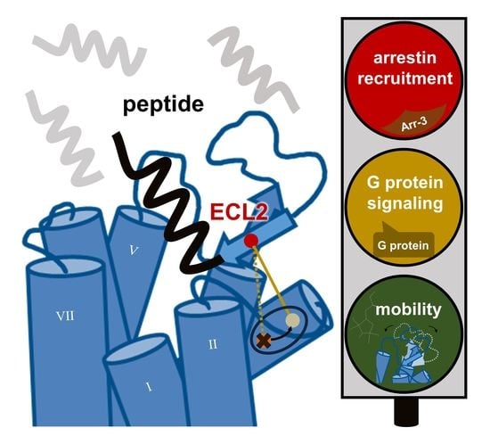
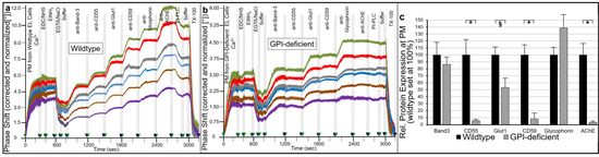
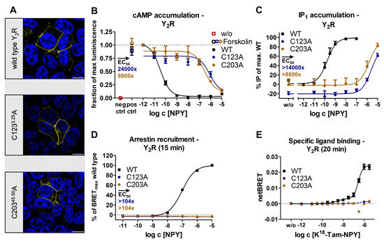
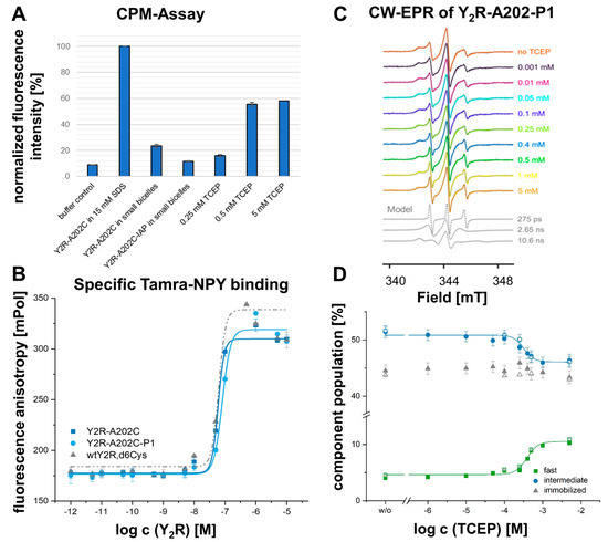
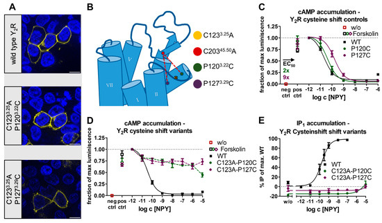
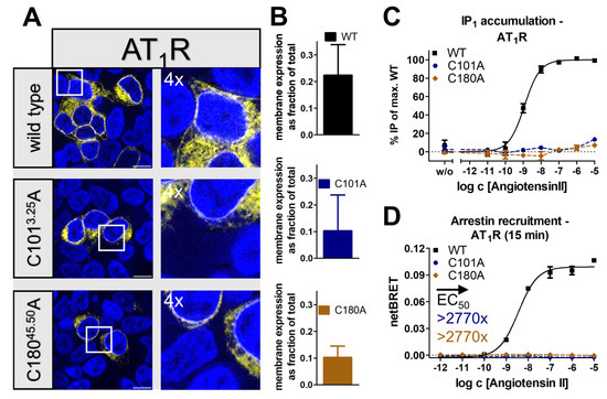
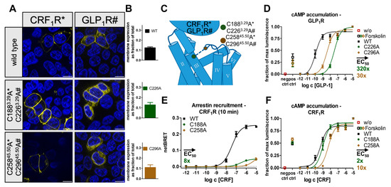
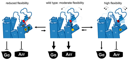
Many peptide-activated rhodopsin-like GPCRs share a β-hairpin folding motif in the extracellular loop 2 (ECL2), which interacts with the peptide ligand while at the same time being connected to transmembrane helix 3 (TM3) via a highly conserved disulfide bond. Currently, it remains unknown whether the coupling of the specifically shaped ECL2 to TM3 influences the activation of peptide-activated GPCRs. We investigated this possibility in a selection of peptide GPCRs with known structures. Most of the receptors with cysteine to alanine mutations folded like the respective wild-type and resided in the cell membrane, challenging pure folding stabilization by the disulfide bridge. G-protein signaling of the disulfide mutants was retained to a greater extent in secretin-like GPCRs than in rhodopsin-like GPCRs, while recruitment of arrestin was completely abolished in both groups, which may be linked to alterations in ligand residence time. We found a correlation between receptor activity of the neuropeptide Y2 receptor and alterations in ECL2 dynamics using engineered disulfide bridges or site-directed spin labeling and EPR spectroscopy. These data highlight the functional importance of the TM3-ECL2 link for the activation of specific signaling pathways in peptide-activated GPCRs, which might have implications for future drug discovery.
Full article
Graphical abstract