The Increasingly Fascinating World of Interleukins
A topical collection in Cells (ISSN 2073-4409). This collection belongs to the section "Cellular Immunology".
Viewed by 37366
Share This Topical Collection
Editor
 Dr. Yasu-Taka Azuma
Dr. Yasu-Taka Azuma
E-Mail
Website
Collection Editor
Laboratory of Veterinary Pharmacology, Division of Veterinary Science, Osaka Prefecture University Graduate School of Life and Environmental Sciences, 1-58 Rinku-ohraikita, Izumisano, Osaka 598-8531, Japan
Interests: macrophage; dendritic cell; T cell; microglia; cytokine; interleukin; gastrointestinal immunology; liver fibrosis; inflammation; etc.
Topical Collection Information
Dear Colleagues,
The number of interleukin types has increased over the years, finally reaching over 40. Many interleukins play a pro-inflammatory role, but some have anti-inflammatory properties. In addition, many interleukins are produced by immune system cells, but some are also produced by non-immune-system cells. It is also interesting to note that several interleukin subfamilies share intracellular signaling. The complexity and sophistication of this system has fascinated many basic and clinical researchers. There are successful examples of bridging basic research to clinical practice; antibody drugs against interleukins and their receptors are used for treatment. In particular, expectations are now rising for the fulfilment of unmet medical needs. Regulating interleukins to control disease was a great achievement, but it is only one success story. It is now becoming clear that interleukins are involved in biological responses before the onset of disease, and that they are involved not only in intercellular networks but also in interorgan networks.
In this Topical Collection of Cells, we seek contributions either in the form of original research articles, reviews, or shorter perspective articles on all aspects related to the subject of “The Increasingly Fascinating World of Interleukins”. We would like to see the fascinating world of interleukins explained by leading experts in their respective fields of research. Papers that demonstrate mechanisms at the cellular or molecular level, in vitro or in vivo, and functional insights in basic or clinical research are welcome.
Dr. Yasu-Taka Azuma
Collection Editor
Manuscript Submission Information
Manuscripts should be submitted online at www.mdpi.com by registering and logging in to this website. Once you are registered, click here to go to the submission form. Manuscripts can be submitted until the deadline. All submissions that pass pre-check are peer-reviewed. Accepted papers will be published continuously in the journal (as soon as accepted) and will be listed together on the collection website. Research articles, review articles as well as short communications are invited. For planned papers, a title and short abstract (about 100 words) can be sent to the Editorial Office for announcement on this website.
Submitted manuscripts should not have been published previously, nor be under consideration for publication elsewhere (except conference proceedings papers). All manuscripts are thoroughly refereed through a single-blind peer-review process. A guide for authors and other relevant information for submission of manuscripts is available on the Instructions for Authors page. Cells is an international peer-reviewed open access semimonthly journal published by MDPI.
Please visit the Instructions for Authors page before submitting a manuscript.
The Article Processing Charge (APC) for publication in this open access journal is 2700 CHF (Swiss Francs).
Submitted papers should be well formatted and use good English. Authors may use MDPI's
English editing service prior to publication or during author revisions.
Keywords
- Interleukin
- Immune system
- Signal transduction
- Target cell
- Target receptor
- Temporal regulation of production
- Therapeutic applications
- Cellular responses
Published Papers (8 papers)
Open AccessArticle
Expression of IL-37 Induces a Regulatory T-Cell-like Phenotype and Function in Jurkat Cells
by
Douglas Grant Osborne, Joanne Domenico and Mayumi Fujita
Cited by 4 | Viewed by 3563
Abstract
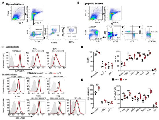
The anti-inflammatory cytokine interleukin-37 (IL-37) plays a key role in inhibiting innate and adaptive immunity. Past results have shown that IL-37 is elevated in human Treg cells compared to other T cell subsets and contributes to enhancing the Treg transcription factor, forkhead box
[...] Read more.
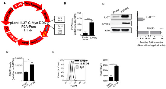
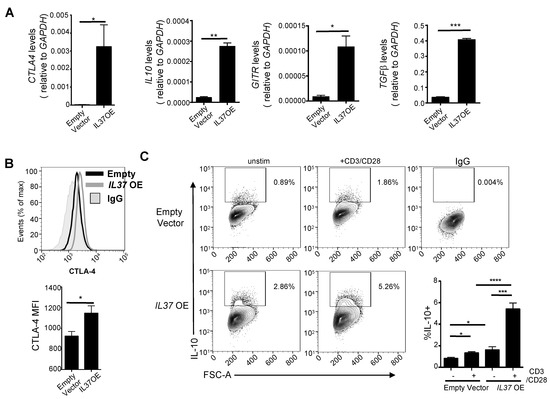
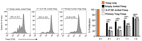
The anti-inflammatory cytokine interleukin-37 (IL-37) plays a key role in inhibiting innate and adaptive immunity. Past results have shown that IL-37 is elevated in human Treg cells compared to other T cell subsets and contributes to enhancing the Treg transcription factor, forkhead box protein P3 (FOXP3). However, it is unknown if ectopic expression of IL-37 in non-Treg CD4+ T cells can lead to the development of Treg phenotype and function. In the present study, we used a PrimeFlow
® RNA assay and confirmed elevated
IL37 expression in human Treg cells. We then stably transfected the non-Treg CD4+ T cell leukemia cell line, E6 Jurkat cells, with
IL37 and found significant induction of the Treg phenotype. These IL-37-expressing Jurkat cells had elevated CTLA-4 and FOXP3 and produced IL-10. In conjunction with the Treg phenotype, IL-37-expressing Jurkat cells suppressed T cell activation/proliferation, comparable to human primary Treg cells. The creation of this stable human Treg-like cell line has the potential to provide further assistance for in vitro studies of human Treg cells, as it is more convenient than the use of primary human Treg cells. Furthermore, it provides insights into Treg cell biology and function.
Full article
►▼
Show Figures
Open AccessReview
Challenging the Paradigm: Anti-Inflammatory Interleukins and Angiogenesis
by
Amanda M. Peluzzo and Michael V. Autieri
Cited by 22 | Viewed by 3714
Abstract
Angiogenesis is a vital biological process, and neovascularization is essential for the development, wound repair, and perfusion of ischemic tissue. Neovascularization and inflammation are independent biological processes that are linked in response to injury and ischemia. While clear that pro-inflammatory factors drive angiogenesis,
[...] Read more.
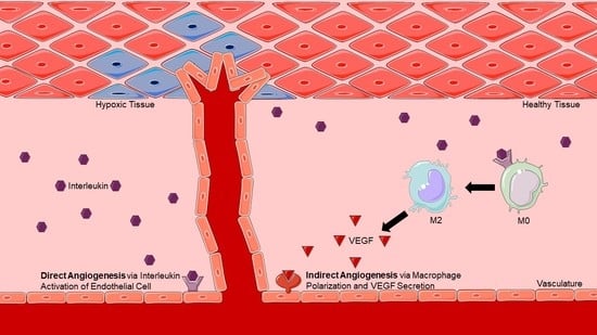
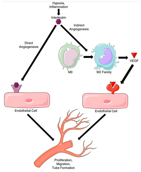
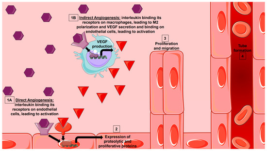
Angiogenesis is a vital biological process, and neovascularization is essential for the development, wound repair, and perfusion of ischemic tissue. Neovascularization and inflammation are independent biological processes that are linked in response to injury and ischemia. While clear that pro-inflammatory factors drive angiogenesis, the role of anti-inflammatory interleukins in angiogenesis remains less defined. An interleukin with anti-inflammatory yet pro-angiogenic effects would hold great promise as a therapeutic modality to treat many disease states where inflammation needs to be limited, but revascularization and reperfusion still need to be supported. As immune modulators, interleukins can polarize macrophages to a pro-angiogenic and reparative phenotype, which indirectly influences angiogenesis. Interleukins could also potentially directly induce angiogenesis by binding and activating its receptor on endothelial cells. Although a great deal of attention is given to the negative effects of pro-inflammatory interleukins, less is described concerning the potential protective effects of anti-inflammatory interleukins on various disease processes. To focus this review, we will consider IL-4, IL-10, IL-13, IL-19, and IL-33 to be anti-inflammatory interleukins, all of which have recognized immunomodulatory effects. This review will summarize current research concerning anti-inflammatory interleukins as potential drivers of direct and indirect angiogenesis, emphasizing their role in future therapeutics.
Full article
►▼
Show Figures
Open AccessArticle
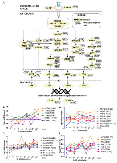
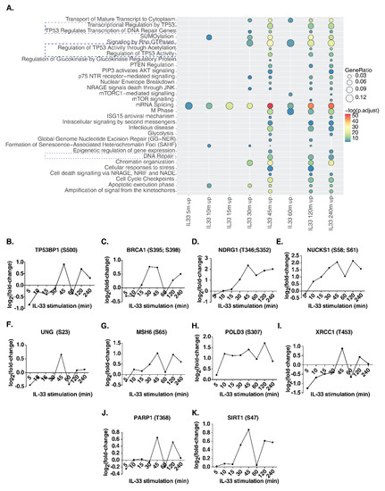
Temporal Quantitative Phosphoproteomics Profiling of Interleukin-33 Signaling Network Reveals Unique Modulators of Monocyte Activation
by
Devasahayam Arokia Balaya Rex, Yashwanth Subbannayya, Prashant Kumar Modi, Akhina Palollathil, Lathika Gopalakrishnan, Yashodhar P. Bhandary, Thottethodi Subrahmanya Keshava Prasad and Sneha M. Pinto
Cited by 3 | Viewed by 3068
Abstract
Interleukin-33 (IL-33), a member of the IL-1 superfamily cytokines, is an endogenous danger signal and a nuclear-associated cytokine. It is one of the essential mediators of both innate and adaptive immune responses. Aberrant IL-33 signaling has been demonstrated to play a defensive role
[...] Read more.
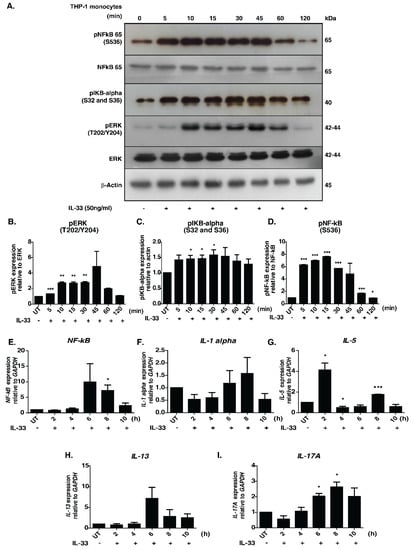
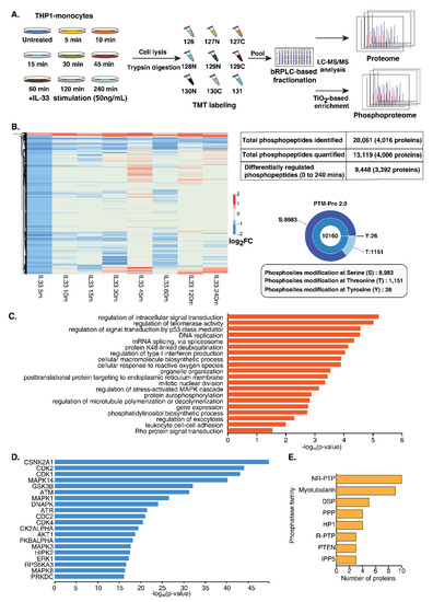
Interleukin-33 (IL-33), a member of the IL-1 superfamily cytokines, is an endogenous danger signal and a nuclear-associated cytokine. It is one of the essential mediators of both innate and adaptive immune responses. Aberrant IL-33 signaling has been demonstrated to play a defensive role against various infectious and inflammatory diseases. Although the signaling responses mediated by IL-33 have been previously reported, the temporal signaling dynamics are yet to be explored. To this end, we applied quantitative temporal phosphoproteomics analysis to elucidate pathways and proteins induced by IL-33 in THP-1 monocytes. Employing a TMT labeling-based quantitation and titanium dioxide (TiO
2)-based phosphopeptide enrichment strategy followed by mass spectrometry analysis, we identified and quantified 9448 unique phosphopeptides corresponding to 3392 proteins that showed differential regulation. Of these, 171 protein kinases, 60 phosphatases and 178 transcription factors were regulated at different phases of IL-33 signaling. In addition to the confirmed activation of canonical signaling modules including MAPK, NFκB, PI3K/AKT modules, pathway analysis of the time-dependent phosphorylation dynamics revealed enrichment of several cellular processes, including leukocyte adhesion, response to reactive oxygen species, cell cycle checkpoints, DNA damage and repair pathways. The detailed quantitative phosphoproteomic map of IL-33 signaling will serve as a potentially useful resource to study its function in the context of inflammatory and pathological conditions.
Full article
►▼
Show Figures
Open AccessReview
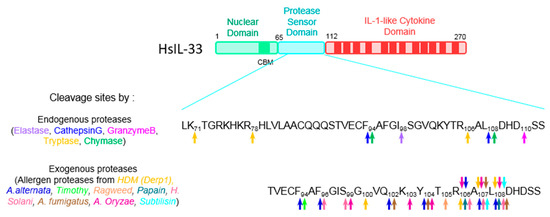
IL-33, an Alarmin of the IL-1 Family Involved in Allergic and Non Allergic Inflammation: Focus on the Mechanisms of Regulation of Its Activity
by
Corinne Cayrol
Cited by 42 | Viewed by 4465
Abstract
Interleukin-33 (IL-33) is a member of the interleukin-1 (IL-1) family that is expressed in the nuclei of endothelial and epithelial cells of barrier tissues, among others. It functions as an alarm signal that is released upon tissue or cellular injury. IL-33 plays a
[...] Read more.
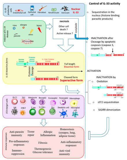
Interleukin-33 (IL-33) is a member of the interleukin-1 (IL-1) family that is expressed in the nuclei of endothelial and epithelial cells of barrier tissues, among others. It functions as an alarm signal that is released upon tissue or cellular injury. IL-33 plays a central role in the initiation and amplification of type 2 innate immune responses and allergic inflammation by activating various target cells expressing its ST2 receptor, including mast cells and type 2 innate lymphoid cells. Depending on the tissue environment, IL-33 plays a wide variety of roles in parasitic and viral host defense, tissue repair and homeostasis. IL-33 has evolved a variety of sophisticated regulatory mechanisms to control its activity, including nuclear sequestration and proteolytic processing. It is involved in many diseases, including allergic, inflammatory and infectious diseases, and is a promising therapeutic target for the treatment of severe asthma. In this review, I will summarize the literature around this fascinating pleiotropic cytokine. In the first part, I will describe the basics of IL-33, from the discovery of interleukin-33 to its function, including its expression, release and signaling pathway. The second part will be devoted to the regulation of IL-33 protein leading to its activation or inactivation.
Full article
►▼
Show Figures
Open AccessFeature PaperArticle
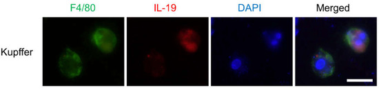
IL-19 Contributes to the Development of Nonalcoholic Steatohepatitis by Altering Lipid Metabolism
by
Yasu-Taka Azuma, Takashi Fujita, Takeshi Izawa, Kana Hirota, Kazuhiro Nishiyama, Airi Ikegami, Tomoko Aoyama, Mikihito Ike, Yumi Ushikai, Mitsuru Kuwamura, Hideki Fujii and Koichi Tsuneyama
Cited by 10 | Viewed by 3816
Abstract
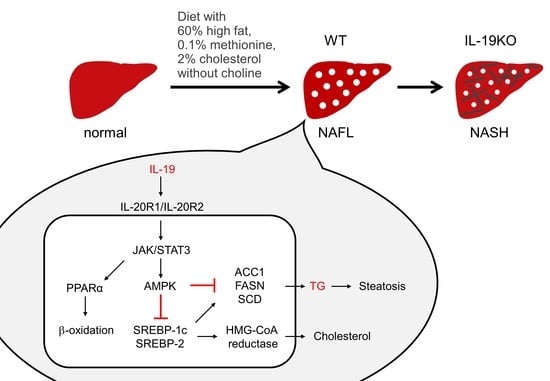
Interleukin (IL)-19, a member of the IL-10 family, is an anti-inflammatory cytokine produced primarily by macrophages. Nonalcoholic steatohepatitis (NASH) is a disease that has progressed from nonalcoholic fatty liver disease (NAFLD) and is characterized by inflammation and fibrosis. We evaluated the functions of
[...] Read more.
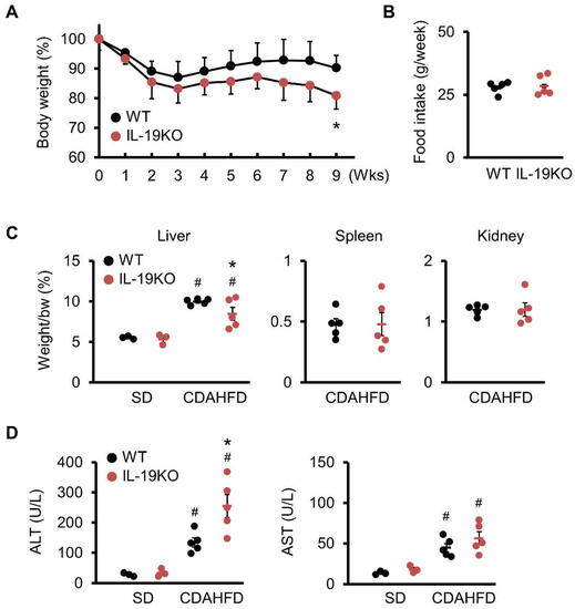
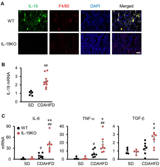
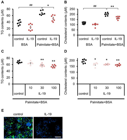
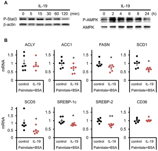
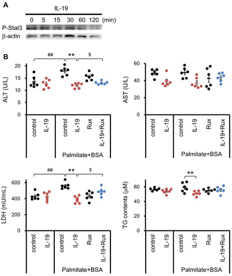
Interleukin (IL)-19, a member of the IL-10 family, is an anti-inflammatory cytokine produced primarily by macrophages. Nonalcoholic steatohepatitis (NASH) is a disease that has progressed from nonalcoholic fatty liver disease (NAFLD) and is characterized by inflammation and fibrosis. We evaluated the functions of IL-19 in a NAFLD/NASH mouse model using a 60% high fat diet with 0.1% methionine, without choline, and with 2% cholesterol (CDAHFD). Wild-type (WT) and IL-19 gene-deficient (KO) mice were fed a CDAHFD or standard diet for 9 weeks. Liver injury, inflammation, and fibrosis induced by CDAHFD were significantly worse in IL-19 KO mice than in WT mice. IL-6, TNF-α, and TGF-β were significantly higher in IL-19 KO mice than in WT mice. As a mechanism using an in vitro experiment, palmitate-induced triglyceride and cholesterol contents were decreased by the addition of IL-19 in HepG2 cells. Furthermore, addition of IL-19 decreased the expression of fatty acid synthesis-related enzymes and increased ATP content in HepG2 cells. The action of IL-19 in vitro suppressed lipid metabolism. In conclusion, IL-19 may play an important role in the development of steatosis and fibrosis by directly regulating liver metabolism and may be a potential target for the treatment of liver diseases.
Full article
►▼
Show Figures
Open AccessReview
The Interleukins Orchestrate Mucosal Immune Responses to Salmonella Infection in the Intestine
by
Fu-Chen Huang
Cited by 17 | Viewed by 3868
Abstract
Salmonella infection remains one of the major public health problems in the world, with increasing resistance to antibiotics. The resolution is to explore the pathogenesis of the infection and search for alternative therapy other than antibiotics. Immune responses to Salmonella infection include innate
[...] Read more.
Salmonella infection remains one of the major public health problems in the world, with increasing resistance to antibiotics. The resolution is to explore the pathogenesis of the infection and search for alternative therapy other than antibiotics. Immune responses to Salmonella infection include innate and adaptive immunity. Flagellin or muramyl dipeptide from Salmonella, recognized by extracellular Toll-like receptors and intracellular nucleotide-binding oligomerization domain2, respectively, induce innate immunity involving intestinal epithelial cells, neutrophils, macrophages, dendric cells and lymphocytes, including natural killer (NK) and natural killer T (NKT) cells. The cytokines, mostly interleukins, produced by the cells involved in innate immunity, stimulate adaptive immunity involving T and B cells. The mucosal epithelium responds to intestinal pathogens through its secretion of inflammatory cytokines, chemokines, and antimicrobial peptides. Chemokines, such as IL-8 and IL-17, recruit neutrophils into the cecal mucosa to defend against the invasion of
Salmonella, but induce excessive inflammation contributing to colitis. Some of the interleukins have anti-inflammatory effects, such as IL-10, while others have pro-inflammatory effects, such as IL-1β, IL-12/IL-23, IL-15, IL-18, and IL-22. Furthermore, some interleukins, such as IL-6 and IL-27, exhibit both pro- and anti-inflammatory functions and anti-microbial defenses. The majority of interleukins secreted by macrophages and lymphocytes contributes antimicrobial defense or protective effects, but IL-8 and IL-10 may promote systemic
Salmonella infection. In this article, we review the interleukins involved in Salmonella infection in the literature.
Full article
Open AccessReview
Significance of Interleukin (IL)-4 and IL-13 in Inflammatory Arthritis
by
Milena Iwaszko, Sylwia Biały and Katarzyna Bogunia-Kubik
Cited by 88 | Viewed by 10705
Abstract
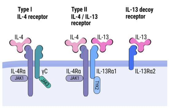
Interleukin (IL)-4 and IL-13 belong to the T helper 2 (Th2) cytokine family, along with IL-3, IL-5, and IL-9. These cytokines are key mediators of allergic inflammation. They have important immunomodulatory activities and exert influence on a wide variety of immune cells, such
[...] Read more.
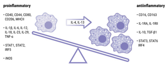
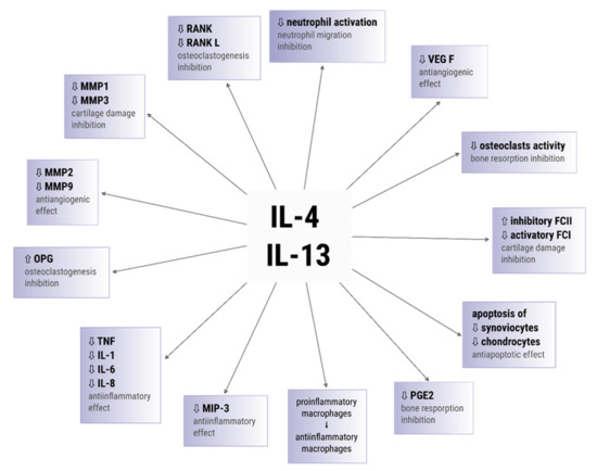
Interleukin (IL)-4 and IL-13 belong to the T helper 2 (Th2) cytokine family, along with IL-3, IL-5, and IL-9. These cytokines are key mediators of allergic inflammation. They have important immunomodulatory activities and exert influence on a wide variety of immune cells, such as B cells, eosinophils, basophils, monocytes, fibroblasts, endothelial cells, airway epithelial cells, smooth muscle cells, and keratinocytes. Recent studies have implicated IL-4 and IL-13 in the development of various autoimmune diseases. Additionally, these cytokines have emerged as potential players in pathogenesis of inflammatory arthritis. Recent findings suggest that the IL-4 and IL-13 might play a significant role in the downregulation of inflammatory processes underlying RA pathology, and beneficially modulate the course of the disease. This review summarizes the biological features of the IL-4 and IL-13 and provides current knowledge regarding the role of these cytokines in inflammatory arthritis.
Full article
►▼
Show Figures
Open AccessArticle
Two Sides to Every Question: Attempts to Activate Chicken Innate Immunity in 2D and 3D Hepatic Cell Cultures
by
Csilla Sebők, Patrik Tráj, Júlia Vörösházi, Máté Mackei, Márton Papp, Péter Gálfi, Zsuzsanna Neogrády and Gábor Mátis
Cited by 8 | Viewed by 2837
Abstract
The liver with resident tissue macrophages is the site of vivid innate immunity, activated also by pathogen-associated molecular patterns (PAMPs) leaking through the intestinal barrier. As gut-derived inflammatory diseases are of outstanding importance in broiler chickens, the present study aimed to establish a
[...] Read more.
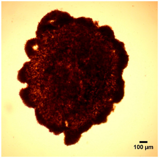
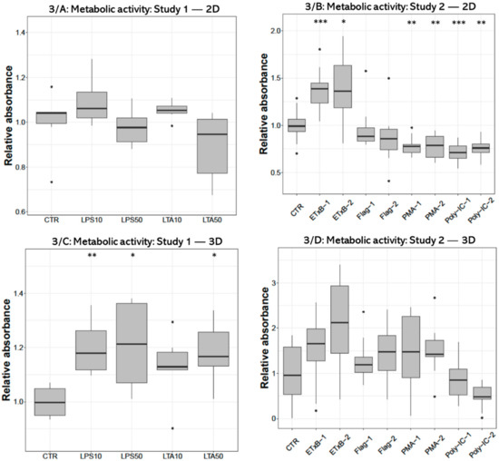
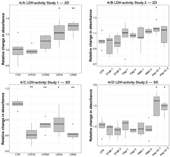
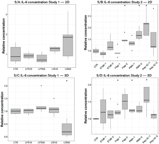
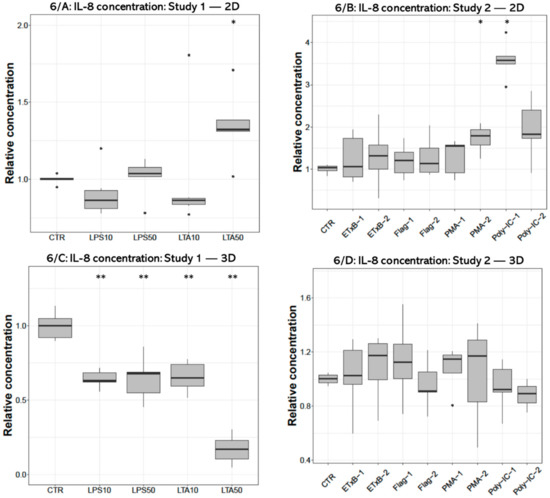
The liver with resident tissue macrophages is the site of vivid innate immunity, activated also by pathogen-associated molecular patterns (PAMPs) leaking through the intestinal barrier. As gut-derived inflammatory diseases are of outstanding importance in broiler chickens, the present study aimed to establish a proper hepatic inflammatory model by comparing the action of different PAMPs from poultry pathogens on chicken 2D and 3D primary hepatocyte—non-parenchymal cell co-cultures, the latter newly developed with a magnetic bioprinting method. The cultures were challenged by the bacterial endotoxins lipopolysaccharide (LPS) from
Escherichia coli, lipoteichoic acid (LTA) from
Staphylococcus aureus and by enterotoxin (ETxB) from
Escherichia coli,
Salmonella Typhimurium derived flagellin, phorbol myristate acetate (PMA) as a model proinflammatory agent and polyinosinic polycytidylic acid (poly I:C) for mimicking viral RNA exposure. Cellular metabolic activity was assessed with the CCK-8 test, membrane damage was monitored with the lactate dehydrogenase (LDH) leakage assay and interleukin-6 and -8 (Il-6 and -8) concentrations were measured in cell culture medium with a chicken specific ELISA. Both LPS and LTA increased the metabolic activity of the 3D cultures, concomitantly decreasing the LDH leakage, while in 2D cultures ETxB stimulated, PMA and poly I:C depressed the metabolic activity. Based on the moderately increased extracellular LDH activity, LTA seemed to diminish cell membrane integrity in 2D and poly I:C in both cell culture models. The applied endotoxins remarkably reduced the IL-8 release of 3D cultured cells, suggesting the effective metabolic adaptation and the presumably initiated anti-inflammatory mechanisms of the 3D spheroids. Notwithstanding that the IL-6 and IL-8 production of 2D cells was mostly not influenced by the endotoxins used, only the higher LTA dose was capable to evoke an IL-8 surge. Flagellin, PMA and poly I:C exerted proinflammatory action in certain concentrations in both 2D and 3D cultures, reflected by the increased cellular IL-6 release. Based on these data, LTA, flagellin, PMA and poly I:C can be considered as potent candidates to induce inflammation in chicken primary hepatic cell cultures, while LPS failed to trigger proinflammatory cytokine production, suggesting the relatively high tolerance of avian liver cells to certain bacterial endotoxins. These results substantiate that the established 3D co-cultures seemed to be proper tools for testing potential proinflammatory molecules; however, the remarkable differences between 2D and 3D models should be addressed and further studied.
Full article
►▼
Show Figures
Planned Papers
The below list represents only planned manuscripts. Some of these
manuscripts have not been received by the Editorial Office yet. Papers
submitted to MDPI journals are subject to peer-review.
Title: IL-20 cytokines controls airway mucosa response at homeostasis and during chronic inflammatory diseases
Authors: Philippe Gosset
Affiliation: CNRS UMR9017, Inserm U1019, University of Lille, Centre Hospitalier Régional Universitaire de Lille, Institut Pasteur, CIIL—Center for Infection and Immunity of Lille, 59000 Lille, France
Title: Regulation of cancer stem cell properties by interleukins
Authors: Eiichi Hinoi
Affiliation: Gifu Pharmaceutical University


