Helicobacter species may cause chronic inflammation of the biliary tract, but its relationship with cancer is controversial. We performed a systematic review and meta-analysis to evaluate the association between
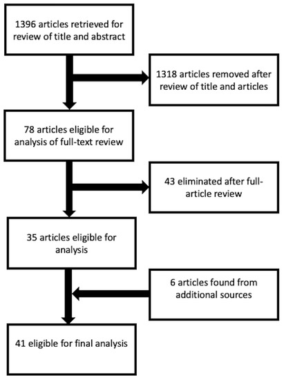
Helicobacter species and hepatobiliary tract malignancies. Twenty-six studies (4083 patients) were included in qualitative
[...] Read more.
Helicobacter species may cause chronic inflammation of the biliary tract, but its relationship with cancer is controversial. We performed a systematic review and meta-analysis to evaluate the association between
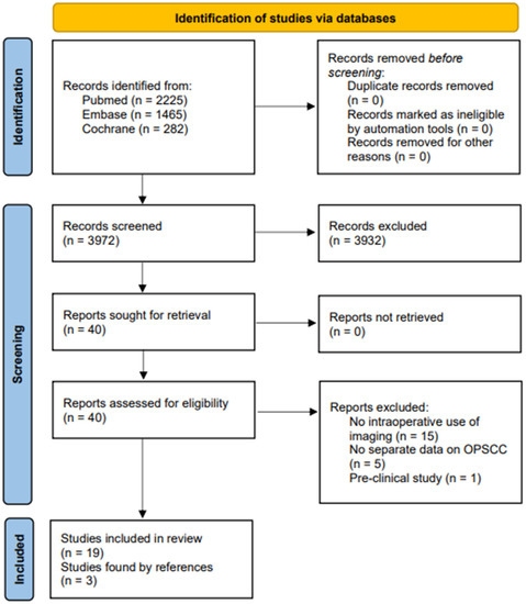
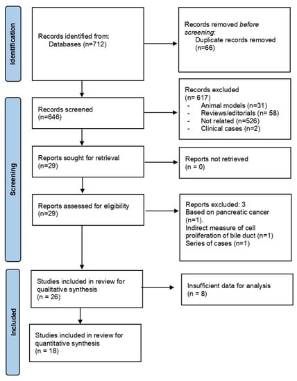
Helicobacter species and hepatobiliary tract malignancies. Twenty-six studies (4083 patients) were included in qualitative synthesis, and 18 studies (
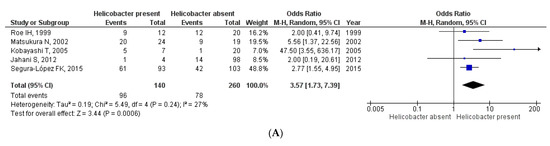
n = 1895 qualified for meta-analysis. All studies were at high-intermediate risk of bias. Most studies combined several direct microbiological methods, mostly PCR (23 studies), culture (8 studies), and/or CLOtest (5 studies). Different specimens alone or in combination were investigated, most frequently bile (16 studies), serum (7 studies), liver/biliary tissue (8 studies), and gastric tissue (3 studies). Patients with
Helicobacter species infection had an increased risk of hepatobiliary tract malignancies (OR = 3.61 [95% CI 2.18–6.00];
p < 0.0001), with high heterogeneity in the analysis (I
2 = 61%;
p = 0.0003). This effect was consistent when
Helicobacter was assessed in bile (OR = 3.57 [95% CI 1.73–7.39];
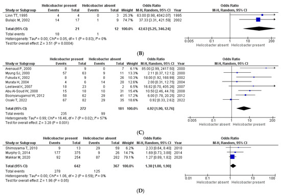
p = 0.0006), gastric tissue (OR = 42.63 [95% CI 5.25–346.24];
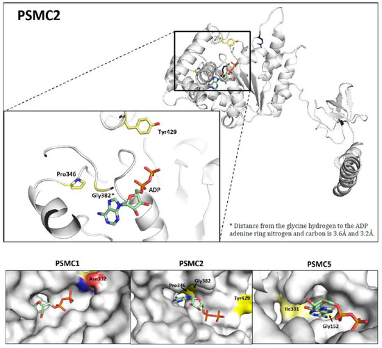
p = 0.0004), liver/biliary tissue (OR = 4.92 [95% CI 1.90–12.76];
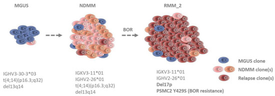
p = 0.001) and serum (OR = 1.38 [95% CI 1.00–1.90];
p = 0.05). Heterogeneity was reduced in these sub-analyses (I
2 = 0–27%;
p = ns), except for liver/biliary tissue (I
2 = 57%;
p = 0.02). In conclusion, based on low-certainty data,
Helicobacter species chronic infection is associated with a tripled risk of hepatobiliary tract malignancy. Prospective studies are required to delineate public health interventions.
Full article


