Systematic Review

Helicobacter Species and Hepato-Biliary Tract Malignancies: A Systematic Review and Meta-Analysis

by
Beatriz Gros
1,2,
Alberto Gómez Pérez
1,
María Pleguezuelo
1,2,
Francisco Javier Serrano Ruiz
1,2,
Manuel de la Mata
1,2,3 and
Manuel Rodríguez-Perálvarez
1,2,3,*
1
Department of Gastroenterology and Hepatology, Hospital Universitario Reina Sofía, 14004 Córdoba, Spain
2
Maimonides Institute of Biomedical Research (IMIBIC), 14004 Córdoba, Spain
3
Centro de Investigación Biomédica en Red de Enfermedades Hepáticas y Digestivas (CIBERehd), 28029 Madrid, Spain
*
Author to whom correspondence should be addressed.
Cancers 2023, 15(3), 595; https://doi.org/10.3390/cancers15030595
Submission received: 25 November 2022 / Revised: 15 January 2023 / Accepted: 16 January 2023 / Published: 18 January 2023
(This article belongs to the Collection Targeting Solid Tumors)

Abstract

:

Simple Summary

Chronic infections are major drivers of cancer. Helicobacter species is one of the most established pro-oncogenic pathogens, but its relationship with hepatobiliary tract malignancies is controversial. We performed a systematic review and meta-analysis of 26 observational studies, including 1203 patients with hepatobiliary tract malignancies and 2880 controls. Helicobacter species chronic infection demonstrated by direct microbiological methods tripled the risk of hepatobiliary tract malignancies, and this effect was consistent for different types of specimens tested, including bile, gastric tissue, hepatic/biliary tissue, and serum. However, available studies were heterogeneous, and the overall quality of the evidence was low, translating into a grade of uncertainty. Prospective studies, randomized, if possible, are required to delineate interventions at a public healthcare scale, particularly in geographic areas with increased incidence of hepatobiliary tumors.

Abstract

Helicobacter species may cause chronic inflammation of the biliary tract, but its relationship with cancer is controversial. We performed a systematic review and meta-analysis to evaluate the association between Helicobacter species and hepatobiliary tract malignancies. Twenty-six studies (4083 patients) were included in qualitative synthesis, and 18 studies (n = 1895 qualified for meta-analysis. All studies were at high-intermediate risk of bias. Most studies combined several direct microbiological methods, mostly PCR (23 studies), culture (8 studies), and/or CLOtest (5 studies). Different specimens alone or in combination were investigated, most frequently bile (16 studies), serum (7 studies), liver/biliary tissue (8 studies), and gastric tissue (3 studies). Patients with Helicobacter species infection had an increased risk of hepatobiliary tract malignancies (OR = 3.61 [95% CI 2.18–6.00]; p < 0.0001), with high heterogeneity in the analysis (I2 = 61%; p = 0.0003). This effect was consistent when Helicobacter was assessed in bile (OR = 3.57 [95% CI 1.73–7.39]; p = 0.0006), gastric tissue (OR = 42.63 [95% CI 5.25–346.24]; p = 0.0004), liver/biliary tissue (OR = 4.92 [95% CI 1.90–12.76]; p = 0.001) and serum (OR = 1.38 [95% CI 1.00–1.90]; p = 0.05). Heterogeneity was reduced in these sub-analyses (I2 = 0–27%; p = ns), except for liver/biliary tissue (I2 = 57%; p = 0.02). In conclusion, based on low-certainty data, Helicobacter species chronic infection is associated with a tripled risk of hepatobiliary tract malignancy. Prospective studies are required to delineate public health interventions.

1. Introduction

Several chronic infections are well-known drivers of cancer in humans [1]. Helicobacter pylori (HP), Opisthorchis viverrini, Clonorchis sinensis, and hepatitis B and C viruses, among other pathogens, have been classified as group 1 carcinogens [2]. In 2018, 2.2 million cancers were attributed to chronic infections worldwide, among which HP was paramount, being responsible for 810.000 newly diagnosed malignancies (mainly non-cardia gastric adenocarcinoma) [3].
Primary liver cancer, which includes mainly hepatocellular carcinoma (HCC) (75–85%) and intrahepatic cholangiocarcinoma (ICC) (10–15%), ranks the sixth malignancy worldwide in terms of prevalence. Indeed, primary liver tumors account for 4.7% of all incident cancers, being the third cause of cancer-related death [4]. Pancreatic cancer and gallbladder cancer represent 2.6% and 0.9% of new cancer diagnoses, respectively, being both characterized by their lethality [5].
There is controversy regarding the epidemiological association between HP (and other Helicobacter species infection) and hepatobiliary tract malignancies [6]. The available evidence relies mostly on case-control studies from Asia, where the highest incidence of these malignancies has been reported [7,8]. The interpretation of observational studies is often challenging due to the high prevalence of other confounding pro-oncogenic infections, such as liver fluke and viral hepatitis [9]. Included cohorts are usually small, and healthy controls are lacking for obvious ethical reasons [10].
The unequivocal assessment of the attributable pro-oncogenic risk of Helicobacter spp. is paramount to delineating public health policies. These data would allow us to identify groups of risk for potential screening programs and to implement therapeutic interventions at a population level. The present systematic review and meta-analysis aim to summarize the available evidence regarding Helicobacter spp. and hepatobiliary tract malignancies with a focus on possible prevention strategies.

2. Materials and Methods

2.1. Search Strategy

We searched MEDLINE, Cochrane Controlled Trial Register (CENTRAL), EMBASE, and Science Citation Index databases, without language restrictions, from inception to May 2022. In addition, the recent literature reviews on the topic, if available, were hand-searched for additional relevant references. We identified studies by using different combinations of the following keywords or equivalent free-text terms: [“helicobacter” OR “helicobacter mustelae” OR “helicobacter hepaticus” OR “helicobacter felis” OR “helicobacter heilmannii” OR “Helicobacter species”] AND [“biliary tract neoplasms” OR “malignancy” OR “cholangiocarcinoma” OR “tumor” OR “cancer”].

2.2. Selection Criteria

The Preferred Reporting Items for Systematic reviews and Meta-Analyses (PRISMA) statement was used to illustrate the flowchart of included and excluded studies [11]. Randomized clinical trials, cohort studies, or case-control studies published in full length comparing the incidence or prevalence of biliary tract malignancies according to the presence of Helicobacter species assessed by direct microbiological methods were included. Exclusion criteria were as follows: studies performed in children or adolescents (<18 years old), uncontrolled studies (mainly case series), editorials or the literature reviews, and studies performed in animal models and in vitro experiments. Studies were also excluded if the approval from an ethics research committee was not disclosed or if the provided data were insufficient to weigh the risk of cancer attributable to Helicobacter species after contact the corresponding author. Two review authors (BG and AGP) independently screened the retrieved references by examining the title, abstract and full texts whenever necessary. Any discrepancy was resolved initially by discussion. Persistent disagreements were resolved by a third review author (MR-P). If there was any doubt as to whether two studies shared the same participants, completely or partially (by identifying common authors, institutions, and overlapping recruiting periods), we contacted the study authors to clarify whether the study report was duplicated. In the absence of a satisfying response, only the study with the larger sample size was considered.

2.3. Data Extraction

Two review authors (BG and AGP) performed the data extraction independently, using a predefined chart specifically designed for this study, with disagreements solved by a third author (MR-P). Data extracted from each study included first author, country and year of publication, sample size, sample collection, method of Helicobacter detection, diagnosis of cases and controls, matching strategy, patient characteristics, type of specimen, organisms identified, number of positive cases for Helicobacter species in each group, and adjusted odds ratios if provided. The corresponding author of each study was contacted in case of relevant missing information. If available, we obtained separate data for participants with different types of biliary malignancies.

2.4. Statistical Analysis and Risk of Bias Assessment

Studies providing sufficient data were included for quantitative synthesis (meta-analysis). We calculated the odds ratio with a 95% credible interval (or Bayesian confidence interval) to estimate the probability of hepatobiliary tract malignancies according to the presence or absence of Helicobacter infection. Since we expected significant clinical heterogeneity due to different methodologies for helicobacter assessment and different types of malignancies, random effects models were implemented. I2 values were used as indicators of the degree of intertrial heterogeneity. The possibility of publication bias was explored with funnel plots. The quality of the evidence was assessed using the checklist provided by the Clarity group at McMaster University, which evaluates the risk of bias in case-control studies by assessing five study domains as follows: quality of assessment of exposure, confidence that cases had developed the outcome of interest but not controls, appropriate cases selection, appropriate control selection and appropriate case-control matching according to important prognostic variables or statistical adjustment. Each of these domains is classified into four categories: low risk of bias, probably low risk of bias, probably high risk of bias, or definitely high risk of bias [12]. Analyses were performed using RevMan version 5.1 (Nordic Cochrane Centre, Copenhagen, Denmark). This systematic review was registered in PROSPERO (registration number 42022348410).

3. Results

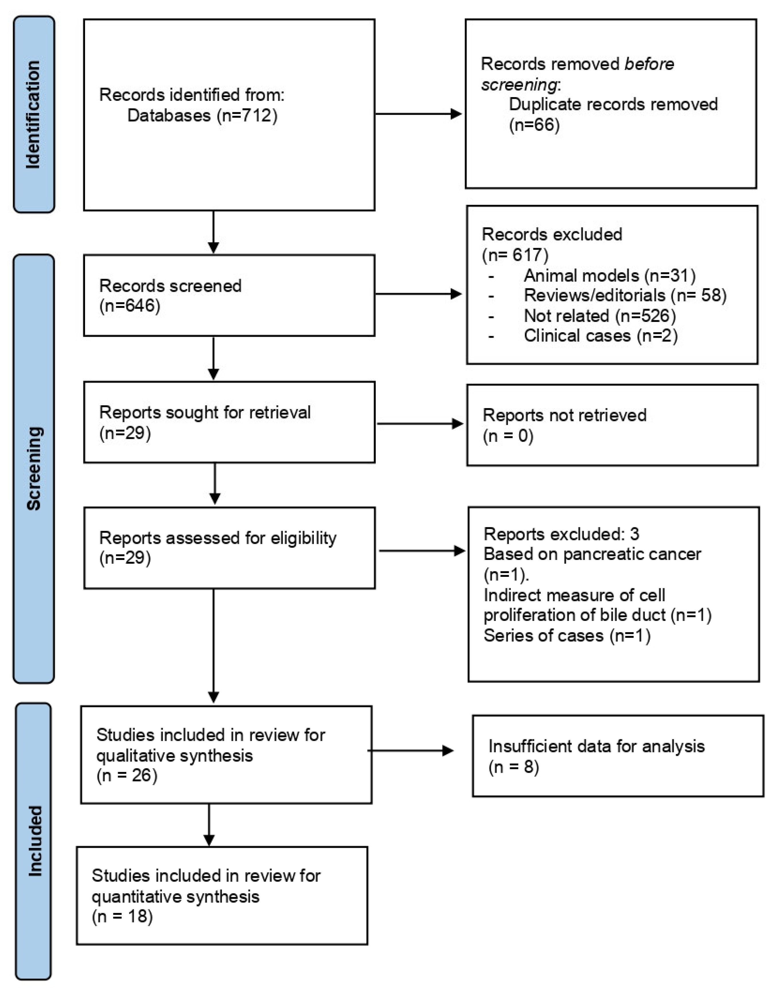
3.1. Description of the Literature Search Strategy

A total of 712 references were obtained from the literature search. The PRISMA flowchart depicting the study selection is shown in Figure 1. After removing duplicates (n = 66), 645 records were screened. Six hundred and seventeen references were excluded because they referred to animal models (n = 31), were the literature reviews, editorials, or case records (n = 60), or were unrelated to the topic of interest (n = 526). Two studies, including an identical number of patients from the same institution and with a widely overlapping enrolment period, were combined to avoid data duplication. Finally, twenty-six articles were included for qualitative synthesis.

3.2. Search Results and Study Characteristics

The main characteristics of the included studies are summarised in Table 1. All studies had a retrospective case-control design and were published between 1995 and 2022. In all, 4083 participants were included, comprising 1203 patients with hepatobiliary tract malignancies, 532 patients with benign hepatobiliary abnormalities, 633 patients with hepatobiliary abnormalities (without differentiating between benign and malignant conditions in three studies), and 1715 healthy controls. Sample size ranged from n = 7 to n = 551. The types of cancers were cholangiocarcinoma (n = 438; 36.4%), hepatocellular carcinoma (n = 231; 19.2%), gallbladder cancer (n = 111, 9.2%), pancreatic cancer (n = 96, 8%), ampulla of Vater cancer (n = 45, 3.7%) colangio-hepatocellular carcinoma (n = 2, 0.2%), cystoadenocarcinoma (n = 1, 0.1%), other bile tract malignancies (n = 279, 23.2%). Eight studies reported “bile tract cancer” comprising gallbladder and bile duct cancer without providing individual data for each tumor location. Participants acting as controls underwent imaging techniques to rule out hepatobiliary malignancies, and they had most frequently other non-malignant biliary diseases such as O. Viverrini negative controls (n = 382, 22.3%), benign biliary diseases (n = 204, 11.9%), gallstones or bile duct stones (n = 194, 11.3%), chronic cholecystitis (n = 90, 5.3%) or other less prevalent biliary diseases such as Caroli disease, primary sclerosing cholangitis, gallbladder polyps, and acute pancreatitis, among others. Of note, 638 (37.2%) patients in the control group were matched controls of large cohort studies. There was a predominance of studies from Asia (Japan [n = 6], Thailand [n = 4], Japan and Thailand [n = 1], Korea [n = 2], Taiwan [n = 1], Pakistan [n = 1], India [n = 1], Iran [n = 1]), followed by Europe (Germany [n = 2], Yugoslavia [n = 1], Finland [n = 1], France [n = 1], Sweden [n = 1]), South America (Mexico [n = 2]), and North America (Canada [n = 1]). The age and gender of the study participants were not disaggregated for cases and controls in 14 and 11 studies, respectively. For the remaining references, the pooled average age was 56.01 years in cases and 54.47 years in controls, the prevalence of women being 51.4% among cases and 47.9% among controls.

3.3. Helicobacter spp. Isolation

As for the microbiological detection methods, the majority of the studies used a single methodology (n = 15). Helicobacter spp. chronic infection was confirmed by means of PCR (n = 23), culture (n = 8), CLOtest (n = 5), histological and/or immunohistochemical (n = 3), ELISA (n = 4), Western Blot (n = 1) and multiplex assay (n = 1). In studies using PCR, the most frequently primer genes were 16rRNA 17/20 (85%) (either to Helicobacter genus or specific Helicobacter specie), urease A 8/20 (40%), cytotoxin-associated gene A (CagA) 7/20 (35%), Vacuolating cytotoxin A (VacA) 6/20 (30%) and glmM gene 3/20 (15%). Regarding the biological specimens analyzed, the most frequent were bile (16 studies), gastric tissue (3 studies), liver tissue (5 studies), gallbladder tissue (4 studies), serum (7 studies), biliary epithelium (2 studies), gallstones (2 studies) and feces (1 study). Invasive procedures to obtain samples were endoscopic retrograde cholangiopancreatography (ERCP) (10 studies), percutaneous transhepatic cholangiography (PTC) (5 studies), surgery (liver resection, cholecystectomy) (10 studies) and liver biopsy (1 study).

3.3.1. Helicobacter spp. under Investigation

The majority of the studies aimed to isolate any Helicobacter spp. (14 studies, n = 2064). However, some studies focused on specific species such as Helicobacter pylori (7 studies, n = 1453), Helicobacter hepaticus (2 studies, n = 322), Helicobacter bilis (2 studies, n = 116) or both H. hepaticus and H. bilis (1 study, n = 125).

3.3.2. Controlling for Confounders and Risk of Bias Assessment

Fourteen studies comprising 1132 participants did not implement any methodology to control for demographic features, clinical characteristics, or laboratory parameters as potential confounders. Two studies were controlled by age and sex (n = 707), and two studies were controlled by age, sex, and body mass index (n = 293). Other studies attempted to control for a myriad of possible confounders, such as hepatitis B chronic infection, hepatitis C chronic infection, liver cirrhosis, smoking habit, alcohol consumption, coffee consumption, drug abuse, hemochromatosis, diabetes, cholesterol level, and educational level. The risk of bias assessment is presented in Table 2. None of the studies obtained a low risk of bias (A category) in all domains. In 16 out of 26 studies, at least one domain showed a high risk of bias (D category). The remaining 10 studies were considered at intermediate risk of bias with 2 or 3 domains classified in B or C categories.

3.3.3. Meta-Analysis

Seventeen studies (65.4%) reported a statistically significant association between Helicobacter infection and hepatobiliary tract abnormalities. Among the remaining nine studies, two studies did not report any patients with Helicobacter spp. infection, neither in patients with cancer nor among controls, [19,24] and seven studies did not perform a statistical comparison between cases and controls. In the meta-analysis including 18 studies and 1895 individuals (933 patients with cancer and 962 controls), the presence of Helicobacter spp. was associated with a significant increase in the risk of hepatobiliary tract malignancies (OR = 3.61 [95% CI 2.18–6]; p < 0.0001), with heterogeneity I2 = 61% across studies (p = 0.0003) (Figure 2). The funnel plot suggested a possible publication bias in this analysis (Figure 3). Regarding geographic distribution, 12 studies in the meta-analysis were performed in Asia (n = 671), obtaining OR = 3.67 (95% CI 1.92–6.99) (p < 0.001), and five studies took place in Europe (n = 1028), obtaining OR = 4.86 (95% CI 1.5–15.7) (p < 0.001). There was only one study from America in the meta-analysis. Heterogeneity (I2) was 38% in Asia (p = 0.09) and 80% in Europe (p = 0.008).
The effect of Helicobacter spp. on the risk of hepatobiliary tract malignancies was consistent irrespective of the type of specimen analyzed (Figure 4). In five studies, including 400 individuals with helicobacter assessed in bile specimens, the effect was OR = 3.57 (95% CI 1.73–7.39); p = 0.0006. Among two studies with 33 participants who underwent Helicobacter assessment in gastric tissue, the effect was OR = 42.63 (95% CI 5.25–346.24); p = 0.0004. In seven studies with 404 individuals with helicobacter assessed in the liver and/or biliary tissue specimens, the effect was OR = 4.92 (95% CI 1.90–12.76); p = 0.001. Finally, among three studies including 1009 participants with Helicobacter assessed in serum, the effect was OR = 1.38 (95% CI 1.00–1.90); p = 0.05. Heterogeneity was low (I2 ranging from 0% to 27%) for bile, gastric tissue, and serum (p = 0.24, p = 0.83, and p = 0.59, respectively). However, there was a significant heterogeneity (I2 = 57%; p = 0.02) when Helicobacter was assessed in liver/biliary tissue specimens.

4. Discussion

Helicobacter spp. may cause chronic inflammation in different organs and tissues, including the hepatobiliary tract [40]. In some types of malignancies, such as gastric cancer, the eradication of HP has been shown to prevent the disease in high-risk populations [41]. In this systematic review and meta-analysis, we showed a direct association between Helicobacter spp. and hepatobiliary tract malignancies, providing relevant data to enable future screening strategies and public health interventions in order to minimize this important health burden.
HP infection is a major global health issue, but there is still little information available on the epidemiology, geographic distribution, and clinical impact of other Helicobacter species [42]. Regardless of its etiology, chronic inflammation and cholestasis are paramount factors in cholangiocarcinogenesis [43]. Chronic inflammation results in an increased exposure of cholangiocytes to interleukin-6, Tumor Necrosis Factor-ɑ, Cyclo-oxygenase-2, and other pro-inflammatory mediators, resulting in gradual mutations in tumor suppressor genes, proto-oncogenes and DNA mismatch-repair genes [44,45,46]. Helicobacter spp. in bile or liver tissue promotes chronic hepatitis in animal models and humans [25,47,48], enhances tissue inflammation in models of acute pancreatitis [49], induces mesenchymal transition in human cholangiocytes in vitro [50], and even hepatocellular and bile tract malignancies in genetically modified mice [51].
On the other hand, a complex interplay exists between Helicobacter spp. and liver fluke in endemic countries, where the latter infection could explain part of the carcinogenic effect [52,53]. Opistorchis viverrini is a well-established risk factor for CCA [54], and infected worms may be a reservoir for HP and HB in the biliary tree [55]. In the present review, two studies included data from O. viverrini co-infection, proving an increased risk for bile tract malignancies when both infections occur [34,36]. A synergistic pro-oncogenic effect of these pathogens is likely in endemic areas, but most of the studies did not report data about O. viverrini co-infection, and therefore we were unable to measure the extent of its interaction with Helicobacter species.
The presence of Helicobacter spp. in bile is able to promote lithiasis [56,57] and, in turn, intrahepatic and gallbladder lithiasis is able to increase the risk of bile duct malignancy and gallbladder cancer [57,58,59]. In this systematic review, ten studies included patients with cholelithiasis in the control group, which could have artificially reduced the expected effect of the infection by Helicobacter spp. alone. In fact, a meta-analysis published in 2014 tried to clarify this by avoiding studies whose controls presented cholelithiasis and concluded that there was a possible association between Helicobacter spp. infection and CCA [10]. Wang Y et al. [60] isolated HP proteins localized into the host cell nucleus of human GB cells and proposed that these could alter the normal function of GB cells, thus triggering carcinogenesis. Two of the analyzed studies included patients with gallbladder polyps, gallbladder adenomyomatosis, or chronic cholecystitis as controls, even when these abnormalities could be considered risk factors for GBC.
Some studies have highlighted not only the possible relation between Helicobacter spp. and GBC [61,62] but also its relation with pancreatic cancer [63,64]. In addition, different meta-analyses have described Helicobacter as a possible risk factor for BTC [6,10]. Compared to previously published meta-analyses on the topic, the most recent including studies from 2001–2018 [6] and the previous one from 1990–2012 [10], our meta-analysis included studies from inception updated to 2022 and performed sub-analyses of different biological specimens, thus providing novel and interesting insights of diagnostic yield for screening purposes. In addition, we considered all Helicobacter species as some of them are enterohepatic and were not included in previous reports.
One of the main sources of heterogeneity in this meta-analysis was the specimen used for the detection of Helicobacter spp., which varied among studies and was usually combined for improved accuracy. Serum may be the most appealing specimen due to its accessibility. However, the extrapolation of serum sample results to the presence of the bacteria in the biliary tract or hepatic tissue is a hard assumption. In our meta-analysis, the presence of Helicobacter spp. in serum obtained the weakest association with hepatobiliary tract malignancies, probably related to the above-referred limitation. On the other hand, bile and gastric tissue specimens were those showing the strongest association with hepatobiliary tract malignancies without a meaningful heterogeneity in the analysis. These specimens are accessible via endoscopy. Since patients with biliary lithiasis are at higher risk of hepatobiliary tract malignancies, bile samples could be obtained routinely if the patient undergoes endoscopic retrograde cholangiopancreatography for any reason, for instance, choledocholithiasis, indicating Helicobacter eradication therapy if the test results positive. There is insufficient information to rely on gastric tissue alone since we could only analyze two studies with 33 participants. Finally, direct detection in the tissue would definitely prove the presence of the bacteria in a non-expected organ, such as the pancreas or liver, that may be accessed by the migration of the bacteria through the bile tract [65].
Nowadays, several direct microbiological techniques for Helicobacter are available, among which polymerase chain reaction (PCR), immunohistochemical (IHC), enzyme-linked immunoassay (ELISA), specific staining, and culture are the most frequently used. The lowest diagnostic yield is obtained with culture techniques as these bacteria are very difficult to grow. Not dedicated stains such as hematoxylin and eosin (H&E), Giemsa, and Warthin-Starry silver may not be appropriate. The ELISA-based approach to identify Helicobacter spp. It could increase the identification rate, but it lacks specificity due to a potential cross-reactivity between Campylobacter and Helicobacter species. PCR offers higher sensitivity and specificity to detect Helicobacter spp. and is more feasible compared to the other methods, thus forming the gold standard for Helicobacter spp. diagnosis [66]. Certain genes of Helicobacter spp. could be targeted by PCR, including the 16S rRNA gene, the 26 K species-specific antigen gene, the glmM gene, ureA, ureB, cagA, and vacA genes. The most sensitive and widely used in this setting is 16S rRNA, which is a housekeeping gene genus-specific present in all bacterial species. Nonetheless, DNA damage could occur during tissue processing and therefore alter the results obtained by PCR. In addition, Helicobacter spp. present high mutation rates, which affect amplification efficiency, and false negative results could be expected [67]. There is also a geographic distribution of Helicobacter mutations, those affecting HP the best characterized in this setting [68].
Therapeutic eradication of Helicobacter spp. in subgroups of patients at increased risk of hepatobiliary tract malignancies with proven bile infection would be tremendously attractive in public health terms since there is no proven screening or intervention for these highly lethal diseases. Of note, none of the studies included in the analysis reported which measures were taken in those patients who tested positive for the bacteria, if any. Another important fact was that none of the studies reported if the patients had prior antibiotic exposure to eradicate HP, which is commonly isolated in the general population, especially in gastritis, duodenitis, and functional dyspepsia [69]. Future studies are required to determine if the same antibiotic approach commonly used for HP eradication would be effective in eliminating Helicobacter spp. from the bile and which tests could be relied on to ensure post-antibiotic eradication.
Our meta-analysis is not without limitations. The quality of the evidence was low, meaning a significant uncertainty of the results and vulnerability to bias. In addition, there is indirect evidence suggesting a possible publication bias. Included studies were heterogeneous in terms of the Helicobacter spp. and specimen analysed, geographical area, and controls used, who often had benign biliary diseases. These discrepancies have been translated into significant heterogeneity in the studies. However, the methodology used provided the strongest and most reliable evidence possible in the field, identifying the knowledge gaps for future study design.

5. Conclusions

Based on low-quality evidence from observational retrospective case-control studies, Helicobacter spp. chronic infection detected by direct microbiological methods is associated with a meaningful increase in the risk of hepatobiliary tract malignancies. PCR analysis on bile specimens would be the optimal approach to screen for Helicobacter spp. considering accessibility, the strength of the association, and heterogeneity. Prospective studies are needed to ascertain which species are specifically associated with the pro-oncogenic risk and to determine if public health interventions on high-risk subpopulations would be cost-effective.

Author Contributions

Conceptualization, M.R.-P. and B.G.; methodology, M.R.-P.; investigation, B.G. and A.G.P.; data curation, B.G. and A.G.P.; writing—original draft preparation, B.G. and M.R.-P.; writing—review and editing, M.R.-P., M.P., F.J.S.R. and M.d.l.M.; supervision, M.R.-P. All authors have read and agreed to the published version of the manuscript.

Funding

This research received no external funding.

Institutional Review Board Statement

This study did not use individual patient data. The sources of information were studies published in scientific journals. Therefore, no ethical approval was required to conduct the study.

Informed Consent Statement

Not applicable.

Data Availability Statement

Data will be available upon reasonable request.

Conflicts of Interest

M.R.-P. received lecture fees from Astellas, Chiesi, and Advanz pharma, none of them related to the present work. The remaining authors have no conflict of interest to disclose.

References

  1. de Martel, C.; Franceschi, S. Infections and Cancer: Established Associations and New Hypotheses. Crit. Rev. Oncol. Hematol. 2009, 70, 183–194. [Google Scholar] [CrossRef] [PubMed]
  2. Murata, M. Inflammation and Cancer. Environ. Health Prev. Med. 2018, 23, 1–8. [Google Scholar] [CrossRef] [PubMed] [Green Version]
  3. de Martel, C.; Georges, D.; Bray, F.; Ferlay, J.; Clifford, G.M. Global Burden of Cancer Attributable to Infections in 2018: A Worldwide Incidence Analysis. Lancet Glob. Heal. 2020, 8, e180–e190. [Google Scholar] [CrossRef] [PubMed] [Green Version]
  4. Sung, H.; Ferlay, J.; Siegel, R.L.; Laversanne, M.; Soerjomataram, I.; Jemal, A.; Bray, F. Global Cancer Statistics 2020: GLOBOCAN Estimates of Incidence and Mortality Worldwide for 36 Cancers in 185 Countries. CA. Cancer J. Clin. 2021, 71, 209–249. [Google Scholar] [CrossRef]
  5. McGuigan, A.; Kelly, P.; Turkington, R.C.; Jones, C.; Coleman, H.G.; McCain, R.S. Pancreatic Cancer: A Review of Clinical Diagnosis, Epidemiology, Treatment and Outcomes. World J. Gastroenterol. 2018, 24, 4846–4861. [Google Scholar] [CrossRef]
  6. Cherif, S.; Bouriat, K.; Rais, H.; Elantri, S.; Amine, A. Helicobacter Pylori and Biliary Tract Cancers: A Meta-Analysis. Can. J. Infect. Dis. Med. Microbiol. 2020, 2020, 9287157. [Google Scholar] [CrossRef] [Green Version]
  7. Chang, J.S.; Tsai, C.R.; Chen, L.T. Medical Risk Factors Associated with Cholangiocarcinoma in Taiwan: A Population-Based Case-Control Study. PLoS ONE 2013, 8, e69981. [Google Scholar] [CrossRef]
  8. Matsukura, N.; Yokomuro, S.; Yamada, S.; Tajiri, T.; Sundo, T.; Hadama, T.; Kamiya, S.; Naito, Z.; Fox, J.G. Association between Helicobacter Bilis in Bile and Biliary Tract Malignancies: H. Bilis in Bile from Japanese and Thai Patients with Benign and Malignant Diseases in the Biliary Tract. Jpn. J. Cancer Res. 2002, 93, 842–847. [Google Scholar] [CrossRef]
  9. Sripa, B.; Deenonpoe, R.; Brindley, P.J. Co-Infections with Liver Fluke and Helicobacter Species: A Paradigm Change in Pathogenesis of Opisthorchiasis and Cholangiocarcinoma? Parasitol. Int. 2017, 66, 383–389. [Google Scholar] [CrossRef]
  10. Xiao, M.; Gao, Y.; Wang, Y. Helicobacter Species Infection May Be Associated with Cholangiocarcinoma: A Meta-Analysis. Int. J. Clin. Pract. 2014, 68, 262–270. [Google Scholar] [CrossRef]
  11. Liberati, A.; Altman, D.G.; Tetzlaff, J.; Mulrow, C.; Gotzsche, P.C.; Ioannidis, J.P.A.; Clarke, M.; Devereaux, P.J.; Kleijnen, J.; Moher, D. The PRISMA Statement for Reporting Systematic Reviews and Meta-Analyses of Studies That Evaluate Healthcare Interventions: Explanation and Elaboration. BMJ 2009, 339, b2700. [Google Scholar] [CrossRef] [Green Version]
  12. Tool to Assess Risk of Bias in Case Control Studies. Contributed by the CLARITY Group at McMaster University. Available online: http://www.distillersr.com/wp-content/uploads/2021/03/Tool-to-Assess-Risk-of-Bias-in-Case-Control-Studies-DistillerSR.pdf (accessed on 1 October 2022).
  13. Lin, T.T.; Yeh, C.T.; Wu, C.S.; Liaw, Y.F. Detection and Partial Sequence Analysis of Helicobacter Pylori DNA in the Bile Samples. Dig. Dis. Sci. 1995, 40, 2214–2219. [Google Scholar] [CrossRef]
  14. Roe, I.H.; Kim, J.T.; Lee, H.S.; Lee, J.H. Detection of Helicobacter DNA in Bile from Bile Duct Diseases. J. Korean Med. Sci. 1999, 14, 182–186. [Google Scholar] [CrossRef] [Green Version]
  15. Myung, S.J.; Kim, M.H.; Shim, K.N.; Kim, Y.S.; Kim, E.O.; Kim, H.J.; Park, E.T.; Yoo, K.S.; Lim, B.C.; Seo, D.W.; et al. Detection of Helicobacter Pylori DNA in Human Biliary Tree and Its Association with Hepatolithiasis. Dig. Dis. Sci. 2000, 45, 1405–1412. [Google Scholar] [CrossRef]
  16. Avenaud, P.; Marais, A.; Monteiro, L.; Le Bail, B.; Sage, P.B.; Balabaud, C.; Mégraud, F. Detection of Helicobacter Species in the Liver of Patients with and without Primary Liver Carcinoma. Cancer 2000, 89, 1431–1439. [Google Scholar] [CrossRef]
  17. Bulajic, M.; Maisonneuve, P.; Schneider-Brachert, W.; Müller, P.; Reischl, U.; Stimec, B.; Lehn, N.; Lowenfels, A.B.; Löhr, M. Helicobacter Pylori and the Risk of Benign and Malignant Biliary Tract Disease. Cancer 2002, 95, 1946–1953. [Google Scholar] [CrossRef]
  18. Fukuda, K.; Kuroki, T.; Tajima, Y.; Tsuneoka, N.; Kitajima, T.; Matsuzaki, S.; Furui, J.; Kanematsu, T. Comparative Analysis of Helicobacter DNAs and Biliary Pathology in Patients with and without Hepatobiliary Cancer. Carcinogenesis 2002, 23, 1927–1931. [Google Scholar] [CrossRef] [Green Version]
  19. Fallone, C.; Tran, S.; Semret, M.; Discepola, F.; Behr, M.; Barkin, A. Helicobacter DNA in Bile: Correlation with Hepato-Biliary Diseases. Aliment. Pharmacol. Ther. 2003, 17, 453–458. [Google Scholar] [CrossRef]
  20. Murata, H.; Tsuji, S.; Tsujii, M.; Fu, H.Y.; Tanimura, H.; Tsujimoto, M.; Matsuura, N.; Kawano, S.; Hori, M. Helicobacter Bilis Infection in Biliary Tract Cancer. Aliment. Pharmacol. Ther. Suppl. 2004, 20, 90–94. [Google Scholar] [CrossRef]
  21. Kobayashi, T.; Harada, K.; Miwa, K.; Nakanuma, Y. Helicobacter Genus DNA Fragments Are Commonly Detectable in Bile from Patients with Extrahepatic Biliary Diseases and Associated with Their Pathogenesis. Dig. Dis. Sci. 2005, 50, 862–867. [Google Scholar] [CrossRef]
  22. Tiwari, S.K.; Khan, A.A.; Ibrahim, M.; Habeeb, M.A.; Habibullah, C.M. Helicobacter Pylori and Other Helicobacter Species DNA in Human Bile Samples from Patients with Various Hepato-Biliary Diseases. World J. Gastroenterol. 2006, 12, 2181–2186. [Google Scholar] [CrossRef] [PubMed]
  23. Leelawat, K.; Suksumek, N.; Leelawat, S.; Lek-Uthai, U. Detection of VacA Gene Specific for Helicobactor Pylori in Hepatocellular Carcinoma and Cholangiocarcinoma Specimens of Thai Patients. Southeast Asian J. Trop. Med. Public Health 2007, 38, 881–885. [Google Scholar]
  24. Bohr, U.R.M.; Kuester, D.; Meyer, F.; Wex, T.; Stillert, M.; Csepregi, A.; Lippert, H.; Roessner, A.; Malfertheiner, P. Low Prevalence of Helicobacteraceae in Gall-Stone Disease and Gall-Bladder Carcinoma in the German Population. Clin. Microbiol. Infect. 2007, 13, 525–531. [Google Scholar] [CrossRef] [Green Version]
  25. Abu Al-Soud, W.; Stenram, U.; Ljungh, Å.; Tranberg, K.G.; Nilsson, H.O.; Wadström, T. DNA of Helicobacter Spp. and Common Gut Bacteria in Primary Liver Carcinoma. Dig. Liver Dis. 2008, 40, 126–131. [Google Scholar] [CrossRef] [PubMed]
  26. Shimoyama, T.; Takahashi, R.; Abe, D.; Mizuki, I.; Endo, T.; Fukuda, S. Serological Analysis of Helicobacter Hepaticus Infection in Patients with Biliary and Pancreatic Diseases. J. Gastroenterol. Hepatol. 2010, 25, S86–S89. [Google Scholar] [CrossRef]
  27. Murakami, K.; Takahashi, R.; Ono, M.; Watanabe, K.; Okimoto, T.; Kodama, M.; Abe, D.; Kimura, M.; Fujioka, T. Serodiagnosis of Helicobacter Hepaticus Infection in Patients with Liver and Gastrointestinal Diseases: Western Blot Analysis and ELISA Using a Highly Specific Monoclonal Antibody for H. Hepaticus Antigen. J. Gastroenterol. 2011, 46, 1120–1126. [Google Scholar] [CrossRef] [PubMed]
  28. Yakoob, J.; Khan, M.R.; Abbas, Z.; Jafri, W.; Azmi, R.; Ahmad, Z.; Naeem, S.; Lubbad, L. Helicobacter Pylori: Association with Gall Bladder Disorders in Pakistan. Br. J. Biomed. Sci. 2011, 68, 59–64. [Google Scholar] [CrossRef] [Green Version]
  29. Boonyanugomol, W.; Chomvarin, C.; Sripa, B.; Bhudhisawasdi, V.; Khuntikeo, N.; Hahnvajanawong, C.; Chamsuwan, A. Helicobacter Pylori in Thai Patients with Cholangiocarcinoma and Its Association with Biliary Inflammation and Proliferation. HPB 2012, 14, 177–184. [Google Scholar] [CrossRef] [Green Version]
  30. Jahani Sherafat, S.; Tajeddin, E.; Majidi, M.R.S.; Vaziri, F.; Alebouyeh, M.; Alizadeh, A.H.M.; Mojarad, E.N.; Zali, M.R. Lack of Association between Helicobacter Pylori Infection and Biliary Tract Diseases. Polish J. Microbiol. 2012, 61, 319–322. [Google Scholar] [CrossRef]
  31. Murphy, G.; Michel, A.; Taylor, P.R.; Albanes, D.; Weinstein, S.J.; Virtamo, J.; Parisi, D.; Snyder, K.; Butt, J.; Mcglynn, K.A.; et al. Association of Seropositivity to Helicobacter Species and Biliary Tract Cancer in the ATBC Study. Hepatology 2014, 60, 1963–1971. [Google Scholar] [CrossRef] [Green Version]
  32. Segura-López, F.K.; Avilés-Jiménez, F.; Güitrón-Cantú, A.; Valdéz-Salazar, H.A.; León-Carballo, S.; Guerrero-Pérez, L.; Fox, J.G.; Torres, J. Infection with Helicobacter Bilis but Not Helicobacter Hepaticus Was Associated with Extrahepatic Cholangiocarcinoma. Helicobacter 2015, 20, 223–230. [Google Scholar] [CrossRef]
  33. Avilés-Jiménez, F.; Guitron, A.; Segura-López, F.; Méndez-Tenorio, A.; Iwai, S.; Hernández-Guerrero, A.; Torres, J. Microbiota Studies in the Bile Duct Strongly Suggest a Role for Helicobacter Pylori in Extrahepatic Cholangiocarcinoma. Clin. Microbiol. Infect. 2016, 22, 178.e11–178.e22. [Google Scholar] [CrossRef] [Green Version]
  34. Deenonpoe, R.; Mairiang, E.; Mairiang, P.; Pairojkul, C.; Chamgramol, Y.; Rinaldi, G.; Loukas, A.; Brindley, P.J.; Sripa, B. Elevated Prevalence of Helicobacter Species and Virulence Factors in Opisthorchiasis and Associated Hepatobiliary Disease. Sci. Rep. 2017, 7, 1–11. [Google Scholar] [CrossRef] [Green Version]
  35. Makkar, R.; Butt, J.; Huang, W.Y.; McGlynn, K.A.; Koshiol, J.; Pawlita, M.; Waterboer, T.; Freedman, N.D.; Murphy, G. Seropositivity for Helicobacter Pylori and Hepatobiliary Cancers in the PLCO Study. Br. J. Cancer 2020, 123, 909–911. [Google Scholar] [CrossRef]
  36. Jala, I.; Almanfaluthi, M.L.; Laha, T.; Kanthawong, S.; Tangkawattana, S.; Saichua, P.; Suttiprapa, S.; Sripa, B. Helicobacter Pylori Groel Seropositivity Is Associated with an Increased Risk of Opisthorchis Viverrini-Associated Hepatobiliary Abnormalities and Cholangiocarcinoma. Korean J. Parasitol. 2021, 59, 363–368. [Google Scholar] [CrossRef]
  37. Osaki, T.; Lin, Y.; Sasahira, N.; Ueno, M.; Yonezawa, H.; Hojo, F.; Okuda, M.; Matsuyama, M.; Sasaki, T.; Kobayashi, S.; et al. Prevalence Estimates of Helicobacter Species Infection in Pancreatic and Biliary Tract Cancers. Helicobacter 2022, 27, e12866. [Google Scholar] [CrossRef]
  38. ATBC Cancer Prevention Study Group. The Alpha-Tocopherol, Beta-Carotene Lung Cancer Prevention Study: Design, Methods, Participant Characteristics, and Compliance. Ann. Epidemiol. 1994, 4, 1–10. [Google Scholar] [CrossRef]
  39. Hayes, R.B.; Reding, D.; Kopp, W.; Subar, A.F.; Bhat, N.; Rothman, N.; Caporaso, N.; Ziegler, R.G.; Johnson, C.C.; Weissfeld, J.L.; et al. Etiologic and Early Marker Studies in the Prostate, Lung, Colorectal and Ovarian (PLCO) Cancer Screening Trial. Control. Clin. Trials 2000, 21, 349S–355S. [Google Scholar] [CrossRef]
  40. Boonyanugomol, W.; Chomvarin, C.; Hahnvajanawong, C.; Sripa, B.; Kaparakis-Liaskos, M.; Ferrero, R.L. Helicobacter Pylori Cag Pathogenicity Island (CagPAI) Involved in Bacterial Internalization and IL-8 Induced Responses via NOD1- and MyD88-Dependent Mechanisms in Human Biliary Epithelial Cells. PLoS ONE 2013, 8, e77358. [Google Scholar] [CrossRef] [Green Version]
  41. Malfertheiner, P.; Megraud, F.; Rokkas, T.; Gisbert, J.P.; Liou, J.M.; Schulz, C.; Gasbarrini, A.; Hunt, R.H.; Leja, M.; O’Morain, C.; et al. Management of Helicobacter Pylori Infection: The Maastricht VI/Florence Consensus Report. Gut 2022, 1724–1762. [Google Scholar] [CrossRef]
  42. Mateos-Muñoz, B.; Pérez-de-la-Serna, J.; Ruiz-de-León, A.; Serrano-Falcón, B.; Casabona-Francés, S.; Velasco-Cerrudo, A.; Rey-Díaz-Rubio, E. Enterohepatic Helicobacter Other than Helicobacter Pylori [Helicobacter Enterohepáticos Distintos de Helicobacter Pylori]. Rev. Esp. Enfermedades Dig. 2013, 105, 477–484. [Google Scholar] [CrossRef] [PubMed]
  43. Ohshima, H.; Bartsch, H. Chronic Infections and Inflammatory Processes as Cancer Risk Factors: Possible Role of Nitric Oxide in Carcinogenesis. Mutat. Res. Fundam. Mol. Mech. Mutagen. 1994, 305, 253–264. [Google Scholar] [CrossRef] [PubMed]
  44. Kawanishi, S.; Ohnishi, S.; Ma, N.; Hiraku, Y.; Oikawa, S.; Murata, M. Nitrative and Oxidative DNA Damage in Infection-Related Carcinogenesis in Relation to Cancer Stem Cells. Genes Environ. 2016, 38, 1–12. [Google Scholar] [CrossRef] [PubMed]
  45. Kawanishi, S.; Hiraku, Y.; Pinlaor, S.; Ma, N. Oxidative and Nitrative DNA Damage in Animals and Patients with Inflammatory Diseases in Relation to Inflammation-Related Carcinogenesis. Biol. Chem. 2006, 387, 365–372. [Google Scholar] [CrossRef] [PubMed]
  46. Labib, P.; Goodchild, G.; Pereira, S. Molecular Pathogenesis of Cholangiocarcinoma. BMC Cancer 2019, 19, 185–201. [Google Scholar] [CrossRef] [Green Version]
  47. Garcí, A.; Erdman, S.E.; Xu, S.; Feng, Y.; Rogers, A.B.; Schrenzel, M.D.; Murphy, J.C.; Fox, J.G. Hepatobiliary Inflammation, Neoplasia, and Argyrophilic Bacteria in a Ferret Colony. Vet. Pathol. 2002, 39, 173–179. [Google Scholar] [CrossRef] [Green Version]
  48. Boonyanugomol, W.; Chomvarin, C.; Song, J.Y.; Kim, K.M.; Kim, J.M.; Cho, M.J.; Lee, W.K.; Kang, H.L.; Rhee, K.H.; Sripa, B.; et al. Effects of Helicobacter Pylori C-Glutamyltranspeptidase on Apoptosis and Inflammation in Human Biliary Cells. Dig. Dis. Sci. 2012, 57, 2615–2624. [Google Scholar] [CrossRef]
  49. Warzecha, Z.; Dembinski, A.; Ceranowicz, P.; Dembinski, M.; Sendur, R.; Wieslawpawlik, W.; Stanislawkonturek, J. Deleterious Effect of Helicobacter Pylori Infection on the Course of Acute Pancreatitis in Rats. Pancreatology 2002, 2, 386–395. [Google Scholar] [CrossRef]
  50. Thanaphongdecha, P.; Karinshak, S.E.; Ittiprasert, W.; Mann, V.H.; Chamgramol, Y.; Pairojkul, C.; Fox, J.G.; Suttiprapa, S.; Sripa, B.; Brindley, P.J. Infection with Helicobacter Pylori Induces Epithelial to Mesenchymal Transition in Human Cholangiocytes. Pathogens 2020, 9, 971. [Google Scholar] [CrossRef]
  51. Avenaud, P.; Le Bail, B.; Mayo, K.; Marais, A.; Fawaz, R.; Bioulac-Sage, P.; Megraud, F. Natural History of Helicobacter Hepaticus Infection in Conventional A/J Mice, with Special Reference to Liver Involvement. Infect. Immun. 2003, 71, 3667–3672. [Google Scholar] [CrossRef] [Green Version]
  52. Scholte, L.L.S.; Pascoal-Xavier, M.A.; Nahum, L.A. Helminths and Cancers from the Evolutionary Perspective. Front. Med. 2018, 5, 90. [Google Scholar] [CrossRef]
  53. Suyapoh, W.; Tirnitz-Parker, J.E.E.; Tangkawattana, S.; Suttiprapa, S.; Sripa, B. Biliary Migration, Colonization, and Pathogenesis of o. Viverrini Co-Infected with Caga+ Helicobacter Pylori. Pathogens 2021, 10, 1089. [Google Scholar] [CrossRef]
  54. Prueksapanich, P.; Piyachaturawat, P.; Aumpansub, P.; Ridtitid, W.; Chaiteerakij, R.; Rerknimitr, R. Liver Fluke-Associated Biliary Tract Cancer. Gut Liver 2018, 12, 236–245. [Google Scholar] [CrossRef] [Green Version]
  55. Deenonpoe, R.; Chomvarin, C.; Pairojkul, C.; Chamgramol, Y.; Loukas, A.; Brindley, P.J.; Sripa, B. The Carcinogenic Liver Fluke Opisthorchis Viverrini Is a Reservoir for Species of Helicobacter. Asian Pacific J. Cancer Prev. 2015, 16, 1751–1758. [Google Scholar] [CrossRef] [Green Version]
  56. Zhou, D.; Zhang, Y.; Gong, W.; Mohamed, S.O.; Ogbomo, H.; Wang, X.; Liu, Y.; Quan, Z. Are Helicobacter Pylori and Other Helicobacter Species Infection Associated with Human Biliary Lithiasis? A Meta-Analysis. PLoS ONE 2011, 6, e27390. [Google Scholar] [CrossRef]
  57. Wang, L.; Chen, J.; Jiang, W.; Cen, L.; Pan, J.; Yu, C.; Li, Y.; Chen, W.; Chen, C.; Shen, Z. The Relationship between Helicobacter Pylori Infection of the Gallbladder and Chronic Cholecystitis and Cholelithiasis: A Systematic Review and Meta-Analysis. Can. J. Gastroenterol. Hepatol. 2021, 2021, 1–11. [Google Scholar] [CrossRef]
  58. Tsuchiya, Y.; Mishra, K.; Kapoor, V.K.; Vishwakarma, R.; Behari, A.; Ikoma, T.; Asai, T.; Endoh, K.; Nakamura, K. Plasma Helicobacter Pylori Antibody Titers and Helicobacter Pylori Infection Positivity Rates in Patients with Gallbladder Cancer or Cholelithiasis: A Hospital-Based Case-Control Study. Asian Pacific J. Cancer Prev. 2018, 19, 1911–1915. [Google Scholar] [CrossRef]
  59. Cen, L.; Pan, J.; Zhou, B.; Yu, C.; Li, Y.; Chen, W.; Shen, Z. Helicobacter Pylori Infection of the Gallbladder and the Risk of Chronic Cholecystitis and Cholelithiasis: A Systematic Review and Meta-Analysis. Helicobacter 2018, 23, e12457. [Google Scholar] [CrossRef] [PubMed]
  60. Wang, Y.; Imran, A.; Shami, A.; Chaudhary, A.A.; Khan, S. Decipher the Helicobacter Pylori Protein Targeting in the Nucleus of Host Cell and Their Implications in Gallbladder Cancer: An Insilico Approach. J. Cancer 2021, 12, 7214–7222. [Google Scholar] [CrossRef]
  61. Sharma, A.; Sharma, K.L.; Gupta, A.; Yadav, A.; Kumar, A. Gallbladder Cancer Epidemiology, Pathogenesis and Molecular Genetics: Recent Update. World J. Gastroenterol. 2017, 23, 3978–3998. [Google Scholar] [CrossRef]
  62. Zhou, D.; Wang, J.; Weng, M.; Zhang, Y.; Wang, X.; Gong, W.; Quan, Z. Infections of Helicobacter Spp. in the Biliary System Are Associated with Biliary Tract Cancer. Eur. J. Gastroenterol. Hepatol. 2013, 25, 447–454. [Google Scholar] [CrossRef] [PubMed]
  63. Schulte, A.; Pandeya, N.; Fawcett, J.; Fritschi, L.; Risch, H.A.; Webb, P.M.; Whiteman, D.C.; Neale, R.E. Association between Helicobacter Pylori and Pancreatic Cancer Risk: A Meta-Analysis. Cancer Causes Control 2015, 26, 1027–1035. [Google Scholar] [CrossRef] [PubMed]
  64. Guo, Y.; Liu, W.; Wu, J. Helicobacter Pylori Infection and Pancreatic Cancer Risk: A Meta-Analysis. J. Cancer Res. Ther. 2016, 12, 229. [Google Scholar] [CrossRef]
  65. Itoh, M.; Wada, K.; Tan, S.; Kitano, Y.; Kai, J.; Makino, I. Antibacterial Action of Bile Acids against Helicobacter Pylori and Changes in Its Ultrastructural Morphology: Effect of Unconjugated Dihydroxy Bile Acid. J. Gastroenterol. 1999, 34, 571–576. [Google Scholar] [CrossRef]
  66. Abdelmalek, S.; Shokry, K.; Hamed, W.; Abdelnaser, M.; Aboubakr, A.; Elenin, S.A.; Ali, M.; Mostafa, M.; Abou-Okada, M. The Validity Evaluation of Different 16srRNA Gene Primers for Helicobacter Detection Urgently Requesting to Design New Specific Primers. Sci. Rep. 2022, 12, 1–11. [Google Scholar] [CrossRef]
  67. Miftahussurur, M.; Yamaoka, Y. Diagnostic Methods of Helicobacter Pylori Infection for Epidemiological Studies: Critical Importance of Indirect Test Validation. Biomed Res. Int. 2016, 2016, 4819423. [Google Scholar] [CrossRef] [Green Version]
  68. Jiang, X.; Xu, Z.; Zhang, T.; Li, Y.; Li, W.; Tan, H. Whole-Genome-Based Helicobacter Pylori Geographic Surveillance: A Visualized and Expandable Webtool. Front. Microbiol. 2021, 12, 1769. [Google Scholar] [CrossRef]
  69. Ford, A.C.; Tsipotis, E.; Yuan, Y.; Leontiadis, G.I.; Moayyedi, P. Efficacy of Helicobacter Pylori Eradication Therapy for Functional Dyspepsia: Updated Systematic Review and Meta-Analysis. Gut 2022, 71, 1967–1975. [Google Scholar] [CrossRef]
Figure 1. Flow diagram of the literature search and study selection.
Cancers 15 00595 g001
Figure 2. Forest plot of 18 studies evaluating the risk of hepatobiliary tract malignancies according to the presence or absence of Helicobacter spp. infection detected by direct microbiological methods using a random-effects model.
Cancers 15 00595 g002
Figure 3. Funnel plot of 18 studies, including evaluating the risk of hepatobiliary tract malignancies according to the presence or absence of Helicobacter spp. infection detected by direct microbiological methods using a random-effects model.
Cancers 15 00595 g003
Figure 4. Forest plots of studies evaluating the risk of hepatobiliary tract malignancies according to the presence or absence of Helicobacter spp. infection assessed in different types of specimens: panel (A): bile; panel (B): gastric tissue; panel (C): liver/biliary tissue; panel (D): serum.
Cancers 15 00595 g004aCancers 15 00595 g004b
Table 1. Characteristics of the studies included in the systematic review.
StudyYearCountrySpecimen TypeDetection MethodType of
Malignancy in Case Group
Control GroupH. spp.
Type
H. spp. + in Case GroupH. spp. + in
Control Group
Results
Lin TT et al. [13]1995TaiwanGastric tissue & bilePCR & CLOtestPC, CCA, GCChronic cholecystitisHPPCR: 4/6
CLOtest 0/6
PCR 0/1
CLOtest 0/1
NR
Roe IH et al. [14]1999KoreaBilePCR &
culture
CCA & PCGSH. spp.PCR 9/21
Culture 0/21
PCR 3/11
CLOtest 0/11
NR
Myung SJ et al. [15]2000KoreaBile, biliary tissue, GS & serumPCR, ELISA, CLOtest & histologyG1: Hepatolithiasis, CCA, BS, papilomatosis, cystoadenocarcinoma.
G2 (cholecystectomies) GS & GB polyp
GCHPSerum G1: 37/43
Serum G2: 20/23
PCR G1: 7/43
PCR G2: 0/23
CLOtest & histology G1 0/43, G2 0/23
Serum 6/8
Histopathology and CLOtest 0/8
NR
Avenaud P et al. [16]2000FranceLiver tissuePCR, culture & histologyHCC, CCA, hepatoCCAPSC, Caroli, Hepatocellular adenoma, focal nodular hyperplasiaH. spp.PCR 8/8 (7/8 H.Pylori & 1/8 H. Felis)
Culture & histology 0/8
PCR 1/8
Culture & histology 0/8
NR
Bulajic M et al. [17]2002YugoslaviaGastric tissue & bilePCR & CLOtestBTC (GBC, CCA, Klatskin) & GSPBC, Caroli, no stones/malignancy or not specifiedH. spp.PCR BTC 14/15
PCR GS 52/63
CLOtest BTC 12/15
CLOtest GS 37/63
PCR 3/11
CLOtest 4/11
BTC OR 9.9 (1.4–70.5)
Fukuda K et al. [18]2002JapanLiver tissue & bilePCR & IHQCCA, GBC & AoVCGS, GB polyps, GB adenomyomatosis,H. spp.PCR bile 6/17
PCR tissue 8/16
IHQ 0/0
PCR 1/19
IHQ 3/19
BTC & H. spp.
F value 5.05 (p = 0.041)
Matsukura N et al. [8]2002Japan & ThailandBilePCRJapan group: GS, BTC (CCA, GBC) Thai group: GS, BTC (CCA, GBC)Non biliary diseaseH. bilisJapan: GS 9/16
Japan: BTC 13/15
Thai: GS 10/26
Thai: BTC 11/14
Japan 4/14Japanese BTC OR 6.5 (1.09–38.63)
Thai BTC
OR 5.86 (1.21–26.33)
Overall OR 6.4 (2.05–20.03)
Fallone CA et al. [19]2003CanadaBilePCRCCA, AoVC, PCGS, biliary stricture, pancreatitis, PSC & others not specifiedH. spp.0/150/110NR
Murata H et al. [20]2004JapanGallbladder tissuePCRGBC, CCA, PCGSH. bilis4/182/16NR
Kobayashi T et al. [21]2005JapanBilePCR &
culture
BTC (GBC, CCA), GSNon biliary diseaseH. spp.BTC: 5/6
GS 16/30
2/21BTC vs. GCC H. spp.
p < 0.05
Tiwari SK et al. [22]2006IndiaGastric tissue & bilePCR, CLOtest & cultureHepatobiliary disorder (CCA, PC & others)Gastric disordersHPBile 29/30
Gastric 26/30
Bile 2/30
Gastric 28/30
NR
Leelawat K et al. [23]2007ThailandLiver tissuePCRHCC & CCALiver metastasis from CRC, IH duct stonesHP VacA18/185/7NR
Bohr URM et al. [24]2007GermanyGallbladder tissuePCR, culture & IHQGBC & GB diseaseCholecystectomyH. spp.GBC 0/20
GBD 1/57
IHQ 0/77
Culture 0/77
0/22NR
Al-Soud WA et al. [25]2008SwedenLiver tissuePCRHCC & CCAHepatocellular adenoma, focal nodular hyperplasia, fatty lesion & hematomaH. spp.HCC 7/12
CCA 8/13
3/24p < 0.01
Shimoyama T et al. [26]2010JapanSerumWestern BlotGS, BTC (CCA, GBC), PCPatients undergoing endoscopy with no gastric ulcer or cancerH. hepaticusGS 11/55
BTC 7/18
PC 2/19
4/34BTC vs. controls p < 0.05
Murakami K et al. [27]2011JapanSerumELISALiver disease, Upper GI disease, Lower GI disease, biliary tract disease, pancreas diseaseHealthy blood donorsH. hepaticusLD 34/69
UGID 6/38
LGID 1/17
BTD 6/26
PD 3/16
8/30H.hepaticus in LD vs. all the other groups p < 0.05 for each comparison
Yakoob J et al. [28]2011PakistanGallbladder tissue & bilePCRGB polyps & GBCChronic cholecystitisH. spp.5/5528/89p = 0.03
Boonyanugamol W et al. [29]2012ThailandBile, liver tissue, gallbladder
tissue & gallstones
PCR
& culture
CCA, GS,
CCA, PC
AutopsiesH. spp.CCA 58/87
GS 22/53
4/16p < 0.05
Jahani S et al. [30]2012IranBilePCR
& culture
BTC, PCGallstones, other diseases not cancerH. spp.PCR 1/15
Culture 0/15
PCR 3/87NR
Murphy G et al. [31]2014FinlandSerumMultiplex assayBTC (GBC, CCA, AoVC), LC (IHDC, HCC)Patients from ATBC study *H. spp.HP +
BTC: 62/64
LC: 115/122
198/224OR for overall cases 2.63 (1.08–6.37)
OR for BTC 5.47 (1.17–25.65)
Segura-López FK et al. [32]2015MexicoBilePCRCCA, AoVC, GBCBenign biliary pathologyH. spp.H.Bilis 44/103
H.Hepaticus 17/103
H.Bilis 19/91
H.Hepaticus 13/91
H. bilis & CCA OR 2.83(1.49–5.32)
H. hepaticus & CCA OR 1.19 (0.54–2.60)
Aviles-Jiménez et al. [33]2016MexicoBilePCRBTCBenign biliary pathologyHPVacA &/or CagA+ 75/100
VacA+ 50/97
CagA+ 46/100
VacA&/or CagA + 52/92
VacA+: 21/86
CagA+: 39/92
HP + p = 0.035
VacA + p = 0.0003
Deenonpoe R et al. [34]2017ThailandFecesPCROV + patientsOV- patientsH. spp.HP+: 190/293
H.Bilis: 86/293
Both HP + HB: 79/293
HP+: 77/260
H.Bilis 14/260
Both HP + HB: 10/260
H. spp. more freq in OV+ p < 0.0001 Advanced periductal fibrosis in OV + HP+ cagA RRR 3.38 (1.52–7.58)
Makkar M et al. [35]2020GermanySerumPCRCCA, HCCPatients from the PLCO trial with no cancer **HPCCA 35/74
HCC 57/105
162/357HP CagA+ HCC OR 1.96 (1.21–2.18)
HP CagA +
BTC OR 2.16 (1.03–4.50)
Jala I et al. [36]2021ThailandSerumELISAOV + with no HB abnormalities; OV+ with HB abnormalities, OV+ CCAOV- patients with no HB abnormalitiesHPpCagA+:
OV+ no HBA: 112/140
OV+ HBA: 119/144
OV + CCA: 117/145
GroEL+
OV+ no HBA: 98/140
OV + HBA: 117/144
OV + CCA: 118/145
pCagA+:
98/122
GroEL+
82/122
HP + OV +
OR for HBA 2.11 (1.20–3.71)
OR for CCA
2.13 (1.21–3.75)
Osaki T et al. [37]2022JapanSerum, bile, and biliary tissuePCR, ELISA & cultureBTC (CCA, AoVC, GBC), PCCholelythiasis, GC, CRC & other suspected cancers not specifiedH. hepaticus & H. bilisPCR H. bilis
BTC 2/35; PC 6/59
PCR H. hepaticus
BTC 2/35; PC 1/59
ELISA H. bilis
BTC 13/37; PC 24/59
ELISA H. hepaticus
BTC 11/37; PC 19/59
PCR H. bilis: 3/21
PCR H. hepaticus 1/21
ELISA H. bilis 8/21
ELISA H. hepaticus 7/21
HB+ and HH+ PC vs. controls p = 0.046
AoVC: Ampulla of Vater cancer; BS: Benign stricture; BTC: biliary tract cancer; CCA: cholangiocarcinoma; CRC: colorectal cancer; GB: gallbladder; GBC: gallbladder cancer; GC: gastric cancer; GS: Gallstones; G1: group 1. G2: group 2; HepatoCCA: Hepato-cholangiocarcinoma.; HCC: hepatocellular carcinoma; HP: Helicobacter pylori; IH: intrahepatic; IHDC: intrahepatic duct cancer; IHQ: immunohistochemical; LC: liver cancer; NR: not reported; OV+: O. viverrini positive; PBC: primary biliary cholangitis; PC: pancreatic cancer; PSC: primary sclerosing cholangitis. * ATBC study: Alpha-Tocopherol Beta-Carotene Cancer Prevention Study is a randomized, double-blind, placebo-controlled trial designed to determine whether daily supplementation with a-tocopherol, b-carotene, or both, would reduce the incidence of lung cancer in male smokers [38]. ** PLCO trial: Prostate, Lung, Colorectal, and Ovarian Cancer Screening Trial was designed be conducted to assess whether screening exams reduce mortality for prostate, lung, colorectal, and ovarian cancers [39].
Table 2. The checklist provided by the CLARITY group at McMaster University evaluates the risk of bias to assess the quality of case-control studies.
StudyAssessment of ExposureOutcome of Interest in Cases but Not
Controls
Cases
Selection
Controls
Selection
Case-Control Matching and
Adjusted for Confounders
Lin TT et al. [13]ACDCD
Roe IH et al. [14]ACCBD
Myung SJ et al. [15]ACBAD
Avenaud P et al. [16]ABBCD
Bulajic M et al. [17]ABABB
Fukuda K et al. [18]ABACC
Matsukura N et al. [8]ABACD
Fallone CA et al. [19]ABACD
Murata H et al. [20]ABACD
Kobayashi T et al. [21]ABABD
Tiwari SK et al. [22]ACACD
Leelawat K et al. [23]BBACD
Bohr URM et al. [24]ABABB
Al-Soud WA et al. [25]AAABD
Shimoyama T et al. [26]BBABD
Murakami K et al. [27]BCCBD
Yakoob J et al. [28]ACBCD
Boonyanugamol W et al. [29]ABABD
Jahani S et al. [30]ABACD
Murphy G et al. [31]BAAAB
Segura-López FK et al. [32]ABABB
Aviles-Jiménez et al. [33]ABABB
Deenonpoe R et al. [34]BABBB
Makkar M et al. [35]BAABA
Jala I et al. [36]BAAAB
Osaki T et al. [37]ABACC
A: Definitely yes (low risk of bias), B: Probably yes, C: Probably no, D: Definitely no (high risk of bias).
Disclaimer/Publisher’s Note: The statements, opinions and data contained in all publications are solely those of the individual author(s) and contributor(s) and not of MDPI and/or the editor(s). MDPI and/or the editor(s) disclaim responsibility for any injury to people or property resulting from any ideas, methods, instructions or products referred to in the content.

