Proceeding Paper

Green Synthesis of Luminescent Carbon Nanomaterials from Porphyridium cruentum Microalgae †

by
Inês Chouzende
1,
Alexandra Isabel Costa
1,2,*,
Patrícia David Barata
1,2,
Sónia Martins
1,3,
Magda Cardoso Semedo
1,3,
Fernando Manuel Henriques Cardoso
4,5,
Maria Luísa Lobo
4,5 and
José Virgílio Prata
1,2
1
Departamento de Engenharia Química, Instituto Superior de Engenharia de Lisboa—Instituto Politécnico de Lisboa, R. Conselheiro Emídio Navarro 1, 1959-007 Lisboa, Portugal
2
Centro de Química-Vila Real, Universidade de Trás-os-Montes e Alto Douro, Quinta de Prados, 5001-801 Vila Real, Portugal
3
Centro GeoBioTec, Faculdade de Ciências e Tecnologia—Universidade Nova de Lisboa, Monte de Caparica, 2829-516 Caparica, Portugal
4
Medical Parasitology Unit, Group of Opportunistic Protozoa/HIV and Other Protozoa, Global Health and Tropical Medicine, Instituto de Higiene e Medicina Tropical—Universidade Nova de Lisboa, R. da Junqueira 100, 1349-008 Lisboa, Portugal
5
Laboratório Associado-Real, Instituto de Higiene e Medicina Tropical—Universidade Nova de Lisboa, R. da Junqueira 100, 1349-008 Lisboa, Portugal
*
Author to whom correspondence should be addressed.
Presented at the 1st International Meeting Molecules 4 Life, Vila Real, Portugal, 20–22 September 2023.
Med. Sci. Forum 2023, 23(1), 3; https://doi.org/10.3390/msf2023023003
Published: 8 December 2023
(This article belongs to the Proceedings of The 1st International Meeting Molecules 4 Life)

Abstract

:
Microalgae (µAlgae) biomass was employed as a sustainable source for the synthesis of fluorescent carbon dots (μAlgae-CNDs) using a hydrothermal carbonization method and ethylenediamine (ED) as a nitrogen additive. The μAlgae-CNDs synthesized with a ratio of 0.64 of ED did not show cytotoxicity against non-tumor NIH 3T3 cells and sarcoma S180 cells, revealing some potential attractive properties for bioimaging.

1. Introduction

Over the past few years, microalgae (μAlgae) have emerged as a valuable source of renewable raw materials rich in high-value products suitable for a wide range of applications, such as animal nutrition, the production of dietary supplements for human consumption, etc [1,2]. The efficient photosynthetic capacity of μAlgae, along with their high growth rate and vast biodiversity, endow this biomass source with tremendous potential for the synthesis of carbon materials with highly diverse physicochemical and morphological characteristics [3,4].
In recent decades, the production of sustainable carbon dots (C-dots) from biomass of various origins, such as agricultural, industrial, and urban waste, has been extensively explored in the field of nanomaterials [5,6,7]. The recognized properties of C-dots, particularly their high luminescence, photostability, water solubility, and small size (diameters < 10 nm), coupled with their low toxicity and ease of surface functionalization, enabling the customization of their characteristics for various applications (e.g., nanomedicine, chemo/biosensing, photocatalysis, optoelectronics devices), have encouraged the development of diverse approaches for their synthesis [8].
In this communication, we report the primary results concerning the sustainable synthesis of carbon nanodots (µAlgae-CNDs) from industrially produced Porphyridium cruentum biomass using one-pot hydrothermal carbonization (HTC) methods. A preliminary evaluation of in vitro cytotoxicity in different cell lines will also be presented.

2. Materials and Methods

2.1. Materials

The biomass of P. cruentum microalgae was produced in Allmicroalgae’s industrial facilities and was kept refrigerated at 5 °C until use. All reagents were used as received. Analytical-grade solvents were used and purified and/or dried by standard methods. In all experiments, we used ultrapure water (Milli-Q, Millipore, Burlington, MA, USA).

2.2. Methods

The hydrothermal carbonization of microalgae biomass was performed in a high-pressure reactor (Parr model 4560) equipped with pressure, temperature, and stirring sensors/controllers (Parr, model 4843) using an inox vessel.
FTIR spectra were taken on a Bruker Vertex 70 as KBr pellets. Band assignments were made by tentatively indicating the nature of the vibration [stretching (str) and bending (ben)].
Absorption measurements of the aqueous dispersions of CNDs were taken on a Jasco UV V-750 spectrophotometer using 1 cm quartz cells.
Excitation and emission fluorescence spectra were obtained using a Perkin Elmer LS45 fluorimeter with a 1 cm quartz cuvette in right angle (RA) geometry at 25 °C and air-equilibrated conditions. Fluorescence quantum yields (ΦF) of aqueous solutions at 25 °C were measured using quinine sulphate in 0.01 M H2SO4 (ΦF = 0.54) as a reference standard [9,10]. To prevent homo-inner filter effects, the optical densities of the samples and reference standard were kept below 0.05 at the excitation wavelength.
Microscopy fluorescence images were obtained using a fluorescence microscope (Olympus BX51, Feasterville, PA, USA).

2.2.1. Synthesis of µAlgae-Carbon Nanodots

The µAlgae-CNDs were synthesized from µAlgae biomass using ED as additive in different ratio ED/biomass, at 250 °C during 6 h, under air-equilibrated conditions. After cooling at room temperature, the reaction mixture was filtered through 0.2 μm cellulose membrane, generating light brown filtrate. The filtrate was extracted with organic solvents.

2.2.2. Cytotoxicity Assays

The highest fluorescent µAlgae-CNDs were selected for cell toxicity studies against normal and tumor mouse cells. NIH/3T3 (ATCC® CRL-1658) and Sarcoma 180 (ATCC® TIB-66) cells were treated with different concentrations of the µAlgae-CNDs and later incubated with tetrazolium salt (MTT) [11]. The blue formazan crystals produced by living cells were quantified in a microtiter plate reader at 570 nm and 650 nm, and cell viability (%) was expressed as a percentage relative to the untreated control cells.

3. Results and Discussion

3.1. Synthesis and Structural Characterization

Eco-friendly methods were applied to produce fluorescent μAlgae-CNDs. The effect of the additive (ED)/biomass ratio on the properties of the C-dots (e.g., fluorescence quantum yield) was explored, maintaining the reaction temperature (250 °C) and the dwell time (6 h) (Table 1).
The presence of ED as an additive proved to have an important impact on the optical properties of μAlgae-CNDs, progressively increasing its quantum yield (ca. a three-fold increase for 0.64 of ED compared to no additive presence; Table 1, entry 1 vs. 4).
The FTIR spectrum was used to assign the functional groups of CNDs, showing a broad band at 3415 cm−1 (O-H str), along with a N-H str shoulder at 3263 cm−1 and weak bands at 2958 and 2938 cm−1 (C-H, str), 1655 cm−1 (C=O, str and C=C, str, overlapped), 1458 cm−1 (CH2, ben), and 1375 cm−1 (CH3, ben).

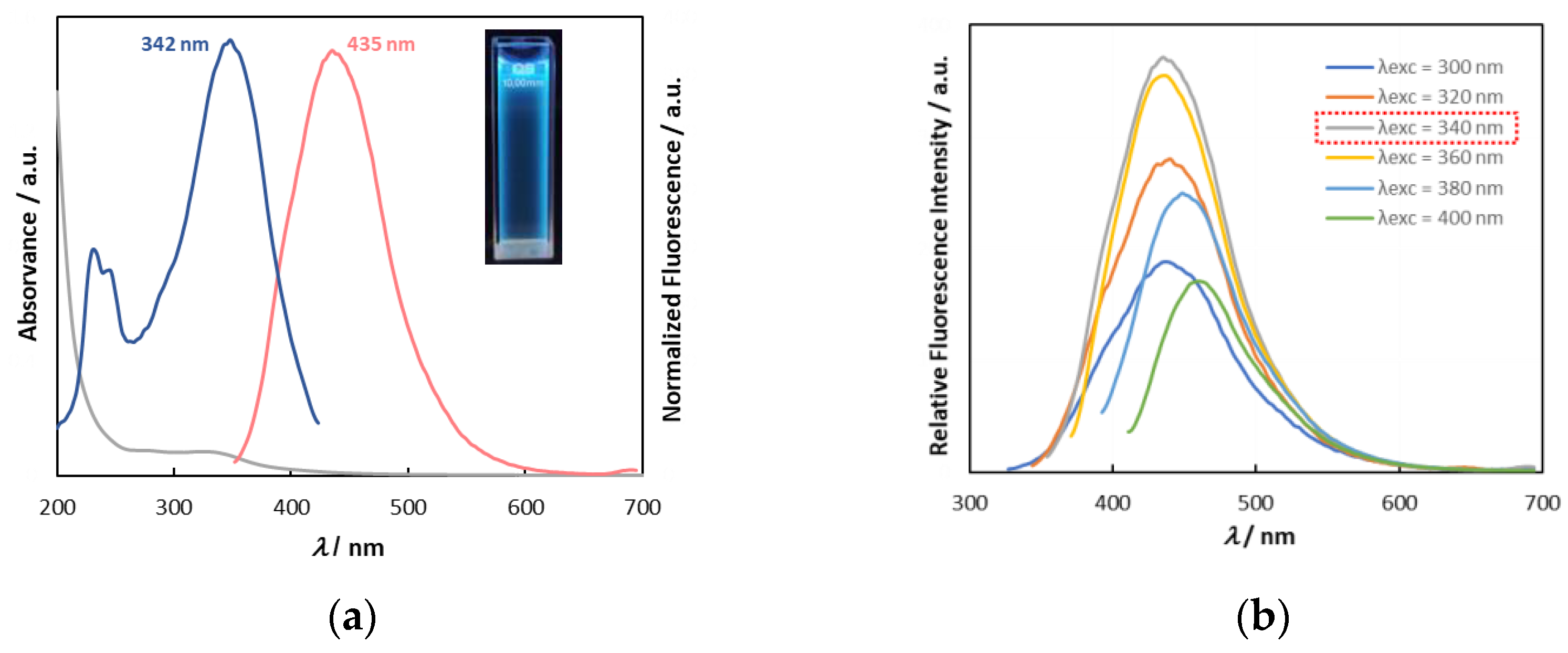
3.2. Optical Properties

Ground-state absorption and steady-state fluorescence spectra are shown in Figure 1a. The excitation spectrum shows that the main chromophores responsible for the emission appear at around 232, 245, and 342 nm. When excited at 340 nm, the emission spectrum revealed a band with a maximum at 435 nm. Figure 1b shows that µAlgae-CNDs display variable emission maxima which are dependent on excitation wavelength.

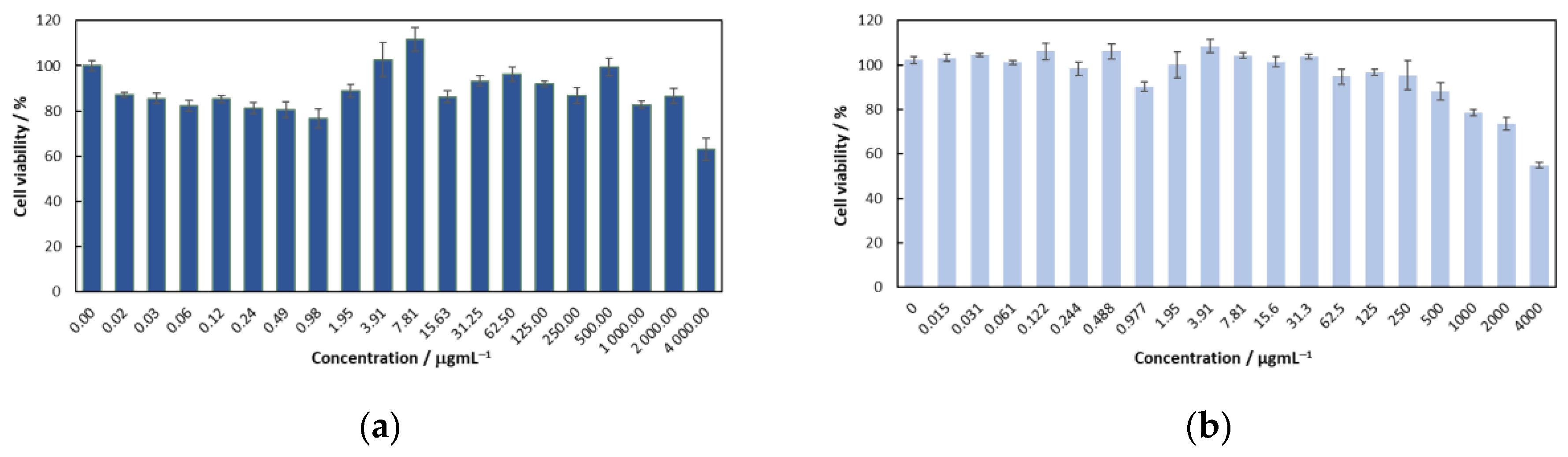
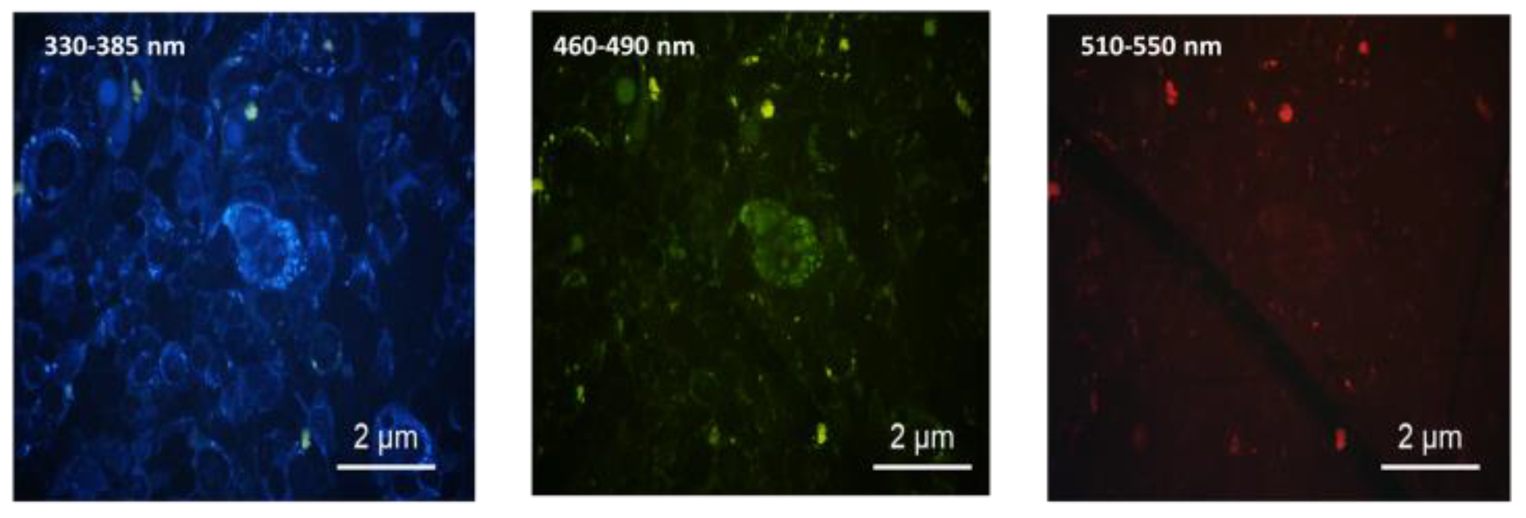
3.3. Cell Viability—Citotoxicity Evaluation

The in vitro cytotoxicity of the μAlgae-CNDs was studied using MTT assays against non-tumor NIH-3T3 cells and sarcoma S180 cells (Figure 2).
The μAlgae-CNDs exhibited no cytotoxicity for the cell lines studied, and for the sarcoma S180 cell lines, the cell viability remained at 80–100%, decreasing only for concentrations above 2 × 10³ μgmL−1.
To explore interactions between μAlgae-CNDs and cells, after incubation for 18 h at 37 °C, fluorescence microscopy was also carried out with sarcoma S180 cells (Figure 3).

4. Conclusions

The biomass from P. cruentum microalgae was a suitable carbon source for obtaining fluorescent CNDs by sustainable one-pot methods. Higher photostability was attained, and fluorescent quantum yield was observed to be dependent on the additive/biomass ratio used in its synthesis. The as-prepared µAlgae-CNDs did not exhibit cytotoxicity in the animal cell lines tested, and the preliminary results reveal good interaction between the µAlgae-CNDs and the cell plasma membrane, as ascertained via fluorescence microscopy.

Author Contributions

Conceptualization, A.I.C., J.V.P. and P.D.B.; methodology, J.V.P., F.M.H.C. and M.L.L.; investigation, I.C., F.M.H.C. and M.L.L.; resources, A.I.C.; data curation, A.I.C., I.C., M.C.S., P.D.B. and S.M.; writing—original draft preparation, A.I.C., P.D.B. and S.M.; writing—review and editing, A.I.C., M.C.S., P.D.B. and S.M. All authors have read and agreed to the published version of the manuscript.

Funding

We are grateful to Fundação para a Ciência e a Tecnologia/Ministério da Ciência, Tecnologia e Ensino Superior (FCT/MCTES) for providing financial support (UIDB/00616/2023, UIDP/00616/2023 and UIDP/04035/2020) and Instituto Politécnico de Lisboa (IPL/IDI&CA_2022/µAlgae2Dot_ISEL).

Institutional Review Board Statement

Not applicable.

Informed Consent Statement

Not applicable.

Data Availability Statement

Not applicable.

Acknowledgments

We thank Allmicroalgae-Natural Products, S.A. for supplying the µAlgae biomass.

Conflicts of Interest

The authors declare no conflict of interest.

References

  1. Balasubramaniam, V.; Gunasegavan, R.D.-N.; Mustar, S.; Lee, J.C.; Noh, M.F.M. Isolation of Industrial Important Bioactive Compounds from Microalgae. Molecules 2021, 26, 943. [Google Scholar] [CrossRef] [PubMed]
  2. Bule, M.H.; Ishtiaq, A.; Faheem, M.; Muhammad, B.; Hafiz, M.N.I. Microalgae as a source of high-value bioactive compounds. Front. Biosci. 2018, 10, 197–216. [Google Scholar] [CrossRef]
  3. Dong, D.; Liu, T.; Liang, D.; Jin, X.; Qi, Z.; Li, A.; Ning, Y. Facile Hydrothermal Synthesis of Chlorella-Derived Environmentally Friendly Fluorescent Carbon Dots for Differentiation of Living and Dead Chlorella. ACS Appl. Bio Mater. 2021, 4, 3697–3705. [Google Scholar] [CrossRef] [PubMed]
  4. Ramanan, V.; Thiyagarajan, S.K.; Raji, K.; Suresh, R.; Sekar, R.; Ramamurth, P. Outright Green Synthesis of Fluorescent Carbon Dots from Eutrophic Algal Blooms for In Vitro Imaging. ACS Sustain. Chem. Eng. 2016, 4, 4724–4731. [Google Scholar] [CrossRef]
  5. Anuar, N.K.K.; Tan, H.L.; Lim, Y.P.; So’aib, M.S.; Bakar, N.F.A. A Review on Multifunctional Carbon-Dots Synthesized from Biomass Waste: Design/Fabrication, Characterization and Applications. Front. Energy Res. 2021, 9, 626549. [Google Scholar] [CrossRef]
  6. Costa, A.I.; Barata, P.D.; Moraes, B.; Prata, J.V. Carbon Dots from Coffee Grounds: Synthesis, Characterization, and Detection of Noxious Nitroanilines. Chemosensors 2022, 10, 113. [Google Scholar] [CrossRef]
  7. Alexandre, M.R.; Costa, A.I.; Berberan-Santos, M.N.; Prata, J.V. Finding Value in Wastewaters from the Cork Industry: Carbon Dots Synthesis and Fluorescence for Hemeprotein Detection. Molecules 2020, 25, 2320. [Google Scholar] [CrossRef] [PubMed]
  8. Bello, G.L.; Bartoli, M.; Giorcelli, M.; Rovere, M.; Tagliaferro, A. Review on the Use of Biochar Derived Carbon Quantum Dots Production for Sensing Applications. Chemosensors 2022, 10, 117. [Google Scholar] [CrossRef]
  9. Eaton, D.F. Reference materials for fluorescence measurement. Pure Appl. Chem. 1988, 60, 1107–1114. [Google Scholar] [CrossRef]
  10. A Guide to Recording Fluorescence Quantum Yields, Horiba Scientific; 29. Available online: https://static.horiba.com/fileadmin/Horiba/Application/Materials/Material_Research/Quantum_Dots/quantumyieldstrad.pdf (accessed on 17 September 2023).
  11. Mosmann, T. Rapid Colorimetric Assay for Cellular Growth and Survival: Application to Proliferation and Cytotoxicity Assays. J. Immunol. Methods 1983, 65, 55–63. [Google Scholar] [CrossRef] [PubMed]
Figure 1. (a) UV-Vis spectra (gray), excitation (blue, monitored at 435 nm), and emission (orange; λexc = 340 nm) of µAlgae-CNDs in aqueous dispersion (0.1 mgmL−1) (inset: UV light at 366 nm); (b) Fluorescence emission spectra of µAlgae-CNDs in aqueous dispersion (0.1 mg mL−1) with different excitation wavelengths. The red dashed line emphasize the excitation wavelength that has the highest intensity.
Msf 23 00003 g001
Figure 2. In vitro cytotoxicity assay of µAlgae-CNDs against NIH-3T3 cells (a) and mouse sarcoma cell line S180 (b) (expressed by the decrease in cell viability).
Msf 23 00003 g002
Figure 3. Fluorescence microscopy images of mouse sarcoma cell line S180 with µAlgae-CNDs (400×).
Msf 23 00003 g003
Table 1. Effect of ED/biomass ratio on fluorescence quantum yield 1.
EntryED/Biomass RatioΦF (λ = 340 nm)
1___0.058
20.160.124
30.320.152
40.640.171
1 Typical reaction conditions: μAlgae biomass (37 mgmL−1), 40 bar, stirring, under air-equilibrated conditions.
Disclaimer/Publisher’s Note: The statements, opinions and data contained in all publications are solely those of the individual author(s) and contributor(s) and not of MDPI and/or the editor(s). MDPI and/or the editor(s) disclaim responsibility for any injury to people or property resulting from any ideas, methods, instructions or products referred to in the content.

