Article

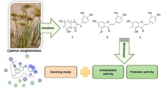
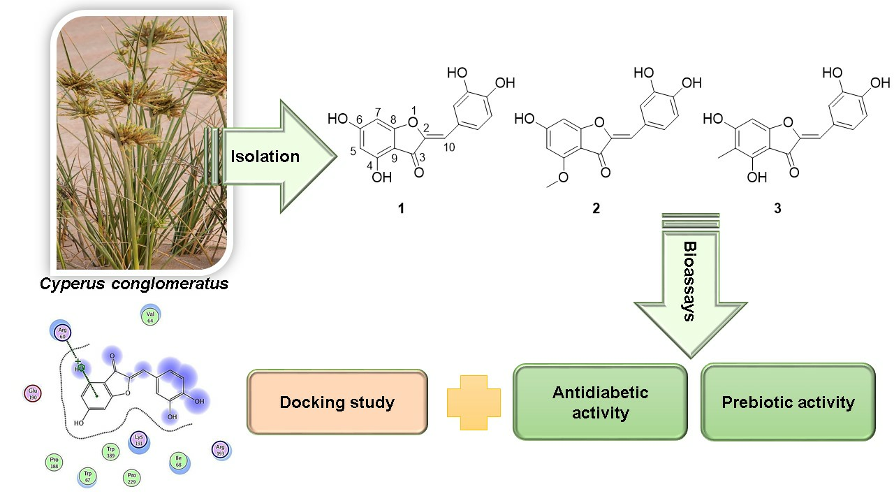
Aurones as Antidiabetic Agents and Their Prebiotic Activities

1
Department of Pharmacognosy, Faculty of Pharmacy, Mansoura University, Mansoura 35516, Egypt
2
Dairy Department, Faculty of Agriculture, Mansoura University, Mansoura 35516, Egypt
3
Veterinary Medicine Directorate, Mansoura 35516, Egypt
4
Department of Food Science and Nutrition, College of Science, Taif University, Taif 21944, Saudi Arabia
5
Department of Pharmaceutical Chemistry, Faculty of Pharmacy, University of Benin, Benin City 300001, Nigeria
*
Authors to whom correspondence should be addressed.
Future Pharmacol. 2023, 3(3), 625-636; https://doi.org/10.3390/futurepharmacol3030040
Submission received: 4 July 2023 / Revised: 23 August 2023 / Accepted: 1 September 2023 / Published: 7 September 2023

Abstract

:
Cyperus conglomeratus has been utilized in traditional medicine as an emollient, diuretic, analgesic, anthelmintic, and for other diseases. Furthermore, several biological activities have been reported for the plant extract and the isolated metabolites. The chromatographic investigation of an ethyl acetate extract of the aerial parts led to the isolation of three aurone derivatives (13) from the plant for the first time. Their structures were identified as aureusidin (1), aureusidin-4-methyl ether (2), and 5-methyl aureusidin (3) using 1D and 2D NMR techniques, along with mass spectrometry. The compounds were tested for their inhibitory activities against enzymes vital in metabolic diseases, especially diabetes, such as α-amylase, α-glucosidase, and glycogen phosphorylase. The results were expressed as percentage inhibition. The inhibitory activity of aurones against the tested enzymes was also analyzed by computational docking studies to provide a rational explanation for the observed results. The tested compounds formed stable interactions in terms of hydrogen bonding and hydrophobic interaction with the active site residues of the tested enzymes, and the results are in agreement with those of the in vitro antidiabetic activity. The compounds were also evaluated for their ability to support the growth and viability of beneficial bacteria in terms of prebiotic activities using two species, Lacticaseibacillus paracasei and Lacticaseibacillus rhamnosus, through the determination of prebiotic activity scores (Pscore). The findings of this study showed that C. conglomeratus is a potential natural source of bioactive agents. There is, however, a need for in vivo testing to evaluate this plant’s efficacy for developing new drug entities in the future.

Graphical Abstract

1. Introduction

Plants are one of the most important and largest sources of natural compounds, which have been proven effective in curing many health problems [1]. Type II diabetes mellitus, which represents 90–95% of total diabetic patients, is a serious chronic disease with numerous complications. The drugs available cause problems such as abdominal disturbance, diarrhea, nausea, flatulence, and other undesirable side effects. Thus, there is a need for an alternative natural drug to solve this problem [2]. One therapeutic protocol for treating diabetes is to reduce postprandial glycemia by inhibiting the enzymes responsible for carbohydrate hydrolyses, such as α-amylase and α-glucosidase [3]. In this respect, the α-amylase and α-glucosidase inhibitors have been exploited as specific targets that help to manage glucose levels in type II diabetes.
Moreover, glycogen phosphorylase is a crucial enzyme in regulating glycogen metabolism. It catalyzes the degradative phosphorolysis of glycogen to glucose 1-phosphate [4]. Thus, glycogen phosphorylase inhibitors have attracted specific attention because they might prevent glycogenolysis at high blood glucose levels in type II diabetes [3].
Natural products, especially purified compounds, have shown good enzyme inhibitory activities [5,6]. Thus, plants constitute a hopeful source in this field. Cyperus conglomeratus Rottb., a perennial herb, family, Cyperaceae, is distributed in temperate, tropical, and subtropical regions [7]. This herb is important in traditional medicine as an emollient, diuretic, analgesic, anthelmintic, and for treating thoracic diseases. Previous studies have described the biological activities of this plant, such as antiulcer activity, antimicrobial activity [8,9], antioxidant and antidiarrheal activities [10], and wound healing activity [11]. In previous phytochemical investigations of C. conglomeratus, several components of different classes were isolated, including essential oil [12], flavonoids [13,14], stilbenes [15], steroids, and triterpene [8]. The oil of C. conglomeratus rhizomes resembled other Cyperus oils in their hydroazulene compounds, such as cyperene, cyperotundone, and α-cyperone. According to Hisham et al. [8], eugenol was found as the principal compound in the volatile oil of C. conglomeratus, with a percentage of 33.3%, which seems rare in Cyperus species. Accordingly, as the major component in the oil, eugenol is a characteristic feature of C. conglomeratus rhizome oil. The oil of C. conglomeratus had moderate antibacterial and antifungal activities against a panel of pathogenic microorganisms (five bacterial and two fungal strains) [8].
Continuing the search for new compounds of natural products origin for treating diabetes mellitus, we describe the isolation of three aurone compounds from C. conglomeratus and their inhibitory activities against enzymes responsible for glucose metabolism. Furthermore, the docking simulation of these compounds was conducted within the active site of α-amylase (PDB ID: 1HNY), α-glucosidase (PDB ID: 2ZE0), and glycogen phosphorylase (PDB ID: 1LWO) to analyze the binding conformation and interactions responsible for their activity.
Aurones are structural isomers of flavonoids but hold a narrow place in nature. They are tricyclic flavonoids with a benzofuranone ring connected to phenyl moiety through a double bond [16]. It has been reported that aurones have therapeutic potential in cancer, Alzheimer’s, and hyperpigmentation. Furthermore, they were discovered to have antiviral, antibacterial, antioxidant, and anti-obesity activities [16]. The antidiabetic activity was also reported for sulfuretin, an aurone derivative isolated from the bark of Rhus verniciflua. It has been identified as an aldose reductase inhibitor and prevented the formation of advanced glycation end products [17].
Prebiotics are the constituents that resist digestion in the upper gastrointestinal tract until they reach the colon undigested or partially digested. Probiotics, or beneficial bacteria, could feed on these constituents and produce short-chain fatty acids, to which the health benefits are attributed. Well-known categories of prebiotics are carbohydrates such as inulin, fructans, and resistant starch [15,16]. Research studies have revealed that compounds other than carbohydrates can improve the growth and activity of probiotic bacteria, such as phenolic compounds and steroids [17,18,19].
It has been reported that prebiotics play an important role in metabolic disorders such as obesity and type II diabetes mellitus. Information obtained from animal studies has indicated that modified gut microbiota might have an impact on the regulation of insulin resistance (IR) and, consequently, type 2 diabetes (T2DM) through a variety of mechanisms [20]. The gut microbiota is primarily engaged in the initial digestion of carbohydrates and the fermentation of indigestible oligosaccharides, producing short-chain fatty acids (SCFA) such as butyrate, propionate, and acetate [21]. Currently, the discourse surrounding the role of SCFA revolves around their potential to stimulate the secretion of glucagon-like peptide-1 (GLP-1) and peptide YY; both are regulators produced by endocrine cells in the colon’s mucosal lining. These hormones influence the gastrointestinal tract, including suppressing the release of gastric juice and gastrointestinal peristalsis, delaying gastric content emptying, and activating the hypothalamus in the central nervous system, thus enhancing sensations of satiety and appetite.
Consequently, this process has the potential to lead to reductions in fasting blood glucose levels, weight, and other markers associated with type II diabetes [22,23]. A study on type II diabetes patients revealed that a daily intake of fermented milk containing probiotics for 6 weeks improved hemoglobin A1c, IL-10, and fructosamine levels. Several meta-analyses demonstrated the beneficial effects of probiotics on hemoglobin A1c and fasting insulin levels. Prebiotics have also been reported to have the potential for type II diabetes mellitus prevention. In a clinical trial on 46 diabetic patients, the daily intake of oligofructose-enriched inulin for 8 weeks improved the lipid profile, glycemic status, and immune biomarkers [24].
The current work aimed at bioprospecting the isolated compounds from C. conglomeratus for their α-amylase, α-glucosidase, and glycogen phosphorylase inhibition activities. Their prebiotic effects in managing patients with type II diabetes mellitus were also investigated.

2. Materials and Methods

2.1. Plant Material

Cyperus conglomeratus Rottb. was collected in Egypt’s Delta region on the Mediterranean coast in May 2018. Plant identification was accomplished in the Department of Pharmacognosy by comparing it to a voucher specimen and verified by the Department of Botany, Faculty of Science, Mansoura University. A voucher sample of the plant under the code CYP-18 was deposited in the Department of Pharmacognosy Herbarium.

2.2. Extraction and Isolation

The shade-dried C. conglomeratus plant was finely powdered (510 g) and extracted with methanol (5 L × 4). The collected methanol extracts were dried using a rotary evaporator. The extract was suspended in 200 mL of distilled water and fractionated with petroleum ether, methylene chloride, ethyl acetate, and n-butanol. The ethyl acetate fraction (2.91 g) was chromatographed over RP18 in a VLC (35 cm L × 4 cm W). The elution was performed using MeOH:H2O mixtures in the following gradients (6:4, 7:3, 8:2, 10:0), two liters each. Fifteen sub-fractions were obtained. Sub-fraction 6 (110 mg) was subjected to silica gel column chromatography (1.5 × 80 cm) and eluted isocratic with EtOAc:CHCl3: CH3OH:H2O (15:8:4:1) to afford compound 1 (6.5 mg). Compound 2 (7.2 mg) was obtained from sub-fraction 7 (71 mg) by using preparative TLC (20 cm × 20 cm, 0.5 mm) using CHCl3:CH3OH (9:1) as a mobile phase. Column chromatography for sub-fraction 11 (182 mg) over silica gel and elution with CHCl3:CH3OH:H2O (8:2:0.25) led to the isolation of compound 3 (8 mg), which was further purified by preparative TLC (20 cm × 20 cm, 0.5 mm) with the same mobile phase.
Compound 1: yellowish brown powder; (MeOH); ES/MS m/z: 287 [M+H]+; UV λmax (MeOH) nm: 198, 255, 270, 335, 399; 1H-NMR (400 MHz, CD3OD, δH) 7.49 (d, J = 2.1 Hz, H-2′), 7.21 (dd, J = 8.2, 2.1 Hz, H-6′), 6.85 (d, J = 8.2 Hz, H-5′), 6.58 (s, H-10), 6.22 (d, J = 1.7 Hz, H-7), and 6.04 (d, J = 1.7 Hz, H-5). 13C-NMR (101 MHz, CD3OD, δc) 182.9 (C-3), 169.6 (C-6), 169.4 (C-8), 159.8 (C-4), 148.9 (C-4′), 147.9 (C-2), 146.6 (C-3′), 125.9 (C-6′), 125.8 (C-1′), 118.7 (C-2′), 116.6 (C-5′), 112.8 (C-10), 104.7 (C-9), 98.6 (C-5), 91.7 (C-7) [25].
Compound 2: yellowish red powder; (MeOH); ES/MS m/z: 301 [M+H]+; UV λmax (MeOH) nm: 198, 255, 270, 335, 399; 1H-NMR (400 MHz, CD3OD, δH) 7.52 (d, J = 8.2 Hz, H-2′), 7.26 (dd, J = 8.2, 2.1 Hz, H-6′), 6.88 (H-5′), 6.61 (s, H-10), 6.32 (s, H-7), 6.18 (s, H-5), 3.93 (s, 4-OCH3). 13C-NMR (101 MHz, CD3OD, δc) 182.0 (C-3), 170.2 (C-6), 169.7 (C-8), 161.3 (C-4), 148.8 (C-4′), 147.9 (C-2), 147.6 (C-3′), 125.8 (C-6′), 125.7 (C-1′), 118.5 (C-2′), 116.4 (C-5′), 113.1 (C-10), 104.7 (C-9), 95.1 (C-5), 92.2 (C-7), 56.2 (4-OCH3) [26].
Compound 3: yellowish brown powder; (MeOH); ES/MS m/z: 301 [M+H]+; UV λmax (MeOH) nm: 214, 259, 266, 270, 325, 400; 1H-NMR (400 MHz, DMSO-d6, δH) 7.41 (s, H-2′), 7.17 (d, J = 8.4 Hz, H-6′), 6.82 (d, J = 8.2 Hz, H-5′), 6.47 (s, H-10), 6.31 (s, H-7), 1.92 (s, 5-CH3). 13C-NMR (101 MHz, DMSO-d6, δc) 180.5 (C-3), 165.6 (C-6), 164.7 (C-8), 155.6 (C-4), 147.9 (C-2), 146.2 (C-4′), 145.8 (C-3′), 124.4 (C-6′), 124.0 (C-1′), 117.9 (C-2′), 116.3 (C-5′), 110.3 (C-10), 106.4 (C-9), 103.7 (C-5), 90.4 (C-7), 8.1 (CH3-5) [27].

2.3. Assessment of Biological Activities

2.3.1. In Vitro Glycogen Phosphorylase Inhibition Assay

The glycogen phosphorylase (GPa) inhibitory activity was evaluated following a previously reported method [25]. Briefly, 50 µL of rabbit muscle GPa (0.38 UmL−1) was mixed with 10 µL of the isolated compounds at a 2 mg/mL concentration in DMSO, then incubated at 37 °C for 15 min. A 45 µL of HEPES solution (50 mM and pH 7.2) containing 0.25 mM glucose 1-phosphate, 0.25 mg/mL glycogen, 100 mM KCl, and 2.5 mM MgCl2, were added to the enzymatic reaction followed by incubation for an additional 15 min at 37 °C. A 130 µL of BIMOL Green, a reagent used for colorimetric phosphate quantification, was added to the enzyme reaction mixture, and the absorbance was measured at 620 nm using a spectrophotometer. The inhibition of mGPa activity was determined using the following equation:
mGPa   inhibition   % = B A C A × 100
A represents the absorbance measured without the enzyme (blank), B represents the absorbance measured with the enzyme and individual compounds, and C represents the control absorbance (measured with the enzyme but without the tested compounds). CP-91149 was used as a positive control.

2.3.2. Inhibition of α-Amylase

The α-amylase inhibitory activity of the isolated compounds was tested by adopting a previously described method [26]. In brief, a mixture containing 200 µL of the sample (at a concentration of 2 mg/mL), α-amylase (10 UmL−1), and 200 µL of 20 mM sodium phosphate buffer (pH 7.0 with 6 mM NaCl) were incubated at 37 °C for 45 min. A 400 µL solution of potato starch (0.5%) was added to each sample, and the mixture was incubated at 37 °C in a shaking water bath (100 rpm) for 10 min. The enzymatic reaction was stopped by adding a dinitro salicylic acid color reagent. The reaction tubes were then transferred to a water bath at 100 °C for 10 min and then cooled to room temperature. The samples were diluted with 3 mL of deionized water, and the absorbance of the solutions was measured at 540 nm using a spectrophotometer. The percentage inhibition of α-amylase was calculated by comparing the test and control samples. Acarbose was employed as a positive control.

2.3.3. Inhibition of α-Glucosidase

The α-glucosidase inhibition activity of the compounds was conducted following the previously described method by Abdallah et al. [26]. In a 96-microwell plate, 25 μL of samples or blank were incubated at 37 °C for 10 min with 50 μL of α-glucosidase from Saccharomyces cerevisiae (0.6 mL−1) in 0.1 M phosphate buffer (pH 7). A 25 μL of 3 mM 4-Nitrophenyl-β-D- glucopyranoside (pNPG) substrate in phosphate buffer (pH 7) was added, and the mixture was incubated for an additional 5 min at 37 °C. Enzyme activity was evaluated by measuring the release of p-nitrophenol from the pNPG substrate at 405 nm using a microplate reader (Omega, Salt Lake City, UT, USA). Acarbose was used as a positive control. The percentage of α-glucosidase inhibition was calculated using the formula:
%   inhibition   = A   blank A   sample A   blank × 100

2.4. Prebiotic Activity

2.4.1. Strains

The bacterial strains used in this study were Lactobacillus rhamnosus ML57, Lactobacillus paracasei MSD108, and Escherichia coli K12. They were obtained from the microbiology laboratory at the Dairy Department, Faculty of Agriculture, Mansoura University, Egypt. The culture media, such as nutrient agar, nutrient broth (NB), de Man, Rogosa, and Sharpe (MRS) broth, and MRS agar, were purchased from Thermo Fisher Scientific (Cairo, Egypt).

2.4.2. Prebiotic Activity Score (Pscore)

A 100 µL portion of the chosen probiotic strains and E. coli K12 was introduced into MRS and NB, respectively, and left to incubate at 37 °C for 24 h. An overnight culture of the probiotic strains and E. coli K12 was also streaked onto MRS and nutrient agar and incubated at 37 °C for 48 h. On the day of the assay, the bacterial cultures were diluted in buffered peptone water to obtain a final cell count of 1.5 × 10⁸ CFU/mL. The effect of compounds (13) on the Pscore of L. rhamnosus and L. paracasei was examined. Overnight cultures of the probiotic strains and E. coli K12 were used to inoculate MRS and NB, respectively, at a concentration of 2% (v/v). In these media, 2% of the glucose was replaced with each compound and then incubated at 37 °C for 24 h. The growth of L. rhamnosus, L. paracasei, and E. coli K12 was evaluated by determining the number of viable colony-forming units (CFU) per mL after 24 h incubation. This assessment was carried out using the pour plate method on MRS agar for probiotic strains [25]. The prebiotic activity scores (Pscore) were determined using the equation of Dawood et al. [18]:
Pscore = Log   P 24 Log   P 0 PC Log   P 24 Log   P 0 Glucose Log   E 24 Log   E 0 PC Log   E 24 Log   E 0   Glucose

2.5. Docking Studies

2.5.1. Preparation of the Target

The crystal structures of α-amylase (PDB ID 1HNY, 1.8 A resolution, [27]), α-glucosidase (PDB ID 2ZE0, 2.0 A resolution, [28]) and glycogen phosphorylase (PDB ID 1LWO, 2.0 A resolution, [29]) were downloaded from the Protein Data Bank. Each protein was protonated with hydrogen atoms in their standard 3D geometry, automatically corrected to check its atom’s connection and type for any errors, and the potential of the receptor and its atoms were fixed, as previously discussed in detail in Samra et al. [30].

2.5.2. Docking of the Tested Molecules to the Enzyme Binding Site

Docking of the database composed of tested compounds together with the positive control acarbose in the case of α-amylase and α-glucosidase proteins and with the positive control CP-91149 in the case of glycogen phosphorylase protein.

2.6. Statistical Analysis

The mean percentages of Pscore, α-Glucosidase, and Glycogen Phosphorylase inhibition and α-amylase inhibition, obtained from three independent repetitions, were subjected to one-way analysis of variance using SAS 2000. Subsequently, pairwise comparisons among means were determined using Duncan’s Multiple Range Test in cases where significant main effects were observed.

3. Results and Discussions

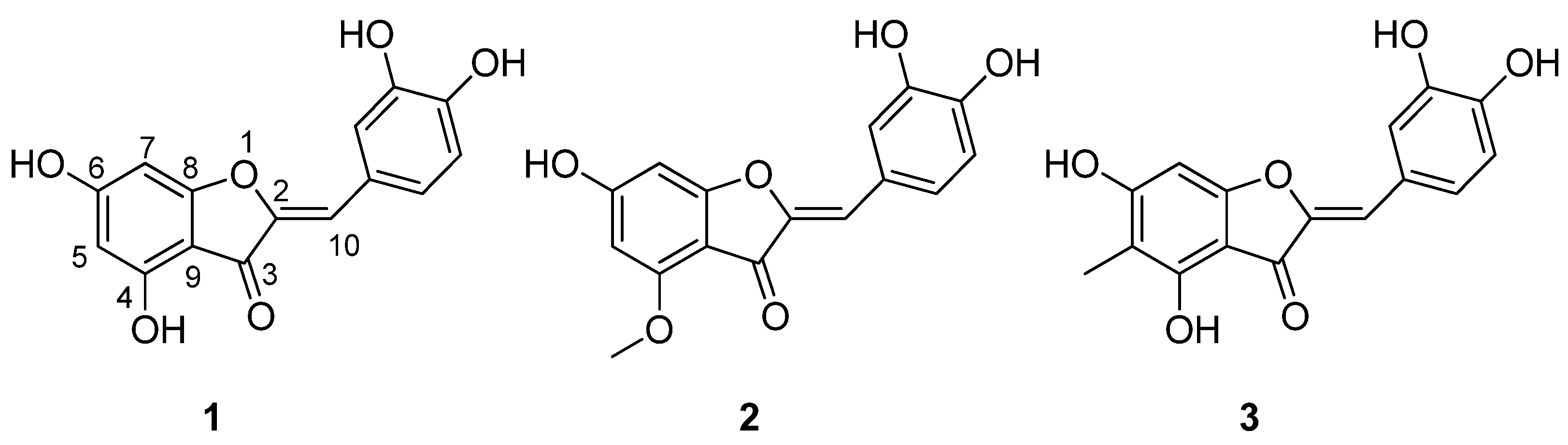
3.1. Identification of Isolated Compounds

The phytochemical investigation of C. conglomeratus extract resulted in the isolation of three aurone derivatives (13) from the plant for the first time. Compound 1 was isolated as a yellowish-brown powder with a molecular weight of 287 [M+H]+, corresponding to the molecular formula C15H10O6. The UV spectral data suggested its structure to be an aurone derivative. The 13C-NMR spectrum showed 15 signals characteristic of a flavonoid skeleton. The presence of a singlet proton signal at δ 6.58 in the 1H-NMR spectrum is characteristic of the flavones. The careful assignment of carbon signals and their corresponding protons in the HSQC spectrum revealed the downfield shifted carbon signal at δ 113.2 (δ 6.58, s, 1H) and a quaternary carbon signal at δ 146.9. These observed signals were assigned to C-10 and C-2. Further, the downfield shifted carbon signals at δ 169.9 and 169.8 were attributed to C-6 and C-4, respectively, of an aurone, a rare type of flavonoid. The full assignment of the structure with 1H, 13C, HSQC, HMBC, and MS spectroscopic analysis, together with a comparison with those previously reported in the literature [31], revealed that compound 1 is aureusidin (the NMR data are shown in Supplementary Materials).
Based on NMR data, compounds 2 and 3 showed the same nucleus as 1; however, they showed additional methoxy and methyl groups, respectively. Compound 2 has a molecular weight of 301 [M+H]+, corresponding to the molecular formula C16H12O6. The 13C and HSQC spectra of 2 revealed the presence of a signal for methoxy at δ 56.2, and the HMBC correlations from methoxyl proton at δ 3.93 and C-4 at δ 161.3 indicated that the methoxy group was located at the 4-position. Thus, compound 2 was shown to be aureusidin 4-methyl ether by comparing spectral data with Sayed et al. [32]. On the other hand, compound 3 had a molecular weight of 301 [M+H]+, corresponding to the molecular formula C16H12O6. The 13C spectrum revealed an additional signal for the methyl group at δ 8.1, which showed HMBC correlation between its proton at δ 1.92 and C-4, C-5, and C-6 at 155.6, 103.7, and165.6, respectively, which proved the location of the methyl group at the C-5 position. In interpreting spectral data and comparing it with the literature [33], compound 3 was characterized as 5-methyl aureusidin. The structures of isolated compounds are shown in Figure 1.

3.2. Assessment of Antidiabetic Activity of the Isolated Compounds

The in vitro antidiabetic activity of the isolated aurones was estimated by assessing their α-amylase and α-glucosidase inhibitory activities, and the results were expressed as percentage inhibition. Acarbose was used as a positive control with a % inhibition of 97.7 ± 0.54 and 97.7 ± 0.88, respectively, against α-glucosidase and α-amylase enzymes. Compounds 1 and 3 had the highest activity with % inhibition of 60.5 + 1.54 and 79.6 + 0.97, respectively, against α-glucosidase enzyme and 58.4 + 1.61 and 53.7 + 1.03, respectively, against α-amylase enzyme. Compound 2 had the lowest inhibitory activity against the tested enzymes. We also investigated the inhibitory activity of the compounds against glycogen phosphorylase. Compound 3 showed potent activity with a % inhibition of 80.4 ± 1.34, compared to CP-91149 84.9 ± 0.81%. Compounds 1 and 2 exhibited low inhibitory activity, with a % inhibition of 27.5 ± 0.64 and 17.3 ± 1.79 against glycogen phosphorylase (Table 1). It is important to note that compound 3 acted as a dual target inhibitor against α-glucosidase and glycogen phosphorylase enzymes, thus inhibiting the absorption of glucose and suppressing its production arising from glycogenolysis. Compound 1 also showed moderate inhibitory activity against the two key starch digestive enzymes. Roshanzamir et al. [34] reported the inhibitory activity of the synthesized aurones on α-amylase, which exhibited a weak-to-moderate inhibitory activity range (9–81% inhibition) up to a concentration of 1000 µM. Sun et al. [35] also reported that a semi-synthesized aurone-coumarin hybrid showed potent α-glucosidase inhibitory activity but moderately against α-amylase. This is the first report proving the preliminary antidiabetic activity of these natural aurone derivatives.

3.3. Prebiotic Activity Score of the Isolated Compounds

The isolated aurones were evaluated for their ability to support the growth and activity of beneficial bacteria in terms of prebiotic activities using two species, Lacticaseibacillus paracasei and Lactobacillus rhamnosus. All Pscore values were positive, indicating that the growth rate of Lb. rhamnosus and Lb. paracasei in different tested compounds was higher than that of E.coli K12 paired with the isolated compounds. Lb. rhamnosus paired with 1, 2, and 3 presented great Pscore values of 3.84 ± 0.67, 3.82 ± 0.73, and 2.36 ± 0.76, respectively. Further, these compounds were found to have a marked effect on the growth of Lb. paracasei. Compounds 13 exhibited noticeable Pscore values of about 1.82 ± 0.14, 3.81 ± 0.56, and 4.54 ± 0.78, respectively (Table 2). There have been no prior studies on the prebiotic activity of this class of flavonoids.

3.4. Docking Studies

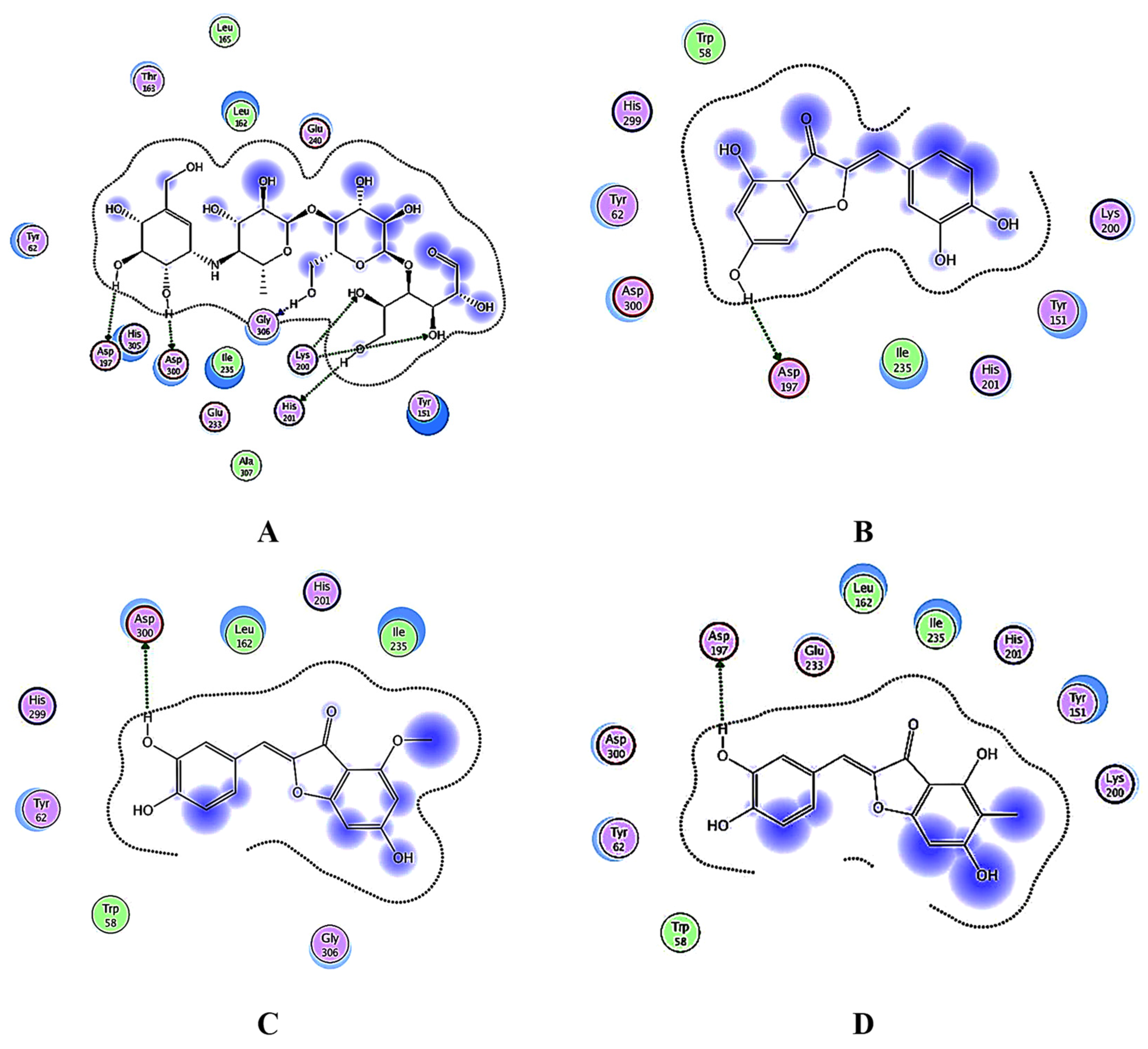
3.4.1. Docking Analysis of the Test Samples and Positive Control (Acarbose) against α-Amylase

The docking of acarbose into the α-amylase binding site indicated that the model structure of ligand-protein interaction had formed six hydrogen bonds with Lys 200, Asp 197, Asp 300, Gly 306, and His 201, as well as hydrophobic interactions with Tyr 62, Tyr 151, Leu 162, and Ile 235. According to [27], the active site residues at the largest domain A of the α-amylase enzyme are Asp 197, Glu 233, and Asp 300. The docking studies revealed that the tested compounds formed stable interactions in terms of hydrogen bonding and hydrophobic interaction with the active site residues of α-amylase (Table S1).
Compound 1 formed a hydrogen bond with the essential amino acid Asp 197 by acting as a hydrogen bond acceptor, in addition to the five hydrophobic interactions with Tyr 62, Tyr 151, Lys 200, Ile 235, and the critical amino acid residue Asp 300. This compound and acarbose illustrate similarity in the binding profiles with α-amylase. Both have potential hydrophobic interactions with Tyr 62, Tyr 151, and Ile 235 residues and hydrogen bonds with Asp 197. Compound 3 exhibited similarity with compound 1 in the binding to Asp 197 by forming a hydrogen bond but with higher binding energy. Furthermore, it interacted with the amino acids Tyr 62, Tyr 151, Leu 162, Ile 235, Glu 233, and Asp 300 of α-amylase by forming hydrophobic interactions.
The α-amylase binding site for compound 2 was mainly formed by amino acid residues Tyr 62, Leu 162, Gly 306, and Ile 235 through hydrophobic interactions. It exhibited one H-bond with the catalytic site, Asp 300. Binding poses with the highest binding affinity for docked compounds are shown in Figure 2. It was observed that substituting the hydroxyl group with methyl or adding methyl to the aurone nucleus may reduce hydrogen bonding, which has a crucial role in inhibitory activity. According to Roshanzamir et al. [34], the most important groups of aurones that are needed for interaction with the catalytic active site are the 6-OH group on the benzofuranone moiety and the OH groups in the 3′ and 4′ positions on the benzylidene ring. These findings generally agree well with our results, as shown in Figure 2.

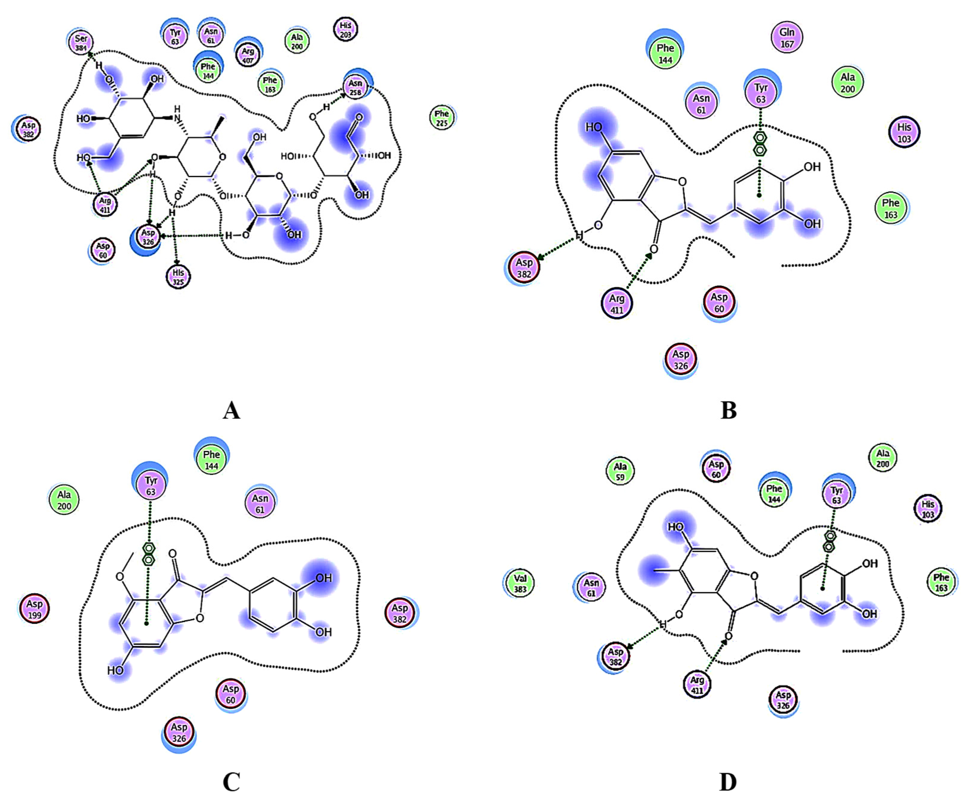
3.4.2. Docking Analysis of α-Glucosidase

According to Shirai et al. [28], the residues important for substrate binding with α-glucosidase are Tyr 63, His 103, and His 325. The control, acarbose, makes eight hydrogen bonds with the active site residues: His 325, Arg 411, Ser 384, Asp 326, Asn 258, and Val 383 and form hydrophobic interactions with Arg 407, Phe 144, Asn 61, Asp 60, and with the essential amino acid residue Tyr 63 (Table S2).
Compounds 1 and 3 displayed almost the same binding modes for the α-glucosidase enzyme. Both compounds formed an arene-arene interaction with the crucial amino acid Tyr 63. They formed two H-bonds with Arg 411 and Asp 382. Additionally, they interacted with Phe 144, Asn 61, and Asp 326 through hydrophobic interactions. It is observed that both the carbonyl and hydroxyl groups of the benzofuranone moiety, as well as the benzylidene ring, are crucial for interaction with the α-glucosidase active site. The presence of methyl in the case of compound 3 may enhance the ability to enter the hydrophobic cavity of the α-glucosidase enzyme, therefore showing a lower binding score than 1. Compound 2 properly accommodated itself into the binding cavity of the α-glucosidase enzyme and developed an arene-arene interaction with the crucial amino acid Tyr 63. Furthermore, hydrophobic interactions were found with Phe 144, Asn 61, and Asp 326 residues. Binding poses with the highest binding affinity for docked aurones are shown in Figure 3.

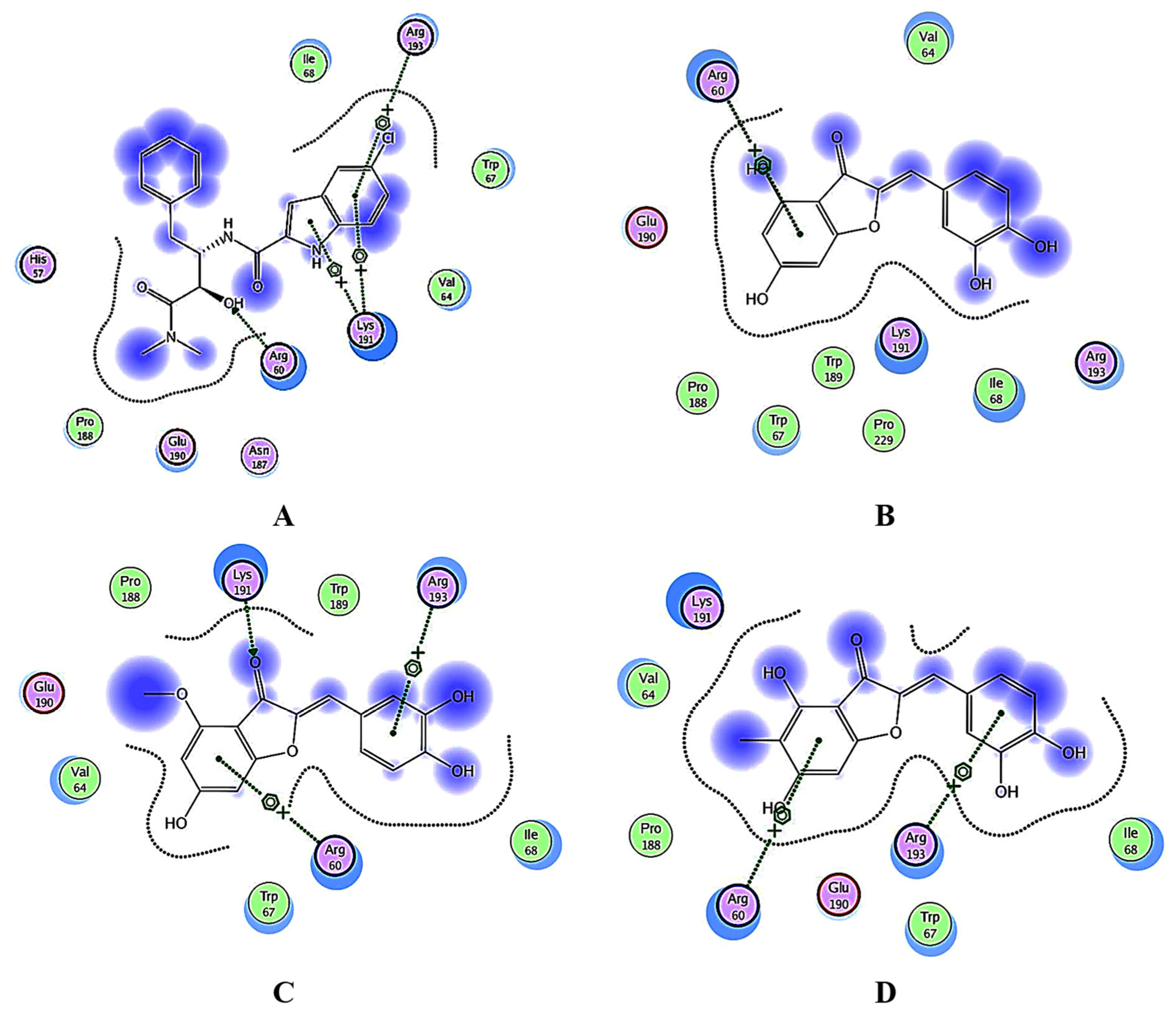
3.4.3. Docking Analysis of Glycogen Phosphorylase

Herowati and Widodo [4] established that amino acid residues of glycogen phosphorylase that are involved in the interaction with CP320626, a potent inhibitor of human glycogen phosphorylase, are Glu 190, Arg 60, and Lys 191. Moreover, hydrophobic interactions were observed between this ligand and residues such as Pro 229, Trp 189, and Trp 67. The positive control, CP-91149, formed three arene-cation interactions with Arg 193, Lys 191, and one H-bond with Arg 60. Additionally, hydrophobic interactions were observed with residues Lys 191, Ile 68, Val 64, and Arg 60 (Table S3). Compound 2 developed two arene-cation interactions with Arg 60 and Arg193, one hydrogen bond with the essential amino acid Lys191, and hydrophobic interactions with Trp 67, Val 64, and Ile 68. Compounds 1 and 3 had a similar interaction profile to CP-91149 but a higher binding score. They formed an arene-cation interaction with Arg 60. Furthermore, hydrophobic interactions were observed with residues Trp 67, Ile 68, Lys 191, and Val 64. Binding poses with the highest binding affinity for docked compounds are shown in Figure 4.

4. Conclusions

Three aurone derivatives (13) were purified from C. conglomeratus. The compounds characterized as aureusidin (1), aureusidin-4-methyl ether (2), and 5-methyl aureusidin (3) were described for the first time from this species. They were screened for anti-diabetic and prebiotic activities. Compound 3 acted as a dual target inhibitor against α-glucosidase and glycogen phosphorylase enzymes. Compound 1 also showed moderate inhibitory activity against the two key starch digestive enzymes. These compounds were found to have a marked prebiotic effect on the growth of Lacticaseibacillus paracasei and Lacticaseibacillus rhamnosus. Docking studies explain the enzymes’ inhibitory activities and agree well with the results of the in-vitro antidiabetic studies. In conclusion, these aurone compounds could serve as potential leads with improved in vitro antidiabetic and prebiotic activities. Still, in vivo evaluations will be required to test these compounds’ efficacy in humans.

Supplementary Materials

The following supporting information can be downloaded at: https://www.mdpi.com/article/10.3390/futurepharmacol3030040/s1, Figure S1: 1H-NMR spectrum (CD3OD, 400 MHz) of aureusidin (1), Figure S2: 13C-NMR spectrum (CD3OD, 101 MHz) of aureusidin (1), Figure S3: HSQC spectrum of aureusidin (1), Figure S4: HMBC spectrum of aureusidin (1), Figure S5: 1H-NMR spectrum (CD3OD, 400 MHz) of aureusidin-4-methyl ether (2), Figure S6: 13C-NMR spectrum (CD3OD, 101 MHz) of aureusidin-4-methyl ether (2), Figure S7: HSQC spectrum of aureusidin-4-methyl ether (2), Figure S8: HMBC spectrum of aureusidin-4-methyl ether (2), Figure S9: 1H-NMR spectrum (DMSO-d6, 400 MHz) of 5-methyl aureusidin (3), Figure S10: 13C-NMR spectrum (DMSO-d6, 101 MHz) of 5-methyl aureusidin (3), Figure S11: HSQC spectrum of 5-methyl aureusidin (3), Figure S12: HMBC spectrum of 5-methyl aureusidin (3), NMR spectra of isolated compounds; Table S1: Receptor interactions and binding energies of the tested compounds, and acarbose inside the binding pocket of α-amylase, Table S2: Receptor interactions and binding energies of the tested compounds, and acarbose inside the binding pocket of α-glucosidase, Table S3: Receptor interactions and binding energies of the tested compounds, and CP-91149 inside the binding pocket of glycogen phosphorylase, Receptor interactions and binding energies of the tested compounds.

Author Contributions

Conceptualization A.A.Z., M.S.D. and R.M.S.; methodology M.S.D., A.A.Z. and R.M.S.; software M.S.D., A.A.Z. and R.M.S.; investigation M.S.D., A.A.Z., R.M.S. and N.A.A.-Z.; resources M.S.D., A.A.Z. and N.A.A.-Z.; data curation M.S.D., A.A.Z., R.M.S., N.A.A.-Z. and E.K.; writing—original draft preparation, A.A.Z., M.S.D. and R.M.S.; writing—review and editing V.O.I., A.A.Z., M.S.D., N.A.A.-Z., E.K. and R.M.S.; visualization, V.O.I., A.A.Z., M.S.D. and R.M.S.; supervision A.A.Z., M.S.D. and R.M.S.; project administration, A.A.Z., M.S.D., E.K. and R.M.S. All authors have read and agreed to the published version of the manuscript.

Funding

This research received no external funding.

Institutional Review Board Statement

Not applicable.

Informed Consent Statement

Not applicable.

Data Availability Statement

Data is contained within the article and Supplementary Materials.

Conflicts of Interest

The authors declare no conflict of interest.

References

  1. Cragg, G.M.; Newman, D.J. Natural products: A continuing source of novel drug leads. Biochim. Biophys. Acta (BBA) Gen. Subj. 2013, 1830, 3670–3695. [Google Scholar] [CrossRef] [PubMed]
  2. Nakib, R.; Ghorab, A.; Ouelhadj, A.; Rodríguez-Flores, S.; Escuredo, O.; Bensouici, C.; Seijo-Coello, C. Chemometric evaluation of antioxidant activity and α-amylase inhibition of selected monofloral honeys from Algeria. J. Apic. Res. 2021, 1–11. [Google Scholar] [CrossRef]
  3. de Souza, P.M.; de Sales, P.M.; Simeoni, L.A.; Silva, E.C.; Silveira, D.; de Oliveira Magalhães, P. Inhibitory activity of α-amylase and α-glucosidase by plant extracts from the Brazilian cerrado. Planta Med. 2012, 78, 393–399. [Google Scholar] [CrossRef] [PubMed]
  4. Herowati, R.; Widodo, G.P. Molecular Docking studies of chemical constituents of Tinospora cordifolia on glycogen phosphorylase. Procedia Chem. 2014, 13, 63–68. [Google Scholar] [CrossRef]
  5. Ajayi, O.S.; Balogun, O.S.; Olawuni, I.J.; October, N.; Adigun, R.; Akinlade, I.G. Alpha Amylase Inhibition and Antioxidant Activities of Bicyclic Diterpenoid Lactones from Andrographis paniculata. Trop. J. Nat. Prod. Res. (TJNPR) 2021, 5, 1110–1117. [Google Scholar] [CrossRef]
  6. Arraki, K.; Totoson, P.; Decendit, A.; Zedet, A.; Maroilley, J.; Badoc, A.; Demougeot, C.; Girard, C. Mammalian Arginase Inhibitory Activity of Methanolic Extracts and Isolated Compounds from Cyperus Species. Molecules 2021, 26, 1694. [Google Scholar] [CrossRef] [PubMed]
  7. Jyoti, P.; Hemali, P.; Nilam, R.; Sumitra, C. Cyperus conglomeratus (Cyperaceae) a halophyte from Gujarat: Physicochemical, Phytochemical and Pharmacognostic studies. J. Phytopharmacol. 2018, 7, 334–340. [Google Scholar] [CrossRef]
  8. Al-Hazmi, G.H.; Awaad, A.S.; Alothman, M.R.; Alqasoumi, S.I. Anticandidal activity of the extract and compounds isolated from Cyperus conglomeratus Rottb. Saudi Pharm. J. 2018, 26, 891–895. [Google Scholar] [CrossRef]
  9. Hisham, A.; Rameshkumar, K.B.; Sherwani, N.; Al-Saidi, S.; Al-Kindy, S. The composition and antimicrobial activities of Cyperus conglomeratus, Desmos chinensis var. lawii and Cyathocalyx zeylanicus essential oils. Nat. Prod. Commun. 2012, 7, 663–666. [Google Scholar] [CrossRef]
  10. Al-Harbi, K.B.; El-Ashmawy, I.M.; Al-Wabel, N.A. The antidiarrheal activity of the methanol extract of some plants native to Al-Qassim Region, Saudi Arabia. J. Food Agric. Environ. 2016, 14, 239. [Google Scholar]
  11. Al-Harbi, K.B.; El-Tigani-Asil, E.; Ahmed, A.F.; El-Ashmawy, I.M.; Al-Wabel, N.A. Wound healing potential of methanolic extracts of some plants native to Al-Qassim Region, Saudi Arabia. J. Food Agric. Environ. 2016, 14, 238–242. [Google Scholar]
  12. Feizbakhsh, A.; Naeemy, A. Chemical composition of the essential oil of Cyperus conglomeratus Rottb. from Iran. J. Chem. 2011, 8, 241325. [Google Scholar]
  13. Abdel-Mogib, M.; Basaif, S.; Ezmirly, S. Two novel flavans from Cyperus conglomeratus. Pharmazie 2000, 55, 693–695. [Google Scholar] [PubMed]
  14. Abdel-Razik, A.F.; Nassar, M.I.; El-Khrisy, E.-D.A.; Dawidar, A.-A.M.; Mabry, T.J. New prenylflavans from Cyperus conglomeratus. Fitoterapia 2005, 76, 762–764. [Google Scholar] [CrossRef] [PubMed]
  15. Zaki, A.A.; Ross, S.A.; El-Amier, Y.A.; Khan, I.A. New flavans and stilbenes from Cyperus conglomeratus. Phytochem. Lett. 2018, 26, 159–163. [Google Scholar] [CrossRef]
  16. Haudecoeur, R.; Boumendjel, A. Recent advances in the medicinal chemistry of aurones. Curr. Med. Chem. 2012, 19, 2861–2875. [Google Scholar] [CrossRef]
  17. Lee, E.H.; Song, D.-G.; Lee, J.Y.; Pan, C.-H.; Um, B.H.; Jung, S.H. Inhibitory effect of the compounds isolated from Rhus verniciflua on aldose reductase and advanced glycation endproducts. Biol. Pharm. Bull. 2008, 31, 1626–1630. [Google Scholar] [CrossRef]
  18. Dawood, D.H.; Darwish, M.S.; El-Awady, A.A.; Mohamed, A.H.; Zaki, A.A.; Taher, M.A. Chemical characterization of Cassia fistula polysaccharide (CFP) and its potential application as a prebiotic in synbiotic preparation. RSC Adv. 2021, 11, 13329–13340. [Google Scholar] [CrossRef]
  19. Megur, A.; Daliri, E.B.-M.; Baltriukienė, D.; Burokas, A. Prebiotics as a tool for the prevention and treatment of obesity and diabetes: Classification and ability to modulate the gut microbiota. Int. J. Mol. Sci. 2022, 23, 6097. [Google Scholar] [CrossRef]
  20. Kobyliak, N.; Virchenko, O.; Falalyeyeva, T. Pathophysiological role of host microbiota in the development of obesity. Nutr. J. 2015, 15, 43. [Google Scholar] [CrossRef]
  21. Jandhyala, S.M.; Talukdar, R.; Subramanyam, C.; Vuyyuru, H.; Sasikala, M.; Reddy, D.N. Role of the normal gut microbiota. World J. Gastroenterol. WJG 2015, 21, 8787. [Google Scholar] [CrossRef]
  22. Combettes, M.M. GLP-1 and type 2 diabetes: Physiology and new clinical advances. Curr. Opin. Pharmacol. 2006, 6, 598–605. [Google Scholar] [CrossRef] [PubMed]
  23. Li, H.-Y.; Zhou, D.-D.; Gan, R.-Y.; Huang, S.-Y.; Zhao, C.-N.; Shang, A.; Xu, X.-Y.; Li, H.-B. Effects and mechanisms of probiotics, prebiotics, synbiotics, and postbiotics on metabolic diseases targeting gut microbiota: A narrative review. Nutrients 2021, 13, 3211. [Google Scholar] [CrossRef]
  24. Elbermawi, A.; Darwish, M.S.; El-Awady, A.A.; Zaki, A.A.; Qiu, L.; Samra, R.M. Isolation and biological activities of compounds from Rumex vesicarius L. and their use as a component of a synbiotic preparation. Food Chem. X 2022, 14, 100306. [Google Scholar] [CrossRef]
  25. Elbermawi, A.; Darwish, M.S.; Zaki, A.A.; Abou-Zeid, N.A.; Taher, M.A.; Khojah, E.; Bokhari, S.A.; Soliman, A.F. In Vitro Antidiabetic, Antioxidant, and Prebiotic Activities of the Chemical Compounds Isolated from Guizotia abyssinica. Antioxidants 2022, 11, 2482. [Google Scholar] [CrossRef] [PubMed]
  26. Abdallah, H.M.; Kashegari, A.T.; Shalabi, A.A.; Darwish, K.M.; El-Halawany, A.M.; Algandaby, M.M.; Ibrahim, S.R.; Mohamed, G.A.; Abdel-Naim, A.B.; Koshak, A.E. Phenolics from Chrozophora oblongifolia Aerial Parts as Inhibitors of α-Glucosidases and Advanced Glycation End Products: In-Vitro Assessment, Molecular Docking and Dynamics Studies. Biology 2022, 11, 762. [Google Scholar] [CrossRef] [PubMed]
  27. Brayer, G.D.; Luo, Y.; Withers, S.G. The structure of human pancreatic α-amylase at 1.8 Å resolution and comparisons with related enzymes. Protein Sci. 1995, 4, 1730–1742. [Google Scholar] [CrossRef] [PubMed]
  28. Shirai, T.; Hung, V.S.; Morinaka, K.; Kobayashi, T.; Ito, S. Crystal structure of GH13 α-glucosidase GSJ from one of the deepest sea bacteria. Proteins Struct. Funct. Bioinform. 2008, 73, 126–133. [Google Scholar] [CrossRef]
  29. Oikonomakos, N.G.; Chrysina, E.D.; Kosmopoulou, M.N.; Leonidas, D.D. Crystal structure of rabbit muscle glycogen phosphorylase a in complex with a potential hypoglycaemic drug at 2.0 Å resolution. Biochim. Biophys. Acta (BBA)-Proteins Proteom. 2003, 1647, 325–332. [Google Scholar] [CrossRef]
  30. Samra, R.M.; Soliman, A.F.; Zaki, A.A.; Ashour, A.; Al-Karmalawy, A.A.; Hassan, M.A.; Zaghloul, A.M. Bioassay-guided isolation of a new cytotoxic ceramide from Cyperus rotundus L. S. Afr. J. Bot. 2021, 139, 210–216. [Google Scholar] [CrossRef]
  31. Shrestha, S.; Lee, D.-Y.; Park, J.-H.; Cho, J.-G.; Lee, D.-S.; Li, B.; Kim, Y.-C.; Jeon, Y.-J.; Yeon, S.-W.; Baek, N.-I. Flavonoids from the fruits of Nepalese sumac (Rhus parviflora) attenuate glutamate-induced neurotoxicity in HT22 cells. Food Sci. Biotechnol. 2013, 22, 895–902. [Google Scholar] [CrossRef]
  32. Sayed, H.M.; Mohamed, M.; Farag, S.; Mohamed, G.; Ebel, R.; Omobuwajo, O.; Proksch, P. Phenolics of Cyperus alopecuroides rottb. Inflorescences and their biological activities. Bull. Pharm. Sci. Assiut 2006, 29, 9–32. [Google Scholar] [CrossRef]
  33. Seabra, R.M.; Silva, A.M.; Andrade, P.B.; Moreira, M.M. Methylaurones from Cyperus capitatus. Phytochemistry 1998, 48, 1429–1432. [Google Scholar] [CrossRef]
  34. Roshanzamir, K.; Kashani-Amin, E.; Ebrahim-Habibi, A.; Navidpour, L. Aurones as new porcine pancreatic α-Amylase inhibitors. Lett. Drug Des. Discov. 2019, 16, 333–340. [Google Scholar] [CrossRef]
  35. Sun, H.; Song, X.; Tao, Y.; Li, M.; Yang, K.; Zheng, H.; Jin, Z.; Dodd, R.H.; Pan, G.; Lu, K. Synthesis & α-glucosidase inhibitory & glucose consumption-promoting activities of flavonoid–coumarin hybrids. Future Med. Chem. 2018, 10, 1055–1066. [Google Scholar]
Figure 1. Chemical Structures of Isolated Aurones.
Futurepharmacol 03 00040 g001
Figure 2. 2D docking representations of the isolated compounds and acarbose against the binding site of α-amylase. (A): acarbose; (BD), compounds 1, 2, and 3, respectively.
Futurepharmacol 03 00040 g002
Figure 3. 2D docking representations of the isolated compounds and acarbose against the binding site of α-glucosidase. (A): acarbose; (BD), compounds 13, respectively.
Futurepharmacol 03 00040 g003
Figure 4. 2D docking representations of the isolated compounds and CP-91149 against the binding site of glycogen phosphorylase. (A): CP-91149; (BD) are compounds 13, respectively.
Futurepharmacol 03 00040 g004
Table 1. Results of enzyme inhibition assays.
% Inhibition
α-Glucosidaseα-AmylaseGP
Acarbose97.7 ± 0.54Acarbose97.7 ± 0.88CP-9114984.9 ± 0.81
160.5 ± 1.54158.4 ± 1.61127.5 ± 0.64
220.91 ± 1.467239.57 ± 0.592217.3 ± 1.79
379.6 ± 0.97353.7 ± 1.03380.4 ± 1.34
Table 2. Results of prebiotic activity of the isolated compounds.
Lb. rhamnosusLb. paracasei
13.84 ± 0.671.82 ± 0.14
23.82 ± 0.733.81 ± 0.56
32.36 ± 0.764.55 ± 0.78
Disclaimer/Publisher’s Note: The statements, opinions and data contained in all publications are solely those of the individual author(s) and contributor(s) and not of MDPI and/or the editor(s). MDPI and/or the editor(s) disclaim responsibility for any injury to people or property resulting from any ideas, methods, instructions or products referred to in the content.

