Review

Moles of Molecules against Mycobacterium abscessus: A Review of Current Research

Department of Biology and Biotechnology “Lazzaro Spallanzani”, University of Pavia, Via A. Ferrata 9, 27100 Pavia, Italy
*
Author to whom correspondence should be addressed.
Future Pharmacol. 2023, 3(3), 637-663; https://doi.org/10.3390/futurepharmacol3030041
Submission received: 10 August 2023 / Revised: 31 August 2023 / Accepted: 8 September 2023 / Published: 11 September 2023

Abstract

:
Mycobacterium abscessus is an emerging opportunistic pathogen that infects mainly the respiratory tract of individuals with pre-existing clinical pictures. In recent years, the incidence of infections of this microorganism has risen, in particular in patients with cystic fibrosis, leading to an exacerbation of their conditions. The actual therapeutic regimen has low efficacy and is extended for long periods since it is mainly based on a combination of repurposed drugs, generally from treatments of Mycobacterium tuberculosis infections. For this reason, it is necessary to develop new drugs or alternative strategies in order to improve the efficacy and shorten the time of treatments. This review aims to give an overview of drugs in the pre-clinical and clinical phases of evaluation against M. abscessus and the molecules that have been in development for the past five years in the early drug-discovery phase.

1. Introduction

Mycobacterium abscessus (Mab) is an environmental rapid-growing non-tuberculous mycobacterium (NTM) [1]. It was isolated in 1953 by Moore and Frerichs from a knee infection, marking the first recorded instance of its discovery [2]. In recent years, this peculiar bacterium has become extensively studied due to its similarities with Mycobacterium tuberculosis (Mtb) and as an opportunistic human pathogen.
In 2009, the Mab genome was sequenced for the first time, revealing a chromosome length of 5 Mb with a G+C content of 64% and approximately 49.920 predicted coding sequences. The bacterium also carries a mercury resistance plasmid identical to the one found in Mycobacterium marinum. Moreover, genes typical of Actinobacteria and Pseudomonas species have been identified, suggesting that they have been acquired by horizontal gene transfer (HGT) [3]. For example, the gene coding for the phospholipase C is more similar to the orthologous of Streptomyces species, Chromobacterium violaceum, and Pseudomonas aeruginosa than to the Mtb homologous, while the mgtC gene seems to be acquired from Actinobacteria by HGT, too [3]. These genes have proved to be important for the bacterium’s survival in the human host [4]. The Mab genome contains an 81 kb full-length prophage, three prophage-like elements, and five inserted sequences [3].
It was hypothesized that this microorganism can be transmitted to humans through upstream water contamination [5]. Its capability to colonialize both the environment and humans is probably due to its ability to change its cell wall features. Indeed, the Mab morphotype can be rough R or smooth S, depending on the presence of glycopeptidolipids (GPLs) in the wall. GPLs facilitate pathogen–host interaction, biofilm formation, motility, and intracellular survival [4].
As an opportunistic pathogen, Mab may establish its infection in immunocompromised individuals or patients with chronic respiratory diseases, such as cystic fibrosis and bronchiectasis, especially in co-infection with other bacteria, such as P. aeruginosa [6].
The Mab mechanism of infection in humans is not fully understood, although several hypotheses have been proposed. The most plausible scenario suggests that Mab may initially colonize human cells, triggering recognition of the innate immune response and the subsequent recruitment of host immune cells, finally leading to the formation of granulomas. Although macrophages and neutrophils phagocytose bacterial cells, Mab can avoid intracellular destruction and continue the infection, leading to immune activation and recruitment of B and T lymphocytes. The irreversible change from S to R variant results in the formation of bacterial cords, which are able to evade phagocytosis and induce the auto-destruction of nearby tissues [7].
Mab infections may also be caused by the so-called M. abscessus complex that involves three organisms: Mab subspecies abscessus, Mab subspecies massiliense, and Mycobacterium bolletii. The complete 16S rRNA gene sequence is shared between Mab and the other two subspecies, but the further differentiation relies on differences in specific genes [8]; thus, drugs specific for the other NTM must be integrated to clear the infection.
Mab, likely Mtb, is resistant to many drugs through a plethora of different mechanisms. Its thick cell wall, for instance, is organized in a peculiar multi-layer complex that results in low permeability for many antibiotics [9]. This wall, together with the efflux pumps, represents the bacterial intrinsic resistance to antimicrobials (AMR). AMR may also be acquired from the environment, for example, by changes in the expression of genes and proteins following exposure to a drug (adaptive resistance) [4]. For example, Mab can modify a drug target to avoid recognition in the drug-binding site through N-acetyltransferase or phosphotransferase activities [10]. Finally, point mutations can alter the susceptibility to drugs thanks to the relative amino acid change that may interfere with the interactions between target and drug [4,11]. All these AMR mechanisms result in difficulties in the treatment of Mab infection.

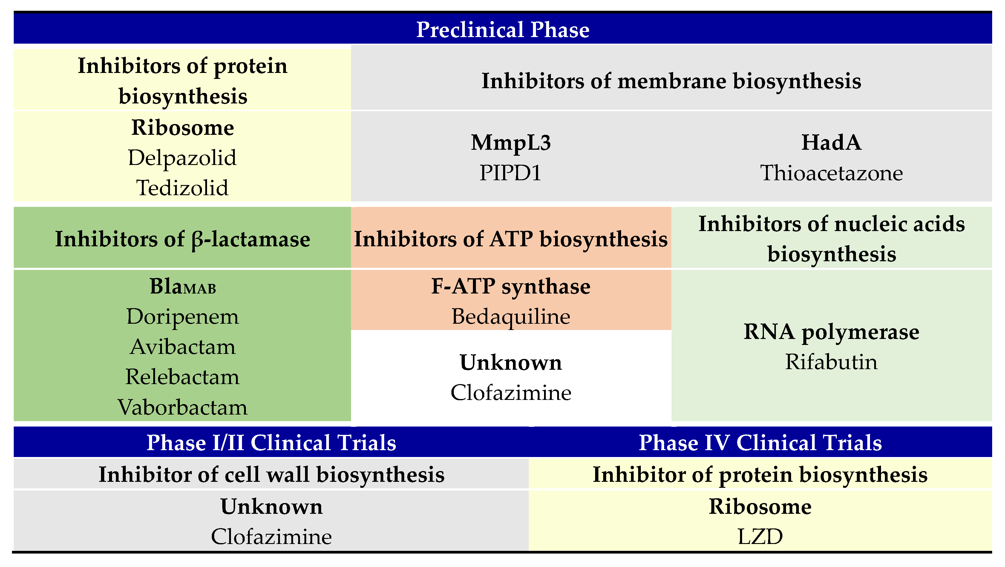
2. Ongoing M. abscessus Therapeutic Regimen

Mab establishes its infection mainly in immunocompromised individuals or patients with chronic respiratory diseases, such as cystic fibrosis and bronchiectasis. In particular, the thick mucus of patients with cystic fibrosis (pwCF) facilitates Mab and other opportunistic pathogens to establish chronic infections and colonialize the respiratory tract [6].
The treatment of Mab infection, as well as other NTM, remains difficult because of its AMR mechanisms and persistence; thus, there is not a consolidated and standardized therapy yet. For example, the actual guideline against NTM pulmonary infections is based on a mix of oral antibiotics and macrolides, depending on the susceptibility of the isolates [12]. However, according to the guidelines published in 2015 by the US Cystic Fibrosis Foundation and European Cystic Fibrosis Society, the current recommendations to treat Mab infections in individuals with pulmonary diseases, such as pwCF, is based on oral macrolide treatment, usually azithromycin, combined with intravenous administration of amikacin (AMK) and one or more additional antimicrobials among the following: tigecycline, imipenem (IMI) and cefoxitin (CEF). Infection is monitored by sputum culture conversion, and the end of the treatment is followed by a continuation phase consisting of administration of oral or inhaled antibiotics for more than 12 months [13].
Drugs currently used against resistant Mab strains are mostly repurposed anti-TB antibiotics that target protein syntheses, such as azithromycins, AMK, and tigecyline, and cell wall biosynthesis, like IMI and CEF. Due to Mab’s capability to acquire resistance, developing new strategies to use in combination with the current antibiotic treatment is a priority. For example, the use of new or repurposed drugs and the combination with phage therapy or other antimicrobial agents, such as small antimicrobial peptides or nitric oxide (NO), could be integrated. NO administration by inhalation is safe and tolerable and showed a reduction of Mab load in sputum and improvement in lung function in initial clinic trials [6,14]. Thanks to these promising results, NO treatment is currently undergoing phase II clinical trials as a supplement to the main therapeutic regimen. Moreover, host-directed therapy in combination with the classical antibiotic approach may be a valid help in Mab infection control [15].
To improve the effectiveness of therapies and shorten the administration time, the development of new drugs against Mab is required. This review aims to provide an overview of the compounds currently in clinical trials for the treatment of Mab infections, as well as the compounds in development in the early drug discovery phase in the past five years.

2.1. Preclinical Phase

Drugs that are classified as in the preclinical phase (Scheme 1) are used in treatments of other mycobacteria infections. They recently underwent evaluation for the treatment of Mab infections.
Delpazolid (LCB01-0371) is an oxazolidinone with a cyclic amidrazone, synthesized from linezolid (LIN) and firstly evaluated against multi-resistant Staphylococcus aureus (MRSA), showing good in vitro and in vivo bacteriostatic activities against Gram-positive bacteria [16]. Currently, in the phase 2 clinical trial for Gram-positive bacteria treatment and Mtb, its effectiveness is under evaluation against NTM infections [17]. In 2017, in vitro and in vivo studies of LCB01-0371 against wild-type Mab and clinical isolates showed that this compound inhibits bacterial growth in mouse lung infection models. Delpazolid was active against AMK, CEF, or clarithromycin (CLA)-resistant Mab with a MIC50 of 1.2 µg/mL. Moreover, it showed an improved safety profile compared to LIN, with lower toxicity even maintaining a good pharmacokinetic profile [18]. LIN inhibits protein synthesis at an early stage by binding to the A-site pocket at the peptidyl transferase center (PTC) of the ribosome 50S subunit, thus preventing the binding of the initiator tRNA, as demonstrated by X-ray crystallization [19]. Isolation of delpazolid-resistant Mab mutants confirmed that this compound shares the same mechanism of action of LIN, although it is still unclear whether delpazolid binds PTC to the same site [18,20].
Indole-2-carboxiamide (IC) is a molecule with potent activity against Mtb [21]. Recently, through structure-activity relationship (SAR) analysis, some derivatives of this compound have been proven to possess a stronger effect against bacteria [21]. Two of them have also been tested against Mab, showing a MIC of 0.125 μg/mL [22].
Similarly, a different series of IC derivatives active against Mab has been tested on acute toxicity mouse models, demonstrating that these molecules are effective in vivo, leading to a reduction of bacterial load in the lungs and good oral bioavailability [23]. It was proven that IC and derivatives disrupt cell wall biosynthesis by inhibiting MmpL3, a transmembrane protein involved in the transport of trehalose monomycolate onto the cell surface. Strains harboring single-point mutations in the MmpL3 gene showed high resistance phenotypes to IC derivatives [22].
PIPD1 is a novel piperidinol derivative that has been identified through the screening of a library of 177 molecules active against Mab. PIPD1 showed bactericidal activity against Mab clinical isolates with a MIC of 0.125 µg/mL, with the same mechanism of action of IC and derivatives. The PIPD1 effect was also evaluated in zebrafish infection models, reducing the bacterial burden and decreasing larval mortality [24].
Thioacetazone (TAC) is a bacteriostatic agent used for Mab treatment in combination with isoniazid (INH) and streptomycin (STR) in Africa and South America. The TAC mechanism of action was identified in Mtb. It is a prodrug activated through oxidation of its thiocarbonyl moiety by the monooxygenase EthA. Once activated, it binds to HadA, a component of the HadABC dehydratase complex involved in fatty acid biosynthesis, resulting in the inhibition of mycolic acid biosynthesis [25]. Mab is intrinsically resistant to this compound, but some TAC derivatives have shown promising inhibitory effects on Mab growth, with MIC values ranging between 1.6 and 6.2 μg/mL [26]. This data suggests that structural modification of the TAC scaffold could provide new effective drugs not only against Mtb but also against NTM.
Clofazimine (CFZ) is a lipophilic drug used for the treatment of Mycobacterium leprae. It acts as an artificial electron acceptor, causing respiratory chain malfunction and the disruption of ion transport across the cell wall. This double effect leads to a reduction of ATP availability and an increase in the reactive oxygen species inside the bacterial cell [27]. The combined treatment of CFZ with other common Mab antibiotics resulted in improved outcomes, with an 81% treatment response rate based on symptoms, but further evaluations of its efficacy are required [26]. Moreover, CFZ showed significant synergistic activity with AMK in vitro in 82% of cases against Mab clinical isolates [28], suggesting its possible use in combination with current anti-Mab drugs.
Bedaquiline (BDQ) is a repurposed anti-tubercular drug that targets the bacterial respiratory chain, inhibiting the mycobacterial F0F1 ATP synthase, also known as F-ATP synthase. It has been proved in vitro and in zebrafish infection models that BDQ possesses a broad-spectrum bacteriostatic activity against NTM. Moreover, it showed MIC values ranging from 0.031 to 0.125 µg/mL against different Mab clinical isolates [29]. The BDQ mechanism of action in NTM was confirmed by isolating low-resistant Mab strains and inserting two single-point mutations in the AtpE gene. The derived mutants present the amino acid substitutions D29V and A64P, showing high resistance to BDQ [29]. Its efficacy against Mab has been largely proved by infecting different mouse models with one Mab clinical isolate from pwCF and then treating it with the drug [30]. In this study, three mouse models were used: the severe combined immunodeficiency model (SCID), the granulocyte–macrophage colony-stimulating factor (GM-CSF) knockout model, and the nude mice model, all showing similar symptoms to human pulmonary Mab infection. In all models, the administration of BDQ for 5 days decreases the bacterial load in the spleen and liver. Moreover, after 8 days of treatment, the mycobacterial load also decreased in the lungs [30]. The use of BDQ in combination with CFZ decreased the mycobacterial burden in the spleen, liver, and lungs only after five days, showing a synergistic effect of the two compounds [30]. Finally, since efflux pumps require the proton motive force and adenosine triphosphate (ATP) to export molecules outside the cell, BDQ indirectly impairs this mechanism of drug extrusion, providing a powerful integration into the actual therapeutic regimen [29].
Rifamycins, such as rifampin, are antitubercular drugs ineffective against Mab since they are inactivated by ribosylation of their scaffold at the C23 hydroxyl position by the enzyme ADP-ribosyltransferase (ArrMab). However, the screening of libraries of approved antitubercular drugs led to the discovery of rifabutin (RIF), a compound of this family that displays bactericidal activity against Mab [31]. It was demonstrated in a macrophage infection model that administration of RIF decreases intra- and extracellular cording of the Mab R morphotype. The same result was observed in Mab-infected zebrafish following RIF treatment, together with a reduced abscess formation [32].
Mab exhibits intrinsic resistance to β-lactams except for CEF and IMI, which are currently used in treatments of Mab infections. This dual β-lactams combination showed a synergist effect against all of the considered Mab clinical isolates, while the combination of Doripenem and IMI resulted in a synergetic effect against most strains. This outcome is probably due to the different targets hit by these two β-lactams, possibly non-redundant enzymes involved in the biosynthesis of the cell wall, consequentially leading to a synergistic effect [33]. Different β-lactams combinations were also tested in vivo in the Mab-infected murine model, according to the synergy showed in vitro. After four weeks of treatment, the average lung CFU decreased by 6 log10 compared to the untreated mice [34]. These results confirmed that the current Mab treatment could benefit from the administration of combined β-lactams. However, the insurgence of resistant strains remains a challenge, limiting the use of such compounds. Resistance to β-lactams in Mab is mediated by a broad-spectrum Ambler-Class A β-lactamase (BlaMAB), which hydrolyses common β-lactamase inhibitors such as clavulanic acid, tazobactam, and sulbactam [35]. Therefore, many studies focused on finding new β-lactamase inhibitors to use in combination with β-lactams. For example, avibactam is a non β-lactam β-lactamase inhibitor used in combination with ceftazidime for the treatment of Gram-negative bacterial infections [36]. Avibactam was tested in combination with several representatives of the three main classes of β-lactams (penams, cephalosporins, and carbapenems) against a set of Mab clinical isolates. In the presence of this molecule, even compounds usually ineffective against Mab inhibited its growth [37]. Moreover, the combination of amoxicillin and avibactam was evaluated ex vivo in the intracellular infection model and in vivo in the zebrafish model. In both cases, the co-administration of avibactam increased the activity of amoxicillin, resulting in an increased survival rate and a decrease in abscess formation in infected zebrafish larvae [37]. Another study evaluated the effect of Avibactam in combination with piperacillin in vitro against Mab and in vivo in a Galleria mellonella infection model. The combination resulted in a lower bacterial burden compared to the larvae treated with piperacillin only. Moreover, the in vitro MIC was 16–32 folds lower when the two compounds were used in combination [38].
Relebactam and vaborbactam are the other two non β-lactam β-lactamase inhibitors recently approved for the treatment of bacterial infection in combination with IMI and meropenem. These compounds were recently evaluated against Mab clinical isolates in combination with a wide range of carbapenems and cephalosporins, resulting in a significative decrease of MIC50 and MIC90 by one or two dilutions, respectively [39]. These results confirm that the combination of non-β-lactam β-lactamase inhibitors with β-lactams is effective for the treatment of Mab infections with positive outcomes and to expand the range of effective β-lactams against these bacteria.
Tedizolid (TDZ) is a next-generation oxazolidinone antibiotic approved by the Food and Drug Administration in 2014 for the treatment of soft tissue and skin infections caused by some Gram-positive bacteria, such as Streptococcus spp. and MRSA. The efficacy of TDZ was evaluated in vitro against a pool of Mab clinical isolates both alone and in combination with other antimicrobials, such as doxycycline, AMK, and CLA. TDZ showed bacteriostatic activity against the considered strains without instances of antagonism with other antimicrobials [40]. Another study confirmed the efficacy of TDZ in vitro with MIC values ranging between 0.25 and 8 mg/L depending on the strain and in an intracellular macrophage infection model. Moreover, at a concentration of 2x MIC, TDZ reduces bacterial load by a factor of 500 compared to the untreated condition [41]. This molecule was also evaluated on twelve patients who met the ATS/IDSA criteria for NTM infections. The treatment was based on a multi-drug therapy, TDZ included, showing benefit from this integration with improvement in symptoms and the clinical outcome [42].

2.2. Phase II Clinical Trial

Despite the lack of any new drug in phase I clinical trials, some studies are focusing on different delivery methods, such as the Liposomal AMK for Inhalation (LAI), which is in phase II of clinical trials. This new formulation is composed of dipalmitoylphosphatidilcholine (DPPC) and cholesterol-containing encapsulated AMK to be delivered through aerosol. AMK is a semi-synthetic aminoglycoside antibiotic derived from kanamycin and commonly used in Mab treatment. It binds the 30S ribosome subunit, inhibiting protein biosynthesis [43].
LAI preparation is capable of lowering drug toxicity compared to systemic AMK administration. Moreover, a higher concentration of AMK is maintained at the site of infection, allowing a lower dosage, thus reducing the potential side effects. LAI was evaluated in vitro against Mab in a macrophage infection model, resulting in better activity compared to free AMK at a dose of 10 mg/L [44]. During a randomized phase II clinical trial, three Mab-infected patients with persistently positive NTM cultures, even after American Thoracic Society/Infectious Diseases Society of America (ATS-IDSA) guidelines-based treatment, were treated with LAI preparation achieving culture conversion [45]. The phase II trial is currently recruiting to further evaluate this formulation in order to confirm its efficacy against Mab and to assess its safety for infected patients [17].

2.3. Phase IV Clinical Trial

LZD is an oxazolidinone currently undergoing phase IV clinical trials for the treatment of NTM infections. Similarly to the two derivatives, TDZ and delpazolid, LZD inhibits protein synthesis by binding the 23S ribosome subunit to the binding site of the initiator tRNA, thus blocking the translation process at its beginning [19]. In 2018, LZD was used to treat a multi-drug-resistant Mab pulmonary infection case, leading to the successful recovery of the patient [46]. Despite its activity against NTM and Gram-positive bacteria, including MRSA, vancomycin-resistant enterococci (VRE), and penicillin-resistant pneumococci, prolonged administration of LZD is advised against due to its toxicity. Indeed, LZD causes long-term side effects, such as reversible myelosuppression, potentially irreversible optic neuropathy, and peripheral neuropathy [47].

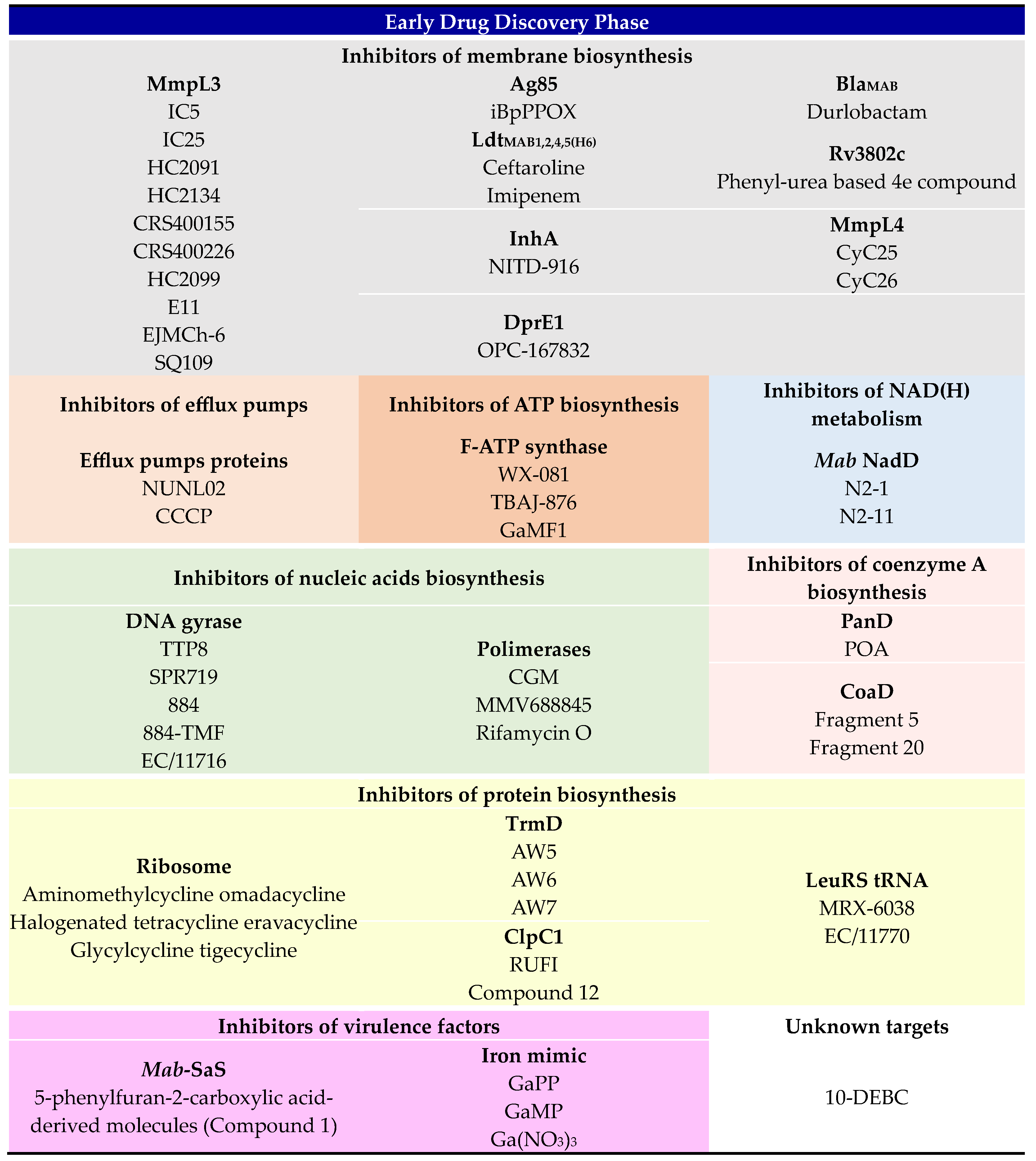
3. Early Drug Discovery Phase

The molecules that have been under development for the last five years in the early drug-discovery phase against Mab are discussed below and shown in Scheme 2. This section is organized according to the metabolic pathway targeted.

3.1. Inhibitors of Membrane Biosynthesis

One of the most interesting strategies to kill mycobacteria is hitting the cell wall biosynthesis. This is possible by exploiting different strategies and targets, such as lipids transporters, penicillin-binding proteins, or mycolic acid metabolism.

3.1.1. Inhibitors of Transporter Proteins

The inhibition of the mycobacterial membrane protein Large 3 (MmpL3), a lipid transporter involved in the translocation of the trehalose mono-mycolate (TMM) towards the cell wall, is by now an attractive and validated strategy to kill mycobacteria [23,48].
The indole-2-carboxamides (ICs) are molecules that interfere with the mycolic acids’ transport across the mycobacterial inner membrane by targeting MmpL3 [21,49,50]. Recently, they have been tested against Mab, too. Two compounds from this class, the IC5 and IC25 shown in Table 1, exhibited MIC values of 0.25 μg/mL and 0.063 μg/mL, respectively, against Mab ATCC19977 with bactericidal effect. The target of these compounds was confirmed in Mab by genetic studies over a mutant strain with a missense mutation on the mmpL3 gene, which showed a high increase of both MICs of ~100 times [23]. These two IC molecules have been well tolerated in vivo, as shown in acute toxicity mouse models at different dosages and for different days of administration without negative side effects. The pharmacokinetic analysis of both molecules showed interesting results in terms of maximum drug levels, half-life of the compounds, plasma clearance, and volume of distribution. Moreover, control-infected mice showed a log10 CFU of 6 for lung, spleen, and liver against the log10 CFU of 3–5 in the same organs of treated mice for both compounds. These results confirm the possibility of repurposing the ICs against Mab infections.
Other derivatives of the same scaffold that had a preliminary evaluation are the N-[2-(4-chlorophenyl)ethyl]-4-thiophen-2-yloxane-4-carboxamide (HC2091, Table 1) exhibiting a MIC50 of 6.25 µM and N-(2-methoxy-5-nitrophenyl)-1-oxo-4-phenylisochromene-3-carboxamide (HC2134) that exhibited a MIC50 of 12.5 µM [51]. Moreover, they have been tested only in vitro against Mab, and further evaluation is necessary to prove that they are also active ex vivo and in vivo in animal models.
The benzothiazole amide derivatives share a similar scaffold and mechanism of action with ICs [52]. The best substituents in benzothiazole-based compounds have been identified in adamantly and cyclohexyl derivatives groups, which effectively inhibit the rapid-growing NTM class, Mab included. These compounds showed great MICs over Mab ATCC19977. From the adamantyl benzothiazole amide, named CRS400226, two other compounds have been developed, further improving the inhibition power of this scaffold. The MICs of these compounds ranged between 0.03 and 0.06 μg/mL for the CRS400359 and CRS400393 and were 0.25 μg/mL for CRS400226 and 0.5 μg/mL for CRS400153 [53]. In particular, CRS400153 and CRS400226 have been exploited for the identification of the mechanism of action monitoring the cell wall biosynthesis. At a concentration of 2X and 10X MIC, they inhibited MmpL3 transport since the mycolic acid was not observed in the mycobacterial cell envelope. This inhibition was proved to be concentration-dependent. Moreover, Mab mutant strains resistant to the benzothiazole amides showed mutations in the MmpL3 transporter, confirming that MmpL3 is the target [53]. In vivo, analysis of CRS400226 in a chronic Mab infection mouse model confirmed that this compound is efficient and tolerable since no side effects have been shown after the infection was cleared [53]. CRS400226 structure is shown in Table 1.
Another derivative of benzothiazole amide under evaluation is the 2-[(6-chloro-1H-benzimidazol-2-yl)sulfanyl]-N,N-di(propan-2-yl)acetamide HC2099, which showed a MIC of 25 µM against Mab [51]. It has been evaluated only in vitro against Mab; hence, further studies are necessary to prove its efficacy in animals and humans.
A different scaffold capable of targeting MmpL3 is well-represented in the acetamide E11 compound (Table 1). It has been well-characterized in Mtb and subsequently evaluated against Mab since their similarities, showing a MIC of 12 µM against the ATCC19977 strain and 25 µM against the Mab subsp. abscessus Bamboo clinical isolate [54]. Additionally, the preliminary studies of E11 are limited to in vitro and in vivo evaluation against the bacteria, so further evaluations are necessary before moving into clinical.
Among the compounds that impair the MmpL3 transporter, the 2-(2-phenalkyl)-1Hbenzo[d]imidazole scaffold has been identified in Mtb and subsequently evaluated over Mab. For example, the 2-(2-cyclohexylethyl)-5,6-dimethyl1H-benzo[d]imidazole (EJMCh-6, Table 1) inhibits the trehalose monomycolate (TMM) transport decreasing the arabinogalactan mycolylation [55]. Indeed, the in vitro evaluation of EJMCh-6 showed Mab growth inhibition with a MIC of 0.125 µg/mL and a bacteriostatic effect. Moreover, it showed MIC values of 0.031 µg/mL against the non-CF R clinical isolates CIP104536 belonging to the Mab complex and 1 µg/mL for the same CF S variant. Nevertheless, similar MIC values resulted from both R and S strains, proving its high efficacy against Mab [55]. EJMCh-6 also showed low toxicity against human THP-1 macrophages; hence, it has been evaluated with a dosage of 3 µg/mL over S Mab infecting macrophages. Results confirmed a reduced mycobacterial load at 3 dpi. In zebrafish, the administration of 0.75 μg/mL of EJMCh-6 resulted in 80% of alive embryos compared with only 40% surviving untreated animals [55]. The presence of single nucleotide polymorphism (SNP) in the mmpL3 gene in EJMCh-6-resistant strains suggests that this transporter is the target. This is further confirmed by the overexpression of mutated mmpL3 strains, which showed increased MIC values [55].
Ethylenediamines are a known class of inhibitors of Mtb mycobacterial membrane biosynthesis. This class of compounds has been recently repurposed for use against Mab. The N-geranyl-N′-(2-adamantyl)ethane-1,2-diamine, also known as SQ109 (Table 1), for example, exhibited a MIC 0.5 µM against Mtb, whereas higher values of 22 µM and 44 µM against R Mab and S Mab, respectively [56]. On the contrary, two analogs characterized by the n-butyl and the benzyl groups showed better MICs of 6 µM each against both morphotypes. A resistant Mab mutant strain in the mmpL3 gene was used to confirm that the related protein is the compound’s main target. This strain, which showed resistance to all the major inhibitors of MmpL3, was sensitive to SQ109, which showed a MIC comparable to wild-type Mab. This finding suggests that MmpL3 may be a secondary target for this class of inhibitors in Mab [56].
The oxadiazolone scaffold has been found to be effective against Mab, too. In particular, the iBpPPOX derivative (Table 1) showed a MIC value of 33.0 µM for the S morphotype and about 50 µM for the R morphotype. It seems clear that the effectiveness of iBpPPOX against Mab depends on the position of the phenoxy group compared to other derivatives of the same class [57]. The intracellular effect of this molecule has been evaluated against the S morphotype in the Raw264.7 murine macrophage infection model, showing that every concentration tested resulted in the same bacterial killing effect of about 58%. This may be due to the stress dictated over the metabolism, which led to bacterial death. Moreover, the intracellular concentration of 30 µM gave the same result, confirming the extracellular MIC [57]. The target of iBpPPOX is the Ag85 protein, which carries out its activity in cell wall biosynthesis. This has been proven by the overexpression of Ag85, which led to an increased MIC, while its genetic inactivation showed no differences compared to the wild type after the iBpPPOX administration [57].

3.1.2. β-Lactamases Inhibitors in Combination with β-Lactams

As briefly discussed in the preclinical phase, the β-lactams disrupt the peptidoglycan biosynthesis, and resistance to these drugs is particularly important in mycobacteria, Mab included [58]. For this reason, different combinations of β-lactams in combination with β-lactamases inhibitors are in development [59].
Molecules belonging to the diazabicyclooctanes (DBOs) class are known inhibitors of β-lactamases enzymes. A brand-new DBO is the durlobactam (DUR), which has been improved compared to the previous DBO ring-featured molecules, and it is currently in clinical trials against other microorganisms in combination with β-lactams [60]. DUR has recently been evaluated against Mab. This molecule targets the BlaMAB β-lactamase enzyme-binding tightly, as demonstrated by its Ki of 4 × 10⁻³ µM, about 75-fold lower than avibactam and relebactam [60]. The MIC of DUR was evaluated against a hundred-one Mab clinical isolates, resulting against all strains in the range of 2–8 µg/mL. In combination with amoxicillin or IMI (ratio 1:1), it decreased at 4 µg/mL [60]. Moreover, the triple combination of DUR, amoxicillin and IMI exhibited a MIC between 0.06 and 2 µg/mL, while the combination of DUR, amoxicillin, and cefuroxime showed a MIC between 0.06 and 1 µg/mL [60]. These results confirm that this novel β-lactamase inhibitor could successfully be integrated into β-lactams treatments to prevent their hydrolysis.
Additionally, not all the combinations of β-lactam and β-lactamase inhibitors are effective. For example, ceftaroline, a β-lactam already in clinical use, has been evaluated against Mab ATCC19977, showing a MIC of 16 µg/mL. A mild synergistic effect in combination with the β-lactamases inhibitors relebactam decreased the MIC of 1–2 dilutions while avibactam decreased the MIC of 4 or more dilutions [61]. In contrast, the combination of IMI, another β-lactam used in the clinic, with both the previous compounds maintained the MIC value unaltered at 2 µg/mL. To better understand these differences in synergy, these β-lactamases inhibitors have been evaluated against the BlaMAB enzyme from a kinetic perspective, confirming that avibactam inhibits the enzyme better than relebactam. Indeed, the avibactam Kd was lower than relebactam, indicating a stronger binding even if they show similar Koff values [61]. Moreover, the two β-lactams have been evaluated against different Mab enzymes that are part of the L-D-transpeptidases family (Ldts), showing that the binding affinities (Ki) of IMI for LdtMAB1, LdtMAB2, and LdtMAB4 are higher than ceftaroline, while both IMI and ceftaroline have a similar affinity for LdtMAB5(H6). These data further confirm that the combination of ceftaroline and IMI with β-lactamases inhibitors may be an effective strategy against Mab [61].

3.1.3. Other Inhibitors of the Cell Wall Metabolism

Rv3802c belongs to a gene cluster that modulates mycolic acid biosynthesis and transport. Recent studies have confirmed that this enzyme hydrolyses the phosphatidylinositol, regulating the membrane lipid content [62]. Considering the importance of his role in cells, Rv3802c has been taken into consideration as a putative drug target to kill mycobacteria. Urea-based molecules have been identified as strong hydrolase inhibitors [63]; hence, heterocyclic urea derivatives have been studied against Mtb hydrolases. Considering the results of these compounds, a selection of derivatives has been evaluated against Mab ATCC19977, too. Only the 4e compound, shown in Table 2, exhibited a moderate MIC of 25 µM, while the toxicity analysis performed over murine cells showed a higher MIC. Despite these preliminary results, this study is a valuable starting point for the development of effective Mab hydrolase inhibitors [62].
Another important target involved in cell wall biosynthesis is the enoyl-ACP reductase (InhA), which is involved in mycolic acid biosynthesis [64]. INH effectively targets Mtb InhA upon activation by the catalase-peroxidase KatG. Recently, this target has also been studied in Mab, using NITD-916 (Table 2), a 4-hydroxy-2-pyridone that bypasses the activation process of the KatG and directly inhibits InhA. The MIC against Mab was variable, depending on the medium used, and ranged from 0.195 μg/mL to 6.25 μg/mL [64]. To note, the killing effect of NITD-916 was not concentration-dependent, while the compound seemed to act as bacteriostatic [64]. NITD-916 was effective ex vivo in the human THP-1 macrophage infection model at a concentration of 12.5 μg/mL, which was below the observed cytotoxic effect. Positive results were also found in airway organoids derived from the lungs of healthy people and pwCF, effectively reducing Mab load in both conditions at a concentration of 15.6 μg/mL. Finally, to prove that NITD-916 is not activated by KatG, its MIC was evaluated in the presence of a Mab strain overexpressing this enzyme. The unaltered MIC against this strain confirmed the direct inhibitory effect of this molecule [64].
One of the most important enzymes involved in the biosynthesis of the mycobacterial cell wall is the decaprenylphosphoryl-b-D-ribose oxidase DprE1, which has been widely characterized in Mtb [65,66]. It catalyzes the formation of a precursor of the arabinogalactan, an essential player in the mycobacterial membrane. Preliminary repurposing attempts of the best molecules developed against Mtb DprE1 have been obtained against the homologous enzyme of Mab. The inhibitor dihydrocarbostyril OPC-167832 (Table 2), for instance, has been evaluated against Mab ATCC19977, showing a MIC of 5.3 µM, hence displaying a good activity against this strain [67]. Other MICs against Mab subsp. Bolletii CCUG 50184T, Mab subsp. Massiliense CCUG 48898T, and a panel of clinical isolates ranged between 5 and 15 µM, suggesting that the repurposing of OPC-167832 was not effective against Mab since the MIC in Mtb was assessed in nanomolar concentration. Moreover, in vivo analysis of OPC-167832 in different mouse models showed a non-statistically significant decrease of bacterial burden in lung and plasma, confirming the previous data [67]. However, the understanding of the molecular interactions between OPC-167832 and its target in Mab will probably allow us to improve its effect or design new inhibitors.
Another appealing target to fight NTM may be the metabolism of GPLs and their translocation on the R strain surface. Indeed, the presence of these glycopeptidolipids in the S morphotype of Mab sensibly decreases its susceptibility to many drugs [68]. For example, the multi-target inhibitors derived from cyclipostins and cyclophostins (CyC) showed higher MIC values against the S Mab morphotype, which has a cell wall rich in GPLs. Indeed, CyC17 and CyC18b against S Mab showed MICs of 6.4 and 6 µg/mL, respectively, while against R Mab, their MICs were 0.18 and 4.9 µg/mL [68]. Among the CyC derivatives that are characterized by a long lipophilic chain and share the phosphate ester scaffold, two features that help the crossing through the membranes, CyC25 and CyC26, showed MIC50 of 13.9 µM and 6.9 µM against R Mab, respectively. The same compounds have MIC50 close to 100 µM against the S morphotype, which means around 10 times higher [69]. The importance of GPLs in Mab resistance was confirmed by inactivation of mmpL4b in S Mab since the MmpL4b protein is involved in the translocation of GPLs in the cell wall. The same complemented strain regained resistance to CyC25 and CyC26 (Table 2), exhibiting high MIC values compared to the ΔmmpL4b S Mab [69]. This data suggests that the absence or reduction of GPLs may induce some changes in the cell-wall fluidity and permeability, possibly favoring the penetration of these inhibitors. Finally, the low toxicity of these molecules allows their use in combination with the well-established antibiotic regimen [69].

3.2. Inhibitors of Efflux Pumps

Efflux pumps are involved in the molecular mechanisms of AMR in bacteria, as previously mentioned in this paper. Because of their role, they are recently considered an attractive target for drug development. Inhibitors of the efflux pumps (EPIs) work synergistically with drugs, improving the accumulation of toxic molecules inside the bacterial cell. Along with the already-known Mtb EPIs such as verapamil, phenothiazines class of molecules, protonophores, valinomycin, and plant-derived EPIs [70], EPIs specifically designed against this pathogen are under development.
Usnic acid, a compound of lichen derivation, showed antimicrobial activity against Mtb and different NTM, Mab included. The increase of ethidium bromide inside Mab cells and the decrease of the MIC of AMI and CLA of 4-fold in the presence of this compound suggest its inhibitory effect on efflux pumps [70].
Recently, the effect of NUNL02 (Table 3), a derivative of tetrahydropyridines and a known inhibitor of the E. coli AcrB efflux pump [71], was assessed against Mab ATCC19977 and three clinical isolates: Mab subsp. abscessus AT 07, Mab subsp. bolletii AT 46, and Mab subsp. bolletii AT 52, showing MIC values ranging from 50 to 200 μg/mL [72]. In this study, the MIC of AMI, ciprofloxacin (CIP), and CLA was evaluated in the presence and absence of NUNL02, and the known EPI verapamil was used as control. The effect of both compounds was assessed at the subinhibitory concentration of ½ MIC, which does not affect the viability of bacteria [72]. Under these conditions, the presence of verapamil reduced the MIC of antimicrobials by 8-fold, while NUNL02 was 16-fold. Additionally, the ethidium bromide accumulation test further suggests that NUNL02 targets the efflux pumps, possibly confirming its mechanism of action in Mab. Additionally, this study lacks any in vivo evaluation to validate the potentiality of NUNL02 as an addition to therapies.
Carbonyl cyanide 3-chlorophenylhydrazone, CCCP (Table 3), is another EPI under investigation against Mab. The accumulation assay showed that CCCP lowers the MIC of CLA in different Mab CLA-resistant clinical isolates by 52.6%, while it did not show any effect against sensitive Mab strains [73]. The only strains resistant to CLA in the presence of CCCP presented the mutation rrl 2270/2271, which confers intrinsic resistance to CLA [73]. Recently, the effect of CCCP was evaluated against 47 Mab strains without the presence of any antibiotics to evaluate its toxicity on bacteria. Data showed MICs lower than 7.5 μg/mL against all strains [74]. The CCCP effect was also evaluated at a concentration of 6 μg/mL in a macrophage infection model. This experiment showed a concentration-dependent inhibition of the intracellular Mab growth of 84.8 ± 8.8% and of the Mab subsp. massiliense growth of 72.5 ± 13.7%. Its cytotoxicity was assessed on THP-1 and U937 cells, demonstrating that the compound is safe at concentrations ≤ 6.25 μg/mL, with over 97% cell viability [74]. Additionally, CCCP lacks the in vivo evaluation before progressing to clinical trial.

3.3. Inhibitors of ATP Biosynthesis

Mycobacteria produce ATP via oxidative phosphorylation through the F-ATP synthase that possesses peculiar features compared to the orthologous of other bacteria. Indeed, the subunit α contains a C-terminal elongated domain that regulates the speed of ATP synthesis. This domain is essential during the bacterial dormancy state [75]. The mycobacterial δ subunit has an inserted domain that interacts with α and β subunits, differently to other bacteria, chloroplast, and mitochondria where the δ orthologous interacts only with the α subunit [76]. Finally, the rotating central γ subunits contain an extra loop of 12–14 polar amino acids that are not present in any other prokaryotic or eukaryotic homologs and are located in the proximity of 4 polar residues of the c ring. This loop is a regulatory element of the F-ATP synthase involved in the coupling of proton translocation with ATP synthesis [77]. These features are unique, attractive elements for drug development. New inhibitors of Mab ATP synthase are being developed to fight resistant strains to BDQ. These molecules may be BDQ analogs or derivatives, or they may exploit different scaffolds.
WX-081 (Table 4), known as sudapyridine, is a structural analog of BDQ, possibly with the same mechanism of action [78]. It was evaluated in vitro against 50 strains of NTM mycobacteria, showing a comparable MIC to BDQ for most of them [79]. In fact, the observed MIC values were mostly below 0.25 μg/mL and in no case above 2 μg/mL. Against Mab, the MIC determined in vitro was 0.25 μg/mL. WX-081 resulted in a similar cytotoxicity to BDQ but with fewer cardiac side effects, making this compound an interesting substitute for the classic drug [79]. However, this preliminary study lacks ex vivo and in vivo evaluation to demonstrate the real efficacy and limitations of WX-081.
Another analog of BDQ is TBAJ-876, a diarylquinoline compound that targets the F-ATP synthase with the same mechanism of action [80]. It showed a bacteriostatic effect against NTM, including various strains of Mab, with a 2-fold lower MIC compared to BDQ [81]. In a murine infection model, it showed a similar effect to BDQ at a dose of 10 mg/Kg. Moreover, it did not exhibit antagonism with commonly used anti-Mab drugs in checkerboard assay [81], suggesting that TBAJ-876 could be a valuable integration into the actual therapeutic regimen.
GaMF1 (Table 4) is a known bactericidal compound selective for Mtb [82]. Its mechanism of action was hypnotized by in silico docking studies. It has been demonstrated that GaMF1 inhibits the rotation of the ATP synthase γ subunit by binding its extra loop. This interaction disrupts the function of the c-ring turbine, interrupts proton flux, and ultimately hinders ATP synthesis [82]. Recently, GaMF1 was evaluated against Mab subsp. abscessus ATCC19977, showing a MIC50 of 33 μM in Middlebrook 7H9 broth and 13 μM in cation-adjusted Mueller–Hinton (CAMH) medium [83]. Similar MIC50 values were observed for the clinical isolate Mab Bamboo. Moreover, the bactericidal effect of GaMF1 was observed 10-fold the MIC50 in both media [83]. GamF1 was analyzed against the enzyme by an intracellular ATP synthesis assay and by inside-out membrane vesicles (IMVs), showing an IC50 of 10 ± 0.9 μM against the ATP synthesis and of 13 ± 2.5 μM against the NADH-driven ATP synthesis [83]. Finally, the combination of GaMF1 with CFZ, RIF, or AMI revealed an increased susceptibility of Mab to these drugs.

3.4. Inhibitors of NAD(H) Metabolism

NAD(H) is an essential redox cofactor for many enzymes, such as the ones involved in glycolysis, and it is an electron donor in the respiratory chain. Depletion of NAD(H) has been shown to affect both these processes, leading to the killing of mycobacteria [84,85,86]. Consequently, NAD biogenesis has been identified as a valuable target to impair mycobacterial growth [87]. In addition, NAD biogenesis inhibition could synergistically impair ATP synthesis [84].
NadD is a nicotinic acid mononucleotide (NaMN) adenylyltransferase implicated in NAD biosynthesis and the most divergent enzyme from its human counterpart [86]. In the literature, different series of molecules selectively hit Mtb NadD are reported. One of this series, named N2, comprises a benzimidazolium core that interacts with the catalytic amino acids Asp109 and Glu160, perturbing the NadD dimer quaternary structure and leading to the inhibition of the enzymatic activity. Although the series was evaluated in vitro against the Mtb homologous enzyme, it was also tested against Mab. The most effective compounds (Table 5) exhibited MIC values in the micromolar range. Specifically, N2-1 showed a high MIC of 61.4 μM while N2-11 had a MIC of 26 μM [87].

3.5. Inhibitors of Nucleic Acids Biosynthesis

Drugs may inhibit different enzymes involved in nucleic acid biosynthesis, eventually killing pathogens. Two strategies are classically exploited: the inhibition of purines and pyrimidines biosynthesis or the impairment of nucleic acids polymerization. Recently, novel strategies have emerged, such as covalently binding the DNA to mechanically impair the enzyme binding. This is, for instance, the mechanism of action of the 5-Nitroimidazoles [88].

3.5.1. Inhibitors of DNA Gyrase

The DNA gyrase is a peculiar type IIA topoisomerase involved in the topology of DNA and a validated druggable target in mycobacteria [89]. This enzyme is a hetero-tetramer composed of two GyrA subunits and two GyrB subunits (A2B2 complex). In the presence of magnesium and upon ATP hydrolysis, it introduces a nick in the double strand to resolve the supercoil during DNA replication and mRNA synthesis. It is also involved in maintaining the homeostasis of negative supercoil DNA [89].
Tricyclic pyrrolopyrimidine (TPP) and SPR719 are inhibitors of the Mtb gyrase. They bind the ATPase domain of the B subunit, impairing the enzymatic activity [90]. TPP8, shown in Table 6, is a derivative of the TPP scaffold that exhibited a MIC in the range of 0.2–0.02 μM against various Mab strains, ATCC19977 included, and a panel of clinical isolates. In vitro DNA supercoiling inhibition studies confirmed that TPP8 impaired the enzymatic activity with an IC50 of 0.3 μM. Finally, the two resistant mutants isolated in the study have been analyzed by whole-genome sequencing. They had a C506A (Thr169Asn) missense mutation in the ATPase domain, confirming the TPP8 mechanism of action [91]. This molecule showed a bacteriostatic effect against Mab in a THP-1-derived macrophage infection model treated with a different MIC multiple.
Similarly, SPR719 (Table 6) targets the ATPase domain of GyrB, acting as a competitive inhibitor of ATP [92]. Its mechanism of action has been identified through the isolation of Mtb-resistant mutants and their genetic analysis. The oral formulation of this molecule already passed the phase 1 clinical trial for safety, tolerability, and pharmacokinetics [93]. A recent evaluation of this molecule against Mab ATCC19977 and Mycobacterium avium showed MIC values of 1.6 µM and 6 µM, respectively. Additionally, SPR719 lacks ex vivo and in vivo experiments to further confirm its efficacy against Mab-infecting cells or animal models.
Piperidine-4-carboxamides are a class of molecules that inhibit mycobacterial gyrase in a different way than TPPs and SPR719. Indeed, resistant mutants suggest that this scaffold targets GyrA instead of GyrB [94]. Two derivatives of this scaffold, molecule 844 and 844-TMF (Table 6), were active against the three reference strains of the Mab complex, and a collection of clinical isolates showing MICs in the range of 6–14 µM for 844 and 1.5–6 µM for 844-TMF. Moreover, both of the molecules had a bactericidal effect on planktonic cultures and biofilm [94]. Despite the positive results of these molecules and their interesting pharmacokinetic properties, it is necessary to further evaluate this molecule to confirm its efficacy in vivo.
A different mechanism of action is explored by the Mab gyrase inhibitor EC/11716 (Table 6). This molecule is classified as part of “novel bacterial topoisomerase inhibitors”. This class of compounds is characterized by a two-part scaffold, the right-hand side, and the left-hand side, linked together by a central unit. These molecules impair the gyrase enzymatic activity by binding a non-catalytic interface pocket between the two GyrA subunits on the right-hand side and intercalating the DNA after cleavage with its left-hand side [95]. EC/11716 was evaluated against Mab ATCC19977 and a pool of clinical isolates showing MICs in the order of a few micromolar against every strain, with a bactericidal effect against planktonic cultures and impairing biofilm formation [96]. Moreover, it decreases 1 log of the bacterial CFU of Mab K21 at a daily dose of 400 mg/Kg after 10 days in the NOD SCID mice infection model, confirming the potentiality of this molecule in vivo.

3.5.2. Inhibitors of Polymerases

Among classical targets of DNA metabolism, DNA polymerase plays a major role. In particular, DNA polymerase III β subunit (DnaN) is involved in DNA replication and repair in bacteria. DnaN is a sliding clamp that acts as protein–protein interaction (PPI) hub by surrounding the double-strand DNA and recruiting a plethora of proteins involved in DNA metabolism [97,98].
Griselimycins are a class of cyclic depsipeptides originally isolated by Streptomyces spp. [99], which binds the PPI cleft, impairing the interaction with the polymerase III α subunit DnaE1 [100]. The cyclohexyl-griselimycins (CGM) analog showed a potent antitubercular effect in vivo in infected TB mouse models and good oral bioavailability [100]. Recently, CGM was evaluated against three reference strains of Mab, ATCC19977, Mab subsp. Massieliense, and Mab subsp. bolletii and against a pool of clinical isolates showing positive results. CGM exhibited MICs against all the considered strains ranging between 0.1 and 0.8 µM with a bactericidal effect in all cases. It was also effective in vivo, decreasing the Mab K21 CFU by 10-fold compared to the same dose of CLA in an infected mouse model [101].
Nα-2-thiophenoyl-d-phenylalanine-2 morpholinoanilide (MMV688845), shown in Table 6, is an interesting inhibitor of RNA polymerase [102]. Its mechanism of action was verified by analysis of six isolated Mab-resistant mutants and by analogy with other inhibitors of the RpoB subunit of the Mtb RNA polymerase sharing the same chemical scaffold [103]. MMV688845 inhibited the growth of different Mab strains in vitro with a MIC in the order of a few µM and ex vivo showing a MIC of 15.9 µM in a macrophage infection model, exhibiting bactericidal effect [102]. Moreover, it showed synergy in combination with CLA, lowering the MIC by 10-fold compared to other antibiotics [102]. Despite these positive results, in vivo studies on mice demonstrated that the plasm concentration of the oral formulation of this compound never reaches the MIC value necessary to eradicate the infection [102], so the improvement of pharmacokinetic properties is required.
RpoB is also targeted by rifamycin, but Mab is naturally resistant to this drug [104]. Rifamycin O (Table 6), the oxidated derivative of the natural rifamycin B obtained by Streptomyces mediterranei [105], showed interesting results even against this microorganism. Indeed, the in vitro evaluation against Mab CIP 104536T, Mab subsp. bolletii CIP108541T and Mab subsp. massiliense CIP108297T showed a MIC90 of 4.0–6.2 µM. Moreover, a concentration of 25 µM of rifamycin O daily used to treat a zebrafish infection model showed a reduction of Mab infection and the increase of zebrafish lifespan of 54% up to 13 days, proving that the molecule is effective in vivo and confirming its low cytotoxicity [104].

3.6. Inhibitors of Protein Biosynthesis

Protein biosynthesis is an essential metabolic mechanism that requires the coordination of many players, such as ribosomes, mRNA, tRNA, and other factors, such as initiation factors IF and elongation factors EF [106,107,108]. The impairment of one of these players blocks protein synthesis; hence, many drugs have been developed exploiting this strategy.

3.6.1. Molecules That Directly Target the tRNA

MRX-6038 is a boron-containing molecule that inhibits the prokaryotic leucyl-tRNA synthetase (LeuRS). This class of inhibitors covalently binds the tRNA at the terminal adenine 76 of the editing site, thereby hindering leucylation and consequentially impairing protein synthesis [109]. MRX-6038 mechanism of action was validated by sequencing resistant mutants, which exhibited different mutations in the editing site [110]. MRX-6038 was evaluated against two Mab reference strains, ATCC19977 and CIP108297, along with 194 clinical isolates. The MICs for all the considered strains ranged between 0.063 and 0.125 mg/L [110]. Furthermore, it was assessed against 10 NTM reference strains, exhibiting MICs below 0.25 mg/L against Mycobacterium smegmatis, Mycobacterium fortuitum, Mycobacterium scrofulaceum, and Mycobacterium peregrinum, and other 33 NTM clinical isolates showing comparable MIC values. Moreover, it showed bactericidal or bacteriostatic effects in a range between 2 and 16 mg/L, depending on the strain. In the J774A.1 macrophage infection model, MRX-6038 reduced the intracellular Mab growth in a dose-dependent manner and showed no cytotoxicity up to 4-fold the MIC. In the mouse lung infection model, a daily administration dose of 10 mg/kg of MRX-6038 reduced Mab infection with a similar effect as LIN and CLA [110].
Other compounds characterized by the benzoxaborole scaffold targeting LeuRS are the nonhalogenated benzocaboroles, such as the so-called epetraborole. Part of this class of compound is EC/11770, a molecule effective against both Mtb and Mab [111]. Its structure is shown in Table 7. EC/11770 mechanism of action was confirmed in Mab by isolation of resistant mutant [111]. Indeed, it showed a MIC against the clinical isolate Mab Bamboo of 1.2 µM and 0.7 µM in 7H9 and CAMH media, respectively. It was evaluated against all the strains of the Mab complex and a panel of clinical isolates exhibiting MICs comparable to Bamboo [111]. EC/11770 was previously defined as bactericidal in Mtb. This effect was confirmed in Mab exhibiting a minimal bactericidal concentration (MBC) of 50 µM against the biofilm and >100 µM against Mab planktonic cells. Nonetheless, since the MIC for the biofilm was 3.1 µM and for planktonic cells 3.0 µM, these data confirm the bacteriostatic effect of this molecule [111]. The effectiveness of this compound was confirmed in vivo in experiments on the murine model, resulting in a decrease of CFU both in the lungs and spleen after the administration of 300 mg/kg [112].

3.6.2. Molecules That Target the Enzymes

Another different tRNA target is the tRNA-(N(1)G37) methyltransferase TrmD. Its role is to avoid +1 translational frameshift errors during protein biosynthesis in prokaryotes. It belongs to the class of the S-adenosyl-L-methionine (SAM)-dependent methyltransferases (Class IV methyltransferases), whereas the eukaryotic counterpart Trm5 belongs to Class I methyltransferases. This difference allows for the exploitation of this protein as a target in bacteria since the putative inhibitors would not target the eukaryotic orthologue [113]. Moreover, the essentiality of TrmD in Mab has been confirmed by mutagenesis experiments, homologous recombination, and allelic replacements of the trmD gene [114]. Some inhibitors of this enzyme have been developed through the so-called fragment-based drug discovery (FBDD). A huge library of small molecules has been screened for the identification of some structures that may interact with the SAM binding pocket of TrmD. The best candidates, which could bind the target in different positions, can be implemented or linked together to improve their potency. In this study, the most effective fragments were merged into the AW1 compound, which exhibited a Kd 0.11 mM and IC50 0.23 mM against the enzyme [114]. Further improvements of AW1 resulted in three more effective derivatives, AW5 (Kd 27 nM, IC50 30 nM), AW6 (Kd 0.49 µM, IC50 1.4 µM), and AW7 (Kd 73 nM, IC50 69 nM), which exhibited MICs of 50 µM each. Moreover, AW6 (Table 7) and AW7 were evaluated ex vivo, showing a decrease in Mab CFU in an infected human macrophage model at a concentration of 25 µM of ~82% and 95%, respectively, without cytotoxic effect up to 150 µM. The specificity of the target has been confirmed by trmD silencing, which resulted in a proportional susceptibility of Mab to AW7 following the increasing silencing of the protein expression [114].
Tetracyclines are known inhibitors of bacterial ribosomes [115]. New derivatives of this class, such as the aminomethylcycline omadacycline [116], the halogenated tetracycline eravacycline [117], and the glycylcycline tigecycline [118], have been evaluated against Mab ATCC19977 and 28 drug-resistant Mab clinical isolates, showing MICs of ≤4 µg/mL [119,120,121]. Although omadacycline seems to have a bacteriostatic effect [120] even at higher concentrations, preliminary pharmacokinetics and dynamics experiments in a static drug concentration scenario showed better behavior compared to tigecycline, suggesting that this molecule could be better tolerated by the human body [119]. Moreover, the aminomethyl substituent at the C-9 position on the tetracycline D-ring allows omadacycline to bypass the common resistant mechanisms to other tetracyclines [121].
The ATP-dependent caseinolytic protein C1 (ClpC1) is an emerging druggable target among the ClpC chaperone proteins [122]. It is involved in the protein degradation processes in complexes with ClpC2. In particular, the importance of ClpC1 rather than ClpC2 stands in its capability of regulating the proteolytic domains of both enzymes [122]. Two mechanisms have been proposed to perturbate this complex: uncoupling the enzymes or overactivating ClpC1 to induce intracellular uncontrolled protein degradation. The cyclic heptapeptide rufomycins (RUFs), also known as ilamycins and originally obtained from Streptomyces atratus and Streptomyces macrosporeus [123], are a class of compounds that target ClpC1. The RUFI derivative, as shown in Table 7, has been well-characterized against Mtb MDR and XDR strains. The repurposing of this molecule showed selective bactericidal activity against Mab, exhibiting a MIC of 0.42 µM and MBC at 1.2 µM. It was also effective in the bone-marrow macrophage infection model, showing a comparable effect to CLR [122]. Isolation of resistant mutants to RUFI showed one to seven mutations on the N-terminal domain of ClpC1, suggesting that this compound may bind the enzyme in this location [122]. Surface plasmon resonance studies on RUFI proved that it targets Mab ClpC1, impairing the proteolytic activity of the complex ClpC1C2 and leaving unaltered the ClpC1 ATPase activity [122]. Apart from RUFI, other RUF analogues showed MICs in the order of a few micromolar. For example, compounds 7, 8, and 12 (Table 7) exhibited MICs against Mab of 0.54 µM, 0.58 µM, and 0.29 µM, respectively [123]. In contrast, compounds 4 and 9 showed worse MICs of >10 µM, suggesting a general high specificity of this class of compound for ClpC1 [123].

3.7. Inhibitors of Coenzyme A Biosynthesis

Mycobacterial L-aspartate α-decarboxylase, PanD, is involved in the coenzyme A (CoA) biosynthesis. It catalyzes the condensation of β-alanine and D-pantoate into pantothenate, a CoA precursor, upon conversion of ATP into ADP [124]. Since the absence of this pantothenate synthase in humans and the essentiality of CoA in all organisms, the enzymes involved in the CoA biosynthetic pathway are interesting drug targets against bacteria. Pyrazinamide (PZA) is a prodrug activated by the pyrazinamide (PncA) into the pyranozoic acid (POA). POA inhibits Mtb PanD, but it is not active against the Mab homologous enzyme, exhibiting only 14% inhibition at 200 µM through β-alanine production assay [125]. Recently, some POA analogs have been evaluated against Mab PanD. Of these, compound 2 resulted in the best inhibition of 72% and IC50 of 55 µM, while other analogs exhibited a milder effect. Anyway, the most effective molecules of this series were characterized by the 3-arylamido substituent in position 3 of POA, whereas other substituents, such as amino, cyclo-alkilamino, benzylamino, and aryl group, resulted in insufficient inhibition percentage of the β-Ala production [125]. Despite the high values of IC50 of compound 2 compared to the best MbtI PanD inhibitors, data suggest that this molecule is a promising starting point for designing new drugs against Mab. This assumption is supported by its MIC50 of 0.7 mM against Mab ATCC19977, comparable to the effect of POA against Mtb, and the MICs ranging between 1 and 1.2 mM assessed against the other Mab species of the complex and against the clinical isolate Bamboo [125].
A different target of mycobacterial CoA biosynthesis is the phosphopantetheine adenylyltransferase (PPAT or CoaD) enzyme involved in the penultimate step of this biosynthetic pathway. A structure-guided fragment-based drug discovery over Mab PPAT led to the identification of 16 potential drug fragments. Besides these hits, fragments already obtained from fragment-based analysis against Mtb PPAT have also been analyzed against Mab. From the first selection, only fragments 5 and 20 (Table 8) had a good dissociation constant against Mab PPAT, inducing important conformational changes in its structure. While compound 5 bound the sub-pocket II of the enzyme with a Kd of 3.2 ± 0.02 mM, compound 20 bound the ATP binding site with a Kd of 0.54 ± 0.02 mM [126]. However, these molecules need to be further developed since they have a weak effect in vitro against Mab. Nonetheless, the parallel studies focused on the binding between the fragments and the enzyme may help the design of effective anti-mycobacterial drug compounds [126].

3.8. Targeting Virulence Factors

An emerging strategy to kill bacteria is targeting virulence factors. Indeed, inhibition of non-essential pathways involved in bacterial growth or disruption of the processes involved in the pathogenesis applies a low evolutive selection compared to targeting essential genes [127]. For instance, targeting the biosynthesis of mycobacterial siderophores, known as mycobactins and involved in host iron scavenging, is an exquisite strategy attracting different study groups of Mtb. In Mab, the first enzyme involved in the mycobactins biosynthesis is the salicylate synthase SaS, which converts the chorismate into salicylate [128]. Since it is homologous in Mtb, the MbtI enzyme has been widely explored [129,130,131], and a library of putative inhibitors has been recently evaluated against Mab-SaS, considering the structural and functional similarities shared by these two proteins. This library was composed of 5-phenylfuran-2-carboxylic acid-derived molecules. Three compounds showed IC50s ranging between 5.3–29.5 µM. Despite the 5-(2,4-bis(trifluoromethyl)phenyl) furan-2-carboxylic acid (compound 1, Table 9) assessed an IC50 of 5.3 ± 1.5 µM against the enzyme and decreased the production of the siderophore in a concentration-dependent manner, it did not show any toxic effect against Mab in liquid culture and iron-limiting condition up to 500 µM [128]. This preliminary data opens the path to new possibilities for the development of effective drugs against Mab.
Other approaches to exploit the mycobacterial iron metabolism as a drug target are ongoing, like mimicking the iron's physical–chemical properties with other metals that can compete for the binding with mycobactins. Iron Fe3+ and gallium Ga3+ have indeed similar features; indeed, gallium-based molecules have been successfully applied to fight other bacterial infections [132,133,134]. Gallium has the ability to disrupt the iron acquisition systems and to impair the function of heme-utilizing hemeproteins; hence, it perturbates multiple aspects of pathogens [127].
Gallium protoporphyrin (GaPP, Table 9), gallium mesoporphyrin (GaMP), and gallium nitrate (Ga(NO3)3) have been tested against Mab in a depleted-iron medium. Ga(NO3)3 showed a MIC of 16 µg/mL, while better results were obtained with porphyrins: the MIC of GaPP was 0.5 µg/mL and GaMP of 1 µg/mL. Moreover, some compound combinations have been evaluated through checkerboard assay to verify a potential synergistic effect. Fractional inhibitory concentrations (FIC) resulted in 0.04 and 0.06 for GaMP/Ga(NO3)3 and GaPP/Ga(NO3)3, respectively, confirming the synergy for these two combinations since the values are ≤0.5 [135]. GaPP has also been evaluated in combination with first-line drugs, resulting in FICs ranging from 0.28 to 0.5, while Ga(NO3)3 in combination with first-line drugs resulted in a mean FIC of 0.5. Despite the positive data of these combinations, they are not comparable with the synergistic effect between Ga-porphyrins and Ga-nitrate. Further experiments against the R morphotype of Mab were conducted in the infected mice model. In this context, the combination of Ga-based compounds inhibited the Mab aconitase enzyme at 1× and 10× concentrations, whereas 1× was 0.5 µg/mL for Ga(NO3)3 and 0.016 µg/mL for GaPP. Moreover, they showed a mild effect against the Mab catalase, but no synergy has been observed against this enzyme. Finally, the synergistic effect of the Ga(NO3)3/GaPP combination showed a significant reduction of Mab infection in mice at two different concentrations, resulting in 100% alive mice at 10x concentration and 60% of alive mice at 1X. The bacterial load in both cases decreased significantly thanks to the synergistic effect compared to the mice population treated with the single compound [135].

3.9. Unknown Targets

Different molecules are under investigation against Mab, but their mechanism of action is still unknown.
Etamycin has shown good efficacy in vitro against different strains and clinical isolates, with MICs ranging between 28.3 and 4.3 μM. Furthermore, it exhibited efficacy against both R and S morphotypes within a similar range. Ex vivo experiments conducted on a macrophage infection model and in vivo experiments on zebrafish confirmed etamycin efficacy in a dose-dependent manner in both models. Interestingly, no cytotoxic effect was observed on various human cell lines up to the concentration of 50 μM [136].
The 10-DEBC is a molecule structurally related to chlorpromazine (Table 10), a phenothiazine compound known for its effectiveness against different drug-resistant strains of Mtb [137]. Although its mechanism of action is not well understood, it has been hypothesized that 10-DEBC may interact with multiple targets in both Mtb and the host cell. Notably, it has been demonstrated that 10-DEBC targets Akt, a serine/threonine kinase involved in the regulation of apoptosis in macrophages. Inhibition of Atk promotes cell survival [138]. The 10-DEBC has been tested in vitro against M. abscessus-LuxG13, exhibiting a comparable efficacy to CLR, AMK, and STR, with a MIC of 12.5 μg/mL. Additionally, it displayed a lower MIC against nine other clinical isolates, ranging from 2.38 to 4.77 μg/mL [139]. The same study proved its efficacy against non-replicating Mab under anaerobic conditions and biofilm-growing Mab with a MIC of 50 μg/mL [139]. The 10-DEBC-inhibited Mab exhibited growth in two infection models: in THP-1 monocytes showing an IC50 of 13.18 μg/mL, and in human embryonic cell-derived macrophages iMACs, with an IC50 of 3.48 μg/mL [139]. Despite the limited understanding of the 10-DEBC mechanism of action, data suggest it could be a valuable addition to the current therapeutic regimen.

4. Conclusions

As previously discussed, the majority of drugs employed to treat Mab infections are mostly repurposed and initially identified against Mtb, such as BDQ, AMK, azithromycin, and tigecyline. However, Mab’s propensity to develop resistance drives the research for alternative options. Therefore, the modification of existing drugs in order to enhance their efficacy against Mab is combined with the development of completely new molecules. For instance, derivatives of ICs, such as IC5 and IC25, showed promising results against this microorganism.
Ongoing research considers all the possible targets, such as membrane biosynthesis, essential enzymes, and virulence factors. As a result, different drugs have been designed to target Mab with high efficacy. This is proved by MICs in the order of a few micromolar and by positive results in in vivo experiments. In the near future, it is possible to expect that a deeper understanding of Mab features associated with a better knowledge of drug targets and the molecular mechanism of action of molecules in development will lead to the improvement of more effective therapies to fight this pathogen.

Author Contributions

Conceptualization, G.S.; writing—original draft preparation, M.C., C.B., S.F. and G.S.; writing—review and editing, G.S.; visualization, M.C. and G.S. All authors have read and agreed to the published version of the manuscript.

Funding

This research received no external funding.

Institutional Review Board Statement

Not applicable.

Informed Consent Statement

Not applicable.

Data Availability Statement

Not applicable.

Conflicts of Interest

The authors declare no conflict of interest.

References

  1. Strnad, L.; Winthrop, K.L. Treatment of Mycobacterium abscessus complex. Semin. Respir. Crit. Care Med. 2018, 39, 362–376. [Google Scholar] [CrossRef] [PubMed]
  2. Moore, M.; Frerichs, J.B. An unusual acid-fast infection of the knee with subcutaneous, abscess-like lesions of the gluteal region; report of a case with a study of the organism, Mycobacterium abscessus, n. sp. J. Investig. Dermatol. 1953, 20, 133–169. [Google Scholar] [CrossRef] [PubMed]
  3. Ripoll, F.; Pasek, S.; Schenowitz, C.; Dossat, C.; Barbe, V.; Rottman, M.; Macheras, E.; Heym, B.; Herrmann, J.L.; Daffe, M.; et al. Non mycobacterial virulence genes in the genome of the emerging pathogen Mycobacterium abscessus. PLoS ONE 2009, 4, e5660. [Google Scholar] [CrossRef] [PubMed]
  4. Johansen, M.D.; Herrmann, J.L.; Kremer, L. Non-tuberculous mycobacteria and the rise of Mycobacterium abscessus. Nat. Rev. Microbiol. 2020, 18, 392–407. [Google Scholar] [CrossRef]
  5. Thomson, R.; Tolson, C.; Carter, R.; Coulter, C.; Huygens, F.; Hargreaves, M. Isolation of nontuberculous mycobacteria (NTM) from household water and shower aerosols in patients with pulmonary disease caused by NTM. J. Clin. Microbiol. 2013, 51, 3006–3011. [Google Scholar] [CrossRef]
  6. Recchia, D.; Stelitano, G.; Stamilla, A.; Gutierrez, D.L.; Degiacomi, G.; Chiarelli, L.R.; Pasca, M.R. Mycobacterium abscessus infections in cystic fibrosis individuals: A review on therapeutic options. Int. J. Mol. Sci. 2023, 24, 4635. [Google Scholar] [CrossRef]
  7. Pawlik, A.; Garnier, G.; Orgeur, M.; Tong, P.; Lohan, A.; Le Chevalier, F.; Sapriel, G.; Roux, A.L.; Conlon, K.; Honore, N.; et al. Identification and characterization of the genetic changes responsible for the characteristic smooth-to-rough morphotype alterations of clinically persistent Mycobacterium abscessus. Mol. Microbiol. 2013, 90, 612–629. [Google Scholar] [CrossRef]
  8. Griffith, D.E.; Brown-Elliott, B.A.; Benwill, J.L.; Wallace, R.J., Jr. Mycobacterium abscessus. Pleased to meet you, hope you guess my name. Ann. Am. Thorac. Soc. 2015, 12, 436–439. [Google Scholar] [CrossRef]
  9. Trias, J.; Jarlier, V.; Benz, R. Porins in the cell wall of mycobacteria. Science 1992, 258, 1479–1481. [Google Scholar] [CrossRef]
  10. Rominski, A.; Selchow, P.; Becker, K.; Brulle, J.K.; Dal Molin, M.; Sander, P. Elucidation of Mycobacterium abscessus aminoglycoside and capreomycin resistance by targeted deletion of three putative resistance genes. J. Antimicrob. Chemother. 2017, 72, 2191–2200. [Google Scholar] [CrossRef]
  11. Victoria, L.; Gupta, A.; Gomez, J.L.; Robledo, J. Mycobacterium abscessus complex: A review of recent developments in an emerging pathogen. Front. Cell Infect. Microbiol. 2021, 11, 659997. [Google Scholar] [CrossRef]
  12. Varley, C.D.; Winthrop, K.L. Nontuberculous mycobacteria: Diagnosis and therapy. Clin. Chest Med. 2022, 43, 89–98. [Google Scholar] [CrossRef] [PubMed]
  13. Floto, R.A.; Olivier, K.N.; Saiman, L.; Daley, C.L.; Herrmann, J.L.; Nick, J.A.; Noone, P.G.; Bilton, D.; Corris, P.; Gibson, R.L.; et al. US Cystic Fibrosis Foundation and European Cystic Fibrosis Society consensus recommendations for the management of non-tuberculous mycobacteria in individuals with cystic fibrosis: Executive summary. Thorax 2016, 71, 88–90. [Google Scholar] [CrossRef] [PubMed]
  14. Bentur, L.; Gur, M.; Ashkenazi, M.; Livnat-Levanon, G.; Mizrahi, M.; Tal, A.; Ghaffari, A.; Geffen, Y.; Aviram, M.; Efrati, O. Pilot study to test inhaled nitric oxide in cystic fibrosis patients with refractory Mycobacterium abscessus lung infection. J. Cyst. Fibros. 2020, 19, 225–231. [Google Scholar] [CrossRef]
  15. Poerio, N.; Riva, C.; Olimpieri, T.; Rossi, M.; Lore, N.I.; De Santis, F.; Henrici De Angelis, L.; Ciciriello, F.; D’Andrea, M.M.; Lucidi, V.; et al. Combined host- and pathogen-directed therapy for the control of Mycobacterium abscessus infection. Microbiol. Spectr. 2022, 10, e0254621. [Google Scholar] [CrossRef]
  16. Jeong, J.W.; Jung, S.J.; Lee, H.H.; Kim, Y.Z.; Park, T.K.; Cho, Y.L.; Chae, S.E.; Baek, S.Y.; Woo, S.H.; Lee, H.S.; et al. In vitro and in vivo activities of LCB01-0371, a new oxazolidinone. Antimicrob. Agents Chemother. 2010, 54, 5359–5362. [Google Scholar] [CrossRef]
  17. Study to Evaluate the Efficacy of Delpazolid as Add-on Therapy in Refractory Mycobacterium Abscessus Complex. Available online: https://clinicaltrials.gov/study/NCT06004037?term=delpazolid%20NTM&rank=1 (accessed on 31 August 2023).
  18. Kim, T.S.; Choe, J.H.; Kim, Y.J.; Yang, C.S.; Kwon, H.J.; Jeong, J.; Kim, G.; Park, D.E.; Jo, E.K.; Cho, Y.L.; et al. Activity of LCB01-0371, a novel oxazolidinone, against Mycobacterium abscessus. Antimicrob. Agents Chemother. 2017, 61, 10–1128. [Google Scholar] [CrossRef]
  19. Foti, C.; Piperno, A.; Scala, A.; Giuffre, O. Oxazolidinone antibiotics: Chemical, biological and analytical aspects. Molecules 2021, 26, 4280. [Google Scholar] [CrossRef]
  20. Zong, Z.; Jing, W.; Shi, J.; Wen, S.; Zhang, T.; Huo, F.; Shang, Y.; Liang, Q.; Huang, H.; Pang, Y. Comparison of in vitro activity and MIC distributions between the novel oxazolidinone delpazolid and linezolid against multidrug-resistant and extensively drug-resistant Mycobacterium tuberculosis in China. Antimicrob. Agents Chemother. 2018, 62, 10–1128. [Google Scholar] [CrossRef]
  21. Stec, J.; Onajole, O.K.; Lun, S.; Guo, H.; Merenbloom, B.; Vistoli, G.; Bishai, W.R.; Kozikowski, A.P. Indole-2-carboxamide-based MmpL3 inhibitors show exceptional antitubercular activity in an animal model of tuberculosis infection. J. Med. Chem. 2016, 59, 6232–6247. [Google Scholar] [CrossRef]
  22. Kozikowski, A.P.; Onajole, O.K.; Stec, J.; Dupont, C.; Viljoen, A.; Richard, M.; Chaira, T.; Lun, S.; Bishai, W.; Raj, V.S.; et al. Targeting mycolic acid transport by indole-2-carboxamides for the treatment of Mycobacterium abscessus infections. J. Med. Chem. 2017, 60, 5876–5888. [Google Scholar] [CrossRef] [PubMed]
  23. Pandya, A.N.; Prathipati, P.K.; Hegde, P.; Li, W.; Graham, K.F.; Mandal, S.; Drescher, K.M.; Destache, C.J.; Ordway, D.; Jackson, M.; et al. Indole-2-carboxamides are active against Mycobacterium abscessus in a mouse model of acute infection. Antimicrob. Agents Chemother. 2019, 63, 10–1128. [Google Scholar] [CrossRef] [PubMed]
  24. Dupont, C.; Viljoen, A.; Dubar, F.; Blaise, M.; Bernut, A.; Pawlik, A.; Bouchier, C.; Brosch, R.; Guerardel, Y.; Lelievre, J.; et al. A new piperidinol derivative targeting mycolic acid transport in Mycobacterium abscessus. Mol. Microbiol. 2016, 101, 515–529. [Google Scholar] [CrossRef]
  25. Dover, L.G.; Alahari, A.; Gratraud, P.; Gomes, J.M.; Bhowruth, V.; Reynolds, R.C.; Besra, G.S.; Kremer, L. EthA, a common activator of thiocarbamide-containing drugs acting on different mycobacterial targets. Antimicrob. Agents Chemother. 2007, 51, 1055–1063. [Google Scholar] [CrossRef]
  26. Halloum, I.; Viljoen, A.; Khanna, V.; Craig, D.; Bouchier, C.; Brosch, R.; Coxon, G.; Kremer, L. Resistance to thiacetazone derivatives active against Mycobacterium abscessus involves mutations in the MmpL5 transcriptional repressor MAB_4384. Antimicrob. Agents Chemother. 2017, 61, 10–1128. [Google Scholar] [CrossRef] [PubMed]
  27. Nasiri, M.J.; Calcagno, T.; Hosseini, S.S.; Hematian, A.; Nojookambari, N.Y.; Karimi-Yazdi, M.; Mirsaeidi, M. Role of Clofazimine in treatment of Mycobacterium avium complex. Front. Med. 2021, 8, 638306. [Google Scholar] [CrossRef]
  28. van Ingen, J.; Totten, S.E.; Helstrom, N.K.; Heifets, L.B.; Boeree, M.J.; Daley, C.L. In vitro synergy between clofazimine and amikacin in treatment of nontuberculous mycobacterial disease. Antimicrob. Agents Chemother. 2012, 56, 6324–6327. [Google Scholar] [CrossRef]
  29. Dupont, C.; Viljoen, A.; Thomas, S.; Roquet-Baneres, F.; Herrmann, J.L.; Pethe, K.; Kremer, L. Bedaquiline inhibits the ATP synthase in Mycobacterium abscessus and is effective in infected zebrafish. Antimicrob. Agents Chemother. 2017, 61, 10–1128. [Google Scholar] [CrossRef]
  30. Obregon-Henao, A.; Arnett, K.A.; Henao-Tamayo, M.; Massoudi, L.; Creissen, E.; Andries, K.; Lenaerts, A.J.; Ordway, D.J. Susceptibility of Mycobacterium abscessus to antimycobacterial drugs in preclinical models. Antimicrob. Agents Chemother. 2015, 59, 6904–6912. [Google Scholar] [CrossRef]
  31. Rominski, A.; Roditscheff, A.; Selchow, P.; Bottger, E.C.; Sander, P. Intrinsic rifamycin resistance of Mycobacterium abscessus is mediated by ADP-ribosyltransferase MAB_0591. J. Antimicrob. Chemother. 2017, 72, 376–384. [Google Scholar] [CrossRef]
  32. Johansen, M.D.; Daher, W.; Roquet-Baneres, F.; Raynaud, C.; Alcaraz, M.; Maurer, F.P.; Kremer, L. Rifabutin is bactericidal against intracellular and extracellular forms of Mycobacterium abscessus. Antimicrob. Agents Chemother. 2020, 64, 10–1128. [Google Scholar] [CrossRef] [PubMed]
  33. Story-Roller, E.; Galanis, C.; Lamichhane, G. Beta-lactam combinations that exhibit synergy against Mycobacteroides abscessus clinical isolates. Antimicrob. Agents Chemother. 2021, 65, 10–1128. [Google Scholar] [CrossRef]
  34. Story-Roller, E.; Maggioncalda, E.C.; Lamichhane, G. Synergistic efficacy of beta-lactam combinations against Mycobacterium abscessus pulmonary infection in mice. Antimicrob. Agents Chemother. 2019, 63, 10–1128. [Google Scholar] [CrossRef]
  35. Soroka, D.; Dubee, V.; Soulier-Escrihuela, O.; Cuinet, G.; Hugonnet, J.E.; Gutmann, L.; Mainardi, J.L.; Arthur, M. Characterization of broad-spectrum Mycobacterium abscessus class A beta-lactamase. J. Antimicrob. Chemother. 2014, 69, 691–696. [Google Scholar] [CrossRef]
  36. Shirley, M. Ceftazidime-avibactam: A review in the treatment of serious Gram-negative bacterial infections. Drugs 2018, 78, 675–692. [Google Scholar] [CrossRef]
  37. Dubee, V.; Bernut, A.; Cortes, M.; Lesne, T.; Dorchene, D.; Lefebvre, A.L.; Hugonnet, J.E.; Gutmann, L.; Mainardi, J.L.; Herrmann, J.L.; et al. Beta-lactamase inhibition by avibactam in Mycobacterium abscessus. J. Antimicrob. Chemother. 2015, 70, 1051–1058. [Google Scholar] [CrossRef]
  38. Meir, M.; Bifani, P.; Barkan, D. The addition of avibactam renders piperacillin an effective treatment for Mycobacterium abscessus infection in an in vivo model. Antimicrob. Resist. Infect. Control 2018, 7, 151. [Google Scholar] [CrossRef]
  39. Kaushik, A.; Ammerman, N.C.; Lee, J.; Martins, O.; Kreiswirth, B.N.; Lamichhane, G.; Parrish, N.M.; Nuermberger, E.L. In vitro activity of the new beta-lactamase inhibitors relebactam and vaborbactam in combination with beta-lactams against Mycobacterium abscessus complex clinical isolates. Antimicrob. Agents Chemother. 2019, 63, 10–1128. [Google Scholar] [CrossRef]
  40. Tang, Y.W.; Cheng, B.; Yeoh, S.F.; Lin, R.T.P.; Teo, J.W.P. Tedizolid activity against clinical Mycobacterium abscessus complex isolates—An in vitro characterization study. Front. Microbiol. 2018, 9, 2095. [Google Scholar] [CrossRef]
  41. Ruth, M.M.; Koeken, V.; Pennings, L.J.; Svensson, E.M.; Wertheim, H.F.L.; Hoefsloot, W.; van Ingen, J. Is there a role for tedizolid in the treatment of non-tuberculous mycobacterial disease? J. Antimicrob. Chemother. 2020, 75, 609–617. [Google Scholar] [CrossRef]
  42. Poon, Y.K.; Monogue, M.; Sanders, J.; Hoz, R.M.L. 1083. Clinical efficacy of tedizolid for the treatment of Mycobacterium abscessus complex infections in solid organ transplant recipients. Open Forum Infect. Dis. 2020, 7 (Suppl. S1), S570–S571. [Google Scholar] [CrossRef]
  43. Lee, S.Y.; Kim, H.Y.; Kim, B.J.; Kim, H.; Seok, S.H.; Kim, B.J.; Kook, Y.H. Effect of amikacin on cell wall glycopeptidolipid synthesis in Mycobacterium abscessus. J. Microbiol. 2017, 55, 640–647. [Google Scholar] [CrossRef]
  44. Rose, S.J.; Neville, M.E.; Gupta, R.; Bermudez, L.E. Delivery of aerosolized liposomal amikacin as a novel approach for the treatment of nontuberculous mycobacteria in an experimental model of pulmonary infection. PLoS ONE 2014, 9, e108703. [Google Scholar] [CrossRef] [PubMed]
  45. Olivier, K.N.; Griffith, D.E.; Eagle, G.; McGinnis, J.P., 2nd; Micioni, L.; Liu, K.; Daley, C.L.; Winthrop, K.L.; Ruoss, S.; Addrizzo-Harris, D.J.; et al. Randomized trial of liposomal amikacin for inhalation in nontuberculous mycobacterial lung disease. Am. J. Respir. Crit. Care Med. 2017, 195, 814–823. [Google Scholar] [CrossRef] [PubMed]
  46. Inoue, T.; Tsunoda, A.; Nishimoto, E.; Nishida, K.; Komatsubara, Y.; Onoe, R.; Saji, J.; Mineshita, M. Successful use of linezolid for refractory Mycobacterium abcessus infection: A case report. Respir. Med. Case Rep. 2018, 23, 43–45. [Google Scholar] [CrossRef] [PubMed]
  47. Cho, Y.L.; Jang, J. Development of delpazolid for the treatment of tuberculosis. Appl. Sci. 2020, 10, 2211. [Google Scholar] [CrossRef]
  48. Bolla, J.R. Targeting MmpL3 for anti-tuberculosis drug development. Biochem. Soc. Trans. 2020, 48, 1463–1472. [Google Scholar] [CrossRef]
  49. Franz, N.D.; Belardinelli, J.M.; Kaminski, M.A.; Dunn, L.C.; Calado Nogueira de Moura, V.; Blaha, M.A.; Truong, D.D.; Li, W.; Jackson, M.; North, E.J. Design, synthesis and evaluation of indole-2-carboxamides with pan anti-mycobacterial activity. Bioorg Med. Chem. 2017, 25, 3746–3755. [Google Scholar] [CrossRef]
  50. Ray, R.; Das, S.; Lobo, M.; Birangal, S.R.; Shenoy, G.G. A holistic molecular modelling approach to design novel indole-2-carboxamide derivatives as potential inhibitors of MmpL3. SAR QSAR Environ. Res. 2022, 33, 551–581. [Google Scholar] [CrossRef]
  51. Williams, J.T.; Haiderer, E.R.; Coulson, G.B.; Conner, K.N.; Ellsworth, E.; Chen, C.; Alvarez-Cabrera, N.; Li, W.; Jackson, M.; Dick, T.; et al. Identification of new MmpL3 inhibitors by untargeted and targeted mutant screens defines MmpL3 domains with differential resistance. Antimicrob. Agents Chemother. 2019, 63, 10–1128. [Google Scholar] [CrossRef]
  52. Ganapathy, U.S.; Dick, T. Why matter matters: Fast-tracking Mycobacterium abscessus drug discovery. Molecules 2022, 27, 6948. [Google Scholar] [CrossRef] [PubMed]
  53. De Groote, M.A.; Jarvis, T.C.; Wong, C.; Graham, J.; Hoang, T.; Young, C.L.; Ribble, W.; Day, J.; Li, W.; Jackson, M.; et al. Optimization and lead selection of benzothiazole amide analogs toward a novel antimycobacterial agent. Front. Microbiol. 2018, 9, 2231. [Google Scholar] [CrossRef] [PubMed]
  54. Shetty, A.; Xu, Z.; Lakshmanan, U.; Hill, J.; Choong, M.L.; Chng, S.S.; Yamada, Y.; Poulsen, A.; Dick, T.; Gengenbacher, M. Novel acetamide indirectly targets mycobacterial transporter MmpL3 by proton motive force disruption. Front. Microbiol. 2018, 9, 2960. [Google Scholar] [CrossRef]
  55. Raynaud, C.; Daher, W.; Johansen, M.D.; Roquet-Baneres, F.; Blaise, M.; Onajole, O.K.; Kozikowski, A.P.; Herrmann, J.L.; Dziadek, J.; Gobis, K.; et al. Active benzimidazole derivatives targeting the MmpL3 transporter in Mycobacterium abscessus. ACS Infect. Dis. 2020, 6, 324–337. [Google Scholar] [CrossRef] [PubMed]
  56. Stampolaki, M.; Malwal, S.R.; Alvarez-Cabrera, N.; Gao, Z.; Moniruzzaman, M.; Babii, S.O.; Naziris, N.; Rey-Cibati, A.; Valladares-Delgado, M.; Turcu, A.L.; et al. Synthesis and testing of analogs of the tuberculosis drug candidate SQ109 against bacteria and protozoa: Identification of lead compounds against Mycobacterium abscessus and malaria parasites. ACS Infect. Dis. 2023, 9, 342–364. [Google Scholar] [CrossRef] [PubMed]
  57. Madani, A.; Mallick, I.; Guy, A.; Crauste, C.; Durand, T.; Fourquet, P.; Audebert, S.; Camoin, L.; Canaan, S.; Cavalier, J.F. Dissecting the antibacterial activity of oxadiazolone-core derivatives against Mycobacterium abscessus. PLoS ONE 2020, 15, e0238178. [Google Scholar] [CrossRef]
  58. Story-Roller, E.; Maggioncalda, E.C.; Cohen, K.A.; Lamichhane, G. Mycobacterium abscessus and beta-lactams: Emerging insights and potential opportunities. Front. Microbiol. 2018, 9, 2273. [Google Scholar] [CrossRef]
  59. Sayed, A.R.M.; Shah, N.R.; Basso, K.B.; Kamat, M.; Jiao, Y.; Moya, B.; Sutaria, D.S.; Lang, Y.; Tao, X.; Liu, W.; et al. First penicillin-binding protein occupancy patterns for 15 β-lactams and β-lactamase inhibitors in Mycobacterium abscessus. Antimicrob. Agents Chemother. 2020, 65, 10–1128. [Google Scholar] [CrossRef]
  60. Dousa, K.M.; Nguyen, D.C.; Kurz, S.G.; Taracila, M.A.; Bethel, C.R.; Schinabeck, W.; Kreiswirth, B.N.; Brown, S.T.; Boom, W.H.; Hotchkiss, R.S.; et al. Inhibiting Mycobacterium abscessus cell wall synthesis: Using a novel diazabicyclooctane beta-lactamase inhibitor to augment beta-lactam action. mBio 2022, 13, e0352921. [Google Scholar] [CrossRef]
  61. Dousa, K.M.; Kurz, S.G.; Taracila, M.A.; Bonfield, T.; Bethel, C.R.; Barnes, M.D.; Selvaraju, S.; Abdelhamed, A.M.; Kreiswirth, B.N.; Boom, W.H.; et al. Insights into the l,d-transpeptidases and d,d-carboxypeptidase of Mycobacterium abscessus: Ceftaroline, imipenem, and novel diazabicyclooctane inhibitors. Antimicrob. Agents Chemother. 2020, 64, 10–1128. [Google Scholar] [CrossRef]
  62. Vartak, A.; Goins, C.; de Moura, V.C.N.; Schreidah, C.M.; Landgraf, A.D.; Lin, B.; Du, J.; Jackson, M.; Ronning, D.R.; Sucheck, S.J. Biochemical and microbiological evaluation of N-aryl urea derivatives against mycobacteria and mycobacterial hydrolases. Medchemcomm 2019, 10, 1197–1204. [Google Scholar] [CrossRef] [PubMed]
  63. la Buscato, E.; Blocher, R.; Lamers, C.; Klingler, F.M.; Hahn, S.; Steinhilber, D.; Schubert-Zsilavecz, M.; Proschak, E. Design and synthesis of dual modulators of soluble epoxide hydrolase and peroxisome proliferator-activated receptors. J. Med. Chem. 2012, 55, 10771–10775. [Google Scholar] [CrossRef] [PubMed]
  64. Alcaraz, M.; Roquet-Baneres, F.; Leon-Icaza, S.A.; Abendroth, J.; Boudehen, Y.M.; Cougoule, C.; Edwards, T.E.; Kremer, L. Efficacy and mode of action of a direct inhibitor of Mycobacterium abscessus InhA. ACS Infect. Dis. 2022, 8, 2171–2186. [Google Scholar] [CrossRef] [PubMed]
  65. Riccardi, G.; Pasca, M.R.; Chiarelli, L.R.; Manina, G.; Mattevi, A.; Binda, C. The DprE1 enzyme, one of the most vulnerable targets of Mycobacterium tuberculosis. Appl. Microbiol. Biotechnol. 2013, 97, 8841–8848. [Google Scholar] [CrossRef]
  66. Mikusova, K.; Makarov, V.; Neres, J. DprE1—From the discovery to the promising tuberculosis drug target. Curr. Pharm. Des. 2014, 20, 4379–4403. [Google Scholar] [CrossRef]
  67. Sarathy, J.P.; Zimmerman, M.D.; Gengenbacher, M.; Dartois, V.; Dick, T. Mycobacterium tuberculosis DprE1 inhibitor OPC-167832 is active against Mycobacterium abscessus in vitro. Antimicrob. Agents Chemother. 2022, 66, e0123722. [Google Scholar] [CrossRef]
  68. Nguyen, P.C.; Madani, A.; Santucci, P.; Martin, B.P.; Paudel, R.R.; Delattre, S.; Herrmann, J.L.; Spilling, C.D.; Kremer, L.; Canaan, S.; et al. Cyclophostin and Cyclipostins analogues, new promising molecules to treat mycobacterial-related diseases. Int. J. Antimicrob. Agents 2018, 51, 651–654. [Google Scholar] [CrossRef]
  69. Madani, A.; Ridenour, J.N.; Martin, B.P.; Paudel, R.R.; Abdul Basir, A.; Le Moigne, V.; Herrmann, J.L.; Audebert, S.; Camoin, L.; Kremer, L.; et al. Cyclipostins and Cyclophostin analogues as multitarget Inhibitors that impair growth of Mycobacterium abscessus. ACS Infect. Dis. 2019, 5, 1597–1608. [Google Scholar] [CrossRef]
  70. Rindi, L. Efflux pump inhibitors against nontuberculous mycobacteria. Int. J. Mol. Sci. 2020, 21, 4191. [Google Scholar] [CrossRef]
  71. Silva, L., Jr.; Carrion, L.L.; von Groll, A.; Costa, S.S.; Junqueira, E.; Ramos, D.F.; Cantos, J.; Seus, V.R.; Couto, I.; Fernandes, L.D.; et al. In vitro and in silico analysis of the efficiency of tetrahydropyridines as drug efflux inhibitors in Escherichia coli. Int. J. Antimicrob. Agents 2017, 49, 308–314. [Google Scholar] [CrossRef]
  72. Vianna, J.S.; Ramis, I.B.; Bierhals, D.; von Groll, A.; Ramos, D.F.; Zanatta, N.; Lourenco, M.C.; Viveiros, M.; Almeida da Silva, P.E. Tetrahydropyridine derivative as efflux inhibitor in Mycobacterium abscessus. J. Glob. Antimicrob. Resist. 2019, 17, 296–299. [Google Scholar] [CrossRef]
  73. Guo, Q.; Chen, J.; Zhang, S.; Zou, Y.; Zhang, Y.; Huang, D.; Zhang, Z.; Li, B.; Chu, H. Efflux pumps contribute to intrinsic clarithromycin resistance in clinical Mycobacterium abscessus isolates. Infect. Drug Resist. 2020, 13, 447–454. [Google Scholar] [CrossRef]
  74. Chen, S.; Teng, T.; Zhang, Z.; Shang, Y.; Xiao, H.; Jiang, G.; Wang, F.; Jia, J.; Dong, L.; Zhao, L.; et al. Carbonyl cyanide 3-chlorophenylhydrazone (CCCP) exhibits direct antibacterial activity against Mycobacterium abscessus. Infect. Drug Resist. 2021, 14, 1199–1208. [Google Scholar] [CrossRef] [PubMed]
  75. Kamariah, N.; Ragunathan, P.; Shin, J.; Saw, W.G.; Wong, C.F.; Dick, T.; Gruber, G. Unique structural and mechanistic properties of mycobacterial F-ATP synthases: Implications for drug design. Prog. Biophys. Mol. Biol. 2020, 152, 64–73. [Google Scholar] [CrossRef] [PubMed]
  76. Kamariah, N.; Huber, R.G.; Nartey, W.; Bhushan, S.; Bond, P.J.; Gruber, G. Structure and subunit arrangement of mycobacterial F(1)F(O) ATP synthase and novel features of the unique mycobacterial subunit delta. J. Struct. Biol. 2019, 207, 199–208. [Google Scholar] [CrossRef] [PubMed]
  77. Hotra, A.; Suter, M.; Biukovic, G.; Ragunathan, P.; Kundu, S.; Dick, T.; Gruber, G. Deletion of a unique loop in the mycobacterial F-ATP synthase gamma subunit sheds light on its inhibitory role in ATP hydrolysis-driven H(+) pumping. FEBS J. 2016, 283, 1947–1961. [Google Scholar] [CrossRef] [PubMed]
  78. Huang, Z.; Luo, W.; Xu, D.; Guo, F.; Yang, M.; Zhu, Y.; Shen, L.; Chen, S.; Tang, D.; Li, L.; et al. Discovery and preclinical profile of sudapyridine (WX-081), a novel anti-tuberculosis agent. Bioorg Med. Chem. Lett. 2022, 71, 128824. [Google Scholar] [CrossRef]
  79. Zhu, R.; Shang, Y.; Chen, S.; Xiao, H.; Ren, R.; Wang, F.; Xue, Y.; Li, L.; Li, Y.; Chu, N.; et al. In vitro activity of the sudapyridine (WX-081) against non-tuberculous mycobacteria isolated in Beijing, China. Microbiol. Spectr. 2022, 10, e0137222. [Google Scholar] [CrossRef]
  80. Sutherland, H.S.; Tong, A.S.T.; Choi, P.J.; Blaser, A.; Conole, D.; Franzblau, S.G.; Lotlikar, M.U.; Cooper, C.B.; Upton, A.M.; Denny, W.A.; et al. 3,5-Dialkoxypyridine analogues of bedaquiline are potent antituberculosis agents with minimal inhibition of the hERG channel. Bioorganic Med. Chem. 2019, 27, 1292–1307. [Google Scholar] [CrossRef]
  81. Sarathy, J.P.; Ganapathy, U.S.; Zimmerman, M.D.; Dartois, V.; Gengenbacher, M.; Dick, T. TBAJ-876, a 3,5-dialkoxypyridine analogue of bedaquiline, is active against Mycobacterium abscessus. Antimicrob. Agents Chemother. 2020, 64, 10–1128. [Google Scholar] [CrossRef]
  82. Hotra, A.; Ragunathan, P.; Ng, P.S.; Seankongsuk, P.; Harikishore, A.; Sarathy, J.P.; Saw, W.G.; Lakshmanan, U.; Sae-Lao, P.; Kalia, N.P.; et al. Discovery of a novel mycobacterial F-ATP synthase inhibitor and its potency in combination with diarylquinolines. Angew. Chem. Int. Ed. Engl. 2020, 59, 13295–13304. [Google Scholar] [CrossRef] [PubMed]
  83. Ragunathan, P.; Dick, T.; Gruber, G. Anti-Mycobacterium abscessus activity of tuberculosis F-ATP synthase inhibitor GaMF1. Antimicrob. Agents Chemother. 2022, 66, e0001822. [Google Scholar] [CrossRef] [PubMed]
  84. Rodionova, I.A.; Schuster, B.M.; Guinn, K.M.; Sorci, L.; Scott, D.A.; Li, X.; Kheterpal, I.; Shoen, C.; Cynamon, M.; Locher, C.; et al. Metabolic and bactericidal effects of targeted suppression of NadD and NadE enzymes in mycobacteria. mBio 2014, 5, 10–1128. [Google Scholar] [CrossRef] [PubMed]
  85. Kim, J.H.; O’Brien, K.M.; Sharma, R.; Boshoff, H.I.; Rehren, G.; Chakraborty, S.; Wallach, J.B.; Monteleone, M.; Wilson, D.J.; Aldrich, C.C.; et al. A genetic strategy to identify targets for the development of drugs that prevent bacterial persistence. Proc. Natl. Acad. Sci. USA 2013, 110, 19095–19100. [Google Scholar] [CrossRef] [PubMed]
  86. Sorci, L.; Blaby, I.; De Ingeniis, J.; Gerdes, S.; Raffaelli, N.; de Crecy Lagard, V.; Osterman, A. Genomics-driven reconstruction of acinetobacter NAD metabolism: Insights for antibacterial target selection. J. Biol. Chem. 2010, 285, 39490–39499. [Google Scholar] [CrossRef]
  87. Osterman, A.L.; Rodionova, I.; Li, X.; Sergienko, E.; Ma, C.T.; Catanzaro, A.; Pettigrove, M.E.; Reed, R.W.; Gupta, R.; Rohde, K.H.; et al. Novel antimycobacterial compounds suppress NAD biogenesis by targeting a unique pocket of NaMN adenylyltransferase. ACS Chem. Biol. 2019, 14, 949–958. [Google Scholar] [CrossRef]
  88. Cambau, E.; Guillard, T. Antimicrobials that affect the synthesis and conformation of nucleic acids. Rev. Sci. Tech. 2012, 31, 65–87. [Google Scholar] [CrossRef]
  89. McKie, S.J.; Neuman, K.C.; Maxwell, A. DNA topoisomerases: Advances in understanding of cellular roles and multi-protein complexes via structure-function analysis. Bioessays 2021, 43, e2000286. [Google Scholar] [CrossRef]
  90. Henderson, S.R.; Stevenson, C.E.M.; Malone, B.; Zholnerovych, Y.; Mitchenall, L.A.; Pichowicz, M.; McGarry, D.H.; Cooper, I.R.; Charrier, C.; Salisbury, A.M.; et al. Structural and mechanistic analysis of ATPase inhibitors targeting mycobacterial DNA gyrase. J. Antimicrob. Chemother. 2020, 75, 2835–2842. [Google Scholar] [CrossRef]
  91. Madani, A.; Negatu, D.A.; El Marrouni, A.; Miller, R.R.; Boyce, C.W.; Murgolo, N.; Bungard, C.J.; Zimmerman, M.D.; Dartois, V.; Gengenbacher, M.; et al. Activity of tricyclic pyrrolopyrimidine Gyrase B inhibitor against Mycobacterium abscessus. Antimicrob. Agents Chemother. 2022, 66, e0066922. [Google Scholar] [CrossRef]
  92. Aragaw, W.W.; Cotroneo, N.; Stokes, S.; Pucci, M.; Critchley, I.; Gengenbacher, M.; Dick, T. In vitro resistance against DNA gyrase inhibitor SPR719 in Mycobacterium avium and Mycobacterium abscessus. Microbiol. Spectr. 2022, 10, e0132121. [Google Scholar] [CrossRef] [PubMed]
  93. Stokes, S.S.; Vemula, R.; Pucci, M.J. Advancement of GyrB inhibitors for treatment of infections caused by Mycobacterium tuberculosis and non-tuberculous mycobacteria. ACS Infect. Dis. 2020, 6, 1323–1331. [Google Scholar] [CrossRef] [PubMed]
  94. Negatu, D.A.; Beuchel, A.; Madani, A.; Alvarez, N.; Chen, C.; Aragaw, W.W.; Zimmerman, M.D.; Laleu, B.; Gengenbacher, M.; Dartois, V.; et al. Piperidine-4-carboxamides target DNA gyrase in Mycobacterium abscessus. Antimicrob. Agents Chemother. 2021, 65, e0067621. [Google Scholar] [CrossRef]
  95. Gibson, E.G.; Bax, B.; Chan, P.F.; Osheroff, N. Mechanistic and structural basis for the actions of the antibacterial gepotidacin against Staphylococcus aureus gyrase. ACS Infect. Dis. 2019, 5, 570–581. [Google Scholar] [CrossRef]
  96. Ganapathy, U.S.; Del Rio, R.G.; Cacho-Izquierdo, M.; Ortega, F.; Lelievre, J.; Barros-Aguirre, D.; Aragaw, W.W.; Zimmerman, M.D.; Lindman, M.; Dartois, V.; et al. A Mycobacterium tuberculosis NBTI DNA gyrase inhibitor is active against Mycobacterium abscessus. Antimicrob. Agents Chemother. 2021, 65, e0151421. [Google Scholar] [CrossRef] [PubMed]
  97. Ditse, Z.; Lamers, M.H.; Warner, D.F. DNA replication in Mycobacterium tuberculosis. Microbiol. Spectr. 2017, 5, 10–1128. [Google Scholar] [CrossRef]
  98. Cossar, P.J.; Lewis, P.J.; McCluskey, A. Protein-protein interactions as antibiotic targets: A medicinal chemistry perspective. Med. Res. Rev. 2020, 40, 469–494. [Google Scholar] [CrossRef]
  99. Terlain, B.; Thomas, J.P. Structure of griselimycin, polypeptide antibiotic extracted from streptomyces cultures. II. Structure of griselimycin. Bull. Soc. Chim. Fr. 1971, 6, 2357–2362. [Google Scholar]
  100. Kling, A.; Lukat, P.; Almeida, D.V.; Bauer, A.; Fontaine, E.; Sordello, S.; Zaburannyi, N.; Herrmann, J.; Wenzel, S.C.; Konig, C.; et al. Antibiotics targeting DnaN for tuberculosis therapy using novel griselimycins. Science 2015, 348, 1106–1112. [Google Scholar] [CrossRef]
  101. Aragaw, W.W.; Roubert, C.; Fontaine, E.; Lagrange, S.; Zimmerman, M.D.; Dartois, V.; Gengenbacher, M.; Dick, T. Cyclohexyl-griselimycin is active against Mycobacterium abscessus in mice. Antimicrob. Agents Chemother. 2022, 66, e0140021. [Google Scholar] [CrossRef]
  102. Mann, L.; Ganapathy, U.S.; Abdelaziz, R.; Lang, M.; Zimmerman, M.D.; Dartois, V.; Dick, T.; Richter, A. In vitro profiling of the synthetic RNA polymerase inhibitor MMV688845 against Mycobacterium abscessus. Microbiol. Spectr. 2022, 10, e0276022. [Google Scholar] [CrossRef]
  103. Lin, W.; Mandal, S.; Degen, D.; Liu, Y.; Ebright, Y.W.; Li, S.; Feng, Y.; Zhang, Y.; Mandal, S.; Jiang, Y.; et al. Structural basis of Mycobacterium tuberculosis transcription and transcription inhibition. Mol. Cell 2017, 66, 169–179. [Google Scholar] [CrossRef]
  104. Hanh, B.T.B.; Park, J.W.; Kim, T.H.; Kim, J.S.; Yang, C.S.; Jang, K.; Cui, J.; Oh, D.C.; Jang, J. Rifamycin O, an alternative anti-Mycobacterium abscessus agent. Molecules 2020, 25, 1597. [Google Scholar] [CrossRef]
  105. Bacchi, A.; Pelizzi, G.; Nebuloni, M.; Ferrari, P. Comprehensive study on structure-activity relationships of rifamycins: Discussion of molecular and crystal structure and spectroscopic and thermochemical properties of rifamycin O. J. Med. Chem. 1998, 41, 2319–2332. [Google Scholar] [CrossRef]
  106. Ramakrishnan, V. Ribosome structure and the mechanism of translation. Cell 2002, 108, 557–572. [Google Scholar] [CrossRef]
  107. Brandi, L.; Fabbretti, A.; Pon, C.L.; Dahlberg, A.E.; Gualerzi, C.O. Initiation of protein synthesis: A target for antimicrobials. Expert. Opin. Ther. Targets 2008, 12, 519–534. [Google Scholar] [CrossRef]
  108. Jayasekera, M.M.; Onheiber, K.; Keith, J.; Venkatesan, H.; Santillan, A.; Stocking, E.M.; Tang, L.; Miller, J.; Gomez, L.; Rhead, B.; et al. Identification of novel inhibitors of bacterial translation elongation factors. Antimicrob. Agents Chemother. 2005, 49, 131–136. [Google Scholar] [CrossRef]
  109. Song, S.; Gao, P.; Sun, L.; Kang, D.; Kongsted, J.; Poongavanam, V.; Zhan, P.; Liu, X. Recent developments in the medicinal chemistry of single boron atom-containing compounds. Acta Pharm. Sin. B 2021, 11, 3035–3059. [Google Scholar] [CrossRef]
  110. Wu, W.; He, S.; Li, A.; Guo, Q.; Tan, Z.; Liu, S.; Wang, X.; Zhang, Z.; Li, B.; Chu, H. A novel leucyl-tRNA synthetase inhibitor, MRX-6038, expresses anti-Mycobacterium abscessus activity in vitro and in vivo. Antimicrob. Agents Chemother. 2022, 66, e0060122. [Google Scholar] [CrossRef]
  111. Ganapathy, U.S.; Del Rio, R.G.; Cacho-Izquierdo, M.; Ortega, F.; Lelievre, J.; Barros-Aguirre, D.; Lindman, M.; Dartois, V.; Gengenbacher, M.; Dick, T. A leucyl-tRNA synthetase inhibitor with broad-spectrum anti-mycobacterial activity. Antimicrob. Agents Chemother. 2021, 65, 10–1128. [Google Scholar] [CrossRef]
  112. Ganapathy, U.S.; Gengenbacher, M.; Dick, T. Epetraborole is active against Mycobacterium abscessus. Antimicrob. Agents Chemother. 2021, 65, e0115621. [Google Scholar] [CrossRef]
  113. Whitehouse, A.J.; Thomas, S.E.; Brown, K.P.; Fanourakis, A.; Chan, D.S.; Libardo, M.D.J.; Mendes, V.; Boshoff, H.I.M.; Floto, R.A.; Abell, C.; et al. Development of inhibitors against Mycobacterium abscessus tRNA (m(1)G37) methyltransferase (TrmD) using fragment-based approaches. J. Med. Chem. 2019, 62, 7210–7232. [Google Scholar] [CrossRef]
  114. Thomas, S.E.; Whitehouse, A.J.; Brown, K.; Burbaud, S.; Belardinelli, J.M.; Sangen, J.; Lahiri, R.; Libardo, M.D.J.; Gupta, P.; Malhotra, S.; et al. Fragment-based discovery of a new class of inhibitors targeting mycobacterial tRNA modification. Nucleic Acids Res. 2020, 48, 8099–8112. [Google Scholar] [CrossRef]
  115. Day, L.E. Tetracycline inhibition of cell-free protein synthesis I binding of tetracycline to components of the system. J. Bacteriol. 1966, 91, 1917–1923. [Google Scholar] [CrossRef]
  116. Honeyman, L.; Ismail, M.; Nelson, M.L.; Bhatia, B.; Bowser, T.E.; Chen, J.; Mechiche, R.; Ohemeng, K.; Verma, A.K.; Cannon, E.P.; et al. Structure-activity relationship of the aminomethylcyclines and the discovery of omadacycline. Antimicrob. Agents Chemother. 2015, 59, 7044–7053. [Google Scholar] [CrossRef]
  117. Ronn, M.; Zhu, Z.; Hogan, P.C.; Zhang, W.-Y.; Niu, J.; Katz, C.E.; Dunwoody, N.; Gilicky, O.; Deng, Y.; Hunt, D.K.; et al. Process R&D of eravacycline: The first fully synthetic fluorocycline in clinical development. Org. Process Res. Dev. 2013, 17, 838–845. [Google Scholar] [CrossRef]
  118. Greer, N.D. Tigecycline (Tygacil): The first in the glycylcycline class of antibiotics. Bayl. Univ. Med. Cent. Proc. 2006, 19, 155–161. [Google Scholar] [CrossRef]
  119. Kaushik, A.; Ammerman, N.C.; Martins, O.; Parrish, N.M.; Nuermberger, E.L. In vitro activity of new tetracycline analogs omadacycline and eravacycline against drug-resistant clinical isolates of Mycobacterium abscessus. Antimicrob. Agents Chemother. 2019, 63, 10–1128. [Google Scholar] [CrossRef]
  120. Bax, H.I.; de Vogel, C.P.; Mouton, J.W.; de Steenwinkel, J.E.M. Omadacycline as a promising new agent for the treatment of infections with Mycobacterium abscessus. J. Antimicrob. Chemother. 2019, 74, 2930–2933. [Google Scholar] [CrossRef]
  121. Brown-Elliott, B.A.; Wallace, R.J., Jr. In vitro susceptibility testing of Omadacycline against nontuberculous mycobacteria. Antimicrob. Agents Chemother. 2021, 65, 10–1128. [Google Scholar] [CrossRef]
  122. Choules, M.P.; Wolf, N.M.; Lee, H.; Anderson, J.R.; Grzelak, E.M.; Wang, Y.; Ma, R.; Gao, W.; McAlpine, J.B.; Jin, Y.Y.; et al. Rufomycin targets ClpC1 proteolysis in Mycobacterium tuberculosis and M. abscessus. Antimicrob. Agents Chemother. 2019, 63, 10–1128. [Google Scholar] [CrossRef] [PubMed]
  123. Zhou, B.; Shetye, G.; Yu, Y.; Santarsiero, B.D.; Klein, L.L.; Abad-Zapatero, C.; Wolf, N.M.; Cheng, J.; Jin, Y.; Lee, H.; et al. Antimycobacterial rufomycin analogues from Streptomyces atratus strain MJM3502. J. Nat. Prod. 2020, 83, 657–667. [Google Scholar] [CrossRef] [PubMed]
  124. Tuck, K.L.; Saldanha, S.A.; Birch, L.M.; Smith, A.G.; Abell, C. The design and synthesis of inhibitors of pantothenate synthetase. Org. Biomol. Chem. 2006, 4, 3598–3610. [Google Scholar] [CrossRef] [PubMed]
  125. Saw, W.G.; Leow, C.Y.; Harikishore, A.; Shin, J.; Cole, M.S.; Aragaw, W.W.; Ragunathan, P.; Hegde, P.; Aldrich, C.C.; Dick, T.; et al. Structural and mechanistic insights into Mycobacterium abscessus aspartate decarboxylase PanD and a pyrazinoic acid-derived inhibitor. ACS Infect. Dis. 2022, 8, 1324–1335. [Google Scholar] [CrossRef]
  126. Thomas, S.E.; McCarthy, W.J.; El Bakali, J.; Brown, K.P.; Kim, S.Y.; Blaszczyk, M.; Mendes, V.; Abell, C.; Floto, R.A.; Coyne, A.G.; et al. Structural characterization of Mycobacterium abscessus phosphopantetheine adenylyl transferase ligand interactions: Implications for fragment-based drug design. Front. Mol. Biosci. 2022, 9, 880432. [Google Scholar] [CrossRef]
  127. Stelitano, G.; Cocorullo, M.; Mori, M.; Villa, S.; Meneghetti, F.; Chiarelli, L.R. Iron acquisition and metabolism as a promising target for antimicrobials (bottlenecks and opportunities): Where do we stand? Int. J. Mol. Sci. 2023, 24, 6181. [Google Scholar] [CrossRef]
  128. Mori, M.; Stelitano, G.; Cazzaniga, G.; Gelain, A.; Tresoldi, A.; Cocorullo, M.; Roversi, M.; Chiarelli, L.R.; Tomaiuolo, M.; Del Re, P.; et al. Targeting siderophore-mediated iron uptake in M. abscessus: A new strategy to limit the virulence of non-tuberculous mycobacteria. Pharmaceutics 2023, 15, 502. [Google Scholar] [CrossRef]
  129. Mori, M.; Stelitano, G.; Griego, A.; Chiarelli, L.R.; Cazzaniga, G.; Gelain, A.; Pini, E.; Camera, M.; Canzano, P.; Fumagalli, A.; et al. Synthesis and assessment of the in vitro and ex vivo activity of salicylate synthase (Mbti) inhibitors as new candidates for the treatment of mycobacterial infections. Pharmaceuticals 2022, 15, 992. [Google Scholar] [CrossRef]
  130. Mori, M.; Stelitano, G.; Gelain, A.; Pini, E.; Chiarelli, L.R.; Sammartino, J.C.; Poli, G.; Tuccinardi, T.; Beretta, G.; Porta, A.; et al. Shedding X-ray light on the role of magnesium in the activity of Mycobacterium tuberculosis salicylate synthase (MbtI) for drug design. J. Med. Chem. 2020, 63, 7066–7080. [Google Scholar] [CrossRef]
  131. Shyam, M.; Shilkar, D.; Verma, H.; Dev, A.; Sinha, B.N.; Brucoli, F.; Bhakta, S.; Jayaprakash, V. The mycobactin biosynthesis pathway: A prospective therapeutic target in the battle against tuberculosis. J. Med. Chem. 2021, 64, 71–100. [Google Scholar] [CrossRef]
  132. Scaccaglia, M.; Rega, M.; Vescovi, M.; Pinelli, S.; Tegoni, M.; Bacci, C.; Pelosi, G.; Bisceglie, F. Gallium(III)-pyridoxal thiosemicarbazone derivatives as nontoxic agents against Gram-negative bacteria. Metallomics 2022, 14, mfac070. [Google Scholar] [CrossRef] [PubMed]
  133. Chitambar, C.R. Gallium and its competing roles with iron in biological systems. Biochim. Biophys. Acta 2016, 1863, 2044–2053. [Google Scholar] [CrossRef] [PubMed]
  134. Kelson, A.B.; Carnevali, M.; Truong-Le, V. Gallium-based anti-infectives: Targeting microbial iron-uptake mechanisms. Curr. Opin. Pharmacol. 2013, 13, 707–716. [Google Scholar] [CrossRef] [PubMed]
  135. Choi, S.R.; Switzer, B.; Britigan, B.E.; Narayanasamy, P. Gallium porphyrin and gallium nitrate synergistically inhibit mycobacterial species by targeting different aspects of iron/heme metabolism. ACS Infect. Dis. 2020, 6, 2582–2591. [Google Scholar] [CrossRef]
  136. Hanh, B.T.B.; Kim, T.H.; Park, J.W.; Lee, D.G.; Kim, J.S.; Du, Y.E.; Yang, C.S.; Oh, D.C.; Jang, J. Etamycin as a novel Mycobacterium abscessus inhibitor. Int. J. Mol. Sci. 2020, 21, 6908. [Google Scholar] [CrossRef]
  137. Han, H.W.; Seo, H.H.; Jo, H.Y.; Han, H.J.; Falcao, V.C.A.; Delorme, V.; Heo, J.; Shum, D.; Choi, J.H.; Lee, J.M.; et al. Drug discovery platform targeting M. tuberculosis with human embryonic stem cell-derived macrophages. Stem Cell Rep. 2019, 13, 980–991. [Google Scholar] [CrossRef] [PubMed]
  138. Thimmaiah, K.N.; Easton, J.B.; Germain, G.S.; Morton, C.L.; Kamath, S.; Buolamwini, J.K.; Houghton, P.J. Identification of N10-substituted phenoxazines as potent and specific inhibitors of Akt signaling. J. Biol. Chem. 2005, 280, 31924–31935. [Google Scholar] [CrossRef]
  139. Lee, D.-G.; Kim, H.-J.; Lee, Y.; Kim, J.-H.; Hwang, Y.; Ha, J.; Ryoo, S. 10-DEBC hydrochloride as a promising new agent against infection of Mycobacterium abscessus. Int. J. Mol. Sci. 2022, 23, 591. [Google Scholar] [CrossRef]
Scheme 1. The molecules under investigation against M. abscessus in the different stages of preclinical and clinical phases are reported in the scheme. The molecules are grouped according to their biological targets.
Futurepharmacol 03 00041 sch001
Scheme 2. The scheme shows the molecules in early phase drug development against M. abscessus grouped according to their biological targets. Each color is associated with a biosynthetic pathway.
Futurepharmacol 03 00041 sch002
Table 1. Structures of the above-discussed inhibitors of the transporter protein.
CodeStructureCodeStructure
IC5Futurepharmacol 03 00041 i001IC25Futurepharmacol 03 00041 i002
HC2091Futurepharmacol 03 00041 i003CRS400226Futurepharmacol 03 00041 i004
E11Futurepharmacol 03 00041 i005EJMCh-6Futurepharmacol 03 00041 i006
SQ109Futurepharmacol 03 00041 i007iBpPPOXFuturepharmacol 03 00041 i008
Table 2. Structures of a selection of the above-discussed inhibitors of enzymes involved in cell wall metabolism.
CodeStructureCodeStructure
4eFuturepharmacol 03 00041 i009NITD-916Futurepharmacol 03 00041 i010
OPC-167832Futurepharmacol 03 00041 i011CysC26Futurepharmacol 03 00041 i012
Table 3. Structures of NUNL02 and carbonyl cyanide 3-chlorophenylhydrazone CCCP, two inhibitors of Mab efflux pumps.
CodeStructureCodeStructure
NUNL02Futurepharmacol 03 00041 i013CCCPFuturepharmacol 03 00041 i014
Table 4. Structures of Mab F-ATP synthase inhibitors WX-081 and GaMF1.
CodeStructureCodeStructure
WX-081Futurepharmacol 03 00041 i015GaMF1Futurepharmacol 03 00041 i016
Table 5. Structures of Mab NadD enzyme inhibitors N2-1 and N2-11.
CodeStructureCodeStructure
N2-1Futurepharmacol 03 00041 i017N2-11Futurepharmacol 03 00041 i018
Table 6. Structures of molecules that target Mab’s nucleic acids biosynthesis.
CodeStructureCodeStructure
TPP8Futurepharmacol 03 00041 i019SPR719Futurepharmacol 03 00041 i020
844-TMFFuturepharmacol 03 00041 i021EC/11716Futurepharmacol 03 00041 i022
MMV688845Futurepharmacol 03 00041 i023Ryfamicin OFuturepharmacol 03 00041 i024
Table 7. Structures of a selection of inhibitors of Mab proteins biosynthesis.
CodeStructureCodeStructure
EC/11770Futurepharmacol 03 00041 i025AW6Futurepharmacol 03 00041 i026
RUFIFuturepharmacol 03 00041 i027Compound 12Futurepharmacol 03 00041 i028
Table 8. Structures of two inhibitors of Mab’s phosphopantetheine adenylyltransferase.
CodeStructureCodeStructure
Fragment 5Futurepharmacol 03 00041 i029Fragment 20Futurepharmacol 03 00041 i030
Table 9. Structures of two inhibitors of Mab’s virulence factors.
CodeStructureCodeStructure
Compound 1Futurepharmacol 03 00041 i031GaPPFuturepharmacol 03 00041 i032
Table 10. Structures of 10-DEBC, one inhibitor of Mab growth. Its molecular target is unknown.
CodeStructure
10-DEBCFuturepharmacol 03 00041 i033
Disclaimer/Publisher’s Note: The statements, opinions and data contained in all publications are solely those of the individual author(s) and contributor(s) and not of MDPI and/or the editor(s). MDPI and/or the editor(s) disclaim responsibility for any injury to people or property resulting from any ideas, methods, instructions or products referred to in the content.

