Article

Complementation of an Escherichia coli K-12 Mutant Strain Deficient in KDO Synthesis by Forming D-Arabinose 5-Phosphate from Glycolaldehyde with Fructose 6-Phosphate Aldolase (FSA)

by
Emma Guitart Font
and
Georg A. Sprenger
*
Institute of Microbiology, University of Stuttgart, D-70569 Stuttgart, Germany
*
Author to whom correspondence should be addressed.
Appl. Microbiol. 2024, 4(1), 470-480; https://doi.org/10.3390/applmicrobiol4010032
Submission received: 27 January 2024 / Revised: 26 February 2024 / Accepted: 1 March 2024 / Published: 3 March 2024

Abstract

:
KDO (2-keto-3-deoxy-D-manno-octulosonate) is a landmark molecule of the Gram-negative outer membrane. Mutants without KDO formation are known to be barely viable. Arabinose 5-phosphate (A5P) is a precursor of KDO biosynthesis and is normally derived from ribulose 5-phosphate by A5P isomerases, encoded by kdsD and gutQ genes in E. coli K-12. We created a kdsD gutQ-deficient double mutant of strain BW25113 and confirmed that these cells are A5P auxotrophs. Fructose 6-phosphate aldolase (FSA) is known to utilize (among other donors such as dihydroxyacetone or hydroxyacetone) glycolaldehyde (GoA) as a donor compound and to provide A5P in vitro when glyceraldehyde 3-phosphate is the acceptor. We show here that this FSA function in vivo fully reverses the growth defect and the A5P deficiency in kdsD gutQ double mutants. Expression of both plasmid-encoded fsaA, fsaAA129S, or fsaB genes as well as a chromosomally integrated form of fsaAA129S led to maximal OD600 values of >2.2 when GoA was added exogenously (together with glucose as a C source) at a concentration of 100 µM (Ks values in the range of 4–10 µM). Thus, a novel bio-orthogonal bypass to overcome an A5P deficiency was opened. Lower GoA concentrations led to lower growth yields. Interestingly, mutant strains with recombinant fsa genes showed considerable growth yields even without exogenous GoA addition, pointing to yet unknown endogenous GoA sources in E. coli metabolism. This is a further example of the usefulness of FSA in rewiring central metabolic pathways in E. coli.

1. Introduction

Cells of Gram-negative bacteria are typically surrounded by the outer membrane (OM) which is an asymmetric bilayer. The OM contributes to the viability of these bacteria. It functions as a permeability barrier as it keeps away various compounds such as antibiotics, enzymes, or detergent-like cholates. Gram-negative bacteria are, thus, less sensitive to a variety of antibiotics or bile acids [1]. Whereas the inner leaflet of the OM consists of glycerophospholipids, the outer leaflet is nearly exclusively composed of the glycolipid lipopolysaccharide (LPS) in E. coli with approximately t 2 × 10⁶ LPS molecules per cell [2]. LPS is embedded in the OM via the so-called lipid A; lipid A is connected with the inner-core oligosaccharide of the LPS by KDO (2-keto-3-deoxy-D-manno-octulosonate) [3,4]. Lipid A and KDO constitute the most conserved part of LPS in Gram-negative bacteria and are essential for cell viability. In E. coli K-12, several different core oligosaccharide glycoforms exist in parallel, with an average of 2.1 molecules of KDO attached to lipid A (so-called KDO2–lipid A) [5].
KDO biosynthesis starts with the enzyme D-arabinose 5-phosphate (A5P) isomerase (API) which converts the pentose phosphate pathway intermediate D-ribulose 5-phosphate in a reversible reaction to A5P. E. coli K-12 hosts two API isoenzymes, encoded by the genes kdsD and gutQ [6,7]. API enzymes are highly conserved among most Gram-negative bacteria and appear to be the only de novo sources of A5P. If both API-encoding genes are knocked out in E. coli, this creates an auxotrophy for A5P even when cells are grown on rich media like LB or YT [3,7,8]. API-deficient mutant strains of E. coli K-12 are severely affected in their anabolism as they are unable to synthesize a proper LPS and, thus, are more sensitive to antibiotics or detergents [3].
For many years, our group has been working with the E. coli enzyme fructose 6-phosphate aldolase (FSA) whose cognate physiological function is still enigmatic [9,10]. Its eponymous function is the reversible aldol formation of fructose 6-phosphate (F6P) from dihydroxyacetone (DHA) and glyceraldehyde 3-phosphate (G3P). E. coli K-12 hosts two genes (fsaA and fsaB) which encode isoenzymes of FSA [9]. FSA, in its recombinant form, has been used for a series of chemo-enzymatic syntheses in vitro as this aldolase is unusual in its broad donor and acceptor spectrum; for an overview and references, see [10]. For example, FSA utilizes other donor compounds (apart from DHA) such as hydroxyacetone [9] or glycolaldehyde (GoA) [11]. When GoA serves as a donor together with the acceptor G3P, A5P is formed in a reversible reaction in vitro [11,12,13]. We wanted to find out whether expression of fsa genes would allow a bypass in an E. coli mutant deficient in the API genes by providing A5P for anabolism. Therefore, we created a kdsD and gutQ double mutant of E. coli K-12. After proving that these cells were indeed A5P auxotrophs, we introduced and expressed fsa genes (either from plasmids or integrated into the chromosome). Thus, we show now that the A5P auxotrophy can be alleviated by adding GoA into the growth medium.

2. Materials and Methods

Bacterial strains of this study are listed in Table 1 and plasmids in Table 2; a list of PCR primers is given in Supplementary Table S1.

2.1. Growth Conditions

Bacteria were grown in Luria–Bertani (LB) medium [14] or MOPS minimal medium [15] with 28 mM glucose as the sole carbon source at 30 or 37 °C (determined by the plasmid used) and 180–200 rpm (INFORS HT, Bottmingen, Switzerland). If required, media were supplemented with antibiotics (100 µg/mL ampicillin, 50 µg/mL kanamycin, or 25 µg/mL chloramphenicol). In the cultures of the strains auxotrophic for A5P (ΔgutQ ΔkdsD), 50 µM A5P and 10 µM G6P were added to the media.
Table 1. E. coli strains. Methods employed to create the strains: (a) [16]; and (b) ∆rbsK::Ptac-fsaAA129S:CRISPR/Cas [17].
StrainGenotypeReference
E. coli K-12
BW25113lacIq rrnBT14 ∆lacZWJ16 hsdR514 ∆araBADAH33 ∆rhaBADLD78[16]
BW25113 ∆gutQBW25113, ΔgutQ::FRT[18]
BW25113 ∆gutQkdsD::kanBW25113, ΔgutQ::FRT ∆kdsD::FRT-kan-FRTThis study a
BW25113 ∆gutQkdsDBW25113, ΔgutQ::FRT ∆kdsD::FRTThis study a
BW25113 ∆gutQkdsDrbsK::Ptac-fsaAA129SBW25113, ΔgutQ::FRT ∆kdsD::FRT ∆rbsK::Ptac-fsaAA129SThis study b

2.2. Recombinant DNA Techniques

Standard molecular biological methods were followed [14]. For the chromosomal deletion of the kdsD gene, a recombineering method was carried out [16]. Yet, the integration of the fsaAA129S gene in the ribose operon was performed with the CRISPR–Cas9 method for E. coli [17]. DNA fragments were amplified by PCR from plasmids and chromosomal DNA. Plasmid constructs and chromosomal modifications were verified by colony PCR, agarose gel electrophoresis (see Figure S1), and custom DNA sequencing (GATC Biotech AG, Konstanz, Germany).
Table 2. Plasmids used during this study.
PlasmidCharacteristicsReference
pJF119EHPtac, lacIq, RBS, AmpR[19]
pJF119fsaAfsaA gene
cloned into pJF119EH (XbaI/HindIII)
[20]
pJF119fsaAA129SfsaAA129S gene
cloned into pJF119EH (XbaI/HindIII)
[20]
pJF119fsaBfsaB gene
cloned into pJF119EH (EcoRI/HindIII)
This study
pKD46repA101 (Ts), araC,
ParaB-ϒ-β-exo (red recombinase), AmpR
[16]
pCO1-catPtac-aroC-aroA, oriR6’ (first 36 bp),
FRT-cat-FRT, lacIq, AmpR, CmR
[21]
pCP20FLP+, λ cl857+, λ pR Repts,
AmpR, CmR
[22]
pCasrepA101 (Ts), Pcas-cas9,
ParaB-ϒ-β-exo (red recombinase),
lacIq, Ptrc-sgRNA-pMB1, KmR
[17]
pTargetF-Cm-rbsKpMB1, sgRNA-rbsK, CmR[23]

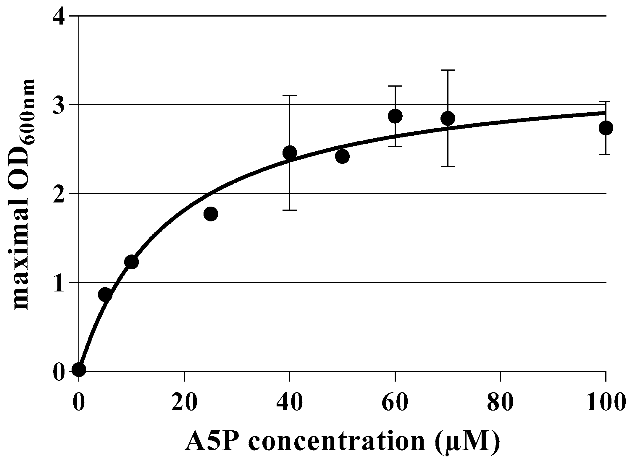
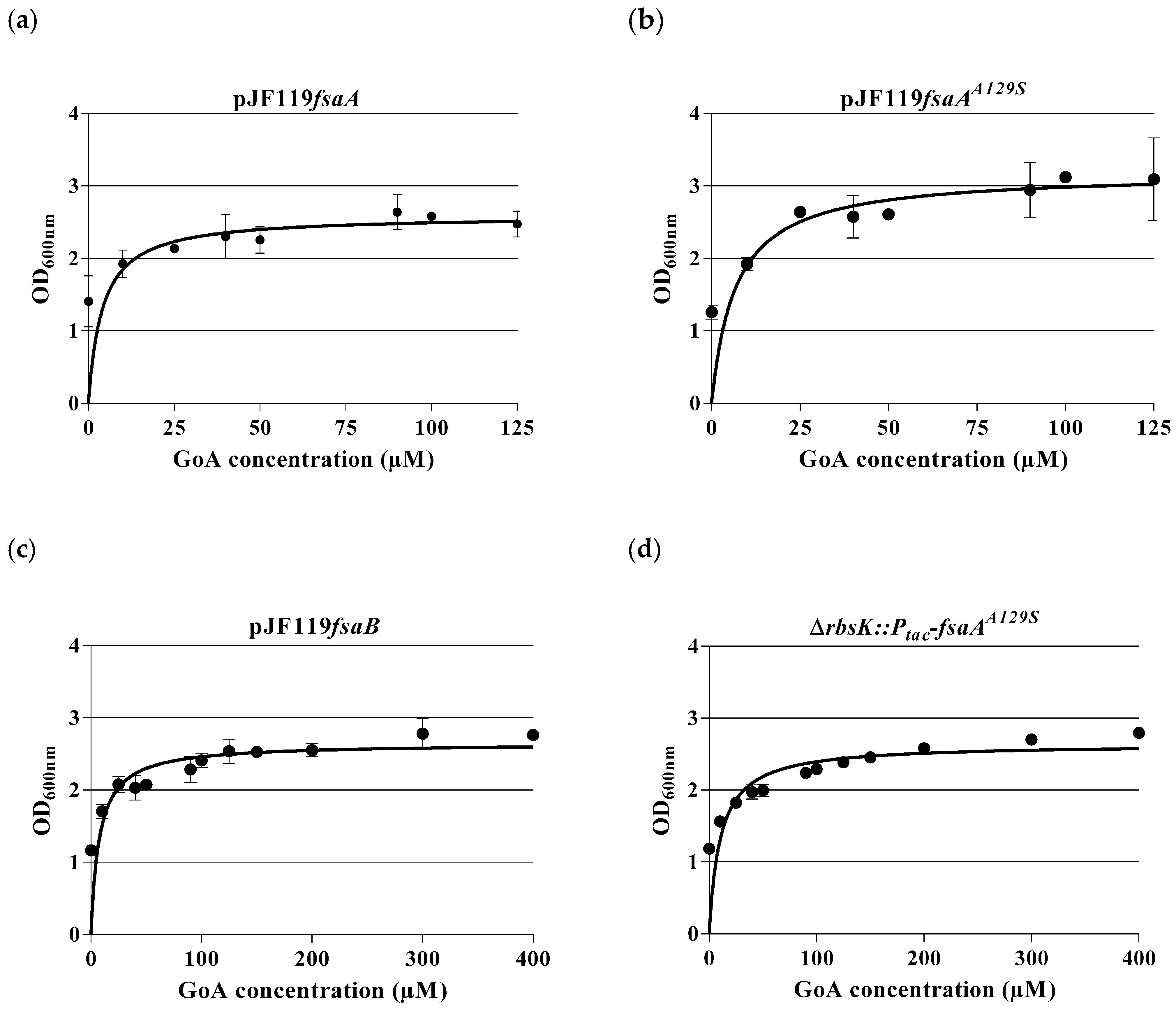
2.3. Dose–Response Curves for A5P and GoA

Dose–response assays were performed using liquid MOPS-MM (containing 28 mM glucose) in shake flasks. The maximal OD600nm achieved after 48 h of incubation at 37 °C (for A5P) or 30 °C (for GoA) was monitored. To assay A5P auxotrophy, an overnight culture of BW25113 ΔgutQ::FRT ΔkdsD::FRT-kan-FRT (10 mL MOPS-MM with 28 mM glucose, 10 µM G6P, 15 µM A5P, and 50 µg/mL Km) was washed twice with MOPS-MM containing 28 mM glucose. Then, cells were inoculated in 100 mL shake flasks containing 10 mL MOPS-MM with 28 mM glucose, 10 µM G6P, 50 µg/mL Km, and different A5P concentrations (0 µM; 5 µM; 10 µM; 25 µM; 40 µM; 50 µM; 60 µM; 70 µM; 100 µM) with an initial OD600nm of 0.05. The shake flasks were incubated at 200 rpm and 37 °C for 48 h. The obtained OD600nm values were plotted as a function of the A5P concentration; this resulted in a dose–response curve. Likewise, dose–response curves for glycolaldehyde were obtained with the A5P auxotroph strains grown on MOPS-MM containing 28 mM glucose, 100 µM IPTG, and different concentrations of GoA. A single colony, grown on MM-Agar with 28 mM glucose, 10 µM G6P, 50 µM A5P, and, if needed, the corresponding antibiotics (ampicillin 100 mg/L, kanamycin 50 mg/L), was inoculated in 10 mL MOPS-MM with 28 mM glucose, 10 µM G6P, 15 µM A5P, and the corresponding antibiotics in a 100 mL shake flask and incubated overnight at 200 rpm and 37 °C. An amount of 2 mL of the preculture was centrifuged at 16,500× g for 1 min and washed twice with 1 mL MOPS-MM with 28 mM glucose. The cell pellet was resuspended in 1 mL MOPS-MM with 28 mM glucose and inoculated with an initial OD600nm of 0.05 in the main culture, which contained 10 mL MOPS-MM with 28 mM glucose, 100 µM IPTG, the corresponding antibiotics, and different GoA concentrations (0 µM; 10 µM; 25 µM; 40 µM; 50 µM; 90 µM; 100 µM; 125 µM). The cell culture was then incubated at 200 rpm and 30 °C for 48 h. The OD600nm was measured when the incubation was finished. KS and the maximal OD600nm values were calculated with the software “GraphPad Prism” version 6.01 from GraphPad Software, Inc. (San Diego, CA, USA).

3. Results

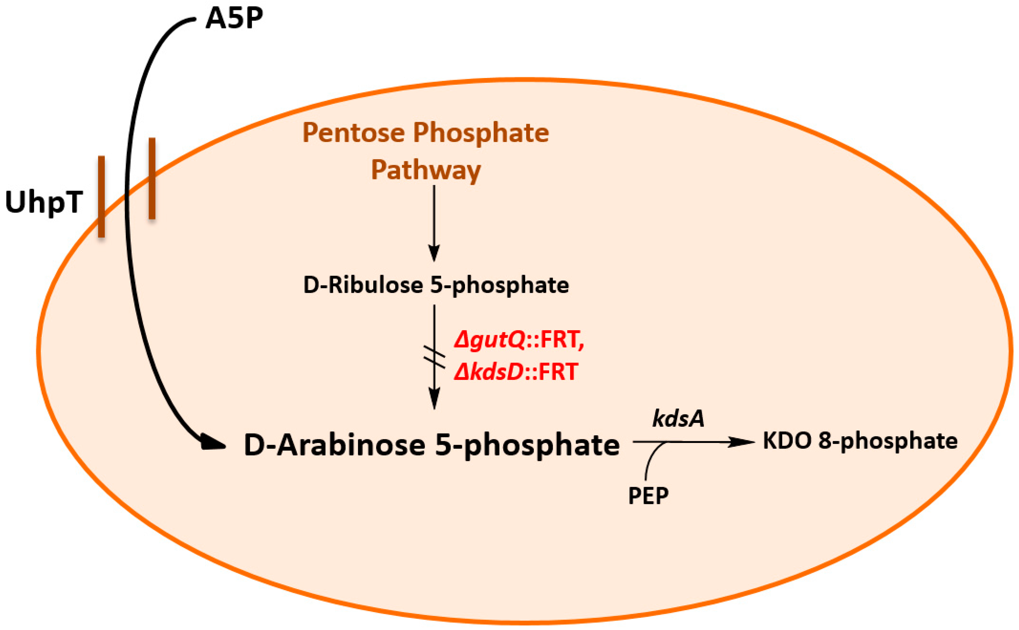
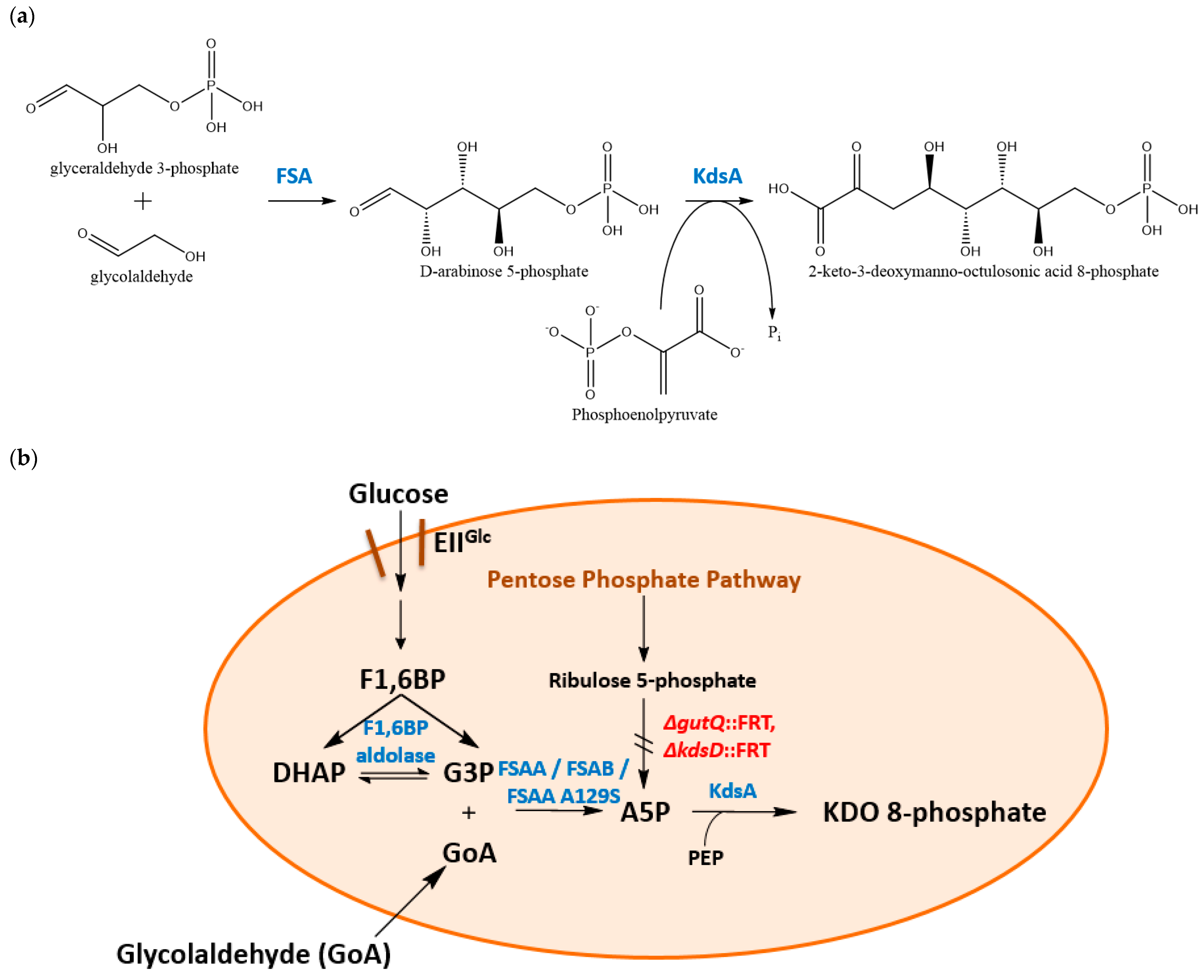
3.1. Construction and Characterization of an E. coli K-12 Double Mutant Strain Deficient in API

E. coli K-12 strain BW25113 (see Table 1 for genotype) [16] was chosen as the parent strain. In order to block KDO formation, the two genes gutQ and kdsD for D-A5P synthesis were knocked out successively by a λ-red recombineering method [16]. Thus, the double mutant (strain BW25113 ΔgutQ::FRT ΔkdsD::FRT-kan-FRT) was obtained. We decided to keep the kanamycin resistance marker for easier detection of the mutant strain. During the last step of the double mutant generation, A5P (50 µM) was added to the cultures together with 10 µM of glucose 6-phosphate to induce the UhpT uptake system which transports A5P [24,25] (see Figure 1). The mutant strain was auxotrophic for supplementation of A5P as expected [7] and A5P had to be added even in complex media such as LB or 2YT, as yeast extract does not contain A5P [3]. We observed that cells of strain BW25113 ΔgutQ ΔkdsD::kan stopped growth after transfer into an A5P-free medium; they clumped together and eventually lysed. Microscopic analysis revealed that cells of this strain—in comparison with the wild-type BW25113—before lysis formed long chains indicating severe problems associated with division (see Figure S2). Therefore, growth curves in shake flasks with strain BW25113 ΔgutQ ΔkdsD::kan could not be obtained in the absence of A5P, a feature which has been reported for analogous E. coli mutants before [7].
A “bioassay” (auxanogram) was carried out with BW25113 ΔgutQ ΔkdsD::kan cells by adding varying concentrations of external A5P to assess the degree of this auxotrophy. Cells from an A5P-containing preculture were transferred (after washing) into in a MOPS-buffered mineral salt medium (MM) with glucose as the sole carbon source. A5P (together with G6P) was added at various concentrations.
As can be seen from Figure 2 and Table S2, as little as 5 µM A5P gave a positive growth signal (increase in the final OD at 600 nm, OD600) while no addition led to no OD increase, cell clumping, and formation of long chains (see Figure S2), and lysis was observed. A positive correlation was found with increasing A5P concentrations, final growth yields of up to OD ~3, with >60 µM of A5P. A KS value of 17.7 ± 4.5 µM was, thus, determined. The wild-type strain BW25113 (without A5P supplementation) reached a maximal OD600 of ~3.

3.2. Physiological Observations in kdsD gutQ Double Mutants with or without FSA Expression

GoA is a donor substrate for FSA [11,26] and A5P synthesis in vitro has been shown before by using FSA with the donor GoA and the acceptor G3P [12]. Further, a synthetic pathway (“glycoptimus”, shown both in vitro and in vivo) from D-glucose, D-xylose, or L-arabinose leading to glycolic acid formation via GoA has been established in E. coli via plasmid-based expression of kdsD and fsaA genes [27].
We wanted to know whether E. coli FSAA might open a bypass reaction in the API-deficient strain BW25113 ΔgutQ ΔkdsD::kan by condensing G3P and GoA to A5P. Therefore, the genes encoding the native FSAA or the variant FSAAA129S (which has an improved catalytic efficiency towards F6P [23,26,28]) were expressed in the E. coli double mutant from the plasmids pJF119fsaA and pJF119fsaAA129S [20,23], respectively. These expression vectors contained an ampicillin resistance marker and the fsa genes were under the control of the Ptac promoter (IPTG-inducible). Thus, IPTG was added for induction (final concentration of 100 µM) and external GoA was added to provide a donor compound for FSA activity. We assumed that the cells would supply the acceptor G3P from glycolysis. As controls, E. coli K-12 wild-type (wt) strain BW25113, BW25113 ΔgutQ ΔkdsD::kan, BW25113 ΔgutQ ΔkdsD, and BW25113 ΔgutQ ΔkdsD::kan pJF119EH (for genotypes, see Table 1) were used.
While the wild-type BW25113 showed, as expected, normal growth, none of the double mutant strains could grow without fsa-bearing plasmids (less than one doubling in OD). Cells of strain BW25113 ΔgutQ ΔkdsD::kan (or its pJF119EH-bearing derivative) clumped and lysed when incubated without the presence of external A5P on MOPS-MM (see Supplementary Figure S2). Addition of various concentrations of GoA did not prevent this. However, cells of BW25113 ΔgutQ ΔkdsD::kan carrying either pJF119fsaA or pJF119fsaAA129S were able to grow in the presence of different GoA concentrations (see Figure 3a,b). Also, pJF119 carrying the other E. coli FSA-encoding gene, fsaB (see Table 2), was able to complement the API deficiency in BW25113 ΔgutQ ΔkdsD (Figure 3c). For data sets, see Table S3.
Furthermore, we wondered if a single chromosomally integrated copy of fsaAA129S might also be sufficient to restore growth. We, therefore, integrated a Ptac-controlled copy of the gene into the rbsK locus. This strain, BW25113 ∆gutQkdsDrbsK::Ptac-fsaAA129S, also grew without external addition of A5P if GoA was provided (Figure 3d).
The four different strains showed a similar reaction versus GoA addition: the higher the GoA concentration present in the medium, the higher the maximal OD600nm reached by these strains. GoA concentrations above 100 µM, however, did not lead to significant growth yield improvement. It is worth noting that even without GoA supplementation, a maximal OD600nm ≥ 1 was reached by all double mutant strains carrying fsa plasmids. This we interpret as a hint that E. coli metabolism provides yet unknown sources of GoA (see Section 4).

4. Discussion

KDO is a landmark molecule of the Gram-negative outer membrane. Mutants without KDO formation are barely viable [3]. We created kdsD and gutQ double mutants of E. coli K-12 strain BW25113 and confirmed, as expected from the work of Woodard’s group, that these cells are A5P auxotrophs. If A5P was removed from the growth media by centrifugation and washing, cells ceased to grow and most of them eventually lysed or formed long chains of cells which ceased to divide (see Supplementary Figure S2); this is also in accordance with earlier reports [7]. Gain-of-function suppressor mutants have been reported to arise from kdsD gutQ double mutants when cultured for prolonged times in liquid media. These mutants grew with a prolonged generation time and showed a growth defect above 42 °C; the LPS layer consisted of lipid IVA free of KDO and the cells were super-susceptible to a variety of antibiotics, SDS, or bile salts. The compensatory suppressor mutants were also considered as endotoxin-free [3]. We did not look for such suppressor mutants, however, as this was not within the scope of the present study.
Our idea was to assay whether expression of fsa genes would complement the growth defects of API-lacking cells. This was performed in the presence or absence of glycolaldehyde (GoA). It turned out that only those double mutant strains with ectopic expression of fsa genes (fsaA, fsaB, or fsaAA129S either from plasmids or inserted under Ptac-control in a chromosomal locus) were able to regain growth in the absence of externally added A5P. Externally added GoA improved growth yields but, remarkably, even without GoA addition, cell densities (measured as OD600) of >1 were obtained. This was not observed with double mutants that did not carry recombinant fsa genes.
FSAA and its variant FSAA A129S have been shown in the past to allow bypass reactions in the central metabolism of E. coli. The group of Stephanopoulos showed that a block in the DXS reaction (providing 1-deoxyxylulose 5-phosphate from pyruvate and G3P for the methylerythritol biosynthesis pathway of isoprenoids in E. coli) could be overcome by FSA by adding exogenous hydroxyacetone with GoA (from an engineered D-arabinose catabolic pathway) to 1-deoxyxylulose [29]; this reaction has been previously shown in vitro [30]. Recently, we reported that FSAA A129S was able to bypass a block by phosphofructokinase in E. coli mutants (ΔpfkA ΔpfkB Δzwf), again following the knowledge that F6P can be cleaved to G3P and DHA. This opened a pathway for the production of DHA and glycerol from glucose [23]. The opposite reaction, e.g., growth on glycerol via DHA to F6P in specific mutants, was achieved as well by the group of the late Arren Bar-Even [31]. While the present manuscript was in preparation, the Walther’s group proved that FSAA can be used to implement a novel in vivo metabolic pathway to yield the product acetyl-CoA. Their starting point was ethylene glycol, which had to be oxidized to GoA with the help of an oxygen-resistant variant of FucO; GoA was then condensed with G3P to A5P by FSAA [32].
As API are also found in several Gram-negative pathogenic bacteria, they are considered as interesting targets for the development of antibacterials [33,34]. With our approach of using gutQ and kdsD mutants, A5P is channeled to KDO8P synthesis starting from glucose (via G3P) and the simple compound GoA. This prevents its mixing with compounds of the PPP (summarized in Figure 4). A5P is, thus, delivered by an orthogonal biosynthetic pathway. This could be used to label KDO8P specifically by using labeled GoA. This could also help in studies where the interaction of KDO2–lipid A with the innate immune system is analyzed. KDO2–lipid A is the conserved structure in LPS of most Gram-negative bacteria. LPS notoriously acts as endotoxin, leading to fever (“pyrogen”) and toxic shock in mammals as it stimulates the innate immune system via the TRL-4 receptor on macrophages. Purified KDO2–lipid A is sufficient to evoke this endotoxic effect [35]. Specific E. coli mutants (with defects in the rfaD gene) have been obtained with improved production of KDO2–lipid A to develop lipid A adjuvants as part of bacterial vaccines [36]. Our findings in A5P formation from inexpensive precursors could help to further improve this production.
Residual growth (after removal of A5P) was found in the control kdsD gutQ double mutants to reach OD values of ~0.3 with the onset of clumping and eventual lysis. The observation that kdsD gutQ double mutants with the expression of recombinant fsa genes could grow up to an OD600 of ~1.2 (starting OD ~0.05) (see Figure 3) in the absence of both external compounds, e.g., A5P or GoA, deserves to be discussed in more detail. This growth feature points to hidden intracellular sources of GoA when cells grow on glucose as a sole carbon source. The literature on in vivo formation of free GoA, however, is scarce or based only on in vitro measurements [37]. GoA is a highly reactive Maillard agent which leads to non-enzymatic post-translational protein modifications in vivo, N6-carboxymethyllysine being the predominant glycated end product [37]. From biochemical studies, a one-substrate reaction of bacterial transketolase on ß-hydroxypyruvate (HPA) is known which could release free GoA; HPA in turn could be formed by action of YeaB from 3-phosphohydroxypyruvate in the L-serine biosynthesis pathway [38].
Exogenously added GoA was shown previously to complement a pyridoxine auxotrophy in an E.coli B mutant WG3. In this strain, the authors found a defect in a glycolaldehyde dehydrogenase (forming GoA by reduction of glycolate) and proposed a possible origin of GoA from glyoxylate via glycolate [39]. More recent work in the literature discusses GoA as precursor of L-4-hydroxythreonine synthesis (aldol reaction of LtaE on glycine and GoA) and, thus, as a precursor of a minor pathway (not the main reaction) to pyridoxal phosphate in E. coli. Here, the endogenous origin of GoA could be 3-phosphohydroxypyruvate (see above) [38]. In the folate biosynthesis pathway, dihydroneopterin aldolase FolB liberates GoA [40]. GoA could also be formed by reduction of the dialdehyde glyoxal via YqhD, a metabolite which is formed by degradation of autoxidized compounds in many organisms [41]. An unspecific monoamine oxidase (MaoA) from E. coli has been shown to provide GoA from ethanolamine in vitro [42]. Further work is necessary to elucidate the internal source(s) of GoA which leads to formation of A5P and complementation of the kdsD and gutQ defects.

5. Conclusions

We have shown that the expression of multifunctional enzyme FSA allows a bypass in an API-deficient mutant strain of E. coli BW25113 by using G3P (from glycolysis) and GoA to form A5P, a precursor of the outer membrane compound KDO. This helped to reinstall full growth and to overcome the detrimental effects on viability caused by KDO deficiency. Full complementation in terms of growth yields took place with exogenously added GoA, while lower growth yields were achieved even without GoA addition, pointing to hidden endogenous sources of GoA. This constitutes another example for the establishment of bio-orthogonal metabolic pathways (bypass reactions) in E. coli with the help of FSA.

Supplementary Materials

The following supporting information can be downloaded at: https://www.mdpi.com/article/10.3390/applmicrobiol4010032/s1, Figure S1: Agarose gel electrophoresis, Figure S2: Light microscopy photos of cells Table S1: PCR primers, Table S2: Dose-response curve for various A5P concentrations, shake flask cultures, OD600 nm measured, Table S3: Dose-response curve for various exogenous GoA concentrations, shake flask cultures, maximal OD600 nm measured.

Author Contributions

Conceptualization, E.G.F. and G.A.S.; methodology, E.G.F.; software, E.G.F.; validation, E.G.F. and G.A.S.; formal analysis, E.G.F.; investigation, E.G.F.; resources, G.A.S.; data curation, E.G.F.; writing—original draft preparation, E.G.F. and G.A.S.; writing—review and editing, E.G.F. and G.A.S.; visualization, E.G.F.; supervision, G.A.S.; project administration, G.A.S.; funding acquisition, none. All authors have read and agreed to the published version of the manuscript.

Funding

This research received no external funding.

Data Availability Statement

All data are available within this paper and the supporting information.

Acknowledgments

We thank members of our institute for help with cloning (Natalie Trachtmann, Katrin Gottlieb).

Conflicts of Interest

The authors declare no conflicts of interest.

References

  1. Nikaido, H. Molecular basis of bacterial outer membrane permeability revisited. Microbiol. Mol. Biol. Rev. 2003, 67, 593–656. [Google Scholar] [CrossRef] [PubMed]
  2. Whitfield, C.; Trent, M.S. Biosynthesis and export of bacterial lipopolysaccharides. Annu. Rev. Biochem. 2014, 83, 99–128. [Google Scholar] [CrossRef] [PubMed]
  3. Meredith, T.C.; Aggarwal, P.; Mamat, U.; Lindner, B.; Woodard, R. Redefining the requisite lipopolysaccharide structure in Escherichia coli. ACS Chem. Biol. 2006, 1, 33–42. [Google Scholar] [CrossRef] [PubMed]
  4. Cipolla, L.; Gabrielli, L.; Bini, D.; Russo, L.; Shaikh, N. Kdo: A critical monosaccharide for bacteria viability. Nat. Prod. Rep. 2010, 27, 1618–1629. [Google Scholar] [CrossRef]
  5. Müller-Loennies, S.; Lindner, B.; Brade, H. Structural analysis of oligosaccharides from lipopolysaccharide (LPS) of Escherichia coli K12 strain W3100 reveals a link between inner and outer core LPS biosynthesis. J. Biol. Chem. 2003, 278, 34090–34101. [Google Scholar] [CrossRef] [PubMed]
  6. Meredith, T.C.; Woodard, R.W. Escherichia coli YrbH is a D-arabinose 5-phosphate isomerase. J. Biol. Chem. 2003, 278, 32771–32777. [Google Scholar] [CrossRef] [PubMed]
  7. Meredith, T.C.; Woodard, R.W. Identification of GutQ from Escherichia coli as a D-arabinose 5-phosphate isomerase. J. Bacteriol. 2005, 187, 6936–6942. [Google Scholar] [CrossRef] [PubMed]
  8. Sperandeo, P.; Pozzi, C.; Dehò, G.; Polissi, A. Non-essential KDO biosynthesis and new essential cell envelope biogenesis genes in the Escherichia coli yrbG-yhbG locus. Res. Microbiol. 2006, 157, 547–558. [Google Scholar] [CrossRef]
  9. Schürmann, M.; Sprenger, G.A. Fructose-6-phosphate aldolase is a novel class I aldolase from Escherichia coli and is related to a novel group of bacterial transaldolases. J. Biol. Chem. 2001, 276, 11055–11061. [Google Scholar] [CrossRef]
  10. Samland, A.K.; Sprenger, G.A. Synthetic potential of dihydroxyacetone-utilizing aldolases. In Industrial Biocatalysis; Grunwald, P., Ed.; Pan Stanford Publishing Pte. Ltd.: Singapore, 2015; pp. 783–816. [Google Scholar]
  11. Garrabou, X.; Castillo, J.A.; Guérard-Hélaine, C.; Parella, T.; Joglar, J.; Lemaire, M.; Clapés, P. Asymmetric self- and cross-aldol reactions of glycolaldehyde catalyzed by D-fructose-6-phosphate aldolase. Angew. Chem. 2009, 121, 5629–5633. [Google Scholar] [CrossRef]
  12. Sánchez-Moreno, I.; Hélaine, V.; Poupard, N.; Charmantray, F.; Légeret, B.; Hecquet, L.; García-Junceda, E.; Wohlgemuth, R.; Guérard-Hélaine, C.; Lemaire, M. One-pot cascade reactions using fructose-6-phosphate aldolase: Efficient synthesis of D-arabinose 5-phosphate, D-fructose 6-phosphate and analogues. Adv. Synth. Catal. 2012, 354, 1725–1730. [Google Scholar] [CrossRef]
  13. Guérard-Hélaine, C.; Debacker, M.; Clapés, P.; Szekrenyi, A.; Hélaine, V.; Lemaire, M. Efficient biocatalytic processes for highly valuable terminally phosphorylated C5 to C9 D-ketoses. Green Chem. 2014, 16, 1109–1113. [Google Scholar] [CrossRef]
  14. Sambrook, J.; Fritsch, E.F.; Maniatis, T. Molecular Cloning: A Laboratory Manual; Cold Spring Harbor Laboratory Press: New York, NY, USA, 1989. [Google Scholar]
  15. Neidhardt, F.C.; Bloch, P.L.; Smith, D.F. Culture medium for enterobacteria. J. Bacteriol. 1974, 119, 736–747. [Google Scholar] [CrossRef] [PubMed]
  16. Datsenko, K.A.; Wanner, B.L. One-step inactivation of chromosomal genes in Escherichia coli K-12 using PCR products. Proc. Natl. Acad. Sci. USA 2000, 97, 6640–6645. [Google Scholar] [CrossRef]
  17. Jiang, Y.; Chen, B.; Duan, C.; Sun, B.; Yang, J.; Yang, S. Multigene editing in the Escherichia coli genome via the CRISPR-Cas9 system. Appl. Environ. Microbiol. 2015, 81, 2506–2514. [Google Scholar] [CrossRef] [PubMed]
  18. Trachtmann, N. (University of Stuttgart, Stuttgart. Personal Communication, 2016.
  19. Fürste, J.P.; Pansegrau, W.; Frank, R.; Blöcker, H.; Scholz, P.; Bagdasarian, M.; Lanka, E. Molecular cloning of the plasmid RP4 primase region in a multi-host-range tacP expression vector. Gene 1986, 48, 119–131. [Google Scholar] [CrossRef]
  20. Inoue, T. Microbial Aldolases as C-C Bonding Enzymes: Investigation of Structural-Functional Characteristics and Application for Stereoselective Reactions. Ph.D. Dissertation, University of Stuttgart, Stuttgart, Germany, 2006. [Google Scholar] [CrossRef]
  21. Trachtmann, N.; Alvarez Fong, K.F.; Guitart Font, E.; Sprenger, G.A. Construction of chromosomally encoded lacZ and gfp reporter strains of Escherichia coli for the study of global regulation of metabolism. Eng. Life Sci. 2016, 16, 675–681. [Google Scholar] [CrossRef]
  22. Cherepanov, P.P.; Wackernagel, W. Gene disruption in Escherichia coli: TcR and KmR cassettes with the option of Flp-catalyzed excision of the antibiotic-resistance determinant. Gene 1995, 158, 9–14. [Google Scholar] [CrossRef]
  23. Guitart Font, E.; Sprenger, G.A. Opening a novel biosynthetic pathway to dihydroxyacetone and glycerol in Escherichia coli mutants through expression of a gene variant (fsaAA129S) for fructose 6-phosphate aldolase. Int. J. Mol. Sci. 2020, 21, 9625. [Google Scholar] [CrossRef]
  24. Eidels, L.; Rick, P.D.; Stimler, N.P.; Osborn, M.J. Transport of D-arabinose-5-phosphate and D-sedoheptulose-7-phosphate by the hexose phosphate transport system of Salmonella typhimurium. J. Bacteriol. 1974, 119, 138–143. [Google Scholar] [CrossRef]
  25. Kadner, R.J.; Murphy, G.P.; Stephens, C.M. Two mechanisms for growth inhibition by elevated transport of sugar phosphates in Escherichia coli. J. Gen. Microbiol. 1992, 138, 2007–2014. [Google Scholar] [CrossRef]
  26. Castillo, J.A.; Guérard-Hélaine, C.; Gutiérrez, M.; Garrabou, X.; Sancelme, M.; Schürmann, M.; Inoue, T.; Hélaine, V.; Charmantray, F.; Gefflaut, T.; et al. A mutant of D-fructose-6-phosphate aldolase (Ala129Ser) with improved affinity towards dihydroxyacetone for the synthesis of polyhydroxylated compounds. Adv. Synth. Catal. 2010, 352, 1039–1046. [Google Scholar] [CrossRef]
  27. Lachaux, C.; Frazao, C.J.R.; Kraußer, F.; Morin, N.; Walther, T.; Francois, J.M. A new synthetic pathway for the bioproduction of glycolic acid from lignocellulosic sugars aimed at maximal carbon conservation. Front. Bioeng. Biotechnol. 2019, 7, 359. [Google Scholar] [CrossRef] [PubMed]
  28. Schürmann, M. Biochemische Charakterisierung und Struktur-Funktionsbeziehungen bakterieller Transaldolasen und Fruktose-6-Phosphat Aldolasen. Ph.D. Dissertation, University of Düsseldorf, Düsseldorf, Germany, 2001. (In Germany). [Google Scholar]
  29. King, J.R.; Woolston, B.M.; Stephanopoulos, G. Designing a new entry point into isoprenoid metabolism by exploiting fructose-6-phosphate aldolase side reactivity of Escherichia coli. ACS Synth. Biol. 2017, 6, 1416–1426. [Google Scholar] [CrossRef] [PubMed]
  30. Schürmann, M.; Schürmann, M.; Sprenger, G.A. Fructose 6-phosphate aldolase and 1-deoxy-D-xylulose 5-phosphate synthase from Escherichia coli as tools in enzymatic synthesis of 1-deoxysugars. J. Mol. Catal. B Enzym. 2002, 19–20, 247–252. [Google Scholar] [CrossRef]
  31. Lindner, S.N.; Aslan, S.; Müller, A.; Hoffart, E.; Behrens, P.; Edlich-Muth, C.; Blombach, B.; Bar-Even, A. A synthetic glycerol assimilation pathway demonstrates biochemical constraints of cellular metabolism. FEBS J. 2020, 287, 160–172. [Google Scholar] [CrossRef] [PubMed]
  32. Wagner, N.; Bade, F.; Straube, E.; Rabe, K.; Frazao, C.J.R.; Walther, T. In vivo implementation of a synthetic metabolic pathway for the carbon-conserving conversion of glycolaldehyde to acetyl-CoA. Front. Bioeng. Biotechnol. 2023, 11, 1125544. [Google Scholar] [CrossRef] [PubMed]
  33. Yep, A.; Sorenson, R.J.; Wilson, M.R.; Showalter, H.D.H.; Larsen, S.D.; Keller, P.R.; Woodard, R.W. Enediol mimics as inhibitors of the D-arabinose 5-phosphate isomerase (KdsD) from Francisella tularensis. Bioorg. Med. Chem. Lett. 2011, 21, 2679–2682. [Google Scholar] [CrossRef] [PubMed]
  34. Gabrielli, L.; Merlo, S.; Airoldi, C.; Sperandeo, P.; Gianera, S.; Polissi, A.; Nicotra, F.; Holler, T.P.; Woodard, R.W.; Cipolla, L. Arabinose 5-phosphate isomerase as a target for antibacterial design: Studies with substrate analogues and inhibitors. Bioorg. Med. Chem. 2014, 22, 2576–2583. [Google Scholar] [CrossRef]
  35. Raetz, C.R.H.; Garrett, T.A.; Reynolds, C.M.; Shaw, W.A.; Moore, J.D.; Smith Jr, D.C.; Ribeiro, A.A.; Murphy, R.C.; Ulevitch, R.J.; Fearns, C.; et al. Kdo2-Lipid A of Escherichia coli, a defined endotoxin that activates macrophages via TLR-4. J. Lipid. Res. 2006, 47, 1097–1111. [Google Scholar] [CrossRef]
  36. Wang, J.; Ma, W.; Wang, Z.; Wang, X. Construction and characterization of an Escherichia coli mutant producing Kdo2-lipid A. Mar. Drugs 2014, 12, 1495–1511. [Google Scholar] [CrossRef] [PubMed]
  37. Klaus, A.; Pfirrmann, T.; Glomb, M.A. Transketolase A from E. coli significantly suppresses protein glycation by glycolaldehyde and glyoxal in vitro. J. Agric. Food Chem. 2017, 65, 8196–8202. [Google Scholar] [CrossRef] [PubMed]
  38. Noda-Garcia, L.; Liebermeister, W.; Tawfik, D.S. Metabolite-enzyme coevolution: From single enzymes to metabolite pathways and networks. Annu. Rev. Biochem. 2018, 87, 187–216. [Google Scholar] [CrossRef] [PubMed]
  39. Tani, Y.; Morita, H.; Ogata, K. Glycolaldehyde synthesizing pathway involved in vitamin B6 biosynthesis in Escherichia coli B. Agric. Biol. Chem. 1977, 41, 1749–1754. [Google Scholar] [CrossRef]
  40. Haussmann, C.; Rohdich, F.; Schmidt, E.; Bacher, A.; Richter, G. Biosynthesis of pteridines in Escherichia coli. J. Biol. Chem. 1998, 273, 17418–17424. [Google Scholar] [CrossRef]
  41. Lee, C.; Kim, I.; Lee, J.; Lee, K.-L.; Min, B.; Park, C. Transcriptional activation of the aldehyde reductase YqhD by YqhC and its implication in glyoxal metabolism of Escherichia coli K-12. J. Bacteriol. 2010, 192, 4205–4214. [Google Scholar] [CrossRef]
  42. Chen, Z.; Huang, J.; Wu, Y.; Liu, D. Metabolic engineering of Corynebacterium glutamicum for the de novo production of ethylene glycol from glucose. Metab. Eng. 2016, 33, 12–18. [Google Scholar] [CrossRef]
Figure 1. Scheme to highlight the role of A5P isomerases (encoded by kdsD and gutQ) and supplementation of an auxotroph for A5P (uptake via the UhpT transporter).
Applmicrobiol 04 00032 g001
Figure 2. Dose–response curve with increasing A5P concentrations. Growth of double mutant BW25113 ΔgutQ ΔkdsD::kan on MOPS-MM with 28 mM of glucose as C source. Dependence of the maximal OD600nm reached by BW25113 ΔgutQ ΔkdsD::kan on varying A5P concentrations. A maximal OD600nm = 3.4 ± 0.3 determined with the software “GraphPad Prism” version 6.01 from GraphPad Software, Inc. (San Diego, CA, USA). For details, see Material and Methods. For uptake of A5P, 10 µM glucose 6-phosphate was added as inducer of UhpT [24]. Data are from two biological replicates with error bars.
Applmicrobiol 04 00032 g002
Figure 3. Dose–response curves for glycolaldehyde in double mutants deficient for API with various fsa-bearing plasmids (ac) or a chromosomal integration (d). Growth yields were determined as final OD600nm values after 48 h of incubation on MOPS-MM containing 28 mM glucose and 100 µM IPTG. (a) BW25113 ΔgutQ ΔkdsD::kan pJF119fsaA (KS = 4.1 ± 3.6; OD600nm max. = 2.6 ± 0.3). (b) BW25113 ΔgutQ ΔkdsD::kan pJF119fsaAA129S (KS = 6.8 ± 3.8; OD600nm max. = 3.2 ± 0.3). (c) BW25113 ΔgutQ ΔkdsD pJF119fsaB (KS = 7.5 ± 3.0; OD600nm max. = 2.6 ± 0.1). (d) BW25113 ΔgutQ ΔkdsD ΔrbsK::Ptac-fsaAA129S (KS = 10.4 ± 3.8; OD600nm max, = 2.6 ± 0.1). Average values of two independent biological replicates with error bars are shown. Note that in Figure 3c,d, the range of GoA is different.
Applmicrobiol 04 00032 g003
Figure 4. (a) Addition of glycolaldehyde (GoA) to glyceraldehyde 3-phosphate (G3P) by FSA enzymes to arabinose 5-phosphate (A5P), and condensation of A5P with phosphoenolpyruvate (PEP) to form 2-keto-3-deoxymanno-octulosonic acid 8-phosphate (KDO 8-phosphate) by KDO 8-phosphate synthetase (KdsA). (b) Scheme of complementation by FSA enzymes in E. coli K-12 kdsD gutQ double mutant by adding external GoA to G3P, thereby providing the cells with A5P, a precursor of KDO.
Applmicrobiol 04 00032 g004
Disclaimer/Publisher’s Note: The statements, opinions and data contained in all publications are solely those of the individual author(s) and contributor(s) and not of MDPI and/or the editor(s). MDPI and/or the editor(s) disclaim responsibility for any injury to people or property resulting from any ideas, methods, instructions or products referred to in the content.

