1. Introduction
The increasing risk of microbiological contamination from aqueous solutions, rinsing and drinking water in food processing, medical application fields and water-bearing systems in buildings and households is arousing great interest in novel decontamination and disinfection methods. The use of the plasma technology for bacterial decontamination achieved high attractiveness. Tap water activated by non-thermal atmospheric pressure plasma to plasma-activated water (PAW) and activated liquids (PAL) has been proposed as a promising approach for a biocidal effect on microorganisms, biofilms and yeast cells [
1,
2,
3,
4,
5,
6,
7,
8].
Plasma, usually produced by electrical discharge in gas [
9], is a source of high-electric fields, energetically charged particles, oxidizing species, reductive species, ultrasound, and UV light [
8,
10]. The numerous reactive species in the plasma are able to react with other molecules and concentrate in water due to convection and diffusion, which interact with water molecules [
11].
In the last decade, the focus of research has expanded to the interaction of plasmas with liquids, which has led to the development of a variety of reactor geometries with different operating principles to produce plasma-activated water [
10,
12,
13,
14]. During the plasma activation process for PAW and PAL production, which was performed in this study, a gas-phase plasma arc in ambient air over the water surface is generated [
15].
Many active and reactive biochemical substances and radicals arise in the water during the process of plasma activation [
16]. In most cases, especially for distilled water [
17], this is associated with a massive decrease in pH and a high increase of the oxidation potential. The method used for plasma activation precisely involves this change in the two named parameters for all used liquids. In this study, ambient air is used during the activation process of water by plasma. This gave rise to secondary species consisting mostly of reactive oxygen and nitrogen species (RONS). The RONS that are considered to be representative in PAWs and PALs are composed, among other things, of short-lived species ·OH, HO
2/O
2−, ONOOH/ONOO
−, and the long-lived species hydrogen peroxide (H
2O
2), nitrite (NO
2−) and nitrate (NO
3−) [
18,
19]. These species are regarded as a basic prerequisite for the chemical, biological and antimicrobial effects of PAWs and PALs [
8,
20]. RONS cause oxidative stress on microorganisms with the consequence of inactivation [
8,
21].
Microorganisms originating from tap water and being able to form biofilms often contaminate water systems. The opportunistic pathogen
Pseudomonas aeruginosa is a waterborne microorganism contaminating aqueous environments and water-bearing systems in buildings, households, and medical facilities. The Gram-negative bacteria adapts very well to the aqueous environment, is robust against environmental factors, and is able to form tolerances to antibiotics and disinfectants [
22]. In terms of pathogenicity,
P. aeruginosa causes a variety of life-threatening infections such as pneumonia and chronic lung infection in cystic patients, bacteremia, wound infection and acute ulcerative keratitis in users of contact lenses [
23,
24]. High-risk clones characterized by multidrug or extensively drug resistance pose an increasing public health threat and
P. aeruginosa is among the top six bacterial pathogens for deaths attributable to antimicrobial resistance [
25,
26,
27].
In this study, we examined the antimicrobial potential of PAW and PAL in post-discharge treatment on inactivation of the indicator microorganism P. aeruginosa. We analyzed the principle of inactivation of microorganisms at different device settings without focusing on the physical and chemical properties of the activated liquids and on the mechanism of action of the plasma device. We investigated the minimal to optimal plasma activation conditions and the resulting biocidal effects in the generated PAWs and PALs consisting of low and high inorganic and organic loads to sufficient cell inactivation. We focused on microbial inactivation in the liquid medium, in which the bacteria were present, and looked at the efficacy of PAWs through dilution effects. To prove inactivation and track it at the cellular level, we followed damages on membrane integrity and on enzymatic activities.
2. Materials and Methods
2.1. Microorganism and Growth Conditions
P. aeruginosa ATCC 27853 originated from American Type Culture Collection and was obtained from the German Collection of Microorganisms and Cell Cultures (Leibniz Institute DSMZ, Braunschweig, Germany). The strain was used as an indicator for waterborne and opportunistic pathogens for evaluation of the antimicrobial activity conditions of plasma-activated liquids as well as for analyses of the mode of action of PAL and PAW. P. aeruginosa was sub-cultivated and maintained on Luria Bertani (LB) broth agar plates (Roth, Karlsruhe, Germany). Characterizing the efficiency of plasma activation on liquids to an antimicrobial effect, PALs were added to the microorganism inoculated in saline as fluid matrix simulating liquid and aqueous environment.
2.2. Plasma Activation Process and Settings for the Generation of Plasma-Activated Water (PAW) and Liquids (PAL)
Water and liquids were treated with plasma by using a lab unit (VitalFluid, Eindhoven, NL, USA) generating a thermal arc in ambient air over the activated liquid, leading to plasma activated water (PAW) and plasma activated liquids (PAL). In the glass reactor, liquids are activated by plasma using a single stainless-steel electrode, which is connected to a high-voltage modulator. During activation, the liquid is stirred continuously. Furthermore, a constant exchange of air is maintained in the reactor chamber. In this study a solid-state dual resonant system was used, which generated up to 80 kV dual resonant high-voltage pulses with an oscillation frequency of up to 2 MHz and a pulse repetition rate of up to 20 kHz according to the manufacturers’ description [
15]. Our use and the settings of the lab unit device was based on information described in great detail [
15].
The electrode was just above the water, while a plasma beam arose between electrode and water by energy application. The device settings were continuously adjustable in terms of activation energy (Watt; W) and activation time (minutes; min). Several conditions and settings were chosen to investigate effects on
P. aeruginosa cell reduction and total inactivation. The electrical discharge power and the respective activation time were set in ranges of 60 to 120 W and from 10 to 120 min, respectively, as indicated. To analyze the influence of plasma-activated liquids with their new characteristic physical and chemical properties on a sufficient antimicrobial effect, various liquids were investigated for plasma activation with respect to cell number reductions. These were tap water, originated from the institute, distilled and filtrated MilliQ water; saline (0.9% [
w/
v] NaCl; Roth, Karlsruhe, Germany), dissolved in MilliQ water; and artificial urine [
28] as liquid with high organic and inorganic loads. In all processes, a volume of approximately 600 mL was activated by plasma in one pass each under rigorous stirring. PAW and PAL for each batch were prepared freshly prior use.
2.3. Determination of Physical and Chemical Parameters of the Plasma-Activated Liquids in Use
A thermal arc appeared in ambient air over the liquid during activation process with the consequence of physical and chemical changes. The pH value and the electric conductivity were determined using a pH and conductivity meter, respectively (Xylem Analytics, Weilheim, Germany). The oxidation-reduction potential (ORP) was measured according to DIN 38404-6 [
29] using a redox potentiometer (Hach, Düsseldorf, Germany). The nitrite concentration was detected photometrically at a wavelength of 540 nm according to DIN EN 26777 [
30]. In this process, nitrite reacts with 4-aminobenzosulphanilamide in the presence of o-phosphoric acid (85%
v/
v) (Merck Millipore, Darmstadt, Germany) forming diazonium salt which changes in the presence of N-naphthyl-1-2 diaminoethane (Roth, Karlsruhe, Germany) to a red–pink dye. The nitrate concentration was determined against reference concentrations according to DIN 38405 [
31]. Ions react with 2,6-dimethylphenol to 4-nitro-2,6-dimethylphenol in which the colour is photometrically determined at 324 nm.
2.4. Treatment of P. aeruginosa with PAL and PAW
P. aeruginosa was sub-cultivated in liquid LB broth (Roth, Karlsruhe, Germany) overnight (approximately 18 h) at 37 °C under shaking conditions (89 rpm). Cells were harvested and washed twice via centrifugation (2500×
g, 15 min, 4 °C), prior to adjusting to a density of approximately 10
8/mL bacteria in saline. PALs were added in the equal volume unit as the cell suspension volume, unless otherwise stated. The mixture was incubated for different periods at room temperature as indicated. The oxidative efficiency reaction of the PALs was terminated by application of a nine-fold volume of neutralization solution comprising 3-L-α-phosphatidylcholine (0.3%), Tween 80 (3%), sodium thiosulfate (0.5%), L-histidine (0.1%), and saponine (3%) (Roth, Karlsruhe, Germany), according to DIN EN 1040 [
32] after defined periods as indicated. Aliquots of a 10-µL volume were spotted in ten-fold dilutions as triplicates on LB agar plates followed by incubation at 37 °C for 24 to 48 h. Bacterial survival was analyzed by determination of the count of colony forming units (cfu).
The original cell concentration was confirmed by plating in tenfold serial dilutions without any treatments. Control experiments indicated a high efficiency of the neutralization solution to inactivate the oxidative suspension and proved no effect on the bacterial suspensions itself. The corresponding non-activated liquids were used as negative controls.
2.5. Live/Dead Analyses by Flow Cytometry (FCM)
To determine the impact of PAL on the membrane integrity and consequently the inactivation rate of the bacteria, samples were stained for live/dead differentiation and analyzed via the flow cytometry (FCM) technique and performed using a NovoCyte Flow Cytometer 2000 R (ACEA Biosciences Inc., San Diego, CA, USA). To exclude an influence of PAL on the dyes through oxidation, the bacterial suspension at a volume of 250 µL was centrifuged (4000×
g, 10 min) and resuspended in saline. Aliquots of 100 µL were transferred to a well of 96-well plates following double staining with SYBR Green I (Invitrogen, Thermo Fisher Scientific, Darmstadt, Germany) and propidium iodide (PI; Roth, Karlsruhe, Germany). SYBR Green I as stock solution in dimethyl sulfoxide (DMSO; Roth, Karlsruhe, Germany) was used in a 100-fold dilution in each assay. PI, suspended in DMSO, was added at a final concentration of 6 µM. For uniform distribution the suspensions were mixed in the plates shortly at 1000 rpm on a horizontal shaker (Bioer, China) following incubation at 38 °C for exactly 12 min in the dark to avoid any impairment of cell viability [
33,
34].
Each sample was analysed at a volume of 50 µL with a flow rate of 35 µL/min in triplicates. A laser at a wavelength of 488 nm was used for excitation, and fluorescence signals were detected with photomultipliers at 520 ± 20 nm in green and at >630 nm in the red spectrum while forward and sideward scattered light signals were also collected. Intrinsic fluorescence of the samples and background and matrix signals were determined with sterile filtrated, fully demineralized water by omitting dyes. The light signals were converted to electronic signals and were finally obtained as logarithmic data. The characteristic gates were adjusted to the signal clusters based on the flow cytometer manufacturer and to initial measurements of untreated bacteria resuspended in tab water. Data collection and analysis were performed using with the NovoExpress 1.3.0 software. No compensation was used for the assays. Background noise by cell debris and ions was separated for the quantification by gating.
2.6. Live/Dead Analyses by Propidium Monoazide (PMA)-RT-PCR
For live/dead differentiation and membrane integrity, another approach was used on a molecular level with propidium monoazide (PMA) staining and the subsequent real-time polymerase reaction (RT-PCR).
PMA, dissolved in DMSO as stock solution (2 mM), was stored at −20 °C in the dark until use. After the respective treatment of P. aeruginosa with PAW under conditions as indicated, bacteria at a 400 µL volume were incubated with PMA (Biotium, Fremont, CA, USA) at a final concentration of 20 µM in a light-transparent tube. For uniform penetration of the dye through the damaged membrane, samples were shaken slowly for 20 min in the dark prior exposure to blue LED lights (1 W per LED) for covalent DNA binding. After 20 min unbound PMA was removed via centrifugation (16,000× g; Eppendorf centrifuge, Eppendorf, Hamburg, Germany). To follow and control a membrane damaging process with bacterial inactivation, cells were heated at 99 °C for 10 min in each assay.
Cell pellets of PMA-treated and untreated samples were resuspended in 100 μL Chelex-InstaGene matrix solution (5% w/v) (Bio-Rad Laboratories, Munich, Germany) and lysed at 99 °C for 10 min under shaking conditions to release the intracellular DNA. Cellular debris was removed via centrifugation (16,000× g, 10 min) and the supernatant containing DNA was used in the RT-PCR assay.
To the Sso fast Eva Green PCR master mix (Bio-Rad Laboratories, Munich, Germany), 0.4 µM of each
P. aeruginosa-specific primers ELA520-F and ELA520-R [
35] and 2 µL DNA were added to a total volume of 10 μL for RT-PCR amplification using an iCycler (version 1.259; Bio-Rad Laboratories, Munich, Germany). The RT-PCR conditions started with a denaturation step at 95 °C for 2 min, followed by 40 cycles of 10 s denaturation at 98 °C, 30 s annealing at 60 °C and 30 s extension at 72 °C. The melting temperature profile, analyzed by gradually increasing temperature from 65 °C to 95 °C (±0.2 °C/5 s), indicated a distinct peak at 90.0 °C ± 0.2 °C proving amplification specificity. The effect of the PMA treatment on each sample was calculated using the difference of the Cq values of PMA untreated and PMA treated samples (∆Cq).
2.7. Detection of Enzymatic Activity after PAW Treatment
The impact of PAW on bacterial enzymatic activities was analyzed using a method of the most portable number (MPN) procedure using the Pseudalert assay (IDEXX, Westbrook, ME, USA) that is used for specific P. aeruginosa detection in water and wastewater according to international standard. The method was modified for microtiter plate application with adaption to very low volumes in the research assay. The assay was based on the enzymatic hydrolysis of a specific substrate associated with a color change to blue-fluorescence detectable under UV light. In the adjusted approach, the substrate was dissolved in saline in half the volume as specified by the manufacturer. P. aeruginosa cells in saline were aliquoted in 100 µL volumes in microtiter 96-well plates followed by addition of 100 µL freshly prepared Pseudalert solution. The final substrate concentration matched the manufacturer’s instructions. Plates incubated at 38 °C for 24 h and the fluorescence was determined under UV-light at 365 nm. Positive fluorescence signals indicated enzymatic activity of active P. aeruginosa, whereby the intensity of the fluorescence does not correlate with cell density.
2.8. Analyses of the Data
All experiments were performed at least three times. The results represent the means of at least three independent experiments performed independently with standard deviations. A total loss in cultivation is indicated by n.d. (not detectable). The statistical analyses were carried out with a two-sided, unpaired t-test without equal variances.
3. Results
3.1. Plasma Activation Conditions for Microbial Effects of PAW
The efficacy of the plasma-activated liquids, suspensions and water on microbial decontamination depends on various factors as setting of the electrical power and the activation time, the volumes added to the bacteria and contact time, the liquid composition for activation following microbial inactivation as well as the bacterial density.
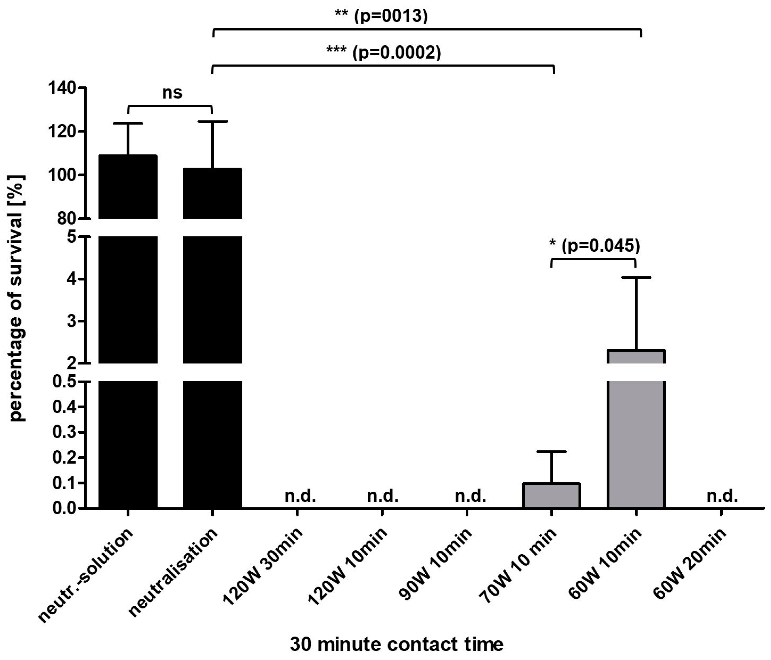
In a first approach tap water was plasma activated using in the lab unit at current settings of 60 to 120 W for 10 to 30 min as indicated.
P. aeruginosa at a high concentration of 10
8 cells/mL, indicating a high contamination level, was added to the PAW for 30 min (
Figure 1).
Four out of six conditions resulted in a total loss of cultivation at which high power settings and short activation times as well as lower power settings and prolonged activation times caused inactivation. Bacteria survived at low rates of 2.32% (±1.72) and 0.097% (±0.13) with PAW treatments at 60 W and 70 W activation, respectively, for 10 min each.
3.2. Reduced Antimicrobial Effects in Diluted PAW
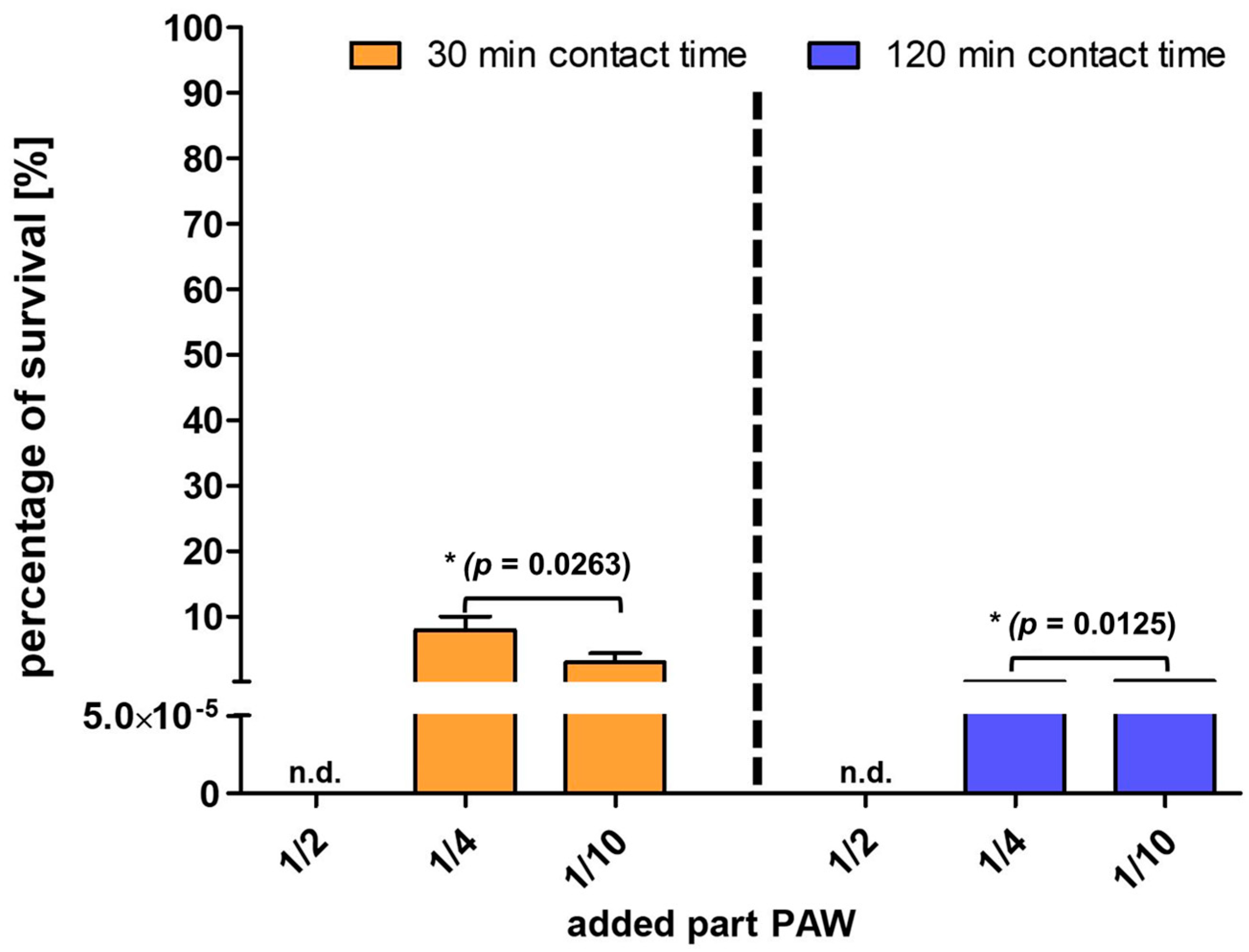
As PAW will be added to bacterial contaminated liquids, dilutions might reduce the oxidative and consequently the bactericidal effects. Thus, the minimum PAW volume unit necessary for sufficient oxidative effects was investigated with PAW (activated for 120 W for 30 min) resulting in an efficient decontamination (
Figure 2).
The application of half the volume unit of PAW to the bacterial suspension volume resulted in maximal reduction and caused total fail in cultivation. With increased dilutions, meaning less PAW volume units, added the survival only reduced to less than 10%. Extended contact times only caused a reduction in cell numbers (survival rate of 0.04% and 0.12% with 1/4 and 1/10 PAW, respectively) but not total inactivation. This result indicated that increasing the PAW dilutions minimized the bactericidal effect, and the antimicrobial effect cannot be achieved even by prolonged confrontation time with bacterial suspensions.
3.3. Physical and Chemical Properties of PAW in Dilutions
Due to the dilution, the physical and chemical properties of PAW also changed. The institute’s own tap water had pH values of approximately 7.5 to 8.0 and conductivities of 501 to 511 µS/cm, which were determined regularly at different times. To follow the physicochemical changes, tap water was activated with 90 W for 30 min as an example. The pH dropped down to 3.13 (±0.27) and the conductivity arose to 1045 µS/cm (±318), which was determined in three independent water samples (
Table 1).
The oxidation/reduction potential (ORP) increased from 441 mV (±2) to 766 mV (±11). The nitrate and nitrite concentrations came up to 364 mg/L (±50) and 85 mg/L (±15), starting from 14 mg/L (±1) and 0 mg/L of tap water, respectively. Dilutions of PAW in tap water and saline resulted in lower conductivity but similar ORP values (
Table 1).
For comparison, plasma-activated saline and plasma-activated MilliQ water demonstrated similar values as plasma-activated tap water regarding pH (2.51 each), ORP (796 mV and 804 mV, respectively) and the nitrate and nitrite contents (326 and 324 mg/L; 83 and 54 mg/L, respectively). The conductivity value was extremely high for saline (16,960 µS/cm) but lower for MilliQ water (1630 µS/cm).
Due to the high PAW susceptibility to microorganisms suspended in saline, the physical and chemical parameters of the PAW solutions mixed with saline were determined. A mixture of the equal volume unit of PAW with non-activated saline simulating the matrix of the bacterial suspension, resulted in changes of pH (3.59; ±0.18), of conductivity (8440 mV; ±251), of ORP (724 µS/cm; ±0.6), of the nitrate (172 mg/L; ±24) and of the nitrite concentrations (46 mg/L; ±8). The physicochemical values of PAW diluted with saline indicated changes accompanying with a decrease in oxidative activities.
3.4. Impact of Organic and Inorganic Loads in Liquids on Activation by Plasma with Antimicrobial Effects
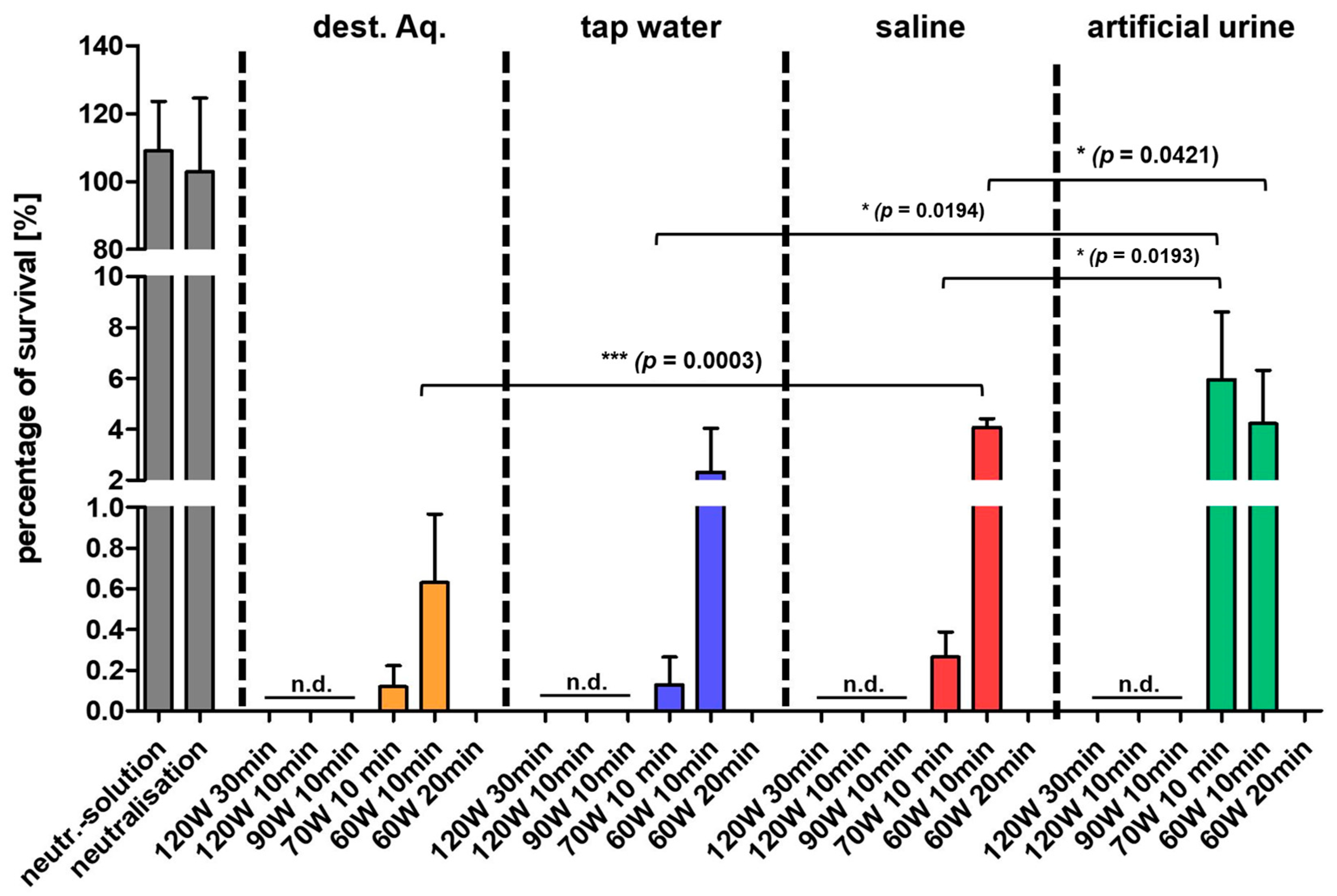
The organic and inorganic composition of the liquid with various mineral salt concentrations is of primary importance for the reactive species yield during the plasma activation process and consequently for antimicrobial effects. Various liquid compositions were activated by plasma treatment with electrical power settings of 60 to 120 W and activation periods of 10 to 30 min, as indicated (
Figure 3). Artificial urine represented a nutrient-rich medium containing lipids and proteins; tap water obtained from the institute indicated a watery environment, and saline, produced based on distilled water as well as distilled water were used comprehending a drop in presence of organic and inorganic compounds.
The antimicrobial effect, indicating a loss of cultivation after 30 min contact time, was achieved with all liquids at settings of 90 W and 120 W and an activation time of at least 10 min (
Figure 3).
A massive reduction in cell counts was found with settings of 70 W/10 min. P. aeruginosa showed survival rates < 1% in distilled water, tap water and saline, and 5.95% in artificial urine proving significant differences of tap water and artificial urine as well as saline and artificial urine. The survival rates increased at the lower activation setting of 60 W/10 min to 2.32% in tap water, 4.07% in saline and 4.23% in urine with significant differences as indicated. Organic and inorganic substances, mainly present in artificial urine, inhibited the efficiency of inactivation. However, a prolongation of the activation time increased the inactivation efficiency, even at lower current settings (60 W, 20 min) and lead to a total loss of cultivability of P. aeruginosa.
3.5. Impact of Storage on PAW Activity Regarding Antibacterial Effects
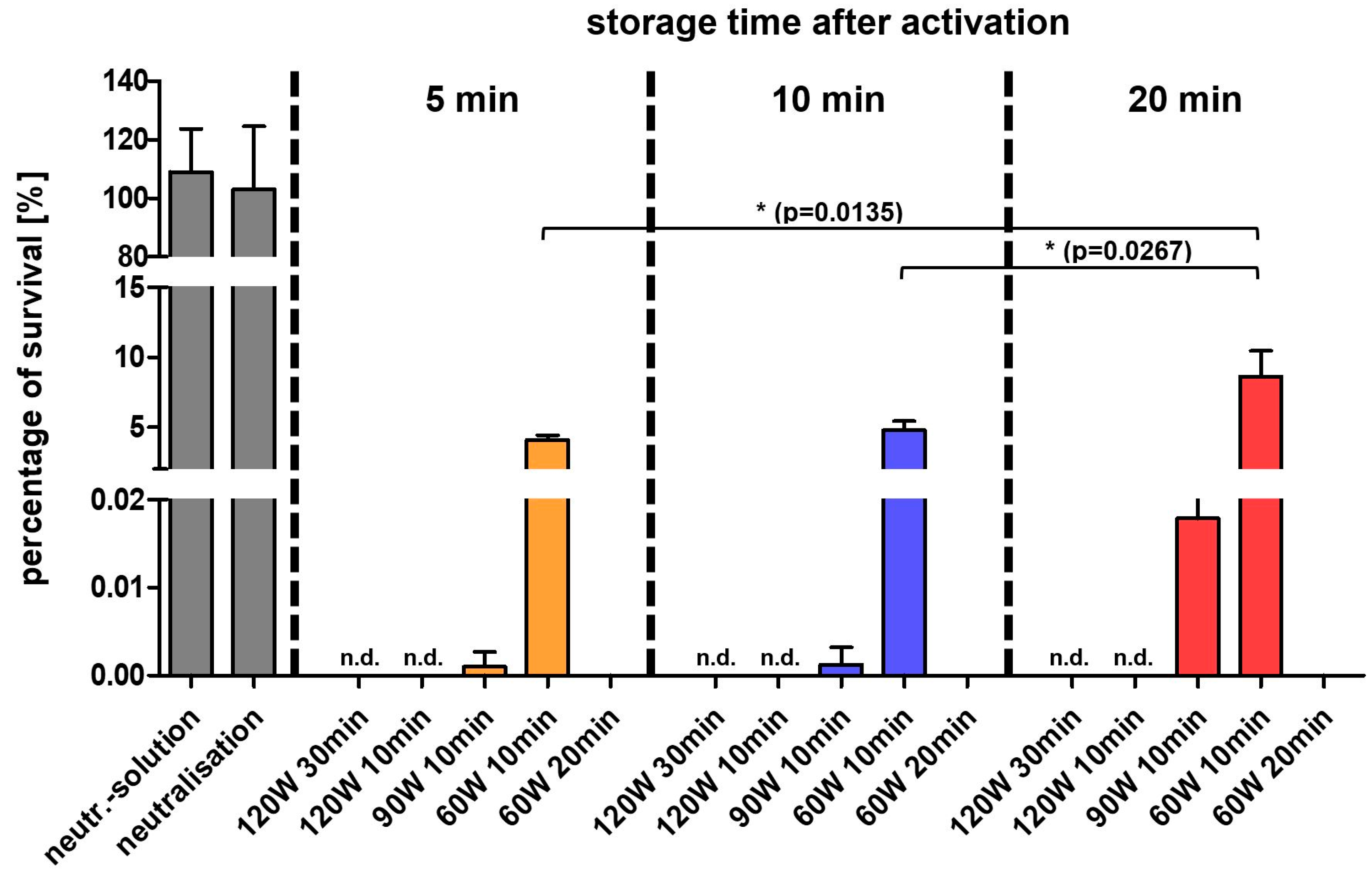
Since PAW proved to be antimicrobial effective, the stability of PAW was investigated because oxidized compounds are often usable only for a short time. For this, freshly prepared PAW samples with various activation powers were stored each for three time-intervals at room temperature as indicated and the antimicrobial efficacy was analyzed after 2 min bacterial contact (
Figure 4).
The microorganism P. aeruginosa failed totally in cultivation after PAW treatment, when activated at 120 W for 10 and 30 min and 60 W/20 min, independent from storage time. However, a decrease in the antimicrobial efficiency was observed with PAW activated at 60 W/10 min as prolonged storage resulted in a significant increase in the survival rate of 4.07% (after 5 min storage) to 8.61% (after 20 min storage). PAW activated at 90 W for 10 min yielded in a reduction of activity within minutes and the bacterial survival rate increased from 0.001% (5 min storage) to 0.02% (20 min storage). In general, the higher the wattage and longer the activation time during plasma activation, the longer the PAW shelf time.
In summary, PAW and PAL activated by high-electric power for a prolonged time resulted in a sufficient activation independent on the liquid composition. PALs in surplus showed a pronounced antimicrobial effect.
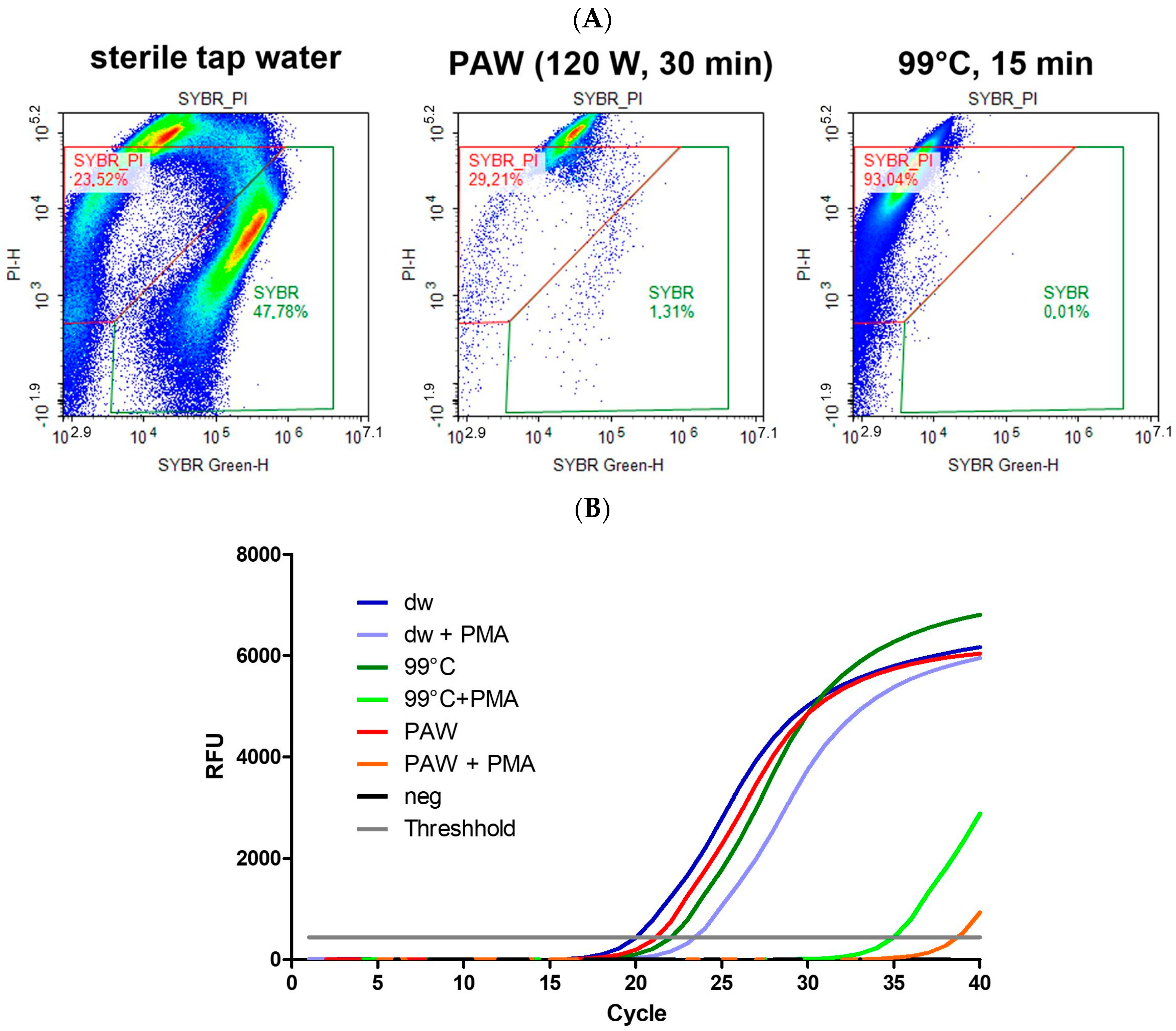
3.6. Cell Damages on Microorganisms by PAW
The antimicrobial effect of PAW on
P. aeruginosa was analyzed on the biological level of the microorganism, mainly focusing on membrane damage and on enzymatic activities. Representation of the cell damages after PAW treatment proved intense membrane damages detected by the flow cytometry and the propidium iodide-RT-PCR techniques (
Figure 5). Only few events were detected as living cells in the SYBR gate after live/dead staining in the flow cytometer by 30 min contact to PAW (120 W/30 min). Most of the events were to be found in the SYBR/PI-gate that represents membrane damaged and dead cells (
Figure 5A). Results of the PMA-RT-PCR proved the bacterial membrane damages caused by PAW. Out of three independent PCR runs, signals came up at 36.65 cycles with a delayed amplification of 15.04 cycles compared with microorganisms without PMA dye (21.61 cycles) after PAW treatment (
Figure 5B).
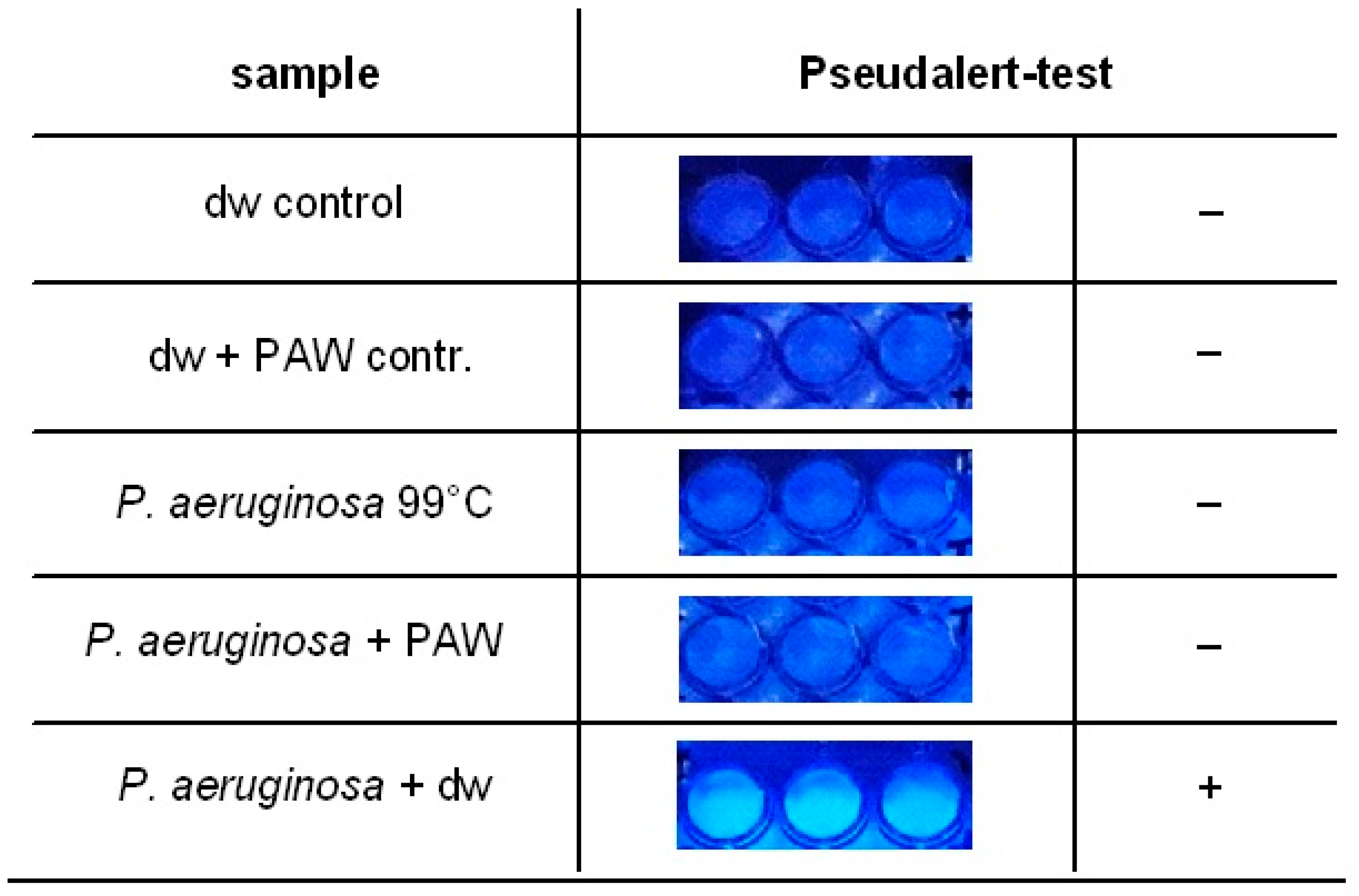
3.7. Enzymatic Inactivation by PAW
The antimicrobial effect of PAW on
P. aeruginosa was analysed on enzymatic level. Based on the Pseudalert system we analysed the enzymatic activity and the growth of
P. aeruginosa cells after PAW treatment using a modified assay adjusted to low volumes in microtiter plates. Intact and living
P. aeruginosa cells expressed an enzyme that hydrolyzed the substrate to a blue-fluorescent compound detectable under UV light. After PAW treatment, a substrate conversion by the enzyme reaction was absent (
Figure 6).
4. Discussion
The activation of tap water and liquids by plasma treatment to oxidative active fluids is a promising tool to reduce successfully microbial contamination of products [
8]. There are numerous devices and variations of plasma sources with the respective setting depending on the application [
8,
11]. In this study, we determined conditions and settings of the laboratory device necessary to inactivate successfully a high bacterial load in liquid media and in an aqueous environment, shown exemplarily by the waterborne microorganism
P. aeruginosa, because a contamination of water bearing systems with this bacterium may lead to major risks for human health [
36,
37,
38].
The plasma activation process generates a high number of reactive oxygen and nitrogen species with the consequence of physical and chemical changes in the water, which are the basis of the antimicrobial effect [
8]. Nitrate and nitrite concentrations rose sharply as well as the ORP and the conductivity, indicating the presence of ions. In contrast, the pH dropped dramatically. The decrease in pH interdepends inversely with the electrical conductivity increase [
39]. PAW in the equal mixing ratio with tap water caused only a slight balance of the physicochemical values with decreases of the nitrate and nitrite concentrations and the conductivity as well as a slight increase in pH. The ORP did not change and the antimicrobial effect was maintained. PAW, activated at 120 W for 30 min and added at least half the volume to tap water, achieved the limit of a successful microbial reduction. A prolonged contact time and exposure to the microorganisms played a subordinate role. However, the bactericidal effect correlated with the increase of the electrical power to generate PAW as well as the plasma-activation duration. The activation of tap water at 120 W was more antimicrobially effective than at 90 W and 60 W, regardless of the respective 10 min activation time. On the other hand, the activation time was also of great importance. The activation duration at 60 W for 20 min indicated a higher bactericidal effect than a treatment at 90 W for only 10 min. Several studies demonstrated increased ORP values with prolonged plasma activation times and treatments [
2,
40,
41,
42]. With higher activation power or prolonged activation periods, the concentration of these RONS increase [
15]. The antimicrobial effect of PAW were mainly based on RONS [
8,
20].
The chemical composition of the liquid being plasma-activated had a significant impact on the activation efficiency. The more organic and inorganic components were present, the lower the antimicrobial effect. Numerous cells survived in activated artificial urine followed by a decreased survival in saline, tap water and distilled water, respectively, after 30 min-treatments with PAWs, which were activated with 70 W and 60 W for 10 min each. This observation, that high organic loads reduce the antimicrobial properties, is in accordance with another study [
42]; as the addition of bovine serum albumin (BSA) to PAW significantly reduced the antimicrobial effect to
S. aureus. It was assumed that single sulfhydryl (SH) groups of amino acids could react with RONS and deactivate RONS as non-specific antioxidants [
42]. This explanation is transferable to the reduced antimicrobial effect of plasma activated artificial urine.
PAW lost activity after being left standing for a prolonged time [
16]. Due to extended lifetime, the concentration of reactive species decreased [
15]. The antimicrobial effect came down with prolonged storage proven with
S. aureus and
S. epidermidis [
15,
43] and shown here with
P. aeruginosa.We proved that prolonged activation times resulted in an increased disinfection efficacy of PAW as described formerly [
6,
44]. The lab device used here has a unit for individual settings as adjustment of electric power and activation period and allows inactivation to be set accordingly with high cell concentrations or when liquids are activated which contain high organic loads. Optimized PAW activation and operating conditions can be adjusted prior to use and to disinfections. This has the advantage of quick and spontaneous production depending on demand.
The antimicrobial effect of PAW and PAL was proven with inactivation of
P. aeruginosa by failing in cultivation, and we followed the effect on the cells in terms of damage. Although, the mechanism of inactivation by PAW has not yet been fully elucidated, it is evident that oxidative stress acts on cells and proteins through the RONS [
21].
The first contact of cells with the oxidative substances is the membrane. The Gram-negative bacterium
P. aeruginosa has a double layer membrane as protection against environmental influences and as an entry portal in the cell interior. The bacterial membrane integrity was damaged by PAW treatment. This was evidenced using fluorescence dyes, which are not able to penetrate intact outer membranes but diffuse inwards when membranes are impaired. Both dyes, propidium iodide (PI) and propidium monoazide (PMA), passed through the membrane after PAW treatment and bound to DNA. Signals of membrane-damaged cells were determined in the inactive cell gate by the FCM technology and in the PMA-PCR with a highly intensive delay in signal amplification. These results are consistent with previous studies using different techniques [
40,
43,
45,
46,
47,
48]. Furthermore, a reduction of the membrane potential was described [
42,
49], which also indicates membrane damage.
Consequently, RONS can penetrate and oxidize intracellular components such as proteins and enzymes. Enzymatic activity was determined in a modified Pseudalert assay for specific P. aeruginosa activity. The enzyme-based test indicates the presence and living status of P. aeruginosa through the hydrolysis of a substrate, which causes fluorescence under UV light. After 30 min contact period with PAW, P. aeruginosa showed no fluorescence indicating inactivation of enzymatic activity.
5. Conclusions
In conclusion, plasma-activated water and plasma-activated fluids are effective against contamination with P. aeruginosa colonizing water and fluid-bearing systems as well as pipes. In addition to low pH, generated PAW and PALs are characterized by high values in electrical conductivity, oxidation-reduction potential and nitrate and nitrite concentrations. It should be highlighted that the organic and inorganic composition of the activated liquid with different mineral salt concentrations is of utmost importance for the yield of reactive species during the plasma activation process and consequently for the antimicrobial effect.
The antimicrobial effect is efficient when PAW and PAL, generated in the lab unit, is added in at least equal but better in surplus volume to bacteria. Furthermore, PAW lost activity after being left standing for a prolonged time, so a direct use after activation is recommended for maximizing the antimicrobial effect. The results demonstrate the potential that PAW and PAL can be used for reduction and inactivation of microorganisms in a fluidic and watery environment whereby more advanced devices may well show better inactivation properties due to a more activation energy. The spontaneous new generated environmental effects in use should be considered to achieve effective results.

 ,
, 


