Review

Nanocomposites of Ferrites with TiO2, SiO2 and Carbon Quantum Dots as Photocatalysts for Degradation of Organic Pollutants and Microbes

1
Department of Chemistry, Punjab Agricultural University, Ludhiana 141004, India
2
Department of Applied Science, Punjab Engineering College, Chandigarh 160012, India
3
Department of Microbiology, Punjab Agricultural University, Ludhiana 141004, India
*
Author to whom correspondence should be addressed.
Magnetochemistry 2023, 9(5), 127; https://doi.org/10.3390/magnetochemistry9050127
Submission received: 7 April 2023 / Revised: 2 May 2023 / Accepted: 7 May 2023 / Published: 9 May 2023

Abstract

:
Ferrites are important magnetic materials used in electronic devices. Nanocomposites of ferrites with TiO2, SiO2 and carbon quantum dots have gained recent interest due to their unique advantages, such as high chemical stability, surface-active sites, high specific surface area, non-toxicity, excellent optical properties, and tunable porosity. In the present review, general and adaptable coprecipitation, sol–gel, hydrothermal, solvothermal, and Stöber methods for the fabrication of nanocomposites are discussed. These materials offer the advantage of magnetic recovery and superior photocatalytic performance. The potential of nanocomposites to act as photocatalysts to eliminate organic pollutants and microbes from water is discussed. Mechanisms involved in these applications are also elaborated upon. The review provides a detailed study of recent applications and future perspectives of nanocomposites in sustainable water treatment.

Graphical Abstract

1. Introduction

For the environment and human life, developing industrialization has been both beneficial and problematic. Many harmful compounds, such as synthetic dyes, heavy-metal ions, and pesticides as well as microorganisms can degrade the quality of drinking water as a result of industrial discharge and other man-made activities, posing a threat to human life. The slow self-deterioration of organic pollutants due to their stable structure is the most important issue. According to a World Health Organization (WHO) report, 785 million people lack access to basic drinking water, and half of the world’s population will be living in water-scarce areas by 2025 [1].
Synthetic dyes, which are used to color plastic, paper, and artificial and natural fabrics, are water-soluble organic pollutants [2]. The usage of dye-tainted water poses a number of health hazards and can injure human organs [3]. Excessive discharge of organic dyes into the water environment prevents sunlight seepage, disrupting photochemical and biological activity of aquatic flora and fauna. They have been found to be very carcinogenic and poisonous [4,5,6].
Pharmaceuticals in water degrade the quality of drinking water supplies, contribute to antibiotic resistance, and are toxic to aquatic organisms. Pharmaceuticals have been placed on the European Union’s and the US Environmental Protection Agency’s water quality watch lists [7]. Tetracycline hydrochloride (TCH) release into the aquatic environment is seen as a concern to humans since it increases antibiotic resistance in some pathogenic bacteria and persists in the environment due to its non-biodegradability.
Surface water is polluted with many infectious microorganisms, such as bacteria (E. coli, Vibrio), viruses (such as Norwalk virus and rotavirus) and protozoans (such as Entamoeba, Giardia) that are capable of causing illnesses in humans, such as bloody diarrhea, affecting human health as well as the environment. Because of their resistance to disinfection, they are the most commonly identified cause of waterborne illness.
Pesticides are another major organic pollutant in water bodies. The agriculture sector is the main source of pesticides into the aquatic environment, as they are used for destroying and controlling pests to protect crops. Their variety, toxicity, persistence and low biodegradability cause threat to humans through pollution of drinking water resources [8].
For the removal of organic contaminants, dyes, and pharmaceuticals from wastewater, a variety of technologies have been developed, including adsorption [9], chemical oxidation [10], advanced oxidation processes (AOPs), and biodegradation. Among them, AOPs have gained much attention because they are eco-friendly and economical. These processes include oxidation, photocatalysis, and electrochemical processes [11]. Photocatalysis is a very economical and extensively used AOP [12]. It requires UV or visible radiation for activation, but does not require massive machinery [13]. Due to its unique potential in deteriorating organic pollutants, it is widely used for wastewater treatment [14]. Due to its numerous favorable qualities, TiO2 emerged as one of the most appropriate materials for environmental protection after the investigation of the Honda–Fujishima effect [15]. TiO2 is non-toxic, and the US Food and Drug Administration has approved it for use in pharmaceuticals, human food, food-contact products, and cosmetics. The size of TiO2 nanoparticles reduces as their surface area increases at the nanoscale level, which is one of the required properties of nanoparticles for photocatalytic applications [16,17,18]. When compared to other photocatalysts, TiO2 is widely used for a variety of applications due to its optical, thermal/chemical stability, abundance, high redox potential, and electrical properties [19,20]. TiO2 can absorb UV light due to a large bandgap (3.2 eV). Solar light has about 50% visible light and 5% UV light. Due to its band potential, TiO2 can efficiently produce reactive oxygen species (ROS) in both oxidative and reductive pathways. There is need for photocatalysts to harvest solar energy for environmental remediation.
Ferrites, which are mixed metal oxides with iron as their major constituent, have emerged as one of the most intensely studied photocatalysts in the last decade due to their remarkable magnetic properties, small bandgap, superparamagnetic, tunable morphologies, high saturation field, non-toxicity, reusability, and chemical stability. Due to their small bandgaps (1.1–2.3 eV), ferrites absorb visible light. They are the most effective photocatalysts because they respond to the visible light spectrum correctly and can trap solar light for pollutant destruction. They have photocatalytic capabilities that are useful in a variety of industrial processes, such as exhaust gas treatment, alcohol and hydrogen peroxide decomposition, oxidative dehydrogenation of hydrocarbons, hydroxylation of phenol, and oxidation of CO, H2, CH4 and chlorobenzene [21,22,23]. Ferrites’ catalytic properties are increased by their magnetic nature and the ease with which they may be recovered using a magnet once the reaction is complete. Iron oxide particles have a high superparamagnetic and surface area-to-volume ratio due to their nanoscale size [24,25]. The vast surface-active sites and great adsorption capacity of iron oxide nanoparticles are because of their large surface area. Iron oxide nanoparticles in their pure, doped, and nanocomposite forms are widely used for environmental protection.
Magnetic nanoparticles have a number of disadvantages due to their aggregation-induced instability, which reduces their surface energy and makes them easier to oxidize in air. When magnetic nanoparticles clump together, they lose their magnetic properties and become dispersible. New ways for chemically stabilizing pure magnetic NPs are needed. Most studies show that coating magnetic NPs with silica (SiO2) prevents deterioration, oxidation of the magnetic core, and aggregation. Silica promotes effective transfer of photoinduced charge carriers, and its porous structure enhances the adsorption capacity and surface area of the system, thus increasing photocatalytic activity.
Because of outstanding physical, mechanical, and chemical properties, carbon nanomaterials such as fullerenes, carbon black, carbon nanotubes, diamond, graphene and carbon quantum dots have gained worldwide attention in the scientific community. Recently, carbon quantum dots (CQDs) with size less than 10 nm have extensive applications in photovoltaic devices, fluorescent probes and bioimaging because of their excellent intrinsic low toxicity, excellent sunlight-harvesting ability, optical properties, eco-friendliness and chemical stability. Hybridization of carbon in CQDs is due to sp3. Because of their conjugated structure, CQDs are good electron acceptors and transporters, and they have up-conversion photoluminescence (PL) characteristics. Functional groups on the surface can be changed depending on synthesis methods to fine-tune the PL of CQDs. CQDs have the ability to alter semiconductors in order to boost photocatalytic activity [26,27,28,29]. As a result, their industrial production and applications in such fields as research, environment, and medicine increased. Because of their low toxicity, high biocompatibility, stability, and ease of use, carbon quantum dots and silica are becoming more popular as coating materials.
Due to the smallness of non-magnetic photocatalysts such as semiconductors (TiO2, SiO2, and CQDs) their separation and recovery after treatment is difficult, expensive, and time-consuming, and as a result, practical applications of non-magnetic photocatalytic semiconductors have decreased [30,31]. Adding spinel ferrite nanoparticles (SFNPs) to non-magnetic photocatalysts improves the ease of recovering photocatalysts from reaction mixtures after they have been employed, as well as the rate of pollutant degradation.
The assembly of nanoparticles of different materials in core–shell nanostructures has become increasingly attractive in recent years. Such nanoplatforms aim to integrate the specific characteristics of each material into a single, multifunctional entity capable of delivering a wide range of features. Potential applications of core–shell nanocomposites comprise heterogeneous catalysis, water remediation, optoelectronics and biomedical applications [32,33]. They have excellent magnetic separation properties, stability and good biocompatibility. They exhibit distinct qualities, such as a high surface area-to-volume ratio, a significant number of reactive sites available to enable the dispersal of organic pollutants on the surface and pores, tiny dimension, and high recovery capacity. Ag2MoO4/BiOBr heterojunction was used for the removal of organic pollutants [34]. Ghamkhari et al. and Mohammadi et al. synthesized a poly(styrene-block-acrylic acid) diblock copolymer-modified Fe3O4 magnetite nanocomposite for removal of penicillin G and ciprofloxacin [35,36]. Neodymium oxide (Nd2O3) nanoparticles and BaFe12O19/CoFe2O4@polyethylene glycol nanocomposites were used for the removal of acid dye [37,38]. Mohafez et al. and Davarpanah et al. used MnCe1.4Fe0.6O4 and BaFe12O19/CoFe2O4 nanocomposites for removal of fungal and bacterial pathogens, respectively [39,40].
Several reviews and chapters have been published on the photocatalytic applications of spinel ferrite nanoparticles and nanocomposites in wastewater treatment, photocatalytic activity of CQDs [41] and on photocatalytic performance of TiO2 [42]. Ghasemi wrote a book on magnetic ferrite and related nanocomposites [43]. However, the research on nanocomposites of ferrites with TiO2, SiO2, and CQDs as photocatalysts for degradation of organic pollutants and microbes has not been compiled as review till now. These three materials are commonly used for making nanocomposites of ferrites due to their ease of synthesis and low cost. The present review envisages the synthesis and structural features of nanocomposites of ferrites with TiO2, SiO2, and CQDs. Their photocatalytic activity for degradation of organic pollutants and microbes is also elaborated upon.

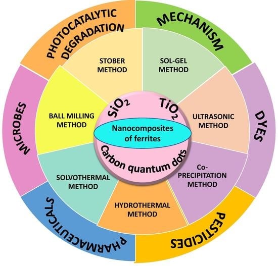
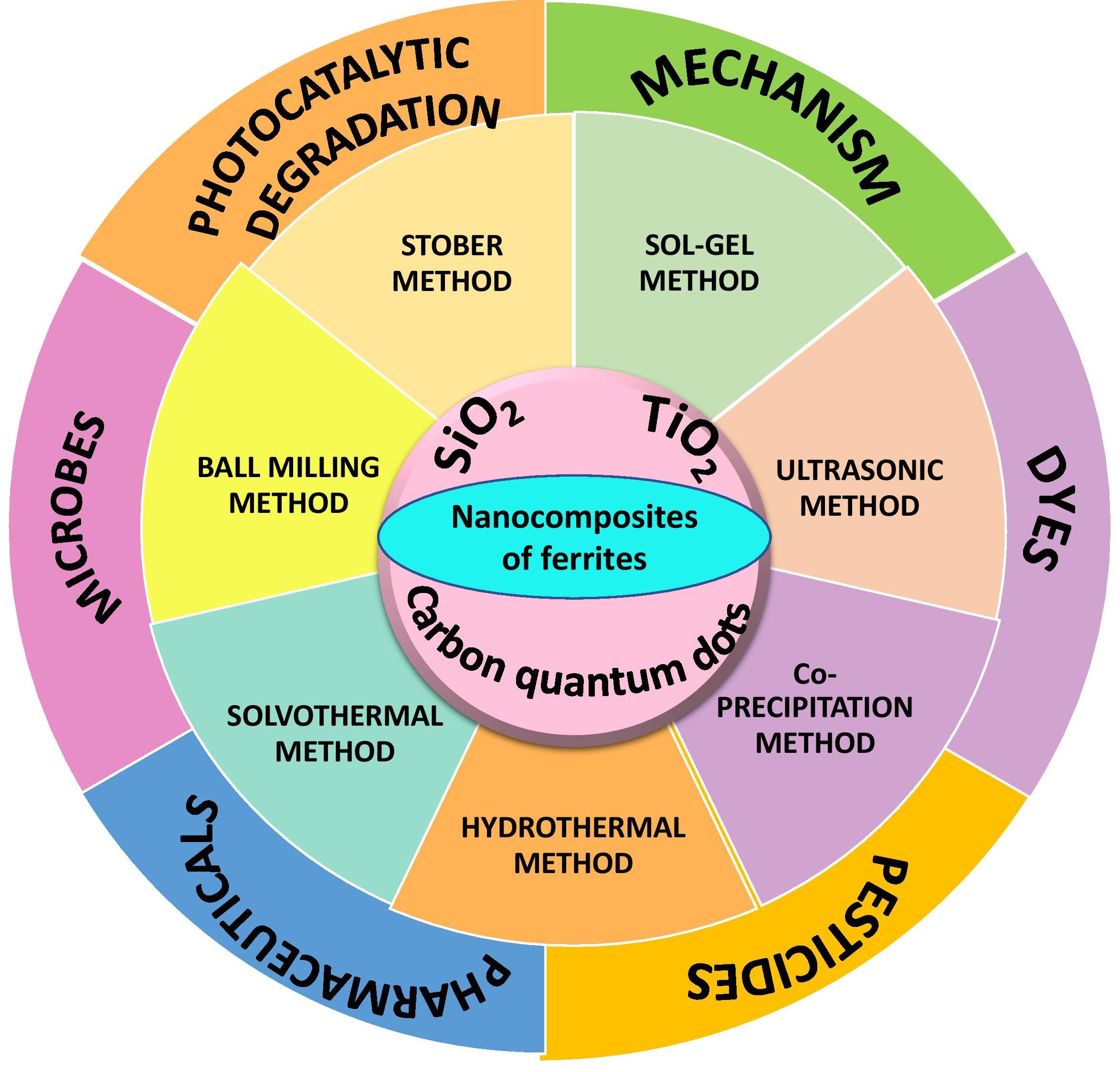
2. Synthesis and Structural Features of Ferrite-Based Nanocomposites

To control size and surface area of materials, methods used to synthesize materials play an important role. Nanocomposites are multiphase solid materials with nanoscale repeat distances between phases and one, two, or three dimensions of less than 100 nm in one of the phases. The successful production of nanocomposites is indicated by homogeneous dispersion of nanometer-sized particles composing the dispersed phase in the matrix. Methods of synthesis of nanocomposites of ferrites with TiO2, SiO2, and CQDs are given in Figure 1.

2.1. Magnetic Features of Ferrite Nanoparticles

Ferrite nanoparticles have chemical and physical characteristics that differ significantly from bulk ingredients, which can be related to their size. Every discrete magnetic nanoparticle behaves like a huge paramagnetic atom at the nanoscale, with a fast response to applied magnetic fields and apparent unique magnetic characteristics. Remnant magnetization (Mr), saturation magnetization (Ms), coercivity (Hc), and magnetic anisotropy are the most essential magnetic characteristics of nanoparticles. Magnetic nanoparticles achieve a perfect magnetic moment in the presence of an external magnetic field (MF) due to parallel orientation of unpaired electrons, which is referred to as Ms. “Mr” refers to the overall residual magnetization of the material after the removal of the MF due to a few unpaired electron orientations. The resistance of a ferromagnetic material towards becoming demagnetized is known as Hc, and it is used to categorize magnetic materials as hard or soft. “Magnetocrystalline anisotropy” (MCA) is another important term used to characterize the magnetic property of nanoparticles, and it represents the orientations of the magnetic moments at a given time in the direction of their ideal easy axes. Spin–orbit interaction is the principal source of MCA, especially in isolated systems, and is responsible for keeping the spins in a certain orientation. It has a direct impact on magnetic characteristics such as Mr, Ms, and Hc. Cation distribution on the octahedral and tetrahedral sites, as well as the size and shape of the nanoparticles, usually regulate these magnetic characteristics. In fact, the total magnetic moment of spinel ferrite nanoparticles is primarily determined by the difference in the exchange interaction of valence electrons of cations located at A and B sites. This is because the magnet’s electron spin is well-organized and parallel inside each lattice site, but antiparallel between the two sublattice sites. However, the contribution of the A-A and B-B interactions is negligible.

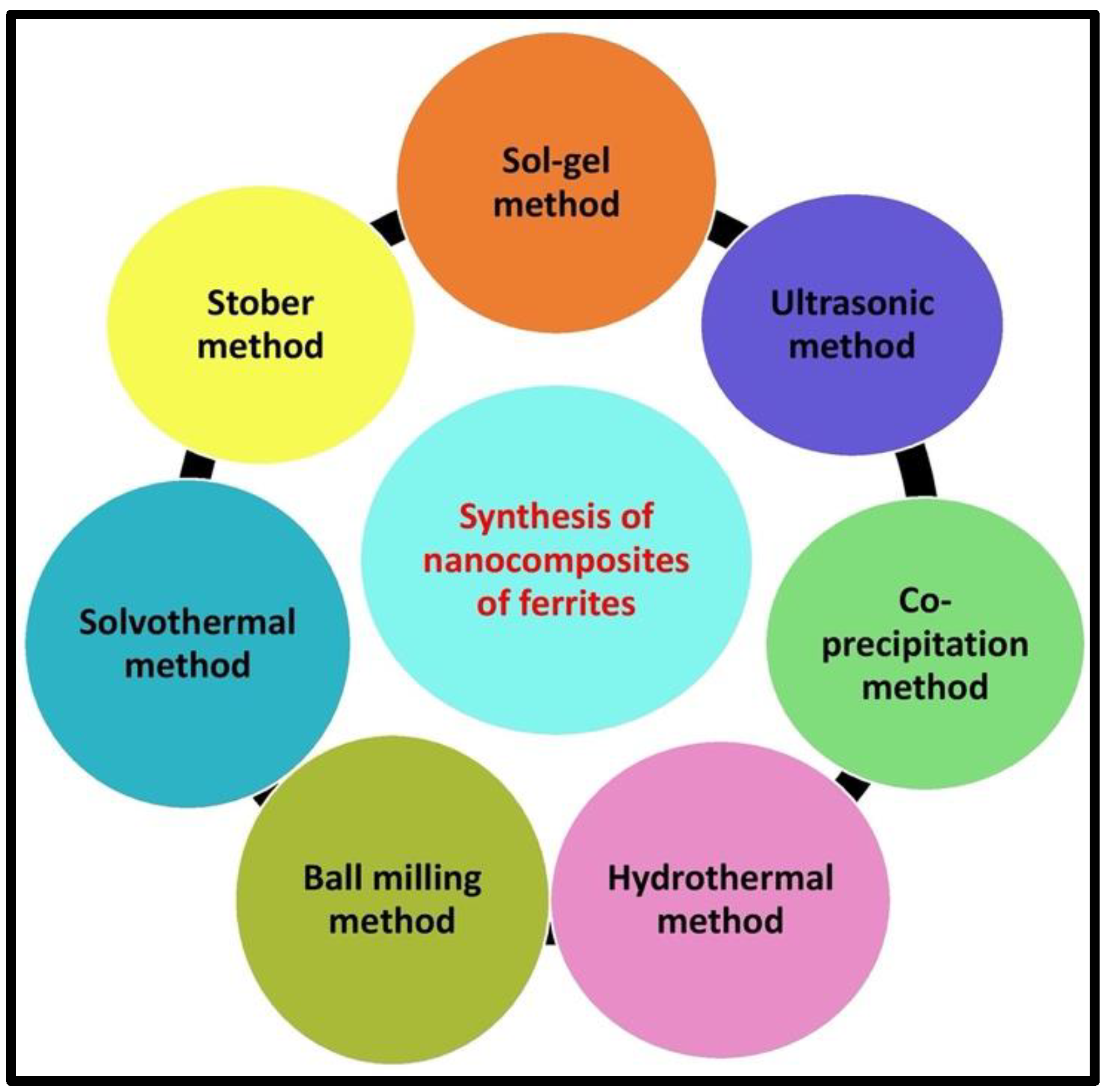
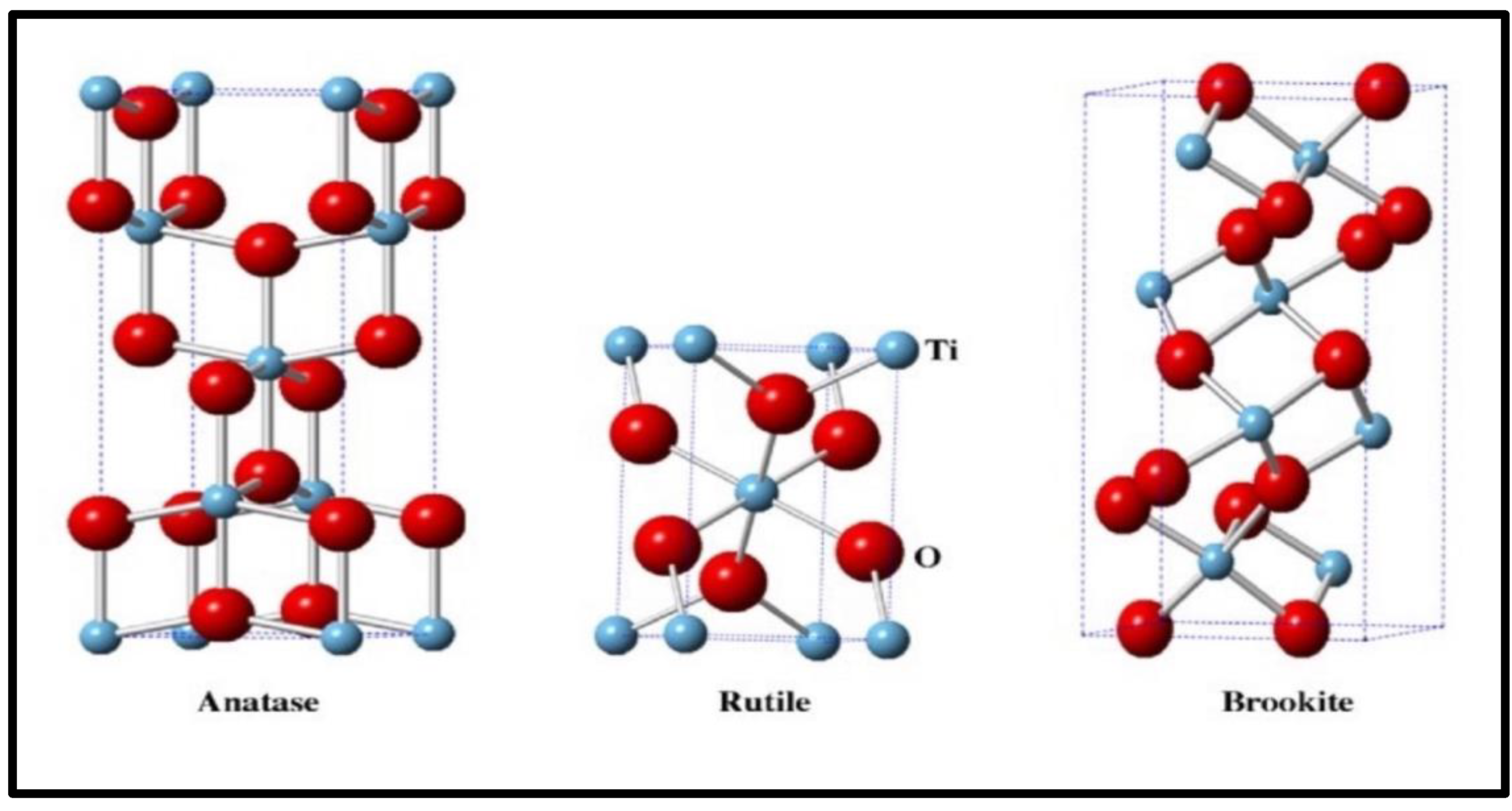
2.2. Nanocomposites of Ferrites with TiO2

TiO2 has four polymorphs: brookite (orthorhombic), rutile (tetragonal), anatase (tetragonal) and TiO2-B, as shown in Figure 2. Anatase, rutile, and brookite phases have bandgaps of 3.2, 3.0, and 3.4 eV, respectively. Brookite is less thermodynamically stable, but rutile is stable and anatase is metastable. At temperatures exceeding 600 °C, irreversible conversion of brookite and anatase TiO2 to rutile occurs [44].
It has been observed that bandgaps of TiO2 nanoparticles with 5–10 nm particles narrow up to 0.2 eV. Anatase is the most photocatalytically effective phase of TiO2 [45] and is widely used in semiconducting materials for environmental applications [46]. It has higher mobility of electron–hole pairs and higher surface hydroxyl density. Brookite-phase activity has not been thoroughly examined [47,48]. Rutile TiO2 is usually investigated in fundamental investigations. Due to the existence of oxygen vacancies, TiO2 is classed as an n-type semiconductor [49]. Because crystalline TiO2 has a less photoexcited electron–hole recombination than amorphous TiO2, it has higher photocatalytic activity [50,51]. Recombination of photogenerated charge carriers is a disadvantage of TiO2 photocatalysts, as it decreases overall reaction quantum efficiency [52]. During the recombination processes, the photoexcited electrons return to the valence band non-radiatively or radiatively [53,54,55]. An attractive feature of the TiO2 photocatalyst is its potential to be activated by visible light. To increase photocatalytic activity and decrease recombination of photoexcited charge carriers, several methods have been followed, such as heterojunction formation and doping with non-metals, metals, and nanosized crystals, which can alter the electronic and optical properties of TiO2 [56,57,58,59,60]. By using dopants in TiO2, its bandgap reduces [61]. Metal doping has long been known to improve visible light absorbance of TiO2 [62,63] and increase its photocatalytic activity under UV irradiation [63,64,65,66], but the introduction of metal ion results in thermal instability, which reduces the reuse of TiO2 photocatalyst and recombination of electron–hole pairs occur at a fast rate. Non-metal doping is more efficient than metal ion doping due to more thermal stability and less formation of recombination centers [67,68,69,70,71].
Nanocomposites of ferrites with TiO2 have been synthesized using different chemical methods. A summary of methods used to synthesize magnetic nanocomposites has been discussed in this section.

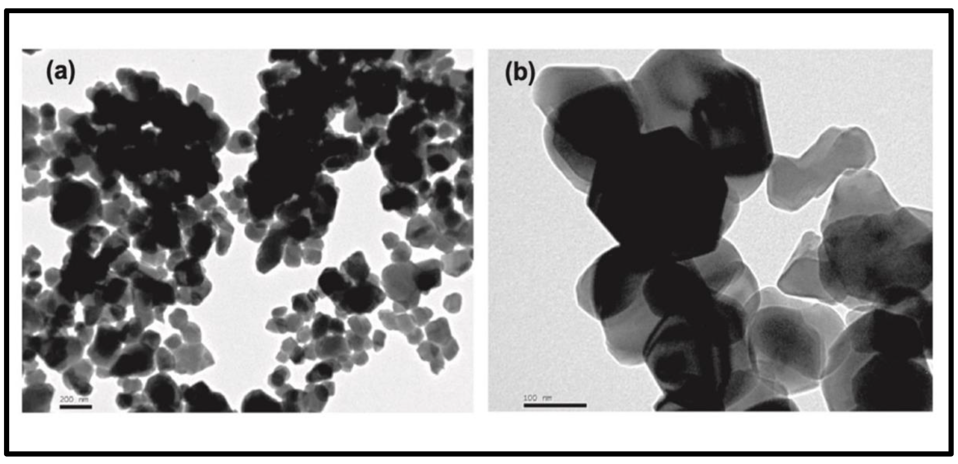
2.2.1. Sol–Gel Method

In typical sol–gel synthesis, formation of gel materials with three-dimensional structure occurs by condensation and hydrolysis reaction of metal ions. It is an irreversible process: once gel is formed, it cannot break down. There is covalent interaction between gel particles. In this method, huge precursors are used during conversions and can be performed at or near room temperature. For chemical catalysts, porous ceramic xerogels with large surface area act as support and thin-film deposition is important for material possessing desired optical and magnetic properties [24]. This method uses simple equipment, produces highly homogeneous and pure products at low temperatures, and allows modification of the surface.
Tatarchuk et al. [72] developed magnetic nanocomposites of CoFe2O4@TiO2 using the Pechini sol–gel method with ethylene glycol and citric acid as chelating agents and titanium (IV) polymeric precursor solution. XRD study of CoFe2O4@TiO2 indicated the presence of 46% and 54% of anatase and rutile crystal phase, respectively, in the titanium dioxide (TiO2) component, and the average size of cobalt ferrite (CoFe2O4) and TiO2 nanoparticles was about 30 and 8 nm, respectively. SEM revealed that because of deposition of TiO2 on the CoFe2O4 surface, particles became large. The IR spectrum of the nanocomposite illustrated the shift of bands, which was the result of isomorphic heterovalent substitution in the octahedral position. Dadfar et al. [73] and Xu et al. [74] fabricated SrFe12O19/TiO2 and TiO2/ZnFe2O4 nanostructures by the sol–gel method. XRD indicated the presence of impurity phases ( α -Fe2O3) at pH 2.5 and 4.5, while pure SrFe12O19 and TiO2 phases were formed at pH 3.5. The production of the rutile phase was prohibited to some extent by highly dispersed ZnFe2O4 nanoparticles in TiO2/ZnFe2O4 nanocomposites. TEM confirmed average particle sizes of 15–75 nm of SrFe12O19/TiO2 and uniform attachment of ZnFe2O4 nanoparticles to TiO2 nanoparticles to produce a coupled semiconductor. The electron diffraction pattern displayed very weak intensity of the electron diffraction pattern for the ZnFe2O4 phase, which was attributed to the dispersion of ZnFe2O4 nanoparticles amid the TiO2 nanoparticles. The value of saturation magnetization, remnant magnetization, and coercivity decreased with increasing amounts of TiO2 in TiO2/ZnFe2O4, due to the contribution of the non-magnetic TiO2 component to total sample volume.
Lahijani et al. [75] fabricated a PbFe12O19-TiO2 nanocomposite using the sol–gel method. From XRD analysis, the average size of crystals was found to be 88 nm. FTIR study indicated that absorption bands at 544, 716, 935 and 1401 nm−1 corresponded to stretching modes of Fe-O, Ti-O, Pb-O and C-C bonds, respectively. Heating the compound in the presence of a polyhydroxy alcohol, such as ethylene glycol, promotes polymerization. The metal ions are equally dispersed inside the organic matrix, resulting in a homogeneous resin—the polymeric precursor [76]. The immobilization of metal complexes in such rigid organic polymeric networks can decrease segregation of specific metals, ensuring molecular compositional uniformity. Polymeric precursors can be used to make ferrite–TiO2 nanocomposites. Mouro et al. [77] used a polymeric precursor technique to make nanometric TiO2/CoFe2O4 composites. X-ray diffraction, Raman spectroscopy, surface area through N2 physisorption, zeta potential, scanning and high-resolution transmission electron microscopy were used to characterize the as-prepared sample. The lack of a rutile phase in the nanocomposites was confirmed by Raman spectroscopy. XRD patterns revealed that thermal treatment did not cause phase segregation. The synthesized nanocomposites showed an increase in surface area, a change in surface charge in relation to pure TiO2, and selectivity in the photodegradation. The materials had photocatalytic activity due to the presence of TiO2 on their surfaces, and CoFe2O4 cores were beneficial for separating and recovering photocatalysts after use in an oxidative process.

2.2.2. Ultrasonic Method

The ultrasonic technique involves using sound energy to agitate the particles in a solution, resulting in physical and chemical changes. This method is found to be the most promising method for manufacturing nanocomposites such as oxides, carbides, alloys and colloids with almost uniform distribution of nanoparticles [78]. Ultrasound irradiation causes unstable bubbles in liquids to form, grow, and collapse rapidly at temperatures as high as 5000 K, pressures as high as 20 MPa, and cooling speeds of 1010 Ks−1 [79]. Ultrasonication was used for the synthesis of titania-coated γ-Fe2O3 magnetic activated carbon and Fe3O4-TiO2 photocatalyst with saturation magnetization value of 2.21 and 14 emug−1, respectively [80,81]. XRD revealed that the titania coated on the magnetic activated carbon maintained the anatase phase, with the same set of characterization peaks for both Fe3O4 and Fe3O4-TiO2, indicating that the ultrasound irradiation had no effect on the crystal structure of the Fe3O4 nanoparticles. The tiny crystallite size was shown by the broad nature peak of the deposited TiO2 on Fe3O4-TiO2. SEM revealed the homogeneous distribution of titania. Superparamagnetic characteristics of Fe3O4-TiO2 photocatalyst at room temperature were confirmed by a magnetization hysteresis loop. HRTEM analysis showed the occurrence of a heterojunction in the Fe3O4-TiO2 nanocomposite. XPS showed that the peaks at 710.5 eV and 458.8 eV originated from Fe 2p and Ti 2p energy levels, respectively, implying that Fe3O4 and TiO2 were largely present as separated phases in Fe3O4-TiO2 composites.

2.2.3. Coprecipitation Method

This approach requires the preparation of a mixed aqueous solution of starting chemicals such as chlorides, nitrates, or sulfites of Fe3+, as well as of divalent metal ions in the requisite stoichiometric ratio. The precipitates are then generated by treating the solution with NaOH, filtration, washing twice, and drying. This approach entails four steps: nucleation, growth, coarsening, and agglomeration, all occurring at the same time [82]. Nanocomposites of ferrites of good quality and phase purity can be obtained by this method. This process offers various advantages such as high yield, high product purity, lack of necessity to use organic solvent, good reproducibility and low cost. It is a quick and easy way to make ultrafine particles that are disseminated in a variety of media. By carefully monitoring the preparation parameters, it is possible to achieve control over the surface morphology, structure, and chemical composition with this procedure.
Coprecipitation was employed for fabrication of CoFe2O4/TiO2 and ZnFe2O4-TiO2 nanocatalysts [83,84]. The average particle size of the coprecipitated nanocatalysts was 50 nm for CoFe2O4 and 150 nm for CoFe2O4/TiO2 according to SEM images, which was consistent with the crystallite size predicted from XRD data. The spherical geometry (9 ± 2 nm) of ZnFe2O4 nanoparticles and spherical-like morphology (11 ± 3 nm) of ZnFe2O4-TiO2 nanocomposites and fine dispersion of black particles (CoFe2O4) on the gray surface (TiO2) of CoFe2O4/TiO2 were depicted in the TEM images (Figure 3). Bandgaps of CoFe2O4/TiO2, CoFe2O4, ZnFe2O4-TiO2, and ZnFe2O4 were found to be 2.8, 1.1, 2.3, and 2.1 eV, respectively, which suggested the formation of a visible light-active photocatalyst. The large bandgap of the composite may be due to the mixing effect of the bandgap and the interfacial coupling effect between ZnFe2O4 and TiO2, CoFe2O4 and TiO2. XRD pattern analysis revealed that in CoFe2O4/TiO2, the conversion of anatase to the rutile phase of TiO2 happened at a TiO2 annealing temperature of ≤600 °C, confirming the purity of ZnFe2O4 and TiO2 phase in ZnFe2O4-TiO2, which was in agreement with HRTEM and SAED investigations. Haw et al. [85] found that in comparison to conventional rutile-phase TiO2 and pure urchin-like TiO2 (3D TiO2) microparticles, nanocomposites of CoFe2O4-3D TiO2 showed an increase in photodegradation of methylene blue, and this nanocomposite demonstrated a generally constant photocatalytic efficacy with low degradation. Two sets of lattice fringes were visible at the TiO2-CoFe2O4 interface, with d110 = 0.322 nm of rutile phase titania and d331 = 0.193 nm of CoFe2O4. The CoFe2O4 nanoparticles were clearly visible on the 3D urchin-like TiO2 structure, and each element was uniformly distributed over the surface of the TiO2 microsphere on scanning transmission electron microscopy. XRD peaks were designated to the rutile phase of 3D urchin-like TiO2 and broader peaks confirmed the presence of nanosized CoFe2O4 in the sample.

2.2.4. Hydrothermal Method

The hydrothermal method is a way of crystallizing a chemical, utilizing an aqueous solution at a high vapor pressure as well as temperature [86]. At standard temperatures (100 °C) and pressures (<1 atm), it is generally depicted as crystal formation or crystal synthesis from insoluble chemicals. Autoclaves are used to carry out the process, which is performed at a controlled pressure and temperature. This enables the utilization of temperatures that are higher than the boiling point of water or an organic solution. The concoction reaction that occurs beyond the dissolvable breaking point and at pressures over bar is known as hydrothermal synthesis. The hydrothermal technique has the following advantages: it is simple to acquire nanotube morphology, variations in the synthesis process may be used to improve TiO2 nanotube characteristics, and it is a method that can be used for a variety of applications.
Hydrothermal deposition of a hard ferromagnetic strontium ferrite (SrFe12O19) over TiO2 can result in a photocatalyst that is both ferromagnetic and visible light-active. The TiO2-supported SrFe12O19 nanocomposite has a strong ferromagnetic property and is quite stable when it comes to losing its magnetic property. XRD images of TiO2-supported SrFe12O19 nanocomposite correspond to mixture of anatase and rutile crystal phases. EDS results confirmed that TiO2-supported SrFe12O19 consists of Fe, Ti, Sr, C, O and Si. The BET-specific surface area of TiO2 was 49.5 m2g−1 with a total pore volume of 0.1553 cm3g−1, which compares with 38.3 m2g−1 and 0.0155 cm3g−1 for TiO2-supported SrFe12O19. Due to the deposition of SrFe12O19 nanoparticles onto the TiO2, the surface area and pore volume of the TiO2 nanocomposite reduces. The paramagnetic impact of TiO2 lowered the coercivity (HC) and saturation magnetization (MS) of TiO2-supported SrFe12O19. The photocatalyst TiO2-supported SrFe12O19 had HC of 2125.5 G. The coercivity of TiO2-supported SrFe12O19 nanocomposite showed that it was a good ferromagnetic material [87]. Nguyen and Doong [88] and Pongwan et al. [89] fabricated ZnFe2O4-TiO2 and CoFe2O4/TiO2 nanostructure photocatalysts by the hydrothermal method. The ZnFe2O4-TiO2 nanocomposite was prepared by coupling 0.2–2 wt % narrow-bandgap material of p-type ZnFe2O4 with n-type anatase TiO2. Transmission electron microscopy (TEM) and high-resolution TEM confirmed average particle sizes of 8–9 nm and 5–35 nm for ZnFe2O4-TiO2 and CoFe2O4/TiO2, respectively. It was observed that ZnFe2O4 and TiO2 were intimately linked, which led to a decrease in electron–hole recombination rate as well as enhanced photocatalytic activity of ZnFe2O4-TiO2 heterostructures under visible light irradiation. When the loading amount of ZnFe2O4 increased from 0.5 to 2 wt %, SEM revealed a slight increase in particle size of ZnFe2O4-TiO2 nanocomposites. The N2 adsorption–desorption isotherms showed that the combination of TiO2 with ZnFe2O4 can increase the specific surface area. XRD analysis indicated that ZnFe2O4-TiO2 can retain the crystallinity of both nanoparticles. Electron diffraction pattern of CoFe2O4/TiO2 displayed the brightness of polymorphic discrete rings of the crystalline particles, suggesting quite a high degree of crystallinity in polycrystals. In this study, the obtained maximum saturation magnetization and coercivity of CoFe2O4 and CoFe2O4/TiO2 nanocomposites were found to be 32.58, 29.64 emug−1 and 0.15, 0.05 kOe, respectively.

2.2.5. Solvothermal Method

The solvothermal technique employs a non-aqueous solvent and a considerably higher temperature, allowing high boiling point solvents to be utilized. The solvothermal method outperforms the hydrothermal method in terms of shape, size control, distribution, and crystallinity of nanoparticles [90]. Organic solvents, which have a low relative permittivity and are free of ionic species, are used in the solvothermal process to produce a product devoid of foreign anions and ionic species. The benefits of both the hydrothermal and sol–gel methods are combined in this technique. This approach may be utilized in the ceramics sector to regulate the particle size of synthesized materials and to fabricate tiny particles, such as magnetic titania photocatalyst [91]. Atacan et al. [92] developed ZnFe2O4/Ag-TiO2 by the solvothermal method. The formation, structure and morphology of prepared samples were characterized by X-ray diffraction, scanning electron microscopy, Fourier-transform infrared spectroscopy and vibrating sample magnetometry. In this study, the obtained value of saturation magnetization of ZnFe2O4/Ag-TiO2 was 5.5 emug−1. XRD patterns indicated that no chemical reaction occurred between ZnFe2O4 and Ag-TiO2, because no peaks related to other impurities were observed. Nguyen and Doong [93] synthesized ZnFe2O4/TiO2 heterostructure by this method.

2.3. Nanocomposites of Ferrites with Silica

Because of the Van der Waals forces of attraction between magnetic particles and their large surface area, they clump together, making it difficult to determine their physicochemical characteristics. One of the excellent methods to reduce particle agglomeration and to stabilize magnetic nanoparticles is the dispersion of magnetic nanoparticles in a silica matrix. Due to biocompatibility and chemical inertness, silica does not affect the redox reaction at core surface and assists the functioning of nanocomposites in a biological environment. This sort of matrix can help to reduce toxicity, increase biocompatibility, and shield encapsulated components from harsh reaction media. The properties of such nanocomposites might provide a number of advantages, including improved catalytic, magnetic, and mechanical capabilities [94,95]. Thus, silica-coated magnetic nanoparticles are becoming a promising and important approach in the development of magnetic nanoparticles for both fundamental studies and technological applications. Therefore, coating magnetic particles with silica allows its surface to conjugate with various functional groups. The optical properties of nanoparticles can also be increased by coating of silica gel [96]. Nanocomposites consisting of nanoscale magnetic particles embedded in an insulating matrix such as silica presented considerable difference in magnetic properties when compared with their equivalent pure and bulk materials. In this section, techniques for synthesis of ferrite–silica nanocomposites are discussed.

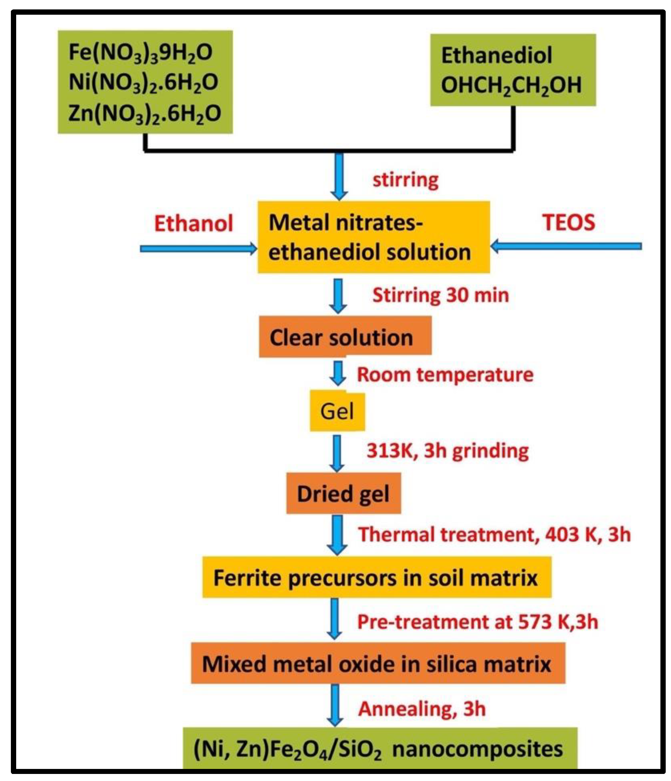
2.3.1. Sol–Gel Auto-Combustion Method

Exothermic chemical reactions occur in the sol–gel process, and self-ignition of the gel occurs as a result of the combustion of the gel, resulting in black–brown powder as a product. Urea, glycine, citric acid, hydrazine, and carbohydrazide are common starting materials for this process, and stoichiometry and crystallite size can be easily adjusted. Colloidal gel is created by combining metal nitrate and an organic solvent, then adding NH4OH to alter the pH. Sol–gel auto-combustion was used to synthesize CoFe2O4-SiO2 nanostructures by using metal nitrates as precursors. Bardapurkar et al. [97] studied (CoFe2O4)x(SiO2)1−x synthesis, structural, spectral, and magnetic characteristics, where x ranged from 0.1 to 1.0. The crystal structure, cation distribution, and band locations of silica matrix were detected in IR spectra. Crystallite size and pore fraction were at their maximum when the sample had the highest silica concentration, due to changes in inter-particle distances and crystallite size following dispersion in the matrix. As the ferrite-silica ratio was increased, the saturation magnetization dropped from 68.7 emug−1 to 4.77 emug−1, indicating that the magnetic characteristics may be changed by altering the ferrite-silica ratio. The reduction in magnetization for the coated sample may be attributed to the presence of a non-magnetic silica layer on the surface of magnetic nanoparticles that reduces the particle–particle interaction and lowers the exchange coupling energy, which in turn reduces the magnetization. In the FTIR spectra, there were strong absorption bands found around 1090 cm−1 and 800 cm−1 that corresponded to silica network. SiO2-coated cobalt ferrite nanocomposites were fabricated by Gharagozlou [98] and Pansambal et al. [99] using tetrakis(2-hydroxyethyl) orthosilicate (THEOS) and tetraethyl orthosilicate (TEOS) as water-soluble silica precursors, respectively. TEM images revealed that spherical, non-agglomerated cobalt ferrite nanoparticles were homogeneously distributed in the silica matrix. Saturation magnetization was found to be less for nanocomposite compared to bare nanoparticles. Peaks present in the XPS spectra of CoFe2O4@SiO2 corresponded to Co 2p, Fe 3p, Fe 2p, Si 2s, Si 2p signals, which confirmed the formation of CoFe2O4@SiO2. There was no unidentified peak in the EDX data, which confirmed the purity, elemental composition and formation of CoFe2O4@SiO2. Specific surface area of CoFe2O4@SiO2 was 9.34 m2g−1 calculated by BET equation. Results indicated that crystallization, saturation magnetization Ms and remanent magnetization Mr escalates as the calcination temperature rises, but the variation in coercivity Hc was not in accordance with that of Ms and Mr, indicating that Hc is not determined only by the crystallinity and size of CoFe2O4 nanoparticles. Figure 4 shows a technological scheme for the synthesis of ferrite–silica nanocomposite. The influence of chelator chain length on precursor formation and decomposition to nanocomposites of ferrite with SiO2 was studied. Dippong et al. [100] prepared CoFe2O4/SiO2 nanocomposite by using three short-chain diols (1,2-ethanediol, 1,3-propanediol, and 1,4-butanediol) as chelators. The influence of the methylene groups’ numbers in the precursors and annealing temperature on the nanocrystallite size was revealed. This method provides some advantages in making silica composite material containing highly dispersed nanoparticles and promotes a good and homogeneous dispersion of particles into silica matrix.

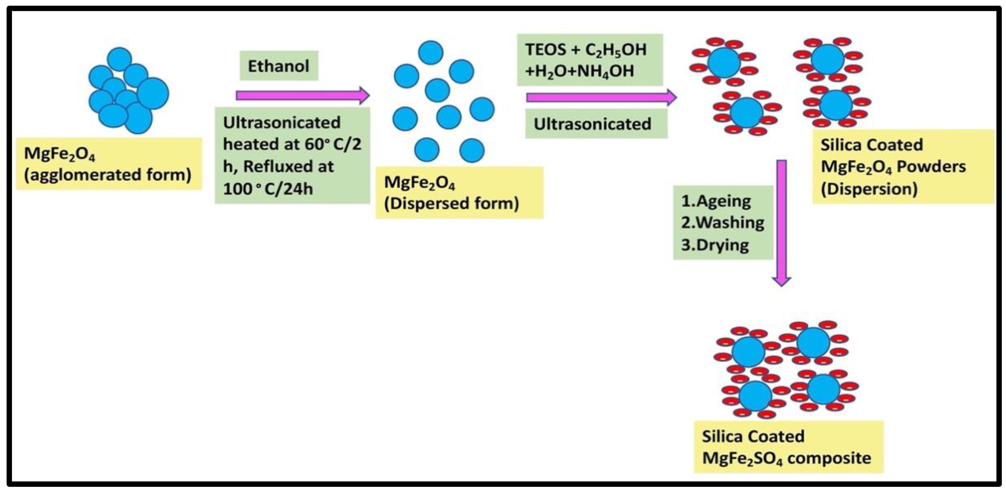
2.3.2. Stöber Method

A system of chemical reactions has been developed that allows the controlled growth of spherical silica particles of uniform size by means of hydrolysis of alkyl silicate and subsequent condensation of silicic acid in alcoholic solutions. The schematic procedure employed for the synthesis of MgFe2O4-SiO2, Co1−xZnxFe2O4/SiO2, CoFe2O4/SiO2 was reported [101]. Eromosele et al. [102] and Khanna and Verma [103] developed SiO2-coated MgFe2O4 and KFeO2 by Stöber method, respectively. Both bare and SiO2-coated KFeO2 exhibited orthorhombic structure, whereas bare and silica-coated magnesium ferrite exhibited a single cubic spinel phase. Association of silica on the surface of MgFe2O4 and KFeO2 nanoparticles was confirmed by EDAX in MgFe2O4-SiO2 and by FTIR in KFeO2/SiO2. It was further confirmed by FTIR data in MgFe2O4-SiO2, because in coated samples, there was a shift in stretching vibration of Fe-O from 560 cm−1 to 574 cm−1 due to silica coating. It was observed that due to silica coating, the crystallite size reduced from 53 to 47 nm in MgFe2O4-SiO2, but in KFeO2/SiO2, it increased from 4–8 nm to 10–22 nm. Values of saturation magnetization of MgFe2O4-SiO2 and KFeO2/SiO2 were 22 and 21.17 emug−1 and possessed ferrimagnetic and superparamagnetic behavior, respectively, at room temperature. Figure 5 shows a schematic representation of SiO2 coating of MgFe2O4.
The influence of aging temperature on the synthesis of nanocomposites of ferrites with silica by the Stöber method was studied. Queiroz et al. [104] investigated various aging temperatures—98, 80, 60, 27/98, and 27 °C—for the synthesis of silica-coated cobalt ferrite. Aging temperatures of 98 and 27/98 °C give the best suited conditions for preparing the single phase of cobalt ferrite. The presence of silica on the particle surface was confirmed using diffuse reflectance infrared Fourier transform, which presented vibrational modes with asymmetric stretching absorption in the region 900–1200 cm−1 associated with the Si-OH and O-Si-O groups and value of saturation magnetization of coated samples of 10.05 emug−1. Girgis et al. [105] studied the magnetic and optical properties of CoFe2O4, ZnFe2O4, Co0.5Zn0.5Fe2O4 coated with silica by heating at 400, 600 and 800 °C. It was observed that by raising the temperature from 400 to 800 °C, average crystallite size of core–shell nanocomposites increase.

2.3.3. Coprecipitation Method

This method was used for the formation of nanocomposites of ferrites with silica using different precursors. SiO2-coated cobalt ferrite nanocomposites were fabricated using metallic chlorides and metal nitrate as precursors and annealing at 100, 200, and 300 °C and 250, 500, and 750 °C, respectively. The coercivity and saturation magnetization of nanocomposite (annealed at 300 °C for 2 h) were much higher than that of bulk cobalt ferrite. CoFe2O4 nanocrystallites were observed in the silica matrix during the annealing of samples, and their size increased with increasing annealing temperature. TEM showed that homogeneously distributed, well-crystallized, and nearly spherical CoFe2O4 nanoparticles were inserted in the silica host matrix (Figure 6). FTIR spectra showed that as annealing temperature increased, the absorption peaks at 1090 cm−1 for Si-O-Si further broadened [106]. The broad hump in the X-ray diffraction pattern at 2 θ   ∼18 ° –23 ° corresponds to the presence of amorphous silica in the sample. An increase in crystallite size of CoFe2O4:SiO2 nanocomposites was observed from 20.26 to 28.95 nm and 38.76 nm with increase in calcination temperature from 250 to 500 °C and 750 °C by the Williamson–Hall method. Moreover, lattice parameter and strain values increased from 8.0321 to 8.0691 Å and 1.01 × 10⁻² to 3.75 × 10⁻³, respectively, as temperature increased from 250 to 750 °C. SEM analysis revealed the formation of well-developed nanoparticles of CoFe2O4:SiO2 with intergranular porosity [107]. Yakob et al. [108] prepared CoFe2O4/SiO2 by coprecipitation using metal nitrate as precursor. The polycrystalline and spinel crystal structure of CoFe2O4/SiO2 with Fd3m group space was determined using XRD analysis. CoFe2O4/SiO2 crystallite nanoparticles were 29.4 ± 0.2 nm in size, whereas CoFe2O4 was 26.8 ± 0.2 nm. Sharma et al. [109] calcined a NiFe2O4/SiO2 composite at temperatures of 300 to 900 °C. It was discovered through Rietveld refining that the powdered combination contains two phases: silicon dioxide and nickel ferrite. As a result, the sample has a cubic phase with space group F d-3 m (227), with Fe atom at 32e site (3/8, 3/8, 3/8), O (oxygen) atom at 32e site (0.387, 0.387 and 0.387) and Ni atom at Wyckoff 8a site (0, 0, 0).

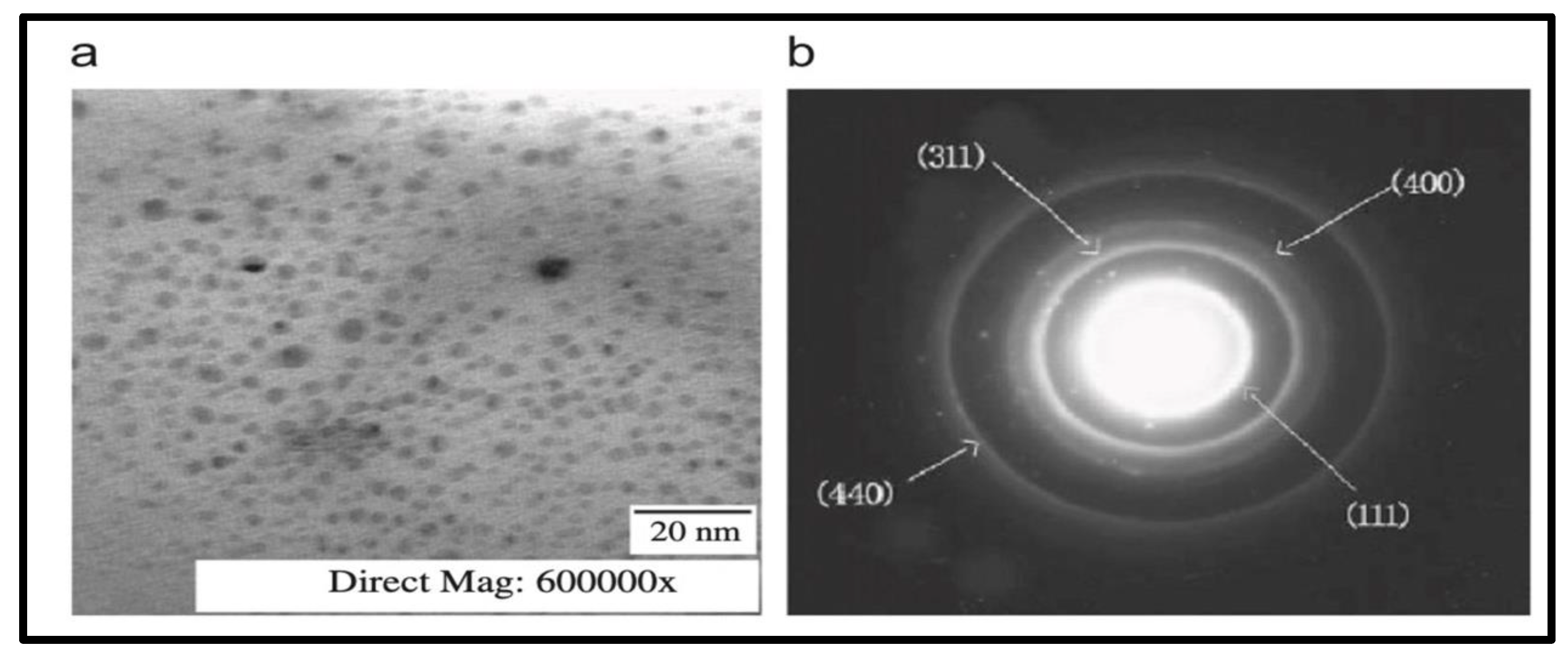
2.3.4. Ball-Milling Method

This is a process of creating new alloys and composites from top-down synthesis. It is a low-cost and quick synthesis approach [110]. Milling causes precise deformation because the internal structure of powders is gradually distorted to the nanoscale level. Gonzalez et al. [111] and Scano et al. [112] synthesized barium ferrite silica and magnetite-silica nanocomposites in molar ratios of 40:60, 50:50, 60:40, 70:30 and silica content of 6, 20, 50 wt %, respectively, by high-energy ball milling. The XRD pattern revealed that a 15 h milling period is sufficient to avoid the formation of hematite phase, but a longer milling period and heat treatment above 900 °C produce hematite phase in BaFe12O19-SiO2 and form uniformly distributed spherical narrow (4–6 nm) Fe3O4 nanoparticles in amorphous SiO2 agglomerate of 100–200 nm in Fe3O4/SiO2. It was discovered that a sample with a compositional ratio of 60 barium ferrite-40 silica milled for 15 h and heated to 900 °C was sufficient to achieve the best magnetic characteristics and excellent dispersion of the hard magnetic phase barium ferrite into the ceramic matrix. In this study, it was found that the value of the highest saturation magnetization and corresponding coercivity (Hc) for BaFe12O19-SiO2 was 43 emug−1 and 3.4 kOe, respectively.

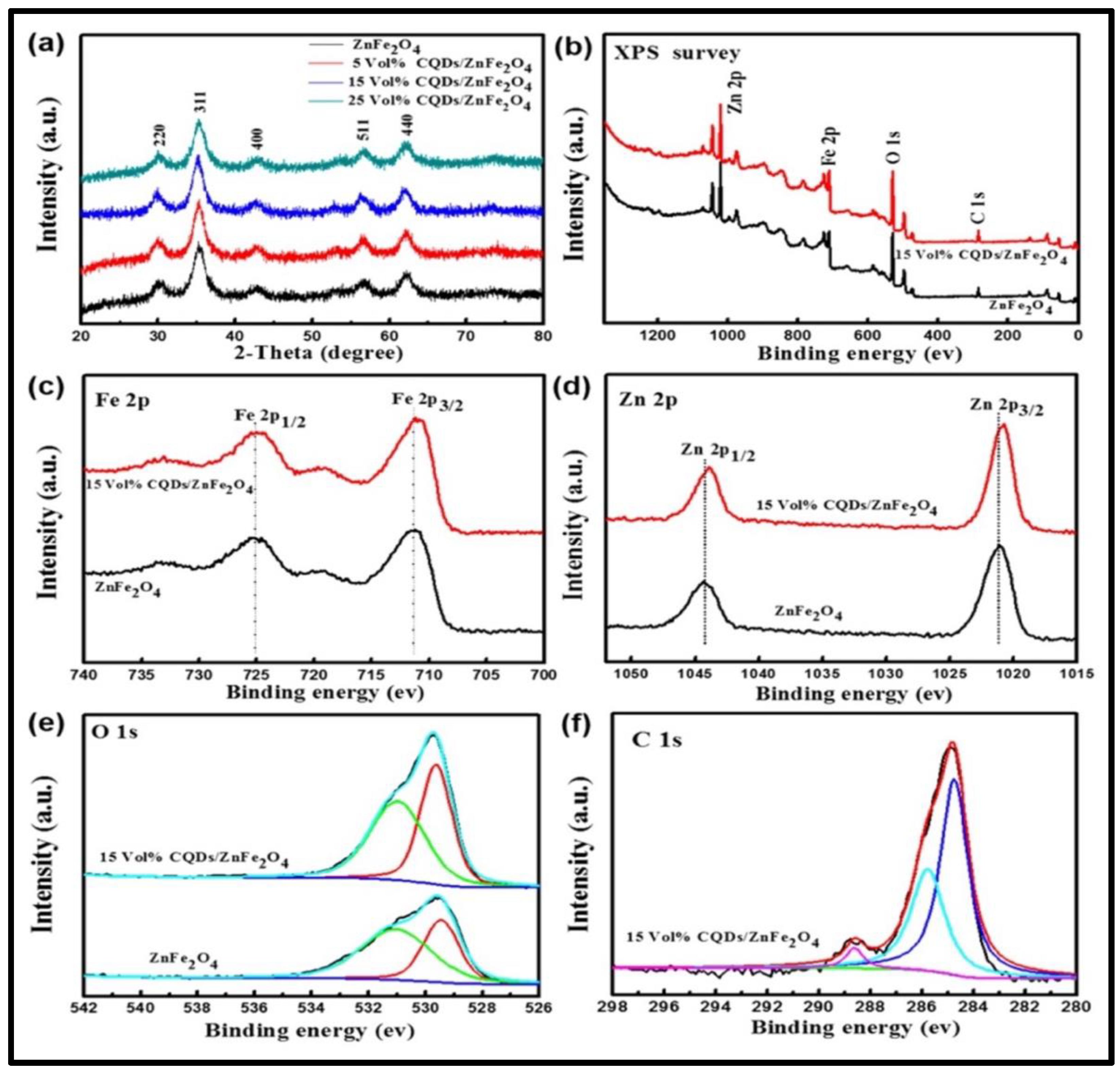
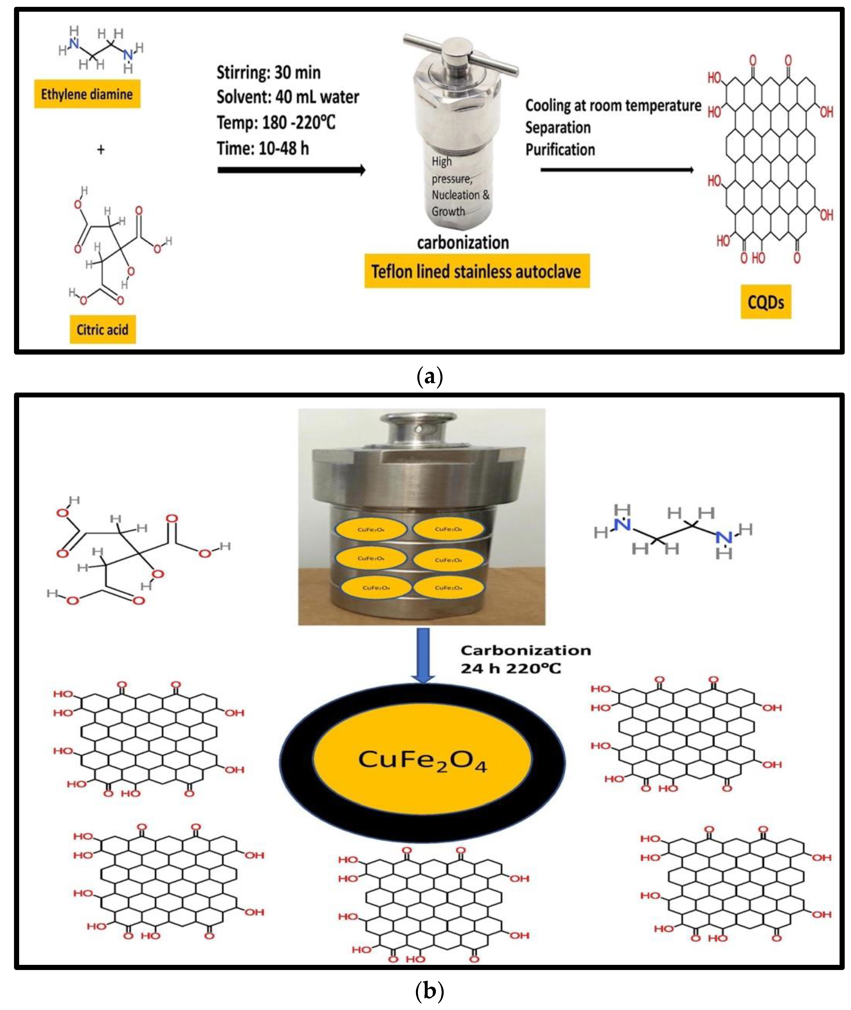
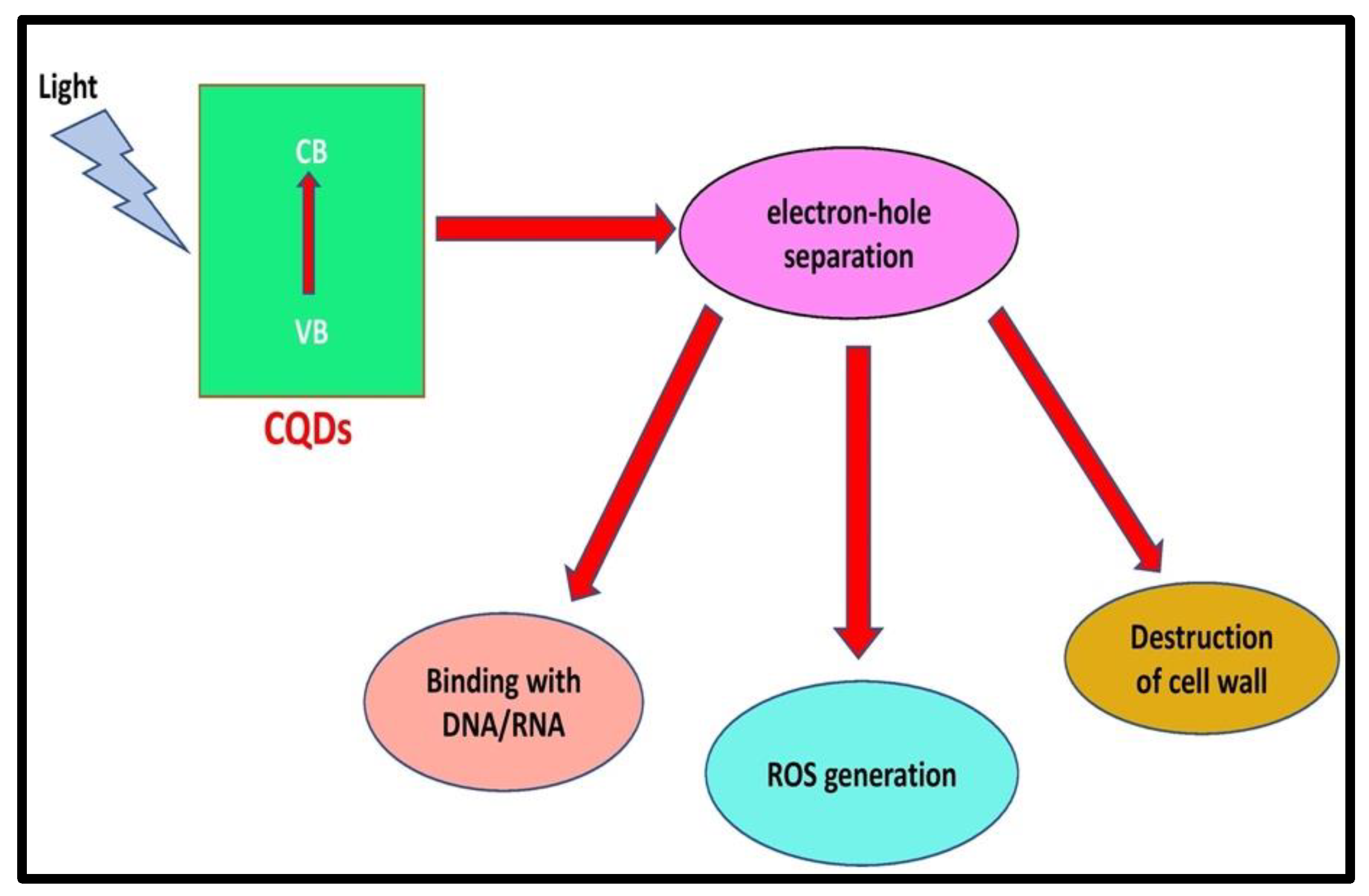
2.4. Ferrites with Carbon Quantum Dots
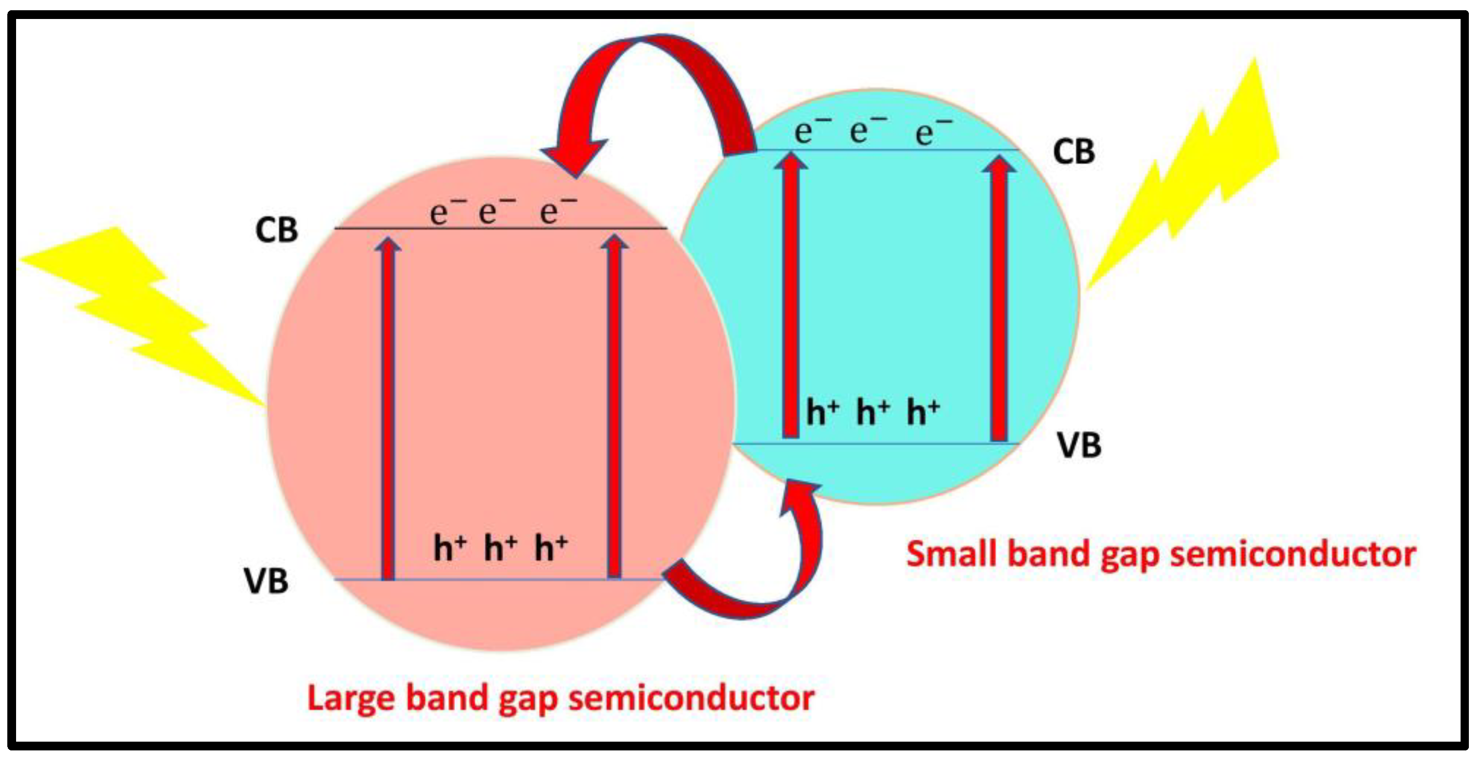
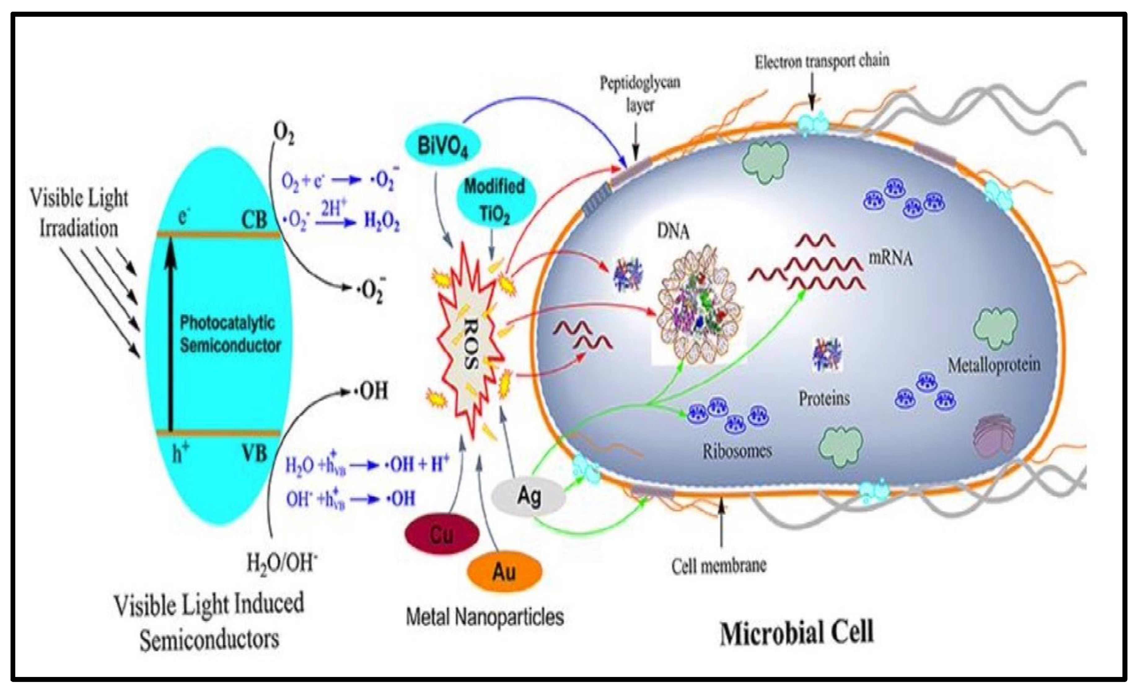
Quantum dots are toxic colloidal semiconductors made up of elements from the periodic groups II–VI, III–V, and IV–VI. Carbon has huge potential in nanoelectronics, catalytic supports, drug delivery, sensors and electrochemical energy storage due to its diverse electron orbital types (sp, sp2, or sp3 hybrid) and size-dependent electric and surface properties [113,114,115,116,117]. Carbon quantum dots are a new type of nanomaterial with diameter less than 10 nm. Carbon quantum dots have unique properties compared to conventional semiconductor quantum dots and organic dyes, including intrinsic low toxicity, chemical stability, excellent optical properties, eco-friendliness, biological labelling, bioimaging, sensing, drug delivery, photocatalysis, biomedicines, low cost, and ease of availability [118,119,120]. The unique and attractive characteristic of CQDs is quantum confinement [121,122,123]. Properties of CQDs such as surface group and size depend intensively on methods of preparation and applied precursors. Based on the synthesis method, CQDs have different oxygen-, nitrogen-, and sulfur-containing functional groups on the surface [124,125,126]. The hydrothermal process is a straightforward, low-cost, and widely used method for preparing ferrite nanocomposites with CQDs. A solution of metal salts and a base is autoclaved under pressure in this process, and crystalline structures are formed from the aqueous solution at high vapor pressure. The precipitated precursor suspensions are then placed in a sealed autoclave and slowly cooked at various temperatures and times. The particle size in this process is determined by the rate of hydrolysis and the solubility of metal oxide. High purity, chemical homogeneity, small and consistent particle size, and controlled particle shape are all advantages of this unconventional technique.
The hydrothermal method was used for fabrication of nanocomposites of MFe2O4 (M=Zn, Cu, Co, Ni) with CQDs. CQDs can be prepared from green sources and chemicals such as turmeric, almond, glucose, L-ascorbic acid, lemon peels, etc. Fini et al. [41] and Fini et al. [127] prepared CoFe2O4–carbon quantum dot and NiFe2O4–carbon dot nanocomposites using turmeric and almond as green starting material, respectively. From hysteresis loop, the obtained values of saturation magnetization and coercivity of CoFe2O4 and CoFe2O4–carbon quantum dots were 35 emug−1 and 585 Oe and 16.8 emug−1 and 647 Oe, respectively, whereas coercivity of NiFe2O4–carbon dots rose from 90 Oe to 220 Oe in comparison to pure NiFe2O4. Increased coercivity of nanocomposites occurred because of increased interactions among nanoparticles. Apparently, magnetic domains are pinned by carbon dots, and as a result, a higher magnetic field is needed for change in magnetic domains of ferrite. In the case of CoFe2O4–carbon quantum dots, TEM analysis revealed that CQDs were uniform in size from 40 to 100 nm in diameter when prepared for 24 h at 180 °C, when prepared for 48 h at 200 °C, CQDs were uniform from 20 to 100 nm in diameter, and when NiFe2O4–carbon quantum dots were autoclaved at 180 °C for 6 h, their size was less than 30 nm.
Huang et al. [128] synthesized CQD/ZnFe2O4 nanocomposites using l-ascorbic acid as a source of CQDs and metal nitrates for preparation of metal ferrite. Formation of CQD/ZnFe2O4 nanocomposites was confirmed by TEM. In CQD/ZnFe2O4, the XRD pattern revealed no typical CQD peak, which could be attributed to the sample’s low concentration and high dispersion of CQDs (Figure 7). The phase structure of ZnFe2O4 was shown to be unaffected by the inclusion of CQDs during the synthesis. XPS proved the presence of CQDs and revealed the chemical states of synthesized materials for the CQD/ZnFe2O4 (15 vol %) and ZnFe2O4 samples. A carbon quantum dot–bismuth ferrite (CQDs/BiFeO3) heterostructure was developed using glucose as a source of CQDs. Attachment of CQDs on the surface of BiFeO3 was confirmed through TEM. Based on XRD analysis, the sites of diffraction peaks of pure BiFeO3 powders were similar to those of CQD/BiFeO3 composite samples (CQD content: 1.2%, 3.3%, and 4.7%), demonstrating that introducing CQDs to BiFeO3 did not modify their crystal structures. As CQD content increased from 1.2% to 4.7%, peaks at 570 cm−1 declined or vanished [129].
Nabiyouni and Ghanbari [130] reported a CuFe2O4–carbon quantum dot nanocomposite as a sensor for detecting Hg (II) Ions. Peaks at 1630, 3410 and 1050 cm−1 in the FTIR spectrum of CuFe2O4–carbon quantum dots correspond to presence of C=O, O-H and C-O group in this nanocomposite. SEM of the product confirmed that CQDs were uniform in size around 50 nm in diameter. From the hysteresis loop of the product, saturation magnetization and coercivity were 8 emug−1 and 500 Oe, showing ferromagnetic behavior. Figure 8 shows the preparation of (a) CQDs and (b) nanocomposites of copper ferrite–carbon quantum dots by the hydrothermal method. The raw materials used in the synthesis of ferrites, TiO2, SiO2, CQDs and their nanocomposites are cheap and easily available. Thus, synthesis of these nanocomposites is cost-effective.

3. Applications

Nanotechnology has been a prominent area of research in science and technology for several decades, including space science, medical technology, coatings, and electronics [131]. It is the study and application of structures smaller than 100 nm [132]. Nanotechnology has the potential to revolutionize the coating industry. Nanoscale coatings are attracting a lot of attention around the world. Nanoparticles are being considered as fillers in coatings with changed surface qualities by the paint and coating industries [133]. Nanocoatings are made by mixing nanoparticles (1–100 nm) with coating formulations to improve certain properties. The large surface area and smallness of nanoparticles present many advantages and opportunities to the paint and coating industries [134].
Nanotechnology has been widely applied in medicine, pharmaceutics, industry, agriculture, and biological sciences, making the 21st century a “golden time” for researchers. Apart from metal and metal oxide nanoparticles, ferrite nanoparticles have raised interest due to their superparamagnetic characteristics and surface area-to-volume ratio, which differ significantly from their bulk counterparts [135]. Ferrites have the advantage of having a bandgap that can absorb visible light, as well as a spinel crystal structure that increases efficiency by providing additional sites due to the crystal lattice. The bandgap of ferrites is narrower than that of other regularly employed visible light catalysts (Figure 9). The size, shape, and number of substituted transition metal ions can be adjusted to modify the properties of ferrite nanoparticles [136,137,138]. Doping of spinel ferrites with other metal ions as mixed ferrites is thought to be a useful engineering method for increasing photocatalytic activity and improving the interaction between contaminants and the photocatalytic surface. In recent years, the application of ferrites as a visible light photocatalyst for the decomposition of pollutants in water has attracted much interest.
In photocatalysis, a semiconductor material interacts with light with higher energy than the bandgap of the semiconductor to excite an electron from the valence band to the conduction band, forming an electron–hole pair, which is responsible for photochemical reactions.
Ferrites have been demonstrated to be effective photocatalysts, generating e/h+ couples on the photocatalyst surface using light energy. The e/h+ pairs are then used for oxidation and reduction reactions, which typically result in the creation of reactive oxygen species such as ·OH and, O 2 . , which aid in the breakdown of pollutants (Figure 10). The addition of oxidants such as H2O2 to the reaction mixture can boost the generation of reactive oxygen species [139].
When H2O2 is introduced as an oxidant, iron cations react with H2O2 according to the processes below.
  Fe 2 + +   H 2 O 2     Fe 3 + +   OH +   OH
  Fe 3 + +   H 2 O 2     Fe 2 + + · OOH   +   H +
  H 2 O 2 +   OH     H 2 O   + · OOH
  H 2 O 2 +   e   OH + · OH
  OH +   h +   H + + · OH
There is also the possibility that once ·OOH is formed, it reacts with Fe3+ to form O2.
  Fe 3 + + · OOH   Fe 2 + +   H + +   O 2
The capacity of ferrites to generate ·OH, which is strongly effective against bacteria, is responsible for its antimicrobial activity. Partial substitution of transition metal in commonly used spinel ferrites improves their antibacterial activity.
For the photocatalytic breakdown of Congo red dye, nanosized iron oxide powder with average crystallite sizes of 35, 100, and 150 nm was utilized. The influence of iron oxide crystal size on the rate of breakdown of Congo red dye was studied both in visible light and in the dark. The results showed that iron oxide particles with crystallite sizes of 35 and 150 nm completely decomposed Congo red dye, whereas iron oxide particles with crystallite sizes of 100 nm were unable to do so [140]. BaFe2O4 was investigated for its visible light photocatalytic activity by decomposition of C3H7OH and H2O. Ag-doped CuFe2O4 NPs showed 97% photodegradation of malachite green, whereas CuFe2O4 degraded only 85% [141]. Table 1 documents the removal of organic pollutants by ferrites [142,143,144,145,146,147]. Ferrites have been found to be efficient in the disinfection of E. coli when coupled with other materials. It has been demonstrated that when ferrites are combined with silver, including Ag/MgFe2O4 and Ag/NiFe2O4, they show antimicrobial activity [148]. Ansari et al. [149] developed chromium-substituted copper ferrite nanoparticles and demonstrated that bacterial growth was inhibited in a size-dependent manner: small nanoparticles possessed strong antibacterial activity, whereas large nanoparticles suppressed bacterial growth at concentrations of more than 16 mg/mL−1. The substitution of Co with Cu in cobalt ferrite nanoparticles strongly influenced the microstructure, crystal structure, and particle diameter, and also improved the antibacterial properties and inactivated E. coli [17,18,150]. Ferrite photocatalyst-inhibition zones were identical to antibiotic-inhibition zones.
The creation of a p–n junction is caused by the connection of semiconductors with distinct bandgaps, which prevents electron–hole recombination and improves their photocatalytic activity (Figure 11). To enhance photocatalytic activity, ferrites have been combined with other photocatalysts, such as TiO2, silica, and CQDs. Because of its high photoreactivity, broad-spectrum antibiosis, and chemical stability, TiO2 is a widely used photocatalyst due to its low cost, abundant availability, biological inertness, photocatalytic stability, and non-toxic behavior toward the environment. It has been used to kill a variety of microorganisms, including bacteria, fungi, and viruses [151,152,153,154,155]. The anatase form of TiO2 has the best photocatalytic activity, followed by the rutile form. Individual ferrites or TiO2 alone have lower photocatalytic efficacy than composite photocatalysts when coupled with TiO2. Furthermore, when ferrites are added to TiO2, the composites become visible light-effective, although TiO2 alone is only UV light-effective and has a large bandgap (Table 2). A magnetic photocatalyst consisting of a magnetic core covered with a photoactive titanium oxide layer was created to solve the problem of catalyst recovery [13,156,157,158,159,160,161,162,163,164]. The magnetic core is utilized to improve the photocatalyst’s separation capabilities from the treated water, while the outside titanium oxide layer is employed to degrade organic pollutants.
The mechanism of photocatalysis in doped TiO2 is shown below:
  TiO 2 +   h ν     TiO 2   ( e cb + h vb + )
O 2   + e cb     O 2
  H 2 O + h vb +     · OH   +   H +  
  · OH   + · OH     H 2 O 2
O 2 . + H 2 O 2   · OH + OH + O 2
  · OH +   Organic + O 2   CO 2 + H 2 O
TiO2 has bactericidal capabilities due to the high redox potential of surface species produced by photoexcitation, which allows a non-selective oxidative attack on bacteria. Microorganisms were photocatalytically inactivated by reactive oxygen species (ROS) such as hydroxyl radical, superoxide radical ( O 2 . ), and hydrogen peroxide (H2O2) [165,166]. Because the rate of generation of ROS is a function of particle size, crystalline phase, isoelectronic point, BET-specific surface area, aggregate size in suspension solution, and other nanostructural characteristics, the type and source of TiO2 play an essential role during bacterial inactivation. The presence of a large number of surface hydroxyl groups is the reason for high antibacterial activity. The bactericidal mode of action of ROS depends on the specific microorganisms involved [167,168]. Nanoparticles can interact with microbial cells through a variety of mechanisms, such as by interrupting transmembrane electron transfer, disruption of the cell wall, and oxidizing cell components [169] (Figure 12).
TiO2 photocatalytic reactions were used to kill a wide range of bacteria [170]. TiO2’s bactericidal and fungicidal activities against E. coli, Staphylococcus aureus, and Pseudomonas putida have been well described [171,172,173]. Food packaging using TiO2 coatings or integrated within it has also attracted a lot of interest [174]. Because infection caused by hazardous microbes has become a severe social concern, the development of simple and efficient antibacterial methods and materials is critical. Thus, wood, medical equipment, fabrics, dental implants, architectural materials, and food packaging coated with TiO2 are efficient for preventing pathogenic infections. Organic dyes such as malachite green, orange G, rhodamine 6G, amido black 10B, alizarin green, solvent red 23, indigo carmine, methylene blue, and methyl orange [175,176,177,178,179] and pesticides such as 3-tert-butyl-5-chloro-6-methyluracil and 2,4,5-tribromoimidazole were decomposed in the presence of TiO2 photocatalyst. The degradation of dyes follows pseudo-first-order kinetics according to the Langmuir–Hinshelwood model. It was found that degradation rates and photonic efficiencies were strongly influenced by pH, substrate, photocatalyst concentration and presence of alternative additives, such as H2O2, KBrO3, (NH4)2S2O8 [180].
Large surface area, easy pore-size management, low cost, rich surface-functional groups, high adsorption capacity, environmental friendliness, and thermal and mechanical stability are all advantages of porous SiO2 [181,182,183,184,185,186,187]. To create well-defined nanocomposites, silica can be used as a support, coating layer, and binding material. When silica coats photocatalysts, it acts as a protective physical barrier, preventing the inside catalysts from dissolving and oxidizing, and therefore improving their chemical stability [188]. Meanwhile, the photocatalyst’s surface property can be modified to boost the surface adsorption of reactants. Because pollutant adsorption on photocatalyst surfaces is frequently the rate-determining step for pollutant degradation, large-surface-area photocatalysts are recommended. Furthermore, nanoparticles supported by silica provide more active sites for catalytic reactions. The photocatalytic activity of photocatalysts can be enhanced by these features of silica.
Water solubility, high-emission quantum yields, size-tunable emission, restricted spectral bands, surface modification for a particular sensing application, and photobleaching resistance are advantages of CQDs. Because of high up- and down-converted photoluminescence, electron-transporting and electron-accepting properties, carbon quantum dots have been used as a photocatalyst. The better electron-transporting and electron-accepting capabilities of CQDs will make it easier to separate electron–hole pairs created by solar light radiation, increasing the life of electrons and holes generated by light [189]. These characteristics help CQDs to enhance photocatalytic activity of semiconductors and use solar light. During the process of photocatalysis in nanocomposites of ferrites with CQDs, the electrons agitate from the valence band to the conduction band of ferrites on irradiation of visible light and create holes on the valence band. When CQDs are absent, recombination of electron–hole pairs occurs quickly and O 2 . radicals are formed by trapping of electrons by O2 or by Fe3+. Due to their strong electronic conductivity, electrons on the conduction band of ferrites generated by light can be transported to CQDs. These electrons are collected on the CQDs and are absorbed by adsorbed O2 on the ferrite surface, which results in superoxide radicals ( O 2 . ). These radicals play an important role during photocatalysis [128] (Figure 13).
CQDs’ inhibitory effect on microorganisms is mediated by three molecular mechanisms (Figure 14):
(1)
Cell membranes and cell walls are destroyed.
(2)
The release of reactive oxygen species (ROS) in order to kill the cells.
(3)
Inhibiting cell proliferation by interacting with nucleic material (DNA/RNA).
CQDs produce a significant number of free electrons and holes due to strong electron transport. Excessive free radicals, such as hydroxide anions ( OH ), superoxide singlet oxygen ( O 2 ), triplet oxygen, and per-hydroxyl anions, are produced by photoactivated CQDs. The buildup of reactive oxygen species (ROS) inside the cell hinders respiration and replication, resulting in microbial cell death [190]. When illuminated by light, CQDs operate as conductive nanoscaffolds, and they are used in nanocomposites of ferrite with CQDs to overcome hematite’s short hole diffusion length and low hole mobility limit. As a result, a large number of holes can drift towards the nanocomposite surface, generating a huge number of ROS at the surface. As a result, antibacterial activity improves [191].

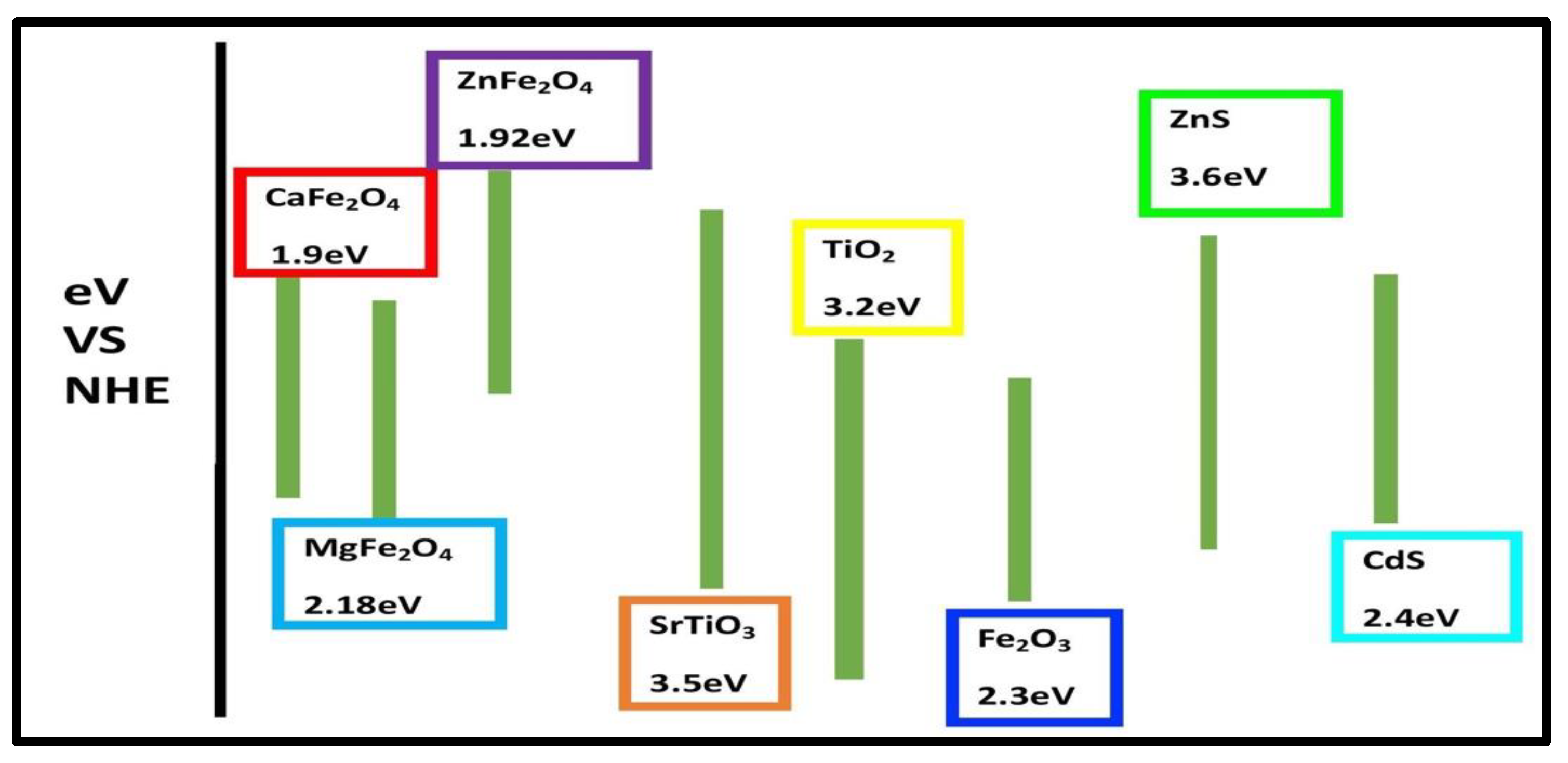
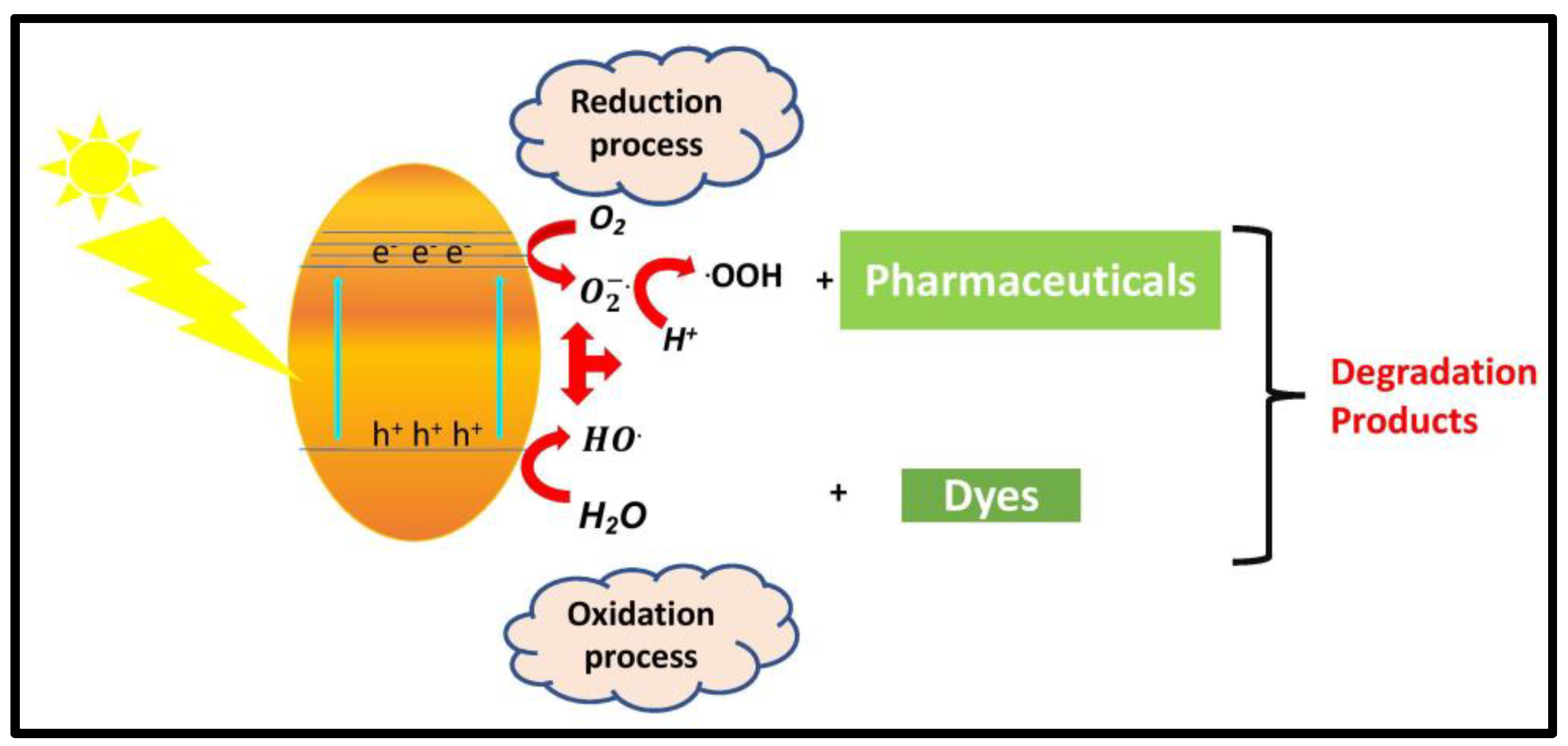
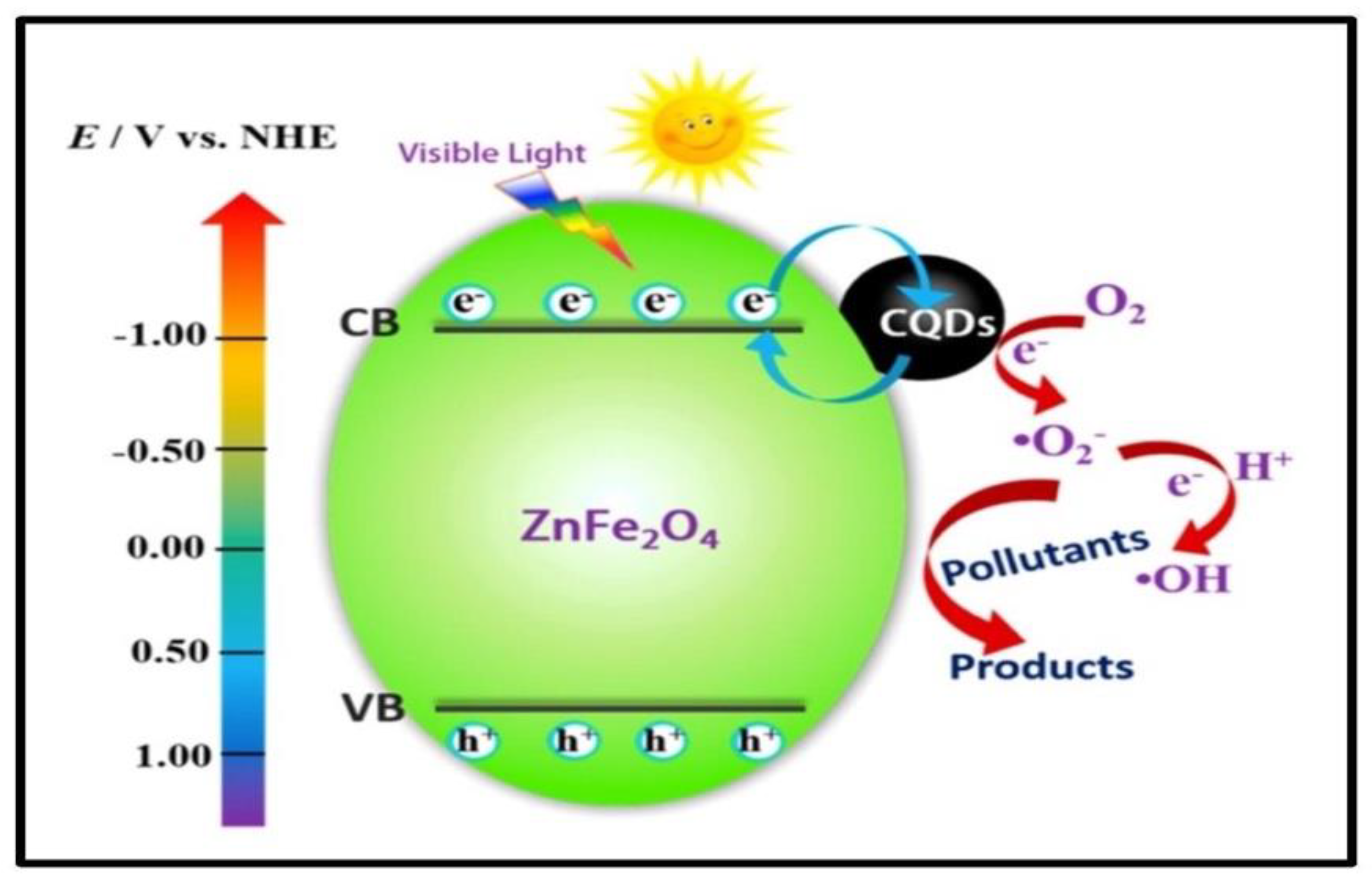
3.1. Photocatalytic Applications for Degradation of Organic Compounds

Extreme pollution of water bodies is a severe risk to both human beings and other life forms. Water contaminated with organic compounds such as pharmaceuticals, dyes, heavy-metal ions, and pesticides has received scientific attention because of its dangerous constituents, and it is necessary to treat to secure environmental safety [192,193]. Of several AOPs, semiconductor-based photocatalysis is a successful and low-cost technique for the treatment of water contaminants and microbes. In photocatalysis, clean and inexhaustible solar energy is converted into useful chemical forms, and all photocatalytic reactions advances through relatively green conditions [194,195,196,197,198]. In this method, complete degradation of organic compounds to H2O and CO2 occurs, and no secondary pollutants are generated.
The influence of doping of Ni/Co and Eu on Fe3O4/TiO2 magnetic core–shell nanocomposites were investigated by degradation of amlodipine [199] and rhodamine B [200] under ultraviolet light irradiation, respectively. It was observed that factors playing a necessary role in decomposition of amlodipine are weight percentage, number of dopants, calcination time, calcination temperature and pH of the amlodipine. Jiang et al. [81] reported that the MS value of TiO2-coated Fe3O4 (14 emu/g) is much lower than that of bare Fe3O4 (63 emu/g). The decrease in saturation magnetization is mainly due to the non-magnetic TiO2 content in the nanocomposites. and has strong photocatalytic activity for photodegradation of rhodamine B solution. Titania-coated magnetite (Fe3O4/TiO2) nanocubes showed excellent efficiency for degradation of methylene blue, and this efficiency was further promoted by addition of hydrogen peroxide within only 5 min of reaction time in the absence of UV irradiation. Even after recycling the sample six times, the introduced catalyst was found to retain as much as 90% initial efficiency [201]. Xu et al. [74] studied the degradation of methylene blue and methyl orange using visible light-active TiO2/ZnFe2O4 photocatalysts. The light-absorbing semiconductor ZnFe2O4 and the wide-bandgap titanium oxide created a linked semiconductor system in the TiO2/ZnFe2O4 photocatalyst, resulting in efficient primary charge separation and photocatalytic activity under visible light irradiation. The influence of calcination temperature and ZnFe2O4 quantity on TiO2/ZnFe2O4 photocatalytic activity was also studied. Golshana et al. [202] reported TiO2@CuFe2O4 as photocatalyst for decomposition of 2,4-dichlorophenoxyacetic acid (2,4-D). Over 97.2% of 2,4-D was degraded within 60 min at 0.3 mM PMS and 0.1 g/L. Difference in energy levels of CuFe2O4 and TiO2 prevents electron–hole recombination, thus improving photocatalytic activity (Figure 15).
The photocatalytic activity of NiFe2O4@TiO2/Pt nanocomposites was demonstrated by degradation of methyl orange and acid brown in an aqueous solution under irradiation with UV light. Maximum wavelengths of methyl orange and acid brown were degraded about 45% and 65% in 90 min under ultraviolet light and in the presence of magnetic photocatalyst [203]. Aziz et al. [87] used a nanocomposite of TiO2 with SrFe12O19 as photocatalyst for degradation of a recalcitrant phenolic compound (2,4-dichlorophenol) under illumination of daylight. Under stronger sunlight illuminance, 2,4-DCP (50 mg/L) was degraded completely in 120 min (supported photocatalyst) and 180 min (unsupported TiO2). Under diffused sunlight, 100% decomposition of 2,4-DCP was accomplished in 240 min for the supported TiO2 and 90% decomposition in 300 min for the unsupported TiO2.
Haw et al. [85] found that in CoFe2O4/TiO2 nanocatalysts, the presence of Co2+ and Fe3+ ions in oxide form on the surface of TiO2 led to visible light absorption in the wavelength range 550–650 nm. The photocatalytic degradation of reactive red 120 was studied by varying its concentration and amount of nanocatalyst in order to attain maximum degradation. The role of electron acceptors in photocatalytic degradation of RR 120 was studied in the presence of a magnetic nanocatalyst. In comparison to commercial rutile phase TiO2 and pure urchin-like TiO2 microparticles, the CoFe2O4-3D TiO2 nanocomposites demonstrated improved photodegradation of methylene blue due to a decreased recombination rate of photoexcited charge carriers [204]. Bavarsiha et al. [205] and Chen et al. [206] studied the degradation of methylene blue by SrFe12O19/SiO2/TiO2 and TiO2/SiO2/Ni–Cu–Zn ferrite composites, respectively. Photodegradation of methylene blue was 80% in the presence of SrFe12O19/SiO2/TiO2 at irradiation time of 180 min and 83.9% in the presence of TiO2/SiO2/Ni–Cu–Zn ferrite composite after 6 h Xe arc lamp irradiation. ZnFe2O4/TiO2 heterojunction was used for the removal of bisphenol A. The close heterojunction interface between p-type ZnFe2O4 and n-type TiO2 can promote charge transfer more easily, resulting in lower e–h+ pair recombination efficiency and higher visible light-responsive photocatalytic activity in ZnFe2O4 heterostructure [93]. Figure 16 shows the mechanism of photodegradation of bisphenol A by ZnFe2O4/TiO2 heterojunction. Path 1 represents the excitation of e from the VB of ZnFe2O4 to CB to produce an e–h+ pair. Path 2 represents the transfer of excited e in ZnFe2O4 across the interface of nanocomposites to the CB of anatase TiO2, and leave holes in the VB of ZnFe2O4. It reduces the recombination rate of holes and electrons. Path 3 indicates that photogenerated holes in VB of ZnFe2O4 can decompose bisphenol A under visible light irradiation, and e in the VB of TiO2 can react with O2 to generate O 2 . and · OOH radicals (Path 4 and 5). The formed radicals can react with electrons and protons to form · OH (hydroxyl radicals) (Path 6). Coromelci et al. [207] developed TiO2/ZnFe2O4 and TiO2/ZnFe1.98La0.02O4 heterostructures for the degradation of methylene blue. Photocatalytic degradation of 97%, 91% and 70% was achieved using TiO2/ZnFe1.98La0.02O4 under UV light, sunlight, and visible light.
Photocatalytic property of CoFe2O4–carbon quantum dot nanocomposites under ultraviolet irradiation was evaluated by degradation of three azo dyes (acid black 24, acid brown 14 and acid red 1). It was observed that the degradation of acid brown 14, acid black 24 and acid red 1 was 90%, 95%, and 60% at 90, 60, and 120 min, respectively [41]. Yakob et al. [108] developed CoFe2O4 and CoFe2O4/SiO2 and evaluated its photocatalytic activity against methylene blue. The CoFe2O4/SiO2 nanocomposites showed a lower optical bandgap than bare CoFe2O4 due to a smaller surface defect and greater particle size. Because of the silica coating on CoFe2O4, CoFe2O4/SiO2 had higher photocatalytic activity than CoFe2O4. Methylene blue photodegradation rate reached 80.6%.
A novel CQDs/ZnFe2O4 composite showed a strong transient photocurrent response, which was much higher than ZnFe2O4, thus indicating better transfer and separation efficiency of photogenerated electron–hole pairs. During the photocatalysis process, CQDs serve as an electron reservoir, a transporter, and a strong energy transfer component. An ESR study found that reactive species contributing towards removal of NO were O 2 . and · OH [128]. The photocatalytic activities of pure BiFeO3, CQDs and CQDs/BiFeO3 composite were investigated against rhodamine B. Due to formation of a heterojunction at the interface between CQD and BiFeO3 and electron-accepting properties of CQDs, the pure BiFeO3 had the poorest photocatalytic activit compared to CQDs and CQDs/BiFeO3. The sample with 3.3 wt % of CQDs in CQDs/BiFeO3 composite had the highest photocatalytic activity, and as a content of CQDs exceeded 3.3 wt %, the photocatalytic activity decreased because too much CQDs covered the surface of BiFeO3 and reduced the adsorption of O2 [129]. Mesoporous silica nanoparticles loaded with different amounts of Fe2O3 nanoparticles synthesized by the sol–gel method was used for the degradation of methylene blue and Congo red dye. Best results were obtained with 20% weight loading of hematite nanoparticles, indicating less agglomeration and availability of more catalytic sites [208]. Titanium-substituted SrFe2O4 and MgFe2O4 nanoparticles were used for the removal of nitroaromatic compounds and rhodamine B dye [209,210]. Sr0.4Ti0.6Fe2O4.6 @ SiO2 nanocomposites removed 96% of pendimethalin [211].
The mechanism of rhodamine B degradation by CQDs/BiFeO3 heterojunction is shown below:
CQDs / BiFeO 3 + h ν     e CQDs + h +   BiFeO 3
e + O 2     O 2 .
h + + OH     · OH
O 2 . + H 2 O     · OOH
· OOH + H 2 O     H 2 O 2   + · OH
H 2 O 2     2   · OH
· OH + RhB     CO 2 + H 2 O

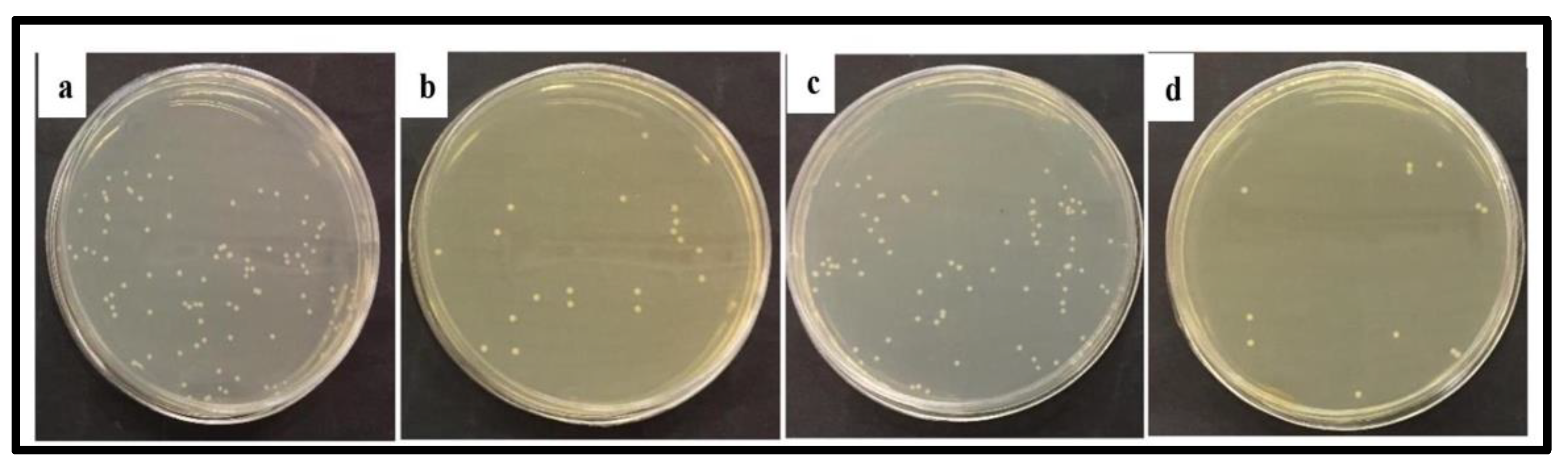
3.2. Photocatalytic Applications for Degradation of Microbes

Bacterial infectious illnesses have long posed a severe threat to human health, and treating them has always been a risk for medical personnel. Antimicrobial resistance (AMR) has emerged as a serious threat to the successful treatment of bacterial illnesses in the last 70 years or more as a result of widespread antibiotic usage. Misuse of antibiotics results in increase and spread of resistant pathogens. Antibiotic-resistant illnesses affect more than 2.8 million individuals in the United States each year, resulting in around 35,000 fatalities [212]. It is projected that by 2050, AMR will cause 10 million deaths per year, costing $100 trillion [213]. MSNs are ideally suited for targeted drug delivery due to their flexible and desired features, such as high drug loading capacity, adjustable pore size and volume, ease of functionalization, and biocompatibility. Silica nanoparticles have a lot of silanol groups (Si-OH) on their surfaces, which can be easily controlled as a surface probe attachment site.
The antibacterial behavior of NiFe2O4@TiO2/Pt nanocomposites was evaluated using degradation of E. coli bacteria. It was found that nanocomposites had a feasible magnetic feature for easy separation and effective photocatalytic properties. As time increased, more bacteria were degraded on the catalyst [203]. Atacan et al. [92] demonstrated the antibacterial activity of ZnFe2O4/Ag-TiO2 nanocomposite against Gram-positive bacteria (S. aureus) and Gram-negative bacteria (E. coli). The degradation rate of S. aureus was higher than E. coli. The inhibition diameter of 15 ± 0.2 mm for ZnFe2O4/Ag-TiO2 nanocomposite was also measured, since the antibacterial activity increased with nanocomposite formation.
To create photokilling agents, Chen et al. [214] utilized two unique properties of titania: its photocatalytic activity and its capacity to self-assemble dopamine onto its surface. The Fe3O4@TiO2 nanocomposites had photocatalytic activity as well as the ability to target bacteria, in addition to their magnetic characteristics. The IgG–Fe3O4@TiO2 nanocomposite was shown to be efficient at inhibiting the cell development of various harmful bacteria when exposed to UV radiation. The photokilling capabilities of synthesized nanocomposites were evaluated using antibiotic-resistant bacterial strains, such as multiantibiotic-resistant S. pyogenes, methicillin-resistant Staphylococcus aureus (MRSA), Staphylococcus saprophyticus, and Streptococcus pyogenes. The antibacterial activity of γ-Fe2O3@SiO2@TiO2–Ag nanocomposites was investigated by Cui et al. [215]. It was demonstrated that the introduction of silver nanoparticles into the TiO2 matrix facilitates charge separation by trapping photogenerated electrons, thereby enhancing biological activity and photoactivity.
The antibacterial effect of NiFe2O4–carbon quantum dots nanocomposite was investigated by degradation of Pseudomonas aeruginosa bacteria. It was found that as time increases, more bacteria were degraded on nanocomposites [127]. Yang et al. [216] found that CQD adsorption causes bacterial cell surface destruction, since the CQDs may change the charge balance of the bacterial surface and can insert into the bacterial surface via the long alkyl chains, which ultimately leads to the inactivation of Gram-positive bacteria. Moradlou et al. [217] (Figure 17) grew thin films of CQD-incorporated hematite (CQDs@ α -Fe2O3) and nanostructured hematite ( α -Fe2O3) on titanium substrate. Under dark and light circumstances, the antibacterial activity of the produced sample was tested against Gram-negative (E. coli) and Gram-positive (S. aureus) bacteria. It was observed that antimicrobial activity of the samples was less in the dark than under light illumination. The antimicrobial investigation revealed that α -Fe2O3 and CQDs@ α -Fe2O3 were toxic to the chosen microbe, and that samples (Ti/ α -Fe2O3, Ti/CQDs@ α -Fe2O3) have higher antibacterial activity against S. aureus than E. coli, owing to the existence of an additional layer in the outer membrane of E. coli. The mechanism of bactericidal activity was the entrance of iron cation into the bacterial cell via its membrane and the formation of reactive oxygen species.
A silver-deposited silanized cobalt ferrite composite (CoFe2O4/SiO2/Ag) was produced and impregnated with streptomycin antibiotic by Kooti et al. [218]. Disk diffusion was used to investigate the antibacterial activity of CoFe2O4/SiO2/Ag alone and in combination with streptomycin against Gram-positive and Gram-negative bacteria. The bactericidal effect of this compound was found to be increased when combined with streptomycin based on the diameter of inhibition zones. It was demonstrated that the bactericidal activity of CoFe2O4/SiO2/Ag in combination with streptomycin against all test stains was higher than each of CoFe2O4/SiO2/Ag and streptomycin applied alone. The DIZ values for streptomycin-impregnated CoFe2O4/SiO2/Ag composite were 19, 17, 15, and 14 mm for B. subtilis, E. coli, S. aureus, and Pseudomonas aeruginosa, respectively, using a composite concentration of 10 mg/mL−1. A summary of applications of nanocomposites of ferrites with TiO2, silica and CQDs for removal of pollutants is given in Table 3 and Table 4.

4. Challenges and Opportunities

Exploring synthetic ways for low-cost large-scale production is essential for the wide use of nanotechnology. The large-scale production of these nanocomposites with uniform and high quality is still a challenging task. There is still a lot of work to be done for synthesis and optimizing the materials for a wide range of applications, and it is critical to assess the toxicity and environmental issues of nanocomposites. Nanocomposites exhibit distinct qualities, such as a high surface area-to-volume ratio, a significant number of reactive sites available to enable the dispersal of organic pollutants on the surface and pores, tininess, and high recovery capacity. High surface area-to-volume ratio property of nanocomposites controls the interactions with bacteria and pollutants. These characteristics provide new opportunities for numerous applications in photocatalysis. Comparative studies on change in the cocatalyst viz. TiO2, SiO2, and CQDs on the photocatalytic activity of ferrites needs to be addressed.

5. Conclusions

Nanocomposites of ferrites with TiO2, SiO2, and CQDs are presently being used for environment protection. In this review, methods of preparation, such as coprecipitation, sol–gel, hydrothermal, solvothermal, and Stöber, structural features and photocatalytic activity of nanocomposites of ferrites with TiO2, SiO2, and CQDs for removal of various toxicants such as dyes, heavy-metal ions, pharmaceuticals, and microbes have been discussed. Mechanisms associated with their applications have also been addressed. Such nanocomposites have become a subject of great scientific interest due to their physiochemical properties, such as thermal stability, tunable shape and size, chemical stability, excellent magnetic characteristics, cost-effectiveness and large surface area. These nanocomposites possess more photocatalytic activity than individual moieties. Among various nanocomposites, nanocomposites of ferrites with CQDs show more photocatalytic activity due to their excellent sunlight-harvesting ability, up-conversion photoluminescence, tunable photoluminescence (PL), and effective promotion of electron–hole separation rate.

Author Contributions

Writing-original draft preparation, A.K. and M.K.; Writing-review and editing, A.K., M.K., P.V. and V.S. All authors have read and agreed to the published version of the manuscript.

Funding

This research received no external funding.

Institutional Review Board Statement

Not applicable.

Informed Consent Statement

Not applicable.

Data Availability Statement

Not applicable.

Conflicts of Interest

The authors declare no conflict of interest.

References

  1. World Health Organization. Drinking Water. 2019. Available online: https://www.who.int/news-room/fact-sheets/detail/drinking-water (accessed on 27 March 2019).
  2. Asfaram, A.; Ghaedi, M.; Goudarzi, A.; Hajati, S. Ultrasound-Assisted Binary Adsorption of Dyes onto Mn@ CuS/ZnS-NC-AC as a Novel Adsorbent: Application of Chemometrics for Optimization and Modeling. J. Ind. Eng. Chem. 2017, 54, 377–388. [Google Scholar] [CrossRef]
  3. Chung, K.-T. Azo Dyes and Human Health: A Review. J. Environ. Sci. Health C 2016, 34, 233–261. [Google Scholar] [CrossRef] [PubMed]
  4. Zhou, Y.; Lu, J.; Zhou, Y.; Liu, Y. Recent Advances for Dyes Removal Using Novel Adsorbents: A Review. Environ. Pollut. 2019, 252, 352–365. [Google Scholar] [CrossRef] [PubMed]
  5. Ojemaye, M.O.; Okoh, A.I. Multiple Nitrogen Functionalized Magnetic Nanoparticles as an Efficient Adsorbent: Synthesis, Kinetics, Isotherm and Thermodynamic Studies for the Removal of Rhodamine B from Aqueous Solution. Sci. Rep. 2019, 9, 9672. [Google Scholar] [CrossRef] [PubMed]
  6. Mazaheri, H.; Ghaedi, M.; Ahmadi Azqhandi, M.H.; Asfaram, A. Application of Machine/Statistical Learning, Artificial Intelligence and Statistical Experimental Design for the Modeling and Optimization of Methylene Blue and Cd(ii) Removal from a Binary Aqueous Solution by Natural Walnut Carbon. Phys. Chem. Chem. Phys. 2017, 19, 11299–11317. [Google Scholar] [CrossRef] [PubMed]
  7. Gerbaldo, M.V.; Marchetti, S.G.; Elías, V.R.; Mendieta, S.N.; Crivello, M.E. Degradation of Anti-Inflammatory Drug Diclofenac Using Cobalt Ferrite as Photocatalyst. Chem. Eng. Res. Des. 2021, 166, 237–247. [Google Scholar] [CrossRef]
  8. Tabasum, A.; Alghuthaymi, M.; Qazi, U.Y.; Shahid, I.; Abbas, Q.; Javaid, R.; Nadeem, N.; Zahid, M. UV-Accelerated Photocatalytic Degradation of Pesticide over Magnetite and Cobalt Ferrite Decorated Graphene Oxide Composite. Plants 2020, 10, 6. [Google Scholar] [CrossRef]
  9. Chatterjee, S.; Guha, N.; Krishnan, S.; Singh, A.K.; Mathur, P.; Rai, D.K. Selective and Recyclable Congo Red Dye Adsorption by Spherical Fe3O4 Nanoparticles Functionalized with 1,2,4,5-Benzenetetracarboxylic Acid. Sci. Rep. 2020, 10, 111. [Google Scholar] [CrossRef]
  10. Anantha, M.S.; Olivera, S.; Hu, C.; Jayanna, B.K.; Reddy, N.; Venkatesh, K.; Muralidhara, H.B.; Naidu, R. Comparison of the Photocatalytic, Adsorption and Electrochemical Methods for the Removal of Cationic Dyes from Aqueous Solutions. Environ. Technol. Innov. 2020, 17, 100612. [Google Scholar] [CrossRef]
  11. Bilińska, L.; Gmurek, M.; Ledakowicz, S. Textile Wastewater Treatment by AOPs for Brine Reuse. Process. Saf. Environ. Prot. 2017, 109, 420–428. [Google Scholar] [CrossRef]
  12. Boutra, B.; Güy, N.; Özacar, M.; Trari, M. Magnetically Separable MnFe2O4/TA/ZnO Nanocomposites for Photocatalytic Degradation of Congo Red under Visible Light. J. Magn. Magn. Mater. 2020, 497, 165994. [Google Scholar] [CrossRef]
  13. Ali, T.; Tripathi, P.; Azam, A.; Raza, W.; Ahmed, A.S.; Ahmed, A.; Muneer, M. Photocatalytic Performance of Fe-Doped TiO2 Nanoparticles under Visible-Light Irradiation. Mater. Res. Express 2017, 4, 015022. [Google Scholar] [CrossRef]
  14. Bora, L.V.; Mewada, R.K. Visible/Solar Light Active Photocatalysts for Organic Effluent Treatment: Fundamentals, Mechanisms and Parametric Review. Renew. Sustain. Energy Rev. 2017, 76, 1393–1421. [Google Scholar] [CrossRef]
  15. Fujishima, A.; Honda, K. Electrochemical Photolysis of Water at a Semiconductor Electrode. Nature 1972, 238, 37–38. [Google Scholar] [CrossRef] [PubMed]
  16. Jeng, M.-J.; Wung, Y.-L.; Chang, L.-B.; Chow, L. Particle Size Effects of TiO2 Layers on the Solar Efficiency of Dye-Sensitized Solar Cells. Int. J. Photoenergy 2013, 2013, 1–9. [Google Scholar] [CrossRef]
  17. Thao, L.; Dang, T.; Khanitchaidecha, W.; Channei, D.; Nakaruk, A. Photocatalytic Degradation of Organic Dye under UV-A Irradiation Using TiO2-Vetiver Multifunctional Nano Particles. Materials 2017, 10, 122. [Google Scholar] [CrossRef]
  18. Saliu, O.D.; Olatunji, G.A.; Yakubu, A.; Arowona, M.T.; Mohammed, A.A. Catalytic Crosslinking of a Regenerated Hydrophobic Benzylated Cellulose and Nano TiO2 Composite for Enhanced Oil Absorbency. e-Polymers 2017, 17, 295–302. [Google Scholar] [CrossRef]
  19. Li, H.; Zhang, T.; Pan, C.; Pu, C.; Hu, Y.; Hu, X.; Liu, E.; Fan, J. Self-Assembled Bi2MoO6/TiO2 Nanofiber Heterojunction Film with Enhanced Photocatalytic Activities. Appl. Surf. Sci. 2017, 391, 303–310. [Google Scholar] [CrossRef]
  20. Wang, Y.; Sunarso, J.; Zhao, B.; Ge, C.; Chen, G. One-Dimensional BiOBr Nanosheets/TiO2 Nanofibers Composite: Controllable Synthesis and Enhanced Visible Photocatalytic Activity. Ceram. Int. 2017, 43, 15769–15776. [Google Scholar] [CrossRef]
  21. Cheng, R.; Fan, X.; Wang, M.; Li, M.; Tian, J.; Zhang, L. Facile Construction of CuFe2O4/g-C3N4 Photocatalyst for Enhanced Visible-Light Hydrogen Evolution. RSC Adv. 2016, 6, 18990–18995. [Google Scholar] [CrossRef]
  22. Kefeni, K.K.; Mamba, B.B.; Msagati, T.A.M. Application of Spinel Ferrite Nanoparticles in Water and Wastewater Treatment: A Review. Sep. Purif. Technol. 2017, 188, 399–422. [Google Scholar] [CrossRef]
  23. Choi, H.; Lee, S.; Kouh, T.; Kim, S.J.; Kim, C.S.; Hahn, E. Synthesis and Characterization of Co-Zn Ferrite Nanoparticles for Application to Magnetic Hyperthermia. J. Korean Phys. Soc. 2017, 70, 89–92. [Google Scholar] [CrossRef]
  24. Zhang, L.; Wu, Y. Sol-Gel Synthesized Magnetic MnFe2O4 Spinel Ferrite Nanoparticles as Novel Catalyst for Oxidative Degradation of Methyl Orange. J. Nanomater. 2013, 2013, 1–6. [Google Scholar] [CrossRef]
  25. Waag, F.; Gökce, B.; Kalapu, C.; Bendt, G.; Salamon, S.; Landers, J.; Hagemann, U.; Heidelmann, M.; Schulz, S.; Wende, H.; et al. Adjusting the Catalytic Properties of Cobalt Ferrite Nanoparticles by Pulsed Laser Fragmentation in Water with Defined Energy Dose. Sci. Rep. 2017, 7, 13161. [Google Scholar] [CrossRef] [PubMed]
  26. Qu, Q.; Zhu, A.; Shao, X.; Shi, G.; Tian, Y. Development of a Carbon Quantum Dots-Based Fluorescent Cu2+ Probe Suitable for Living Cell Imaging. Chem. Commun. 2012, 48, 5473. [Google Scholar] [CrossRef]
  27. Mirtchev, P.; Henderson, E.J.; Soheilnia, N.; Yip, C.M.; Ozin, G.A. Solution Phase Synthesis of Carbon Quantum Dots as Sensitizers for Nanocrystalline TiO2 Solar Cells. J. Mater. Chem. 2012, 22, 1265–1269. [Google Scholar] [CrossRef]
  28. Li, H.; Kang, Z.; Liu, Y.; Lee, S.-T. Carbon Nanodots: Synthesis, Properties and Applications. J. Mater. Chem. 2012, 22, 24230. [Google Scholar] [CrossRef]
  29. Di, J.; Xia, J.; Ge, Y.; Li, H.; Ji, H.; Xu, H.; Zhang, Q.; Li, H.; Li, M. Novel Visible-Light-Driven CQDs/Bi2WO6 Hybrid Materials with Enhanced Photocatalytic Activity toward Organic Pollutants Degradation and Mechanism Insight. Appl. Catal. B Environ. 2015, 168–169, 51–61. [Google Scholar] [CrossRef]
  30. Mamba, G.; Mishra, A. Advances in Magnetically Separable Photocatalysts: Smart, Recyclable Materials for Water Pollution Mitigation. Catalysts 2016, 6, 79. [Google Scholar] [CrossRef]
  31. Govan, J.; Gun’ko, Y. Recent Advances in the Application of Magnetic Nanoparticles as a Support for Homogeneous Catalysts. Nanomaterials 2014, 4, 222–241. [Google Scholar] [CrossRef]
  32. Rodriguez-Arco, L.; Rodriguez, I.A.; Carriel, V.; Bonhome-Espinosa, A.B.; Campos, F.; Kuzhir, P.; Duran, J.D.G.; Lopez-Lopez, M.T. Biocompatible Magnetic Core–Shell Nanocomposites for Engineered Magnetic Tissues. Nanoscale 2016, 8, 8138–8150. [Google Scholar] [CrossRef] [PubMed]
  33. Hakami, T.M.; Davarpanah, A.M.; Rahdar, A.; Barrett, S.D. Structural and Magnetic Study and Cytotoxicity Evaluation of Tetra-Metallic Nanoparticles of Co0.5Ni0.5CrxFe2-XO4 Prepared by Co-Precipitation. J. Mol. Struct. 2018, 1165, 344–348. [Google Scholar] [CrossRef]
  34. Wang, Y.; Sun, X.; Yi, Z.; Wu, X.; Liu, G.; Pu, Z.; Yang, H. Construction of a Z-Scheme Ag 2 MoO4/BiOBr Heterojunction for Photocatalytically Removing Organic Pollutants. Dalton Trans. 2022, 51, 18652–18666. [Google Scholar] [CrossRef] [PubMed]
  35. Ghamkhari, A.; Mohamadi, L.; Kazemzadeh, S.; Zafar, M.N.; Rahdar, A.; Khaksefidi, R. Synthesis and Characterization of Poly(Styrene-Block-Acrylic Acid) Diblock Copolymer Modified Magnetite Nanocomposite for Efficient Removal of Penicillin G. Compos. B: Eng. 2020, 182, 107643. [Google Scholar] [CrossRef]
  36. Mohammadi, L.; Rahdar, A.; Khaksefidi, R.; Ghamkhari, A.; Fytianos, G.; Kyzas, G.Z. Polystyrene Magnetic Nanocomposites as Antibiotic Adsorbents. Polymers 2020, 12, 1313. [Google Scholar] [CrossRef]
  37. Ahmadi, S.; Mohammadi, L.; Rahdar, A.; Rahdar, S.; Dehghani, R.; Adaobi Igwegbe, C.; Kyzas, G.Z. Acid Dye Removal from Aqueous Solution by Using Neodymium(III) Oxide Nanoadsorbents. Nanomaterials 2020, 10, 556. [Google Scholar] [CrossRef]
  38. Rahdar, S.; Rahdar, A.; Sattari, M.; Hafshejani, L.D.; Tolkou, A.K.; Kyzas, G.Z. Barium/Cobalt@Polyethylene Glycol Nanocomposites for Dye Removal from Aqueous Solutions. Polymers 2021, 13, 1161. [Google Scholar] [CrossRef]
  39. Mohafez, F.S.; Davarpanah, A.M.; Rahdar, A.; Beyzaei, H.; Zeybek, O.; Barrett, S.D. Structural, Magnetic, and in Vitro Inhibitory Characteristics of Ce-Substituted MnFe2O4 Nanoparticles. Appl. Phys. A 2021, 127, 600. [Google Scholar] [CrossRef]
  40. Davarpanah, A.M.; Rahdar, A.; Dastnae, M.A.; Zeybek, O.; Beyzaei, H. (1−x)BaFe12O19/xCoFe2O4 Hard/Soft Magnetic Nanocomposites: Synthesis, Physical Characterization, and Antibacterial Activities Study. J. Mol. Struct. 2019, 1175, 445–449. [Google Scholar] [CrossRef]
  41. Ahmadian-Fard-Fini, S.; Salavati-Niasari, M.; Safardoust-Hojaghan, H. Hydrothermal Green Synthesis and Photocatalytic Activity of Magnetic CoFe2O4–Carbon Quantum Dots Nanocomposite by Turmeric Precursor. J. Mater. Sci. Mater. Electron. 2017, 28, 16205–16214. [Google Scholar] [CrossRef]
  42. Kefeni, K.K.; Mamba, B.B. Photocatalytic Application of Spinel Ferrite Nanoparticles and Nanocomposites in Wastewater Treatment: Review. Sustain. Mater. Technol. 2020, 23, e00140. [Google Scholar] [CrossRef]
  43. Ghasemi, A. Magnetic Ferrites and Related Nanocomposites, 1st ed.; Elsevier: Amsterdam, The Netherlands, 2021; p. 300. [Google Scholar]
  44. Katal, R.; Masudy-Panah, S.; Tanhaei, M.; Farahani, M.H.D.A.; Jiangyong, H. A Review on the Synthesis of the Various Types of Anatase TiO2 Facets and Their Applications for Photocatalysis. Chem. Eng. J. 2020, 384, 123384. [Google Scholar] [CrossRef]
  45. Kim, C.-S.; Shin, J.-W.; Cho, Y.-H.; Jang, H.-D.; Byun, H.-S.; Kim, T.-O. Synthesis and Characterization of Cu/N-Doped Mesoporous TiO2 Visible Light Photocatalysts. Appl. Catal. A Gen. 2013, 455, 211–218. [Google Scholar] [CrossRef]
  46. Min, K.S.; Kumar, R.S.; Lee, J.H.; Kim, K.S.; Lee, S.G.; Son, Y.-A. Synthesis of New TiO2/Porphyrin-Based Composites and Photocatalytic Studies on Methylene Blue Degradation. Dyes Pigm. 2019, 160, 37–47. [Google Scholar] [CrossRef]
  47. Min, K.S.; Manivannan, R.; Son, Y.-A. Porphyrin Dye/TiO2 Imbedded PET to Improve Visible-Light Photocatalytic Activity and Organosilicon Attachment to Enrich Hydrophobicity to Attain an Efficient Self-Cleaning Material. Dyes Pigm. 2019, 162, 8–17. [Google Scholar] [CrossRef]
  48. Wu, Q.; Zhang, Z. The Preparation of Self-Floating Sm/N Co-Doped TiO2/Diatomite Hybrid Pellet with Enhanced Visible-Light-Responsive Photoactivity and Reusability. Adv. Powder Technol. 2019, 30, 415–422. [Google Scholar] [CrossRef]
  49. Wang, G.; Ma, X.; Wei, S.; Li, S.; Qiao, J.; Wang, J.; Song, Y. Highly Efficient Visible-Light Driven Photocatalytic Hydrogen Production from a Novel Z-Scheme Er3+:YAlO3/Ta2O5-V5+||Fe3+-TiO2/Au Coated Composite. J. Power Sources 2018, 373, 161–171. [Google Scholar] [CrossRef]
  50. Lee, H.U.; Lee, S.C.; Choi, S.; Son, B.; Lee, S.M.; Kim, H.J.; Lee, J. Efficient Visible-Light Induced Photocatalysis on Nanoporous Nitrogen-Doped Titanium Dioxide Catalysts. Chem. Eng. J. 2013, 228, 756–764. [Google Scholar] [CrossRef]
  51. Krasil’Nikov, V.N.; Shalaeva, E.V.; Baklanova, I.V.; Melkozerova, M.A.; Kuznetsov, M.V.; Zabolotskaya, E.V.; Gyrdasova, O.I.; Buldakova, L.Y.; Murzakaev, A.M. Synthesis, Structure and Spectroscopic Characteristics of Ti(O,C)2/Carbon Nanostructured Globules with Visible Light Photocatalytic Activity. Bull. Mater. Sci. 2016, 39, 1569–1579. [Google Scholar] [CrossRef]
  52. Ju, T.; Lee, H.; Kang, M. The Photovoltaic Efficiency of Dye Sensitized Solar Cell Assembled Using Carbon Capsulated TiO2 Electrode. J. Ind. Eng. Chem. 2014, 20, 2636–2640. [Google Scholar] [CrossRef]
  53. Chen, P.; Wang, F.; Chen, Z.-F.; Zhang, Q.; Su, Y.; Shen, L.; Yao, K.; Liu, Y.; Cai, Z.; Lv, W.; et al. Study on the Photocatalytic Mechanism and Detoxicity of Gemfibrozil by a Sunlight-Driven TiO2/Carbon Dots Photocatalyst: The Significant Roles of Reactive Oxygen Species. Appl. Catal. B Environ. 2017, 204, 250–259. [Google Scholar] [CrossRef]
  54. Surenjan, A.; Sambandam, B.; Pradeep, T.; Philip, L. Synthesis, Characterization and Performance of Visible Light Active C-TiO2 for Pharmaceutical Photodegradation. J. Environ. Chem. Eng. 2017, 5, 757–767. [Google Scholar] [CrossRef]
  55. Li, X.-W.; Yuan, Y.; Huang, X.; Lin, K.-F. Visible-Infrared Optical Properties Measurement of the Mesoporous TiO2/Carbon Materials. Int. J. Hydrog. Energy 2016, 41, 15638–15645. [Google Scholar] [CrossRef]
  56. Wanag, A.; Kusiak-Nejman, E.; Kapica-Kozar, J.; Morawski, A.W. Photocatalytic Performance of Thermally Prepared TiO2/C Photocatalysts under Artificial Solar Light. Micro Nano Lett. 2016, 11, 202–206. [Google Scholar] [CrossRef]
  57. Neville, E.M.; Ziegler, J.; Don MacElroy, J.M.; Ravindranathan Thampi, K.; Sullivan, J.A. Serendipity Following Attempts to Prepare C-Doped Rutile TiO2. Appl. Catal. A Gen. 2014, 470, 434–441. [Google Scholar] [CrossRef]
  58. Teh, C.Y.; Wu, T.Y.; Juan, J.C. An Application of Ultrasound Technology in Synthesis of Titania-Based Photocatalyst for Degrading Pollutant. Chem. Eng. J. 2017, 317, 586–612. [Google Scholar] [CrossRef]
  59. Reli, M.; Kobielusz, M.; Matějová, L.; Daniš, S.; Macyk, W.; Obalová, L.; Kuśtrowski, P.; Rokicińska, A.; Kočí, K. TiO2 Processed by Pressurized Hot Solvents as a Novel Photocatalyst for Photocatalytic Reduction of Carbon Dioxide. Appl. Surf. Sci. 2017, 391, 282–287. [Google Scholar] [CrossRef]
  60. Rahimi, N.; Pax, R.A.; Gray, E.; Mac, A. Review of Functional Titanium Oxides. I: TiO2 and Its Modifications. Prog. Solid State Chem. 2016, 44, 86–105. [Google Scholar] [CrossRef]
  61. Mikrut, P.; Kobielusz, M.; Macyk, W. Spectroelectrochemical Characterization of Euhedral Anatase TiO2 Crystals—Implications for Photoelectrochemical and Photocatalytic Properties of {001} {100} and {101} Facets. Electrochim. Acta 2019, 310, 256–265. [Google Scholar] [CrossRef]
  62. Surówka, M.; Kobielusz, M.; Trochowski, M.; Buchalska, M.; Kruczała, K.; Broś, P.; Macyk, W. Iron and Other Metal Species as Phase-Composition Controllers Influencing the Photocatalytic Activity of TiO2 Materials. Appl. Catal. B Environ. 2019, 247, 173–181. [Google Scholar] [CrossRef]
  63. Sadowski, R.; Wach, A.; Buchalska, M.; Kuśtrowski, P.; Macyk, W. Photosensitized TiO2 Films on Polymers—Titania-Polymer Interactions and Visible Light Induced Photoactivity. Appl. Surf. Sci. 2019, 475, 710–719. [Google Scholar] [CrossRef]
  64. Karthik, K.; Vijayalakshmi, S.; Phuruangrat, A.; Revathi, V.; Verma, U. Multifunctional Applications of Microwave-Assisted Biogenic TiO2 Nanoparticles. J. Clust. Sci. 2019, 30, 965–972. [Google Scholar] [CrossRef]
  65. Mahdavi-Shakib, A.; Husremovic, S.; Ki, S.; Glynn, J.; Babb, L.; Sempel, J.; Stavrinoudis, I.; Arce-Ramos, J.-M.; Nelson, R.; Grabow, L.C.; et al. Titania Surface Chemistry and Its Influence on Supported Metal Catalysts. Polyhedron 2019, 170, 41–50. [Google Scholar] [CrossRef]
  66. Fatima, R.; Afridi, M.N.; Kumar, V.; Lee, J.; Ali, I.; Kim, K.-H.; Kim, J.-O. Photocatalytic Degradation Performance of Various Types of Modified TiO2 against Nitrophenols in Aqueous Systems. J. Clean. Prod. 2019, 231, 899–912. [Google Scholar] [CrossRef]
  67. Boro, B.; Gogoi, B.; Rajbongshi, B.M.; Ramchiary, A. Nano-Structured TiO2/ZnO Nanocomposite for Dye-Sensitized Solar Cells Application: A Review. Renew. Sustain. Energy Rev. 2018, 81, 2264–2270. [Google Scholar] [CrossRef]
  68. Akhter, P.; Farkhondehfal, M.A.; Hernández, S.; Hussain, M.; Fina, A.; Saracco, G.; Khan, A.U.; Russo, N. Environmental Issues Regarding CO2 and Recent Strategies for Alternative Fuels through Photocatalytic Reduction with Titania-Based Materials. J. Environ. Chem. Eng. 2016, 4, 3934–3953. [Google Scholar] [CrossRef]
  69. Ashraf, S.; Siddiqa, A.; Shahida, S.; Qaisar, S. Titanium-Based Nanocomposite Materials for Arsenic Removal from Water: A Review. Heliyon 2019, 5, e01577. [Google Scholar] [CrossRef]
  70. Etacheri, V.; Michlits, G.; Seery, M.K.; Hinder, S.J.; Pillai, S.C. A Highly Efficient TiO2–xCx Nano-Heterojunction Photocatalyst for Visible Light Induced Antibacterial Applications. ACS Appl. Mater. Interfaces 2013, 5, 1663–1672. [Google Scholar] [CrossRef]
  71. Di Valentin, C.; Pacchioni, G. Trends in Non-Metal Doping of Anatase TiO2: B, C, N and F. Catal. Today 2013, 206, 12–18. [Google Scholar] [CrossRef]
  72. Tatarchuk, T.; Mironyuk, I.; Kotsyubynsky, V.; Shyichuk, A.; Myslin, M.; Boychuk, V. Structure, Morphology and Adsorption Properties of Titania Shell Immobilized onto Cobalt Ferrite Nanoparticle Core. J. Mol. Liq. 2020, 297, 111757. [Google Scholar] [CrossRef]
  73. Dadfar, M.R.; Seyyed Ebrahimi, S.A.; Masoudpanah, S.M. Sol–Gel Synthesis and Characterization of SrFe12O19/TiO2 Nanocomposites. J. Supercond. Nov. Magn. 2015, 28, 89–94. [Google Scholar] [CrossRef]
  74. Xu, S.; Feng, D.; Shangguan, W. Preparations and Photocatalytic Properties of Visible-Light-Active Zinc Ferrite-Doped TiO2 Photocatalyst. J. Phys. Chem. C 2009, 113, 2463–2467. [Google Scholar] [CrossRef]
  75. Lahijani, B.; Hedayati, K.; Goodarzi, M. Magnetic PbFe12O19-TiO2 Nanocomposites and Their Photocatalytic Performance in the Removal of Toxic Pollutants. Main Group Met. Chem. 2018, 41, 53–62. [Google Scholar] [CrossRef]
  76. De Lucena, P.R.; Pessoa-Neto, O.D.; Dos Santos, I.M.G.; Souza, A.G.; Longo, E.; Varela, J.A. Synthesis by the Polymeric Precursor Method and Characterization of Undoped and Sn, Cr and V-Doped ZrTiO. J. Alloys Compd. 2005, 397, 255–259. [Google Scholar] [CrossRef]
  77. Mourão, H.A.J.L.; Malagutti, A.R.; Ribeiro, C. Synthesis of TiO2-Coated CoFe2O4 Photocatalysts Applied to the Photodegradation of Atrazine and Rhodamine B in Water. Appl. Catal. A Gen. 2010, 382, 284–292. [Google Scholar] [CrossRef]
  78. Mohanty, P.; Mahapatra, R.; Padhi, P.; Ramana, C.V.V.; Mishra, D.K. Ultrasonic Cavitation: An Approach to Synthesize Uniformly Dispersed Metal Matrix Nanocomposites—A Review. Nano Struct. Nano Objects 2020, 23, 100475. [Google Scholar] [CrossRef]
  79. Ley, S.V.; Low, C.M.R. Ultrasound in Synthesis; Springer: Berlin/Heidelberg, Germany, 1989; p. 133. [Google Scholar]
  80. Ao, Y.; Xu, J.; Fu, D.; Shen, X.; Yuan, C. A Novel Magnetically Separable Composite Photocatalyst: Titania-Coated Magnetic Activated Carbon. Sep. Purif. Technol. 2008, 61, 436–441. [Google Scholar] [CrossRef]
  81. Jiang, W.; Zhang, X.; Gong, X.; Yan, F.; Zhang, Z. Sonochemical Synthesis and Characterization of Magnetic Separable Fe3O4-TiO2 Nanocomposites and Their Catalytic Properties. Int. J. Smart Nano Mater. 2010, 1, 278–287. [Google Scholar] [CrossRef]
  82. Rane, A.V.; Kanny, K.; Abitha, V.K.; Thomas, S. Synthesis of Inorganic Nanomaterials; Bhagyaraj, S.M., Oluwafemi, O.S., Kalarikkal, N., Thomas, S., Eds.; Elsevier: Amsterdam, The Netherlands, 2018; pp. 121–139. [Google Scholar]
  83. Sathishkumar, P.; Mangalaraja, R.V.; Anandan, S.; Ashokkumar, M. CoFe2O4/TiO2 Nanocatalysts for the Photocatalytic Degradation of Reactive Red 120 in Aqueous Solutions in the Presence and Absence of Electron Acceptors. Chem. Eng. J. 2013, 220, 302–310. [Google Scholar] [CrossRef]
  84. Chandrika, M.; Ravindra, A.V.; Rajesh, C.; Ramarao, S.D.; Ju, S. Studies on Structural and Optical Properties of Nano ZnFe2O4 and ZnFe2O4-TiO2 Composite Synthesized by Co-Precipitation Route. Mater. Chem. Phys. 2019, 230, 107–113. [Google Scholar] [CrossRef]
  85. Haw, C.; Chiu, W.; Abdul Rahman, S.; Khiew, P.; Radiman, S.; Abdul Shukor, R.; Hamid, M.A.A.; Ghazali, N. The Design of New Magnetic-Photocatalyst Nanocomposites (CoFe2O4–TiO2) as Smart Nanomaterials for Recyclable-Photocatalysis Applications. New J. Chem. 2016, 40, 1124–1136. [Google Scholar] [CrossRef]
  86. Allaedini, G.; Tasirin, S.M.; Aminayi, P. Magnetic Properties of Cobalt Ferrite Synthesized by Hydrothermal Method. Int. Nano Lett. 2015, 5, 183–186. [Google Scholar] [CrossRef]
  87. Aziz, A.A.; Puma, G.L.; Ibrahim, S.; Saravanan, P. Preparation, Characterisation and Solar Photoactivity of Titania Supported Strontium Ferrite Nanocomposite Photocatalyst. J. Exp. Nanosci. 2013, 8, 295–310. [Google Scholar] [CrossRef]
  88. Nguyen, T.B.; Doong, R. Fabrication of Highly Visible-Light-Responsive ZnFe2O4/TiO2 Heterostructures for the Enhanced Photocatalytic Degradation of Organic Dyes. RSC Adv. 2016, 6, 103428–103437. [Google Scholar] [CrossRef]
  89. Pongwan, P.; Inceesungvorn, B.; Phanichphant, S.; Kangwansupamonkon, W.; Wetchakun, N. Synthesis and Characterization of a Magnetically Separable CoFe2O4/TiO2 Nanocomposite for the Photomineralization of Formic Acid. Ferroelectrics 2013, 453, 133–140. [Google Scholar] [CrossRef]
  90. Sun, J.; Fu, Y.; Xiong, P.; Sun, X.; Xu, B.; Wang, X. A Magnetically Separable P25/CoFe2O4/Graphene Catalyst with Enhanced Adsorption Capacity and Visible-Light-Driven Photocatalytic Activity. RSC Adv. 2013, 3, 22490. [Google Scholar] [CrossRef]
  91. Etacheri, V.; Di Valentin, C.; Schneider, J.; Bahnemann, D.; Pillai, S.C. Visible-Light Activation of TiO2 Photocatalysts: Advances in Theory and Experiments. J. Photochem. Photobiol. C Photochem. Rev. 2015, 25, 1–29. [Google Scholar] [CrossRef]
  92. Atacan, K.; Güy, N.; Çakar, S. Preparation and Antibacterial Activity of Solvothermal Synthesized ZnFe2O4/Ag-TiO2 Nanocomposite. Sakarya Uni. J. Sci. 2018, 22, 1720–1726. [Google Scholar] [CrossRef]
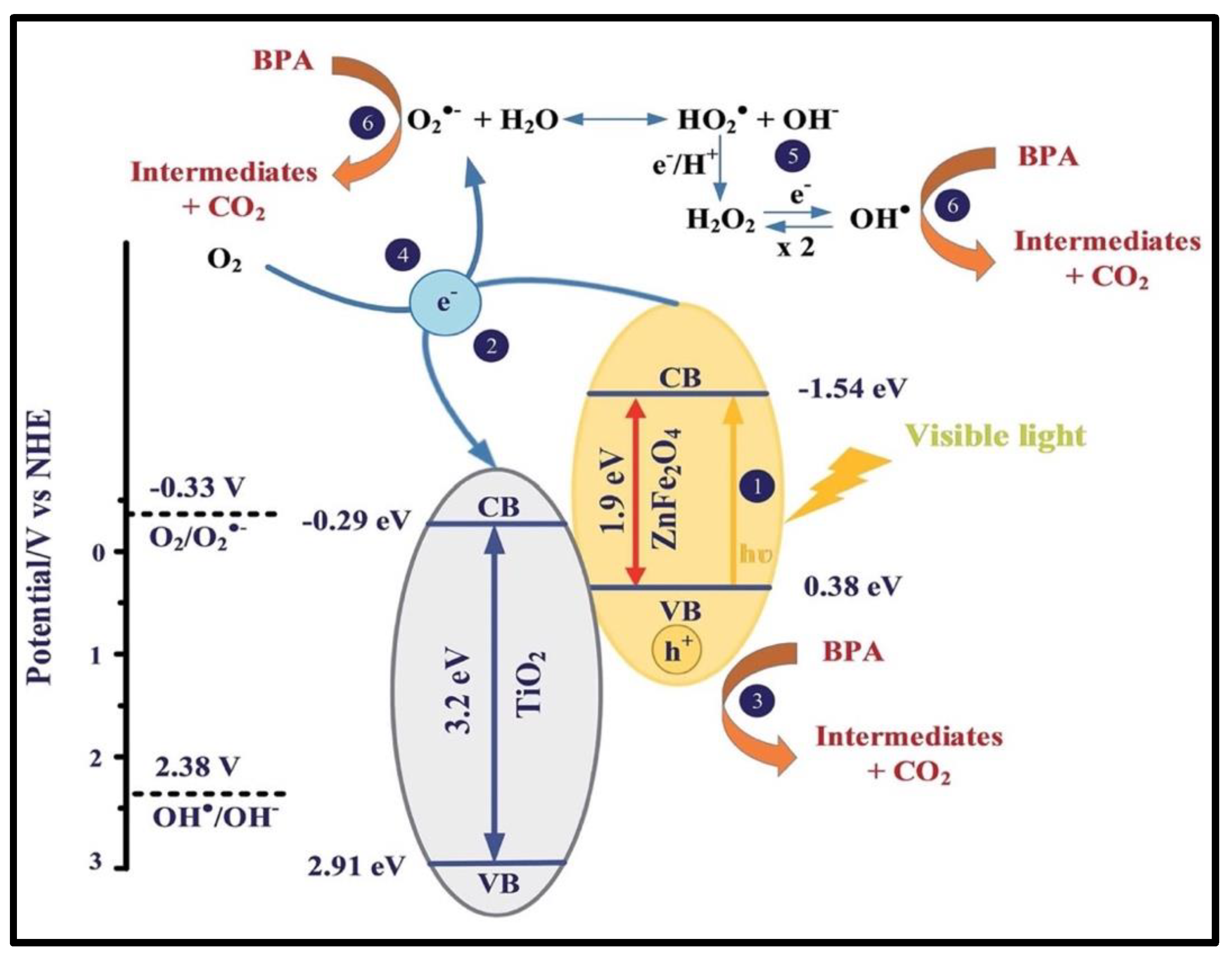
  93. Nguyen, T.B.; Doong, R. Heterostructured ZnFe2O4/TiO2 Nanocomposites with a Highly Recyclable Visible-Light-Response for Bisphenol A Degradation. RSC Adv. 2017, 7, 50006–50016. [Google Scholar] [CrossRef]
  94. Deng, Y.-H.; Wang, C.-C.; Hu, J.-H.; Yang, W.-L.; Fu, S.-K. Investigation of Formation of Silica-Coated Magnetite Nanoparticles via Sol–Gel Approach. Colloids Surf. A Physicochem. Eng. Asp. 2005, 262, 87–93. [Google Scholar] [CrossRef]
  95. Sounderya, N.; Zhang, Y. Use of Core/Shell Structured Nanoparticles for Biomedical Applications. Biomedical 2008, 1, 34–42. [Google Scholar] [CrossRef]
  96. Morales, A.E.; Mora, E.S.; Pal, U. Use of diffuse reflectance spectroscopy for optical characterization of unsupported nanostructures. Rev. Mex. Fis. 2007, 53, 18–22. [Google Scholar]
  97. Bardapurkar, P.P.; Shewale, S.S.; Barde, N.P.; Jadhav, K.M. Structural, Magnetic and Catalytical Properties of Cobalt Ferrite Nanoparticles Dispersed in Silica Matrix. Mater. Res. Express. 2019, 6, 045055. [Google Scholar] [CrossRef]
  98. Gharagozlou, M. Influence of Calcination Temperature on Structural and Magnetic Properties of Nanocomposites Formed by Co-Ferrite Dispersed in Sol-Gel Silica Matrix Using Tetrakis(2-Hydroxyethyl) Orthosilicate as Precursor. Chem. Cent. J. 2011, 5, 19. [Google Scholar] [CrossRef] [PubMed]
  99. Pansambal, S.; Ghotekar, S.; Shewale, S.; Deshmukh, K.; Barde, N.; Bardapurkar, P. Efficient Synthesis of Magnetically Separable CoFe2O4@SiO2 Nanoparticles and Its Potent Catalytic Applications for the Synthesis of 5-Aryl-1,2,4-Triazolidine-3-Thione Derivatives. J. Water Environ. Nanotechnol. 2019, 4, 174–186. [Google Scholar] [CrossRef]
  100. Dippong, T.; Levei, E.A.; Cadar, O. Preparation of CoFe2O4/SiO2 Nanocomposites at Low Temperatures Using Short Chain Diols. J. Chem. 2017, 2017, 1–11. [Google Scholar] [CrossRef]
  101. Stöber, W.; Fink, A.; Bohn, E. Controlled Growth of Monodisperse Silica Spheres in the Micron Size Range. J. Colloid Interface Sci. 1968, 26, 62–69. [Google Scholar] [CrossRef]
  102. Ehi-Eromosele, C.O.; Ita, B.I.; Iweala, E.E.J.; Ogunniran, K.O.; Adekoya, J.A.; Siyanbola, T.O. Silica Functionalized Magnesium Ferrite Nanocomposites for Potential Biomedical Applications: Preparation, Characterization and Enhanced Colloidal Stability Studies. J. Nano Res. 2016, 40, 146–157. [Google Scholar] [CrossRef]
  103. Khanna, L.; Verma, N.K. Silica/Potassium Ferrite Nanocomposite: Structural, Morphological, Magnetic, Thermal and in Vitro Cytotoxicity Analysis. Mater. Sci. Eng. B 2013, 178, 1230–1239. [Google Scholar] [CrossRef]
  104. Flood-Garibay, J.A.; Méndez-Rojas, M.A. Synthesis and Characterization of Magnetic Nanoparticles of Cobalt Ferrite Coated with Silica. Biointerface Res. Appl. Chem. 2019, 10, 4908–4913. [Google Scholar] [CrossRef]
  105. Girgis, E.; Wahsh, M.M.; Othman, A.G.; Bandhu, L.; Rao, K. Synthesis, Magnetic and Optical Properties of Core/Shell Co1−xZnx Fe2O4/SiO2 Nanoparticles. Nanoscale Res. Lett. 2011, 6, 460. [Google Scholar] [CrossRef] [PubMed]
  106. Rohilla, S.; Kumar, S.; Aghamkar, P.; Sunder, S.; Agarwal, A. Investigations on Structural and Magnetic Properties of Cobalt Ferrite/Silica Nanocomposites Prepared by the Coprecipitation Method. J. Magn. Magn. Mater. 2011, 323, 897–902. [Google Scholar] [CrossRef]
  107. Bansal, M.; Ahlawat, D.S.; Singh, A.; Kumar, V.; Rathee, S.P. Effect of Heat Treatment on the Microstructural Properties of Silica Embedded Cobalt Ferrite Nanocomposites. Nanocomposites 2020, 6, 158–164. [Google Scholar] [CrossRef]
  108. Yakob, M.; Umar, H.; Wahyuningsih, P.; Putra, R.A. Characterization of microstructural and optical CoFe2O4/SiO2 ferrite nanocomposite for photodegradation of methylene blue. AIMS Mater. Sci. 2019, 6, 45–51. [Google Scholar] [CrossRef]
  109. Sharma, S.; Rohilla, S. Synthesis of Nanocomposites of NiFe2O4/SiO2 through Coprecipitation Method and Structural Characterization Using Rietveld Refinement. In Proceedings of the AIP Conference Proceedings, Seoul, Republic of Korea, 2 November 2020; p. 110023. [Google Scholar]
  110. Kong, L.B.; Zhang, T.S.; Ma, J.; Boey, F. Progress in Synthesis of Ferroelectric Ceramic Materials via High-Energy Mechanochemical Technique. Prog. Mater. Sci. 2008, 53, 207–322. [Google Scholar] [CrossRef]
  111. Aguilar-González, M.A.; Mendoza-Suárez, G.; Padmasree, K.P. Synthesis and Characterization of Barium Ferrite–Silica Nanocomposites. Mater. Charact. 2013, 84, 175–181. [Google Scholar] [CrossRef]
  112. Scano, A.; Cabras, V.; Marongiu, F.; Peddis, D.; Pilloni, M.; Ennas, G. New Opportunities in the Preparation of Nanocomposites for Biomedical Applications: Revised Mechanosynthesis of Magnetite–Silica Nanocomposites. Mater. Res. Express 2017, 4, 025004. [Google Scholar] [CrossRef]
  113. Xu, Y.; Tang, C.-J.; Huang, H.; Sun, C.-Q.; Zhang, Y.-K.; Ye, Q.-F.; Wang, A.-J. Green Synthesis of Fluorescent Carbon Quantum Dots for Detection of Hg2+. Chin. J. Anal. Chem. 2014, 42, 1252–1258. [Google Scholar] [CrossRef]
  114. De, B.; Karak, N. A Green and Facile Approach for the Synthesis of Water Soluble Fluorescent Carbon Dots from Banana Juice. RSC Adv. 2013, 3, 8286. [Google Scholar] [CrossRef]
  115. Gao, Z.; Wang, X.; Chang, J.; Wu, D.; Wang, L.; Liu, X.; Xu, F.; Guo, Y.; Jiang, K. Fluorescent Carbon Quantum Dots, Capacitance and Catalysis Active Porous Carbon Microspheres from Beer. RSC Adv. 2015, 5, 48665–48674. [Google Scholar] [CrossRef]
  116. Wee, S.S.; Ng, Y.H.; Ng, S.M. Synthesis of Fluorescent Carbon Dots via Simple Acid Hydrolysis of Bovine Serum Albumin and Its Potential as Sensitive Sensing Probe for Lead (II) Ions. Talanta 2013, 116, 71–76. [Google Scholar] [CrossRef] [PubMed]
  117. Wang, Q.; Zhang, C.; Shen, G.; Liu, H.; Fu, H.; Cui, D. Fluorescent Carbon Dots as an Efficient SiRNA Nanocarrier for Its Interference Therapy in Gastric Cancer Cells. J. Nanobiotechnol. 2014, 12, 58. [Google Scholar] [CrossRef] [PubMed]
  118. Vitha, T.S. Quantitative Evaluation of Pytocompounds from Terminalia Chebula by High Performance Thin Layer Chromatography (Hptlc) Method and Its Antibiofilm Activity. Int. J. Pharma Bio Sci. 2017, 8, 214–224. [Google Scholar] [CrossRef]
  119. Moradi, B.; Nabiyouni, G.; Ghanbari, D. Rapid Photo-Degradation of Toxic Dye Pollutants: Green Synthesis of Mono-Disperse Fe3O4–CeO2 Nanocomposites in the Presence of Lemon Extract. J. Mater. Sci. Mater. Electron. 2018, 29, 11065–11080. [Google Scholar] [CrossRef]
  120. Barman, M.K.; Patra, A. Current Status and Prospects on Chemical Structure Driven Photoluminescence Behaviour of Carbon Dots. J. Photochem. Photobiol. C 2018, 37, 1–22. [Google Scholar] [CrossRef]
  121. Das, R.; Bandyopadhyay, R.; Pramanik, P. Carbon Quantum Dots from Natural Resource: A Review. Mater. Today Chem. 2018, 8, 96–109. [Google Scholar] [CrossRef]
  122. Ahmadian-Fard-Fini, S.; Salavati-Niasari, M.; Ghanbari, D. Hydrothermal Green Synthesis of Magnetic Fe3O4-Carbon Dots by Lemon and Grape Fruit Extracts and as a Photoluminescence Sensor for Detecting of E. Coli Bacteria. Spectrochim. Acta Mol. Biomol. Spectrosc. 2018, 203, 481–493. [Google Scholar] [CrossRef]
  123. Baragau, I.-A.; Power, N.P.; Morgan, D.J.; Heil, T.; Lobo, R.A.; Roberts, C.S.; Titirici, M.-M.; Dunn, S.; Kellici, S. Continuous Hydrothermal Flow Synthesis of Blue-Luminescent, Excitation-Independent Nitrogen-Doped Carbon Quantum Dots as Nanosensors. J. Mater. Chem. A 2020, 8, 3270–3279. [Google Scholar] [CrossRef]
  124. Wang, Y.; Hu, A. Carbon Quantum Dots: Synthesis, Properties and Applications. J. Mater. Chem. C 2014, 2, 6921. [Google Scholar] [CrossRef]
  125. Liu, J.-H.; Cao, L.; LeCroy, G.E.; Wang, P.; Meziani, M.J.; Dong, Y.; Liu, Y.; Luo, P.G.; Sun, Y.-P. Carbon “Quantum” Dots for Fluorescence Labeling of Cells. ACS Appl. Mater. Interfaces 2015, 7, 19439–19445. [Google Scholar] [CrossRef]
  126. Dong, X.; Liang, W.; Meziani, M.J.; Sun, Y.-P.; Yang, L. Carbon Dots as Potent Antimicrobial Agents. Theranostics 2020, 10, 671–686. [Google Scholar] [CrossRef]
  127. Ahmadian-Fard-Fini, S.; Ghanbari, D.; Salavati-Niasari, M. Photoluminescence Carbon Dot as a Sensor for Detecting of Pseudomonas Aeruginosa Bacteria: Hydrothermal Synthesis of Magnetic Hollow NiFe2O4-Carbon Dots Nanocomposite Material. Compos. Part B Eng. 2019, 161, 564–577. [Google Scholar] [CrossRef]
  128. Huang, Y.; Liang, Y.; Rao, Y.; Zhu, D.; Cao, J.; Shen, Z.; Ho, W.; Lee, S.C. Environment-Friendly Carbon Quantum Dots/ZnFe2O4 Photocatalysts: Characterization, Biocompatibility, and Mechanisms for NO Removal. Environ. Sci. Technol. 2017, 51, 2924–2933. [Google Scholar] [CrossRef] [PubMed]
  129. Chen, C.C.; Fan, T. Study on Carbon Quantum Dots/BiFeO3 Heterostructures and Their Enhanced Photocatalytic Activities under Visible Light Irradiation. J. Mater. Sci. Mater. Electron. 2017, 28, 10019–10027. [Google Scholar] [CrossRef]
  130. Nabiyouni, G.; Ghanbari, D. Hydrothermal Synthesis of Magnetic and Photoluminescence CuFe2O4-Carbon Dots Nanocomposite as a Sensor for Detecting of HgII Ions. J. Nanostruct. 2020, 10, 760–768. [Google Scholar] [CrossRef]
  131. Rahman, I.A.; Padavettan, V. Synthesis of Silica Nanoparticles by Sol-Gel: Size-Dependent Properties, Surface Modification, and Applications in Silica-Polymer Nanocomposites—A Review. J. Nanomater. 2012, 2012, 1–15. [Google Scholar] [CrossRef]
  132. Hagens, W.I.; Oomen, A.G.; De Jong, W.H.; Cassee, F.R.; Sips, A.J.A.M. What Do We (Need to) Know about the Kinetic Properties of Nanoparticles in the Body? J. Regul. Toxicol. Pharmacol. 2007, 49, 217–229. [Google Scholar] [CrossRef]
  133. Mathiazhagan, A.; Joseph, R. Nanotechnology—A New Prospective in Organic Coating-Review. Int. J. Chem. Eng. Appl. 2011, 2, 225–237. [Google Scholar] [CrossRef]
  134. Nalathambi, V.; Suresh, G. Contribution of Nanotechnology in The Paints and Coatings. Int. J. Chem. Eng. Res. 2014, 1, 116–186. [Google Scholar] [CrossRef]
  135. Singh, R.; Thirupathi, G. Manganese-Zinc Spinel Ferrite Nanoparticles and Ferrofluids; Singh, R., Thirupathied, G., Eds.; InTech: London, UK, 2017; pp. 140–159. [Google Scholar]
  136. Peng, Y.; Wang, Z.; Liu, W.; Zhang, H.; Zuo, W.; Tang, H.; Chen, F.; Wang, B. Size-and Shape-Dependent Peroxidase-like Catalytic Activity of MnFe2O4 Nanoparticles and Their Applications in Highly Efficient Colorimetric Detection of Target Cancer Cells. Dalton Trans. 2015, 44, 12871–12877. [Google Scholar] [CrossRef]
  137. Reddy, S.; Kumara Swamy, B.E.; Chandra, U.; Mahathesha, K.R.; Sathisha, T.V.; Jayadevappa, H. Synthesis of MgFe2O4 Nanoparticles and MgFe2O4 Nanoparticles/CPE for Electrochemical Investigation of Dopamine. Anal. Methods 2011, 3, 2792. [Google Scholar] [CrossRef]
  138. Céspedes, E.; Byrne, J.M.; Farrow, N.; Moise, S.; Coker, V.S.; Bencsik, M.; Lloyd, J.R.; Telling, N.D. Bacterially Synthesized Ferrite Nanoparticles for Magnetic Hyperthermia Applications. Nanoscale 2014, 6, 12958–12970. [Google Scholar] [CrossRef] [PubMed]
  139. Pardeshi, S.K.; Pawar, R.Y. SrFe2O4 Complex Oxide an Effective and Environmentally Benign Catalyst for Selective Oxidation of Styrene. J. Mol. Catal. A Chem. 2011, 334, 35–43. [Google Scholar] [CrossRef]
  140. Khedr, M.H.; Abdel Halim, K.S.; Soliman, N.K. Synthesis and Photocatalytic Activity of Nano-Sized Iron Oxides. Mater. Lett. 2009, 63, 598–601. [Google Scholar] [CrossRef]
  141. Surendra, B.S. Green Engineered Synthesis of Ag-Doped CuFe2O4: Characterization, Cyclic Voltammetry and Photocatalytic Studies. J. Sci. Adv. Mater. Dev. 2018, 3, 44–50. [Google Scholar] [CrossRef]
  142. Diao, Y.; Yan, Z.; Guo, M.; Wang, X. Magnetic Multi-Metal Co-Doped Magnesium Ferrite Nanoparticles: An Efficient Visible Light-Assisted Heterogeneous Fenton-like Catalyst Synthesized from Saprolite Laterite Ore. J. Hazard. Mater. 2018, 344, 829–838. [Google Scholar] [CrossRef]
  143. Han, X.; Zhang, H.; Chen, T.; Zhang, M.; Guo, M. Facile Synthesis of Metal-Doped Magnesium Ferrite from Saprolite Laterite as an Effective Heterogeneous Fenton-like Catalyst. J. Mol. Liq. 2018, 272, 43–52. [Google Scholar] [CrossRef]
  144. Feng, Y.; Liao, C.; Shih, K. Copper-Promoted Circumneutral Activation of H2O2 by Magnetic CuFe2O4 Spinel Nanoparticles: Mechanism, Stoichiometric Efficiency, and Pathway of Degrading Sulfanilamide. Chemosphere 2016, 154, 573–582. [Google Scholar] [CrossRef]
  145. Wang, G.; Zhao, D.; Kou, F.; Ouyang, Q.; Chen, J.; Fang, Z. Removal of Norfloxacin by Surface Fenton System (MnFe2O4/H2O2): Kinetics, Mechanism and Degradation Pathway. Chem. Eng. J. 2018, 351, 747–755. [Google Scholar] [CrossRef]
  146. Guo, X.; Wang, D. Photo-Fenton Degradation of Methylene Blue by Synergistic Action of Oxalic Acid and Hydrogen Peroxide with NiFe2O4 Hollow Nanospheres Catalyst. J. Environ. Chem. Eng. 2019, 7, 102814. [Google Scholar] [CrossRef]
  147. Moreno-Castilla, C.; López-Ramón, M.V.; Fontecha-Cámara, M.Á.; Álvarez, M.A.; Mateus, L. Removal of Phenolic Compounds from Water Using Copper Ferrite Nanosphere Composites as Fenton Catalysts. Nanomaterials 2019, 9, 901. [Google Scholar] [CrossRef] [PubMed]
  148. Yao, H.; Fan, M.; Wang, Y.; Luo, G.; Fei, W. Magnetic Titanium Dioxide Based Nanomaterials: Synthesis, Characteristics, and Photocatalytic Application in Pollutant Degradation. J. Mater. Chem. A 2015, 3, 17511–17524. [Google Scholar] [CrossRef]
  149. Schneider, J.; Matsuoka, M.; Takeuchi, M.; Zhang, J.; Horiuchi, Y.; Anpo, M.; Bahnemann, D.W. Understanding TiO2 Photocatalysis: Mechanisms and Materials. Chem. Rev. 2014, 114, 9919–9986. [Google Scholar] [CrossRef] [PubMed]
  150. Yeganeh, M.; Shahtahmasebi, N.; Kompany, A.; Karimipour, M.; Razavi, F.; Nasralla, N.H.S.; Šiller, L. The Magnetic Characterization of Fe Doped TiO2 Semiconducting Oxide Nanoparticles Synthesized by Sol–Gel Method. Physical. B Condens. Matter 2017, 511, 89–98. [Google Scholar] [CrossRef]
  151. Lin, Z.; Xu, Y.; Zhen, Z.; Fu, Y.; Liu, Y.; Li, W.; Luo, C.; Ding, A.; Zhang, D. Application and Reactivation of Magnetic Nanoparticles in Microcystis Aeruginosa Harvesting. Bioresour. Technol. 2015, 190, 82–88. [Google Scholar] [CrossRef]
  152. Wang, X.; Zhao, X.; Li, H.; Jia, J.; Liu, Y.; Ejenavi, O.; Ding, A.; Sun, Y.; Zhang, D. Separating and Characterizing Functional Alkane Degraders from Crude-Oil-Contaminated Sites via Magnetic Nanoparticle-Mediated Isolation. Res. Microbiol. 2016, 167, 731–744. [Google Scholar] [CrossRef]
  153. Zhang, P.; Mo, Z.; Han, L.; Zhu, X.; Wang, B.; Zhang, C. Preparation and Photocatalytic Performance of Magnetic TiO2/Montmorillonite/Fe3O4 Nanocomposites. Ind. Eng. Chem. Res. 2014, 53, 8057–8061. [Google Scholar] [CrossRef]
  154. Chen, Z.; Ma, Y.; Geng, B.; Wang, M.; Sun, X. Photocatalytic Performance and Magnetic Separation of TiO2-Functionalized γ-Fe2O3, Fe, and Fe/Fe2O3 Magnetic Particles. J. Alloys Compd. 2017, 700, 113–121. [Google Scholar] [CrossRef]
  155. Chen, C.-C.; Jaihindh, D.; Hu, S.-H.; Fu, Y.-P. Magnetic Recyclable Photocatalysts of Ni-Cu-Zn Ferrite@SiO2@TiO2@Ag and Their Photocatalytic Activities. J. Photochem. Photobiol. A Chem. 2017, 334, 74–85. [Google Scholar] [CrossRef]
  156. Moreira, F.C.; Boaventura, R.A.R.; Brillas, E.; Vilar, V.J.P. Electrochemical Advanced Oxidation Processes: A Review on Their Application to Synthetic and Real Wastewaters. Appl. Catal. B Environ. 2017, 202, 217–261. [Google Scholar] [CrossRef]
  157. Boczkaj, G.; Fernandes, A. Wastewater Treatment by Means of Advanced Oxidation Processes at Basic PH Conditions: A Review. Chem. Eng. J. 2017, 320, 608–633. [Google Scholar] [CrossRef]
  158. Dewil, R.; Mantzavinos, D.; Poulios, I.; Rodrigo, M.A. New Perspectives for Advanced Oxidation Processes. J. Environ. Manag. 2017, 195, 93–99. [Google Scholar] [CrossRef] [PubMed]
  159. Suzuki, H.; Araki, S.; Yamamoto, H. Evaluation of Advanced Oxidation Processes (AOP) Using O3, UV, and TiO2 for the Degradation of Phenol in Water. J. Water Process. Eng. 2015, 7, 54–60. [Google Scholar] [CrossRef]
  160. Moellmann, J.; Ehrlich, S.; Tonner, R.; Grimme, S. A DFT-D Study of Structural and Energetic Properties of TiO2 Modifications. J. Phys. Condens. Matter. 2012, 24, 424206. [Google Scholar] [CrossRef] [PubMed]
  161. Scanlon, D.O.; Dunnill, C.W.; Buckeridge, J.; Shevlin, S.A.; Logsdail, A.J.; Woodley, S.M.; Catlow, C.R.A.; Powell Michael, J.; Palgrave, R.G.; Parkin, I.P.; et al. Band Alignment of Rutile and Anatase TiO2. Nat. Mater. 2013, 12, 798–801. [Google Scholar] [CrossRef]
  162. Graça, M.P.F.; Nico, C.; Peres, M.; Valente, M.A.; Monteiro, T. Study of the Optical and Dielectric Properties of TiO2 Nanocrystals Prepared by the Pechini Method. J. Nanosci. Nanotechnol. 2012, 12, 8600–8606. [Google Scholar] [CrossRef]
  163. Habila, M.A.; ALOthman, Z.A.; El-Toni, A.M.; Labis, J.P.; Soylak, M. Synthesis and Application of Fe3O4@SiO2@TiO2 for Photocatalytic Decomposition of Organic Matrix Simultaneously with Magnetic Solid Phase Extraction of Heavy Metals Prior to ICP-MS Analysis. Talanta 2016, 154, 539–547. [Google Scholar] [CrossRef]
  164. Wilson, M.; Cheng, C.Y.C.; Oswald, G.; Srivastava, R.; Beaumont, S.K.; Badyal, J.P.S. Magnetic Recyclable Microcomposite Silica-Steel Core with TiO2 Nanocomposite Shell Photocatalysts for Sustainable Water Purification. Colloids Surf. A Physicochem. Eng. Asp. 2017, 523, 27–37. [Google Scholar] [CrossRef]
  165. Chung, I.-M.; Park, I.; Seung-Hyun, K.; Thiruvengadam, M.; Rajakumar, G. Plant-Mediated Synthesis of Silver Nanoparticles: Their Characteristic Properties and Therapeutic Applications. Nanoscale Res. Lett. 2016, 11, 40. [Google Scholar] [CrossRef]
  166. Ravikumar, P.; Kumar, S.S. Antifungal activity of extracellularly synthesized silver nanoparticles from Morinda citrifolia. Int. J. Tech. Res. Appl. 2014, 2, 108–111. [Google Scholar]
  167. Inbathamizh, L.; Ponnu, T.M.; Mary, E.J. In Vitro Evaluation of Antioxidant and Anticancer Potential of Morinda Pubescens Synthesized Silver Nanoparticles. J. Pharm. Res. 2013, 6, 32–38. [Google Scholar] [CrossRef]
  168. He, Z.; Cai, Q.; Fang, H.; Situ, G.; Qiu, J.; Song, S.; Chen, J. Photocatalytic Activity of TiO2 Containing Anatase Nanoparticles and Rutile Nanoflower Structure Consisting of Nanorods. J. Environ. Sci. 2013, 25, 2460–2468. [Google Scholar] [CrossRef] [PubMed]
  169. Hooshmand, S.; Kargozar, S.; Ghorbani, A.; Darroudi, M.; Keshavarz, M.; Baino, F.; Kim, H.-W. Biomedical Waste Management by Using Nanophotocatalysts: The Need for New Options. Materials 2020, 13, 3511. [Google Scholar] [CrossRef] [PubMed]
  170. Vimbela, G.V.; Ngo, S.M.; Fraze, C.; Yang, L.; Stout, D.A. Antibacterial properties and toxicity from metallic nanomaterials. Int. J. Nanomed. 2017, 12, 3941–3965. [Google Scholar] [CrossRef] [PubMed]
  171. Verdier, T.; Coutand, M.; Bertron, A.; Roques, C. Antibacterial Activity of TiO2 Photocatalyst Alone or in Coatings on E. Coli: The Influence of Methodological Aspects. Coatings 2014, 4, 670–686. [Google Scholar] [CrossRef]
  172. Xie, J.; Hung, Y.-C. Methodology to Evaluate the Antimicrobial Effectiveness of UV-Activated TiO2 Nanoparticle-Embedded Cellulose Acetate Film. Food Control 2019, 106, 106690. [Google Scholar] [CrossRef]
  173. Reddy, A.K.; Kambalyal, P.B.; Shanmugasundaram, K.; Rajesh, V.; Donthula, S.; Patil, S.R. Comparative Evaluation of Antimicrobial Efficacy of Silver, Titanium Dioxide and Zinc Oxide Nanoparticles against Streptococcus Mutans. Pesqui. Bras. Odontopediatria Clín. Integr. 2018, 18, e4150. [Google Scholar] [CrossRef]
  174. Long, M.; Wang, J.; Zhuang, H.; Zhang, Y.; Wu, H.; Zhang, J. Performance and Mechanism of Standard Nano-TiO2 (P-25) in Photocatalytic Disinfection of Foodborne Microorganisms—Salmonella Typhimurium and Listeria Monocytogenes. Food Control 2014, 39, 68–74. [Google Scholar] [CrossRef]
  175. Altın, İ.; Sökmen, M. Preparation of TiO2-Polystyrene Photocatalyst from Waste Material and Its Usability for Removal of Various Pollutants. Appl. Catal. B Environ. 2014, 144, 694–701. [Google Scholar] [CrossRef]
  176. Bonetta, S.; Bonetta, S.; Motta, F.; Strini, A.; Carraro, E. Photocatalytic Bacterial Inactivation by TiO2-Coated Surfaces. AMB Expr. 2013, 3, 59. [Google Scholar] [CrossRef]
  177. Yao, N.; Lun Yeung, K. Investigation of the Performance of TiO2 Photocatalytic Coatings. Chem. Eng. J. 2011, 167, 13–21. [Google Scholar] [CrossRef] [PubMed]
  178. Luo, Z.S.; Ye, Q.Y.; Li, D.D. Influence of nano-TiO2 modified LDPE film packaging on quality of strawberry. Mod. Food Sci. Technol. 2013, 29, 2340–2344. [Google Scholar]
  179. Gumiero, M.; Peressini, D.; Pizzariello, A.; Sensidoni, A.; Iacumin, L.; Comi, G.; Toniolo, R. Effect of TiO2 Photocatalytic Activity in a HDPE-Based Food Packaging on the Structural and Microbiological Stability of a Short-Ripened Cheese. Food Chem. 2013, 138, 1633–1640. [Google Scholar] [CrossRef] [PubMed]
  180. Wang, J.; Li, C.; Zhuang, H.; Zhang, J. Photocatalytic Degradation of Methylene Blue and Inactivation of Gram-Negative Bacteria by TiO2 Nanoparticles in Aqueous Suspension. Food Control 2013, 34, 372–377. [Google Scholar] [CrossRef]
  181. Yue, Q.; Sun, J.; Kang, Y.; Deng, Y. Advances in the Interfacial Assembly of Mesoporous Silica on Magnetite Particles. Angew. Chem. Int. Ed. 2020, 59, 15804–15817. [Google Scholar] [CrossRef] [PubMed]
  182. Liang, J.; Liang, Z.; Zou, R.; Zhao, Y. Heterogeneous Catalysis in Zeolites, Mesoporous Silica, and Metal-Organic Frameworks. Adv. Mater. 2017, 29, 1701139. [Google Scholar] [CrossRef] [PubMed]
  183. Fujiwara, K.; Kuwahara, Y.; Sumida, Y.; Yamashita, H. Fabrication of Photocatalytic Paper Using TiO2 Nanoparticles Confined in Hollow Silica Capsules. Langmuir 2017, 33, 288–295. [Google Scholar] [CrossRef]
  184. Wang, D.; Han, D.; Shi, Z.; Wang, J.; Yang, J.; Li, X.; Song, H. Optimized Design of Three-Dimensional Multi-Shell Fe3O4/SiO2/ZnO/ZnSe Microspheres with Type II Heterostructure for Photocatalytic Applications. Appl. Catal. B Environ. 2018, 227, 61–69. [Google Scholar] [CrossRef]
  185. Tang, X.; Feng, Q.; Liu, K.; Luo, X.; Huang, J.; Li, Z. A simple and innovative route to remarkably enhance the photocatalytic performance of TiO2: Using micro-meso porous silica nanofibers as carrier to support highly-dispersed TiO2 nanoparticles. Micropor. Mesopor. Mater. 2018, 258, 251–261. [Google Scholar] [CrossRef]
  186. Mohanty, S.; Babu, P.; Parida, K.; Naik, B. Surface-Plasmon-Resonance-Induced Photocatalysis by Core–Shell SiO2@Ag NCs@Ag3PO4 toward Water-Splitting and Phenol Oxidation Reactions. Inorg. Chem. 2019, 58, 9643–9654. [Google Scholar] [CrossRef]
  187. Singh, R.; Bapat, R.; Qin, L.; Feng, H.; Polshettiwar, V. Atomic Layer Deposited (ALD) TiO2 on Fibrous Nano-Silica (KCC-1) for Photocatalysis: Nanoparticle Formation and Size Quantization Effect. ACS Catal. 2016, 6, 2770–2784. [Google Scholar] [CrossRef]
  188. Yin, Q.; Wu, W.; Qiao, R.; Ke, X.; Hu, Y.; Li, Z. Glucose-Assisted Transformation of Ni-Doped-ZnO@carbon to a Ni-Doped-ZnO@void@SiO2 Core–Shell Nanocomposite Photocatalyst. RSC Adv. 2016, 6, 38653–38661. [Google Scholar] [CrossRef]
  189. Zhang, X.; Huang, H.; Liu, J.; Liu, Y.; Kang, Z. Carbon Quantum Dots Serving as Spectral Converters through Broadband Upconversion of Near-Infrared Photons for Photoelectrochemical Hydrogen Generation. J. Mater. Chem. A 2013, 1, 11529. [Google Scholar] [CrossRef]
  190. Rajendiran, K.; Zhao, Z.; Pei, D.-S.; Fu, A. Antimicrobial Activity and Mechanism of Functionalized Quantum Dots. Polymers 2019, 11, 1670. [Google Scholar] [CrossRef]
  191. Moradlou, O.; Rabiei, Z.; Banazadeh, A.; Warzywoda, J.; Zirak, M. Carbon Quantum Dots as Nano-Scaffolds for α-Fe2O3 Growth: Preparation of Ti/CQD@α-Fe2O3 Photoanode for Water Splitting under Visible Light Irradiation. Appl. Catal. B Environ. 2018, 227, 178–189. [Google Scholar] [CrossRef]
  192. Lops, C.; Ancona, A.; Di Cesare, K.; Dumontel, B.; Garino, N.; Canavese, G.; Hérnandez, S.; Cauda, V. Sonophotocatalytic Degradation Mechanisms of Rhodamine B Dye via Radicals Generation by Micro-and Nano-Particles of ZnO. Appl. Catal. B Environ. 2019, 243, 629–640. [Google Scholar] [CrossRef]
  193. Madhav, S.; Ahamad, A.; Singh, P.; Mishra, P.K. A Review of Textile Industry: Wet Processing, Environmental Impacts, and Effluent Treatment Methods. Environ. Qual. Manag. 2018, 27, 31–41. [Google Scholar] [CrossRef]
  194. Wang, S.; Xu, M.; Peng, T.; Zhang, C.; Li, T.; Hussain, I.; Wang, J.; Tan, B. Porous Hypercrosslinked Polymer-TiO2-Graphene Composite Photocatalysts for Visible-Light-Driven CO2 Conversion. Nat. Commun. 2019, 10, 676. [Google Scholar] [CrossRef]
  195. Tang, Y.; Zhou, P.; Wang, K.; Lin, F.; Lai, J.; Chao, Y.; Li, H.; Guo, S. BiOCl/Ultrathin Polyaniline Core/Shell Nanosheets with a Sensitization Mechanism for Efficient Visible-Light-Driven Photocatalysis. Sci. China Mater. 2019, 62, 95–102. [Google Scholar] [CrossRef]
  196. Fu, J.; Yu, J.; Jiang, C.; Cheng, B. G-C3N4-Based Heterostructured Photocatalysts. Adv. Energy Mater. 2018, 8, 1701503. [Google Scholar] [CrossRef]
  197. Zhang, X.; Wei, W.; Zhang, S.; Wen, B.; Su, Z. Advanced 3D Nanohybrid Foam Based on Graphene Oxide: Facile Fabrication Strategy, Interfacial Synergetic Mechanism, and Excellent Photocatalytic Performance. Sci. China Mater. 2019, 62, 1888–1897. [Google Scholar] [CrossRef]
  198. Chen, S.; Yu, J.; Zhang, J. Enhanced Photocatalytic CO2 Reduction Activity of MOF-Derived ZnO/NiO Porous Hollow Spheres. J. CO2 Util. 2018, 24, 548–554. [Google Scholar] [CrossRef]
  199. Teimouri, M.; Husain, S.W.; Saber-Tehrani, M.; Aberoomand-Azar, P. Preparation of Novel Ni/Co Co-Doping Fe3O4/TiO2 Core–Shell Nanocomposites and Their Use in Effective Photocatalytic Degradation of Amlodipine Drug. Sep. Sci. Technol. 2019, 54, 634–641. [Google Scholar] [CrossRef]
  200. Stefan, M.; Leostean, C.; Pana, O.; Toloman, D.; Popa, A.; Perhaita, I.; Senilă, M.; Marincas, O.; Barbu-Tudoran, L. Magnetic Recoverable Fe3O4-TiO2:Eu Composite Nanoparticles with Enhanced Photocatalytic Activity. Appl. Surf. Sci. 2016, 390, 248–259. [Google Scholar] [CrossRef]
  201. Abbas, M.; Rao, B.P.; Reddy, V.; Kim, C. Fe3O4/TiO2 Core/Shell Nanocubes: Single-Batch Surfactantless Synthesis, Characterization and Efficient Catalysts for Methylene Blue Degradation. Ceram. Int. 2014, 40, 11177–11186. [Google Scholar] [CrossRef]
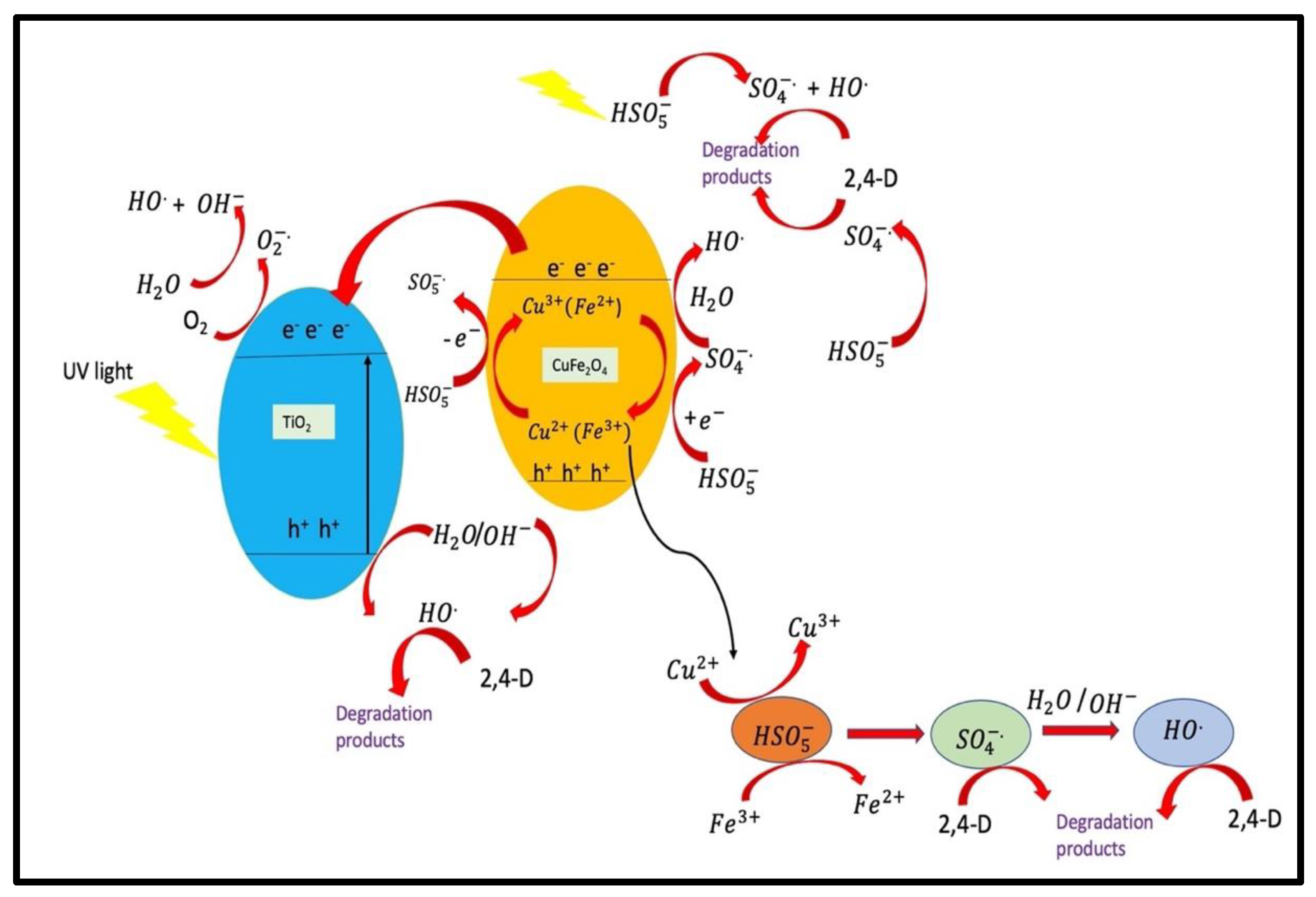
  202. Golshan, M.; Kakavandi, B.; Ahmadi, M.; Azizi, M. Photocatalytic Activation of Peroxymonosulfate by TiO2 Anchored on Cupper Ferrite (TiO2@CuFe2O4) into 2,4-D Degradation: Process Feasibility, Mechanism and Pathway. J. Hazard. Mater. 2018, 359, 325–337. [Google Scholar] [CrossRef]
  203. Dung, N.T. Preparation of Magnetic Antibacterial Composite Beads Fe3O4/Alginate/Ag. Vietnam. J. Sci. Technol. 2018, 56, 192. [Google Scholar] [CrossRef]
  204. Moosavi, S.M.; Molla-Abbasi, P.; Haji-Aghajani, Z. Photo-Catalyst CoFe2O4–TiO2: Application in Photo-Degradation of Organic Dyes and Magnetic Nanocomposite Preparation. J. Mater. Sci. Mater. Electron. 2016, 27, 4879–4886. [Google Scholar] [CrossRef]
  205. Bavarsiha, F.; Rajabi, M.; Montazeri-Pour, M. Synthesis of SrFe12O19/SiO2/TiO2 Composites with Core/Shell/Shell Nano-Structure and Evaluation of Their Photo-Catalytic Efficiency for Degradation of Methylene Blue. J. Mater. Sci. Mater. Electron. 2018, 29, 1877–1887. [Google Scholar] [CrossRef]
  206. Chen, C.-C.; Fu, Y.-P.; Hu, S.-H. Characterizations of TiO2/SiO2/Ni-Cu-Zn Ferrite Composite for Magnetic Photocatalysts. J. Am. Ceram. Soc. 2015, 98, 2803–2811. [Google Scholar] [CrossRef]
  207. Coromelci, C.; Neamtu, M.; Ignat, M.; Samoila, P.; Zaltariov, M.F.; Palamaru, M. Ultrasound Assisted Synthesis of Heterostructured TiO2/ZnFe2O4 and TiO2/ZnFe1.98La0.02O4 Systems as Tunable Photocatalysts for Efficient Organic Pollutants Removal. Ceram. Int. 2022, 48, 4829–4840. [Google Scholar] [CrossRef]
  208. Mandal, S.; Adhikari, S.; Pu, S.; Wang, X.; Kim, D.-H.; Patel, R.K. Interactive Fe2O3/Porous SiO2 Nanospheres for Photocatalytic Degradation of Organic Pollutants: Kinetic and Mechanistic Approach. Chemosphere 2019, 234, 596–607. [Google Scholar] [CrossRef] [PubMed]
  209. Grewal, J.K.; Kaur, M.; Sharma, R.K.; Oliveira, A.C.; Garg, V.K.; Sharma, V.K. Structural and Photocatalytic Studies on Oxygen Hyperstoichiometric Titanium-Substituted Strontium Ferrite Nanoparticles. Magnetochemistry 2022, 8, 120. [Google Scholar] [CrossRef]
  210. Singh, G.; Kaur, M.; Kumar Garg, V.; Oliveira, A.C. Oxygen Hyper Stoichiometric Trimetallic Titanium Doped Magnesium Ferrite: Structural and Photocatalytic Studies. Ceram. Int. 2022, 48, 24476–24484. [Google Scholar] [CrossRef]
  211. Grewal, J.K.; Kaur, M.; Ubhi, M.K.; Oliveira, A.C.; Garg, V.K.; Sharma, V.K. Structural, Magnetic, and Photocatalytic Properties of Core–Shell Reversal Nanocomposites of Titanium-Doped Strontium Ferrite and Silica. J. Mater. Res. 2023, 38, 1019–1034. [Google Scholar] [CrossRef]
  212. Redfield, R.R. Antibiotic Resistance Threats in the United States. (U.S. Centers for Disease Control and Prevention). Am. Fam. Physician 2014, 89, 938–941. [Google Scholar] [CrossRef]
  213. O’Neill, J. Tackling Drug-Resistant Infections Globally: Final Report and Recommendations. London: Review on Antimicrobial Resistance; Government of United Kingdom: London, UK, 2016; pp. 1–84. [Google Scholar]
  214. Chen, W.-J.; Tsai, P.-J.; Chen, Y.-C. Functional Fe3O4/TiO2 Core/Shell Magnetic Nanoparticles as Photokilling Agents for Pathogenic Bacteria. Small 2008, 4, 485–491. [Google Scholar] [CrossRef]
  215. Cui, B.; Peng, H.; Xia, H.; Guo, X.; Guo, H. Magnetically Recoverable Core–Shell Nanocomposites γ-Fe2O3@SiO2@TiO2–Ag with Enhanced Photocatalytic Activity and Antibacterial Activity. Separ. Purif. Technol. 2013, 103, 251–257. [Google Scholar] [CrossRef]
  216. Yang, J.; Gao, G.; Zhang, X.; Ma, Y.-H.; Chen, X.; Wu, F.-G. One-Step Synthesis of Carbon Dots with Bacterial Contact-Enhanced Fluorescence Emission: Fast Gram-Type Identification and Selective Gram-Positive Bacterial Inactivation. Carbon 2019, 146, 827–839. [Google Scholar] [CrossRef]
  217. Moradlou, O.; Rabiei, Z.; Delavari, N. Antibacterial Effects of Carbon Quantum Dots@hematite Nanostructures Deposited on Titanium against Gram-Positive and Gram-Negative Bacteria. J. Photochem. Photobiol. A Chem. 2019, 379, 144–149. [Google Scholar] [CrossRef]
  218. Kooti, M.; Gharineh, S.; Mehrkhah, M.; Shaker, A.; Motamedi, H. Preparation and Antibacterial Activity of CoFe2O4/SiO2/Ag Composite Impregnated with Streptomycin. Chem. Eng. J. 2015, 259, 34–42. [Google Scholar] [CrossRef]
Figure 1. Methods of synthesis of NCs of ferrites with TiO2, SiO2, and CQDs.
Magnetochemistry 09 00127 g001
Figure 2. Three polymorphs of TiO2 (adapted from with the permission from Ref. [44], 2020, Chemical Engineering Journal).
Magnetochemistry 09 00127 g002
Figure 3. TEM photographs of CoFe2O4/TiO2 nanocatalysts confirms the fine dispersion of black particles (CoFe2O4) on the gray surface (TiO2) at (a) 200 nm and (b) 100 nm (adapted from with the permission from Ref. [83], 2013, Chemical Engineering Journal).
Magnetochemistry 09 00127 g003
Figure 4. Scheme for the synthesis of ferrite–silica nanocomposite via sol–gel method.
Magnetochemistry 09 00127 g004
Figure 5. Schematic representation of silica coating of MgFe2O4.
Magnetochemistry 09 00127 g005
Figure 6. TEM images of (a) CoFe2O4/SiO2 (b) electron diffraction pattern (adapted from with the permission from Ref. [106], 2011, Journal of Magnetism and Magnetic Materials).
Magnetochemistry 09 00127 g006
Figure 7. (a) XRD patterns of ZnFe2O4 and CQD/ZnFe2O4 with different CQDs loading. (b) XPS survey spectra of ZnFe2O4 and CQD/ZnFe2O4 (15 vol %). High-resolution XPS spectra of (c) Fe 2p, (d) Zn 2p, (e) O 1s, and (f) C 1s in corresponding samples (adapted from with the permission from Ref. [128], 2017, Environmental Science and Technology).
Magnetochemistry 09 00127 g007
Figure 8. (a) Method of preparation of CQDs. (b) Schematic of preparation of copper-ferrite carbon quantum dot nanocomposite.
Magnetochemistry 09 00127 g008
Figure 9. Bandgap energies (eV vs. NHE) for selected ferrites.
Magnetochemistry 09 00127 g009
Figure 10. Schematic of reactions occurring via photocatalysis to produce ·OH to react with contaminants.
Magnetochemistry 09 00127 g010
Figure 11. Electron–hole separation in coupled semiconductors.
Magnetochemistry 09 00127 g011
Figure 12. Schematic representation of the antimicrobial mechanism proposed for different photocatalytic semiconductors (adapted from with the permission from Ref. [169], 2020, Materials).
Magnetochemistry 09 00127 g012
Figure 13. Schematic of separation and transfer of photogenerated charges in CQD/ZnFe2O4 nanocomposite (adapted from with the permission from Ref. [128], 2017, Environmental Science and Technology).
Magnetochemistry 09 00127 g013
Figure 14. The inhibitory action of CQDs on microbial organisms mainly takes place via three molecular mechanisms.
Magnetochemistry 09 00127 g014
Figure 15. Mechanism of degradation of 2,4-D by TiO2@CuFe2O4 nanocomposites.
Magnetochemistry 09 00127 g015
Figure 16. Mechanism of degradation of bisphenol A by ZnFe2O4/TiO2 heterojunction (adapted from with the permission from Ref. [93], 2017, the Royal Society of Chemistry).
Magnetochemistry 09 00127 g016
Figure 17. Colonies growing on agar plates for samples Ti/α-Fe2O3 (a,b) and Ti/CQD@α-Fe2O3 (c,d) against E. coli (a,c) and S. aureus (b,d) bacterial strains (adapted from with the permission from Ref. [217], 2019, Journal of Photochemistry and Photobiology A: Chemistry).
Magnetochemistry 09 00127 g017
Table 1. List of ferrites for removal of pollutants.
S.No.FerritesPreparation MethodSize (nm)Bandgap (eV)Crystal StructureDegradation (%)References
1.Fe2O3Coprecipitation35-cubic77 (Congo red)[140]
2.Codoped MgFe2O4Hydrothermal521.92cubic96.8 (Rhodamine B)[142]
3.Doped MgFe2O4Hydrothermal96-cubic97.8 (Rhodamine B)[143]
4.CuFe2O4Sol–gel15-cubic100 (Sulfanilamide)[144]
5.MnFe2O4Sol–gel281.12.8cubic90.6 (Norfloxacin)[145]
6.NiFe2O4Coprecipitation155–1851.56cubic98.5 (Methylene blue)[146]
7.CuFe2O4Solvothermal150-cubic95 (Phenolic compound)[147]
Table 2. Bandgap of nanocomposites of ferrites with TiO2.
S. No.Nanocomposites of Ferrites with TiO2Bandgap (eV)References
1.TiO23.2[35]
2.CoFe2O4/TiO22.8[66]
3.ZnFe2O4-TiO22.3[74]
4.TiO2/ZnFe2O41.92[75]
5.TiO2-SrFe12O192.26[78]
6.ZnFe2O4-TiO21.9[79]
Table 3. List of ferrites and their nanocomposites with TiO2, Silica and CQDs for removal of pollutants.
S.No.PhotocatalystPollutantDegradation
(%)
Time
(min)
Light SourceReferences
1.CoFe2O4 -CQDAcid black 24, acid brown 14, acid red 195, 90, 6560, 90, 120UV[41]
2.CoFe2O4-TiO2Reactive red 12098.89360UV[85]
3.TiO2/SrFe12O192,4-dichlorophenoxyacetic acid100240sunlight[87]
4.CoFe2O4/SiO2Methylene blue80.6120UV[108]
5.CQD/BiFeO3Rhodamine B-60Visible[129]
6..Ni/Co-Fe3O4/TiO2Amlodipine drug92.4990UV[198]
7.Fe3O4/TiO2: EuRhodamine B85.3180Visible[199]
8.Fe3O4/TiO2Methylene blue816UV[201]
9.TiO2@CuFe2O42,4-dichlorophenoxyacetic acid69.760UV[202]
10.NiFe2O4@TiO2/PtMethyl orange, acid brown45, 6590UV[203]
11.SrFe12O19/SiO2/TiO2Methylene blue80180UV[205]
12.TiO2/SiO2/Ni–Cu–Zn ferriteMethylene blue83.9360Visible[206]
13.Fe2O3/SiO2Methylene blue and congo red88, 88180, 240Visible[208]
14.Sr1−xTixFe2O4+δp-nitrophenol, pendimethalin, martius yellow91.3, 94.4, 87.2120Visible[209]
15.Mg1−xTixFe2O4+δRhodamine-B98120Visible[210]
16.Sr0.4Ti0.6Fe2O4.6@SiO2pendimethalin96120Visible[211]
17.γ-Fe2O3@SiO2@TiO2-AgMethyl orange8460UV[215]
Table 4. List of various nanocomposites of ferrites with TiO2, silica and CQDs for removal of microbes.
S. No.PhotocatalystMicrobesDegradationLight SourceReferences
1.ZnFe2O4/Ag-TiO2S. aureus, E. coliZone of inhibition
15 ± 0.2, 12 ± 0.3 mm respectively
Dark[92]
2.Ig-G-Fe3O4/TiO2Staphylococcus saprophyticus, Streptococcus pyogenes, S. aureus79.15%, 82.87%
82.40%
-[214]
3.γ-Fe2O3@SiO2@TiO2-AgE. coli75%,
97%
Dark,
visible
[215]
4.Ti/CQD@hematiteS. aureus, E.coli70%, 20%
80%, 35%
Dark,
Visible
[217]
5.CoFe2O4/SiO2/AgS. aureus, Bacillus subtills, E. coli, Pseudomonas aeruginosaDiameter of inhibition zone
20, 21, 17, 18 mm respectively
-[218]
Disclaimer/Publisher’s Note: The statements, opinions and data contained in all publications are solely those of the individual author(s) and contributor(s) and not of MDPI and/or the editor(s). MDPI and/or the editor(s) disclaim responsibility for any injury to people or property resulting from any ideas, methods, instructions or products referred to in the content.

