Review

Aloe vera-Based Hydrogels for Wound Healing: Properties and Therapeutic Effects

by
Mariana Chelu
,
Adina Magdalena Musuc
*,
Monica Popa
and
Jose Calderon Moreno
*
“Ilie Murgulescu” Institute of Physical Chemistry, 202 Splaiul Independentei, 060021 Bucharest, Romania
*
Authors to whom correspondence should be addressed.
These authors contributed equally to this work.
Gels 2023, 9(7), 539; https://doi.org/10.3390/gels9070539
Submission received: 15 June 2023 / Revised: 26 June 2023 / Accepted: 30 June 2023 / Published: 3 July 2023
(This article belongs to the Special Issue Hydrogel for Sustained Delivery of Therapeutic Agents)

Abstract

:
Aloe vera-based hydrogels have emerged as promising platforms for the delivery of therapeutic agents in wound dressings due to their biocompatibility and unique wound-healing properties. The present study provides a comprehensive overview of recent advances in the application of Aloe vera-based hydrogels for wound healing. The synthesis methods, structural characteristics, and properties of Aloe vera-based hydrogels are discussed. Mechanisms of therapeutic agents released from Aloe vera-based hydrogels, including diffusion, swelling, and degradation, are also analyzed. In addition, the therapeutic effects of Aloe vera-based hydrogels on wound healing, as well as the reduction of inflammation, antimicrobial activity, and tissue regeneration, are highlighted. The incorporation of various therapeutic agents, such as antimicrobial and anti-inflammatory ones, into Aloe vera-based hydrogels is reviewed in detail. Furthermore, challenges and future prospects of Aloe vera-based hydrogels for wound dressing applications are considered. This review provides valuable information on the current status of Aloe vera-based hydrogels for the delivery of therapeutic agents in wound dressings and highlights their potential to improve wound healing outcomes.

1. Introduction

Medicinal plants have been used since ancient times. It has even been estimated that nearly 80% of the world’s population relies on traditional herbal medicine for primary health care [1]. Herbal therapies have recently shown an upward trend for a variety of ailments in parallel with the development of modern medicine. Many new drugs and treatments derived from medicinal plants are being developed and prescribed today. According to the World Health Organization (WHO), almost 25% of modern medicines are derived from plants that were used in traditional medicine. Additionally, many drugs are synthetic analogues obtained from model compounds isolated from plants [2]. This review summarizes the preparation, structural features, and properties of Aloe vera-based hydrogels and recent advances in Aloe vera-based hydrogels for wound dressing applications.
Aloe vera (AV) belongs to the Liliaceae family, of which the best-known species is Aloe Barbadensis Miller, and has been used for thousands of years in traditional medicine [3]. Being one of the most famous medicinal plants in the world, it is considered a miracle gift of nature due to its many therapeutic benefits [4].
References to the medicinal use of the AV plant date back 4000 years, but the first inscriptions mentioning the plant were found on a collection of Sumerian clay tablets from 2100 BC [5,6]. Additionally, in the Egyptian Ebers Papyrus of 1552 BC, the plant was mentioned as a laxative [5]. The first populations to identify and appreciate the healing properties of Aloe plants were the Egyptians, Romans, Greeks, Arabs, and Indians [7]. There were many legends, which said that the Aloe plant was used by the Egyptian Queen Nefertiti (1353 BC), considered “the most beautiful woman who ever lived”, and by Queen Cleopatra VII (69–30 BC) in their usual beauty treatments, but also as medicine. According to legend, in 333 BC, Aristotle advised Alexander the Great to capture the island of Socotra in the Indian Ocean for its famous AV plantations, which were needed to treat his wounded soldiers [5].
Starting in the 1950s, Aloe leaf gel began to be industrialized and commercialized. The global AV extracts market size is projected to grow from USD 2.65 billion in 2023 to USD 4.55 billion by 2030 at a compound annual growth rate of 8.0% during the forecast period [8]. The market demand for AV products is now widespread globally and has been steadily increasing, driven by consumer awareness of its various health benefits associated with medicinal and cosmetic properties and the growing preference for natural and organic herbal products, including (i) health and wellness products such as dietary supplements, herbal remedies, and functional beverages, for their potential health benefits such as aiding digestion and supporting the immune system; (ii) skin care products and cosmetics, e.g., lotions, creams, gels, and face masks, due to its soothing and moisturizing properties; (iii) pharmaceuticals: AV extracts are used in the production of ointments, creams, and oral medications for burns, wounds, psoriasis, and gastrointestinal disorders; (iv) agriculture and farming: AV is used in soil improvement and as a natural fertilizer. Gel-based pharmaceutical and skin care products account for approximately 80% of the market size.
AV is a shrubby plant with fleshy green leaves, conical and filled with a clear, viscous gel. It grows perennially in many areas of the globe [9,10]. AV gel has been used for curative and therapeutic purposes, and numerous bioactive components have been discovered in the inner gel. It was believed that the special biological activities of AV gel are due to the synergistic effect of the multitude of biochemical components present in its composition. It exhibits numerous biological benefits such as astringent, anti-diabetic, anti-ulcer, antibacterial, anti-inflammatory, antimicrobial, antioxidant, hemostatic, and anti-carcinogenic properties and also effectiveness in treating gastrointestinal disorders [11,12].
AV is a plant often cultivated in people’s homes around the world as a natural compound intended for widespread use by both adults and children and recognized in clinical practice as a tool for wound healing [13,14,15,16,17]. AV gel has been particularly associated with the treatment of skin injuries such as cuts, burns, frostbite, radiation, and electrical injuries [18,19,20,21].
Depending on the evolution of the recovery process, wounds can be classified into two broad categories: acute and chronic wounds [22,23]. Acute wounds are injuries with complete healing within up to 12 weeks [24,25]. In contrast, chronic wounds take more than three months to heal. This may be due to repeated tissue damage or associated physiological conditions such as poor primary treatment, infections, diabetes, malignancy, severe injury, or a compromised immune system [26,27,28].
Wound care is necessary to prevent or mitigate possible infection, the most common complication for compromised skin. Dressings are mainly applied to prevent microorganisms from reaching the wound, to keep the wounded area hydrated, and to absorb exudates [29,30]. Traditionally, sterile gauze dressings have been widely applied to wounded areas [31,32,33]. However, they are not always effective because they do not provide hydration, and sometimes their removal becomes painful because they stick to the wounds. Additionally, to prevent the development of infections, different creams and ointments with antimicrobial action are used, which must be removed and reapplied constantly [34,35,36]. Modern dressings are adapted to different types of injuries and patient typologies to avoid infection and promote scarless healing. They are designed to provide hydration and interact with wounds by releasing bioactive molecules to accelerate the wound-healing process [37,38].
With the adaptation of synthesis methods and the evolution towards ecological chemistry, it is absolutely necessary to use non-toxic solvents for the production of dressings. Thus, dressings such as dermal patches, foams, hydrogels, hydrocolloids, nanoparticles, nanofibers, films, membranes, and three-dimensional (3D) printed scaffolds can be obtained with various bio-based adaptive features [39,40,41,42,43,44].
Hydrogels are a class of materials often applied in the soft tissue engineering of skin, blood vessels, and muscles [45,46]. With a three-dimensional porous structure, hydrogels are formed by physically or chemically crosslinked bonds of hydrophilic polymers [47,48,49]. They are also insoluble and have an exceptional capacity to absorb wound exudates and allow oxygen diffusion to accelerate healing [25,50,51,52]. They can retain several times more water compared to their dry weight and maintain good hydration in the injured area [53,54]. Due to these unique physical properties, hydrogels are the most suitable dressings to cover skin wounds [55,56,57]. Hydrogel design and development can provide a platform for the encapsulation of cells, antibacterial agents, or bioactive factors. As dressings, hydrogels must be biocompatible, have suitable physical and mechanical properties, and ensure cell proliferation in wounds [58,59,60].
Throughout history, humans have used native AV gel, which has been shown to have exceptional properties in the wound-healing process and in promoting tissue regeneration. The huge potential of AV gel is due to the advantages of the biocompatible, bioavailable, and biodegradable matrix, as well as the ability to heal wounds easily and effectively without leaving scars [52,61,62]. Native AV gel not only releases bioactive components but also moisturizes the wound to increase flexibility, acts as a barrier against foreign microbes, and helps reduce pain at nerve endings [21].

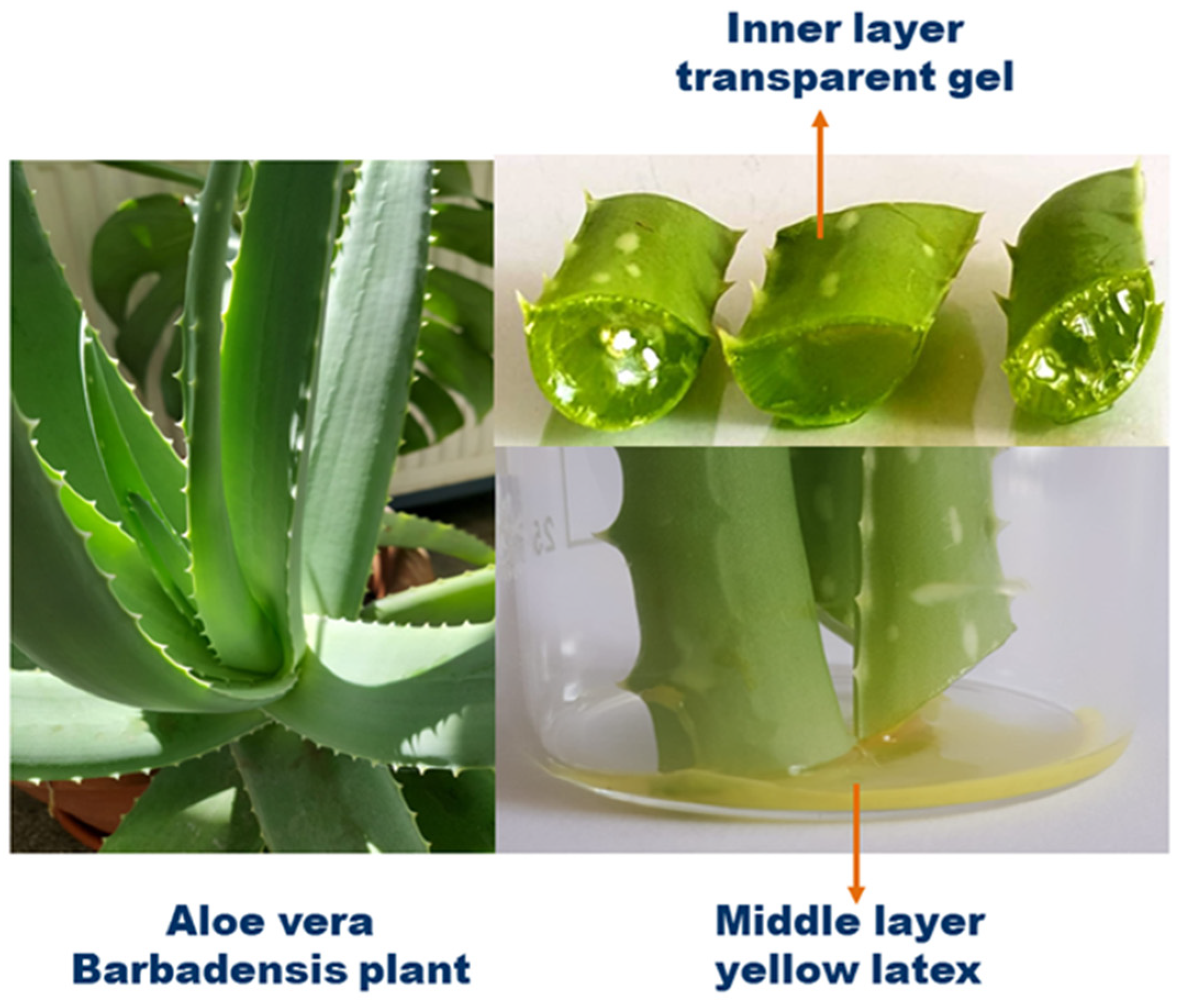
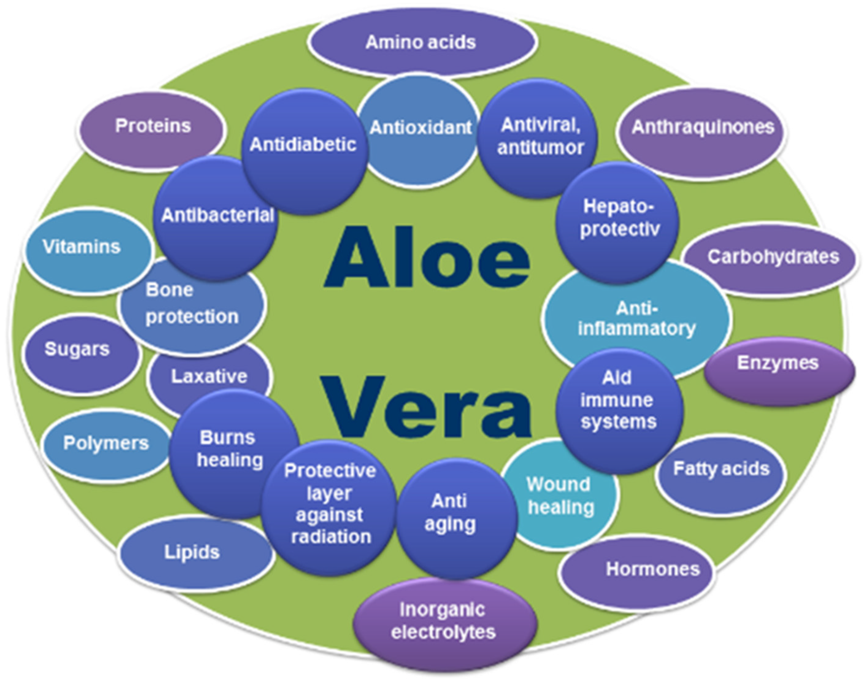
2. Phytochemical Constituents of Aloe vera

Numerous studies have demonstrated the exceptional healing potential of AV and identified the many bioactive compounds responsible for wound healing. The structure of the Aloe leaf is configured in the form of three layers. The inner layer consists of a transparent gel containing 99% water and 1% solid matter that compresses over 75 different compounds (such as glucomannans, amino acids, lipids, sterols, and vitamins), the middle layer is a bitter latex in the form of yellow juice rich in glycosides and anthraquinones, and the outer layer is a thick cortex that produces carbohydrates and proteins (Figure 1) [63,64,65,66,67].
Depending on the species, the influence of climatic conditions, and the diversity of the ecosystem to which they belong, the phytochemical constituents can be different in AV plants. Harvested from the inside of the leaves of the AV plant, the gel is a gelatinous substance that contains a complex variety of several bioactive compounds, and the analysis of the dry matter of the dry AV gel showed that it mainly contains polysaccharides (approx. 55%), sugars (approx. 17%), minerals (approx. 16%), proteins (approx. 7%), lipids (4%), and phenolic compounds (approx. 1%) [4,68,69,70,71,72]. One of the most important compounds of the gel is acemannan, which is used in many pharmacological and biological applications in medical and industrial fields, such as dentistry [73], metabolic disorders [74], cardiovascular diseases [75], and tumor diseases [76]. It has also been used for wound treatment [77] and drug delivery [78,79]. Other constituents, such as amino acids, are building blocks for body and muscle proteins; sugars control cholesterol levels, proper digestion, liver function, and help strengthen bones. Anthraquinones have an antiviral effect, enzymes catalyze the biochemical reactions, inorganic compounds have a role in the proper functioning of several enzymes in various metabolic pathways, vitamins have a strong antioxidant action in neutralizing free radicals, proteins have an antitumor effect, and hormones and sterols promote wound healing.
It is believed that the power to adjust the various biological and therapeutic implications of AV gel is due to the synergistic effect of all the active phytochemical components. This unique composition enabled the gel to harmoniously integrate into human tissues, promoting natural healing and regeneration processes. Applied topically to a wound, AV gel acts gently but as a potent antimicrobial and anti-inflammatory agent, inhibiting bacterial growth and reducing inflammation [21,80,81,82,83,84]. Table 1 summarizes the main biocomponents of AV. Additionally, the active compounds of the gel stimulate the production of new cells and collagen, which is an essential protein in the process of tissue regeneration (Figure 2) [68,85]. Thus, wounds treated with AV gel heal faster and without leaving unsightly scars.

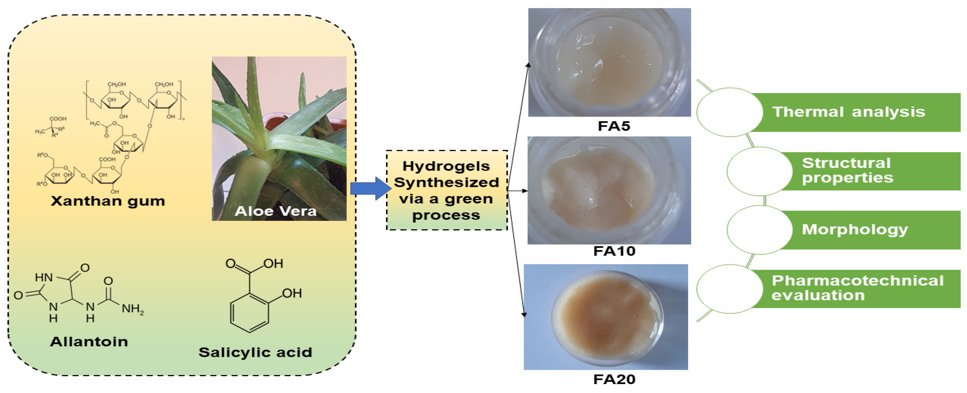
3. Preparation of Aloe vera Hydrogels

AV gel can serve as a natural and biocompatible matrix for hydrogel. It can be obtained by extracting the gel from mature AV leaves that are healthy and free from any damage or discoloration, removal of the yellow latex layer, which can be irritant, and processing the clear gel in the inner leaf to remove any impurities by washing with distilled water or ethanol. After purification and excess water draining (a concentration of 1–10% (w/v) is typically used for hydrogel formulations), the gel can be mixed with a cross-linking agent, such as a suitable polymer, considering factors such as gelation time, biocompatibility, and stability of the cross-linked hydrogel, to form a hydrogel. Finally, the gel is washed with distilled water to remove any unreacted cross-linking agent or by-products and stored refrigerated in a moisture-sealed container to maintain its moisture content (Figure 3). In Figure 4, the procedure for the AV hydrogel network preparation for its use in regenerative medicine is represented [86,87].
The specific procedure for preparing AV-based hydrogels can vary depending on the desired application and the chosen cross-linking method. It is essential to follow good laboratory practices and refer to relevant literature or established protocols to ensure the reproducibility and quality of the hydrogel preparation. It is worth mentioning that the incorporation of therapeutic agents, such as antimicrobial and anti-inflammatory agents, into AV-based hydrogels can enhance their potential for wound healing and other biomedical applications. By combining AV medicinal properties and wound healing effects with the controlled release capabilities of hydrogels, it is possible to develop advanced biomaterials with improved therapeutic outcomes. Therapeutic agents can be added to the AV gel solution before or during the cross-linking process. This can be achieved by dissolving the agents in a suitable solvent and then mixing them with the gel solution. The concentration of the agents can be varied to control the release rate and dosage. Additionally, the incorporation of therapeutic agents can be attained by the selection of appropriate therapeutic agents with desired antimicrobial and anti-inflammatory effects based on the specific application. Examples of antimicrobial agents include silver nanoparticles [88], antibiotics [89], or natural antimicrobial compounds [90], while anti-inflammatory agents may include corticosteroids [91] or non-steroidal anti-inflammatory drugs (NSAIDs) [92].
The cross-linking and gelation of AV hydrogels are crucial steps in the preparation process to convert the AV gel solution into a solid hydrogel matrix. Cross-linking is the process of creating covalent or physical bonds between polymer chains, resulting in a three-dimensional network that gives the hydrogel its structural stability and enhanced mechanical properties. Gelation refers to the transformation of the liquid gel solution into a solid gel form. The gelation process involves mixing the AV gel solution with an appropriate concentration of cross-linking agent and allowing it to react for a specific period. The AV gel solution containing therapeutic agents can be cross-linked using a suitable method, such as chemical cross-linking or physical cross-linking. Cross-linking agents are substances that promote the formation of covalent bonds between polymer chains, resulting in a three-dimensional network structure. This network improves the gel’s strength, elasticity, and resistance to dissolution in aqueous environments, making it suitable for wound healing applications. Various cross-linking mechanisms and agents can be utilized for AV-based hydrogels. Chemical cross-linking may involve the addition of a cross-linking agent that reacts with hydroxyl groups in AV to form covalent bonds, leading to gelation; while physical cross-linking can be achieved through temperature, pH-incorporating temperature, or pH-responsive polymers, the hydrogel forms as the polymer chains undergo a conformational change by simply heating the gel solution to a specific temperature or adjusting the pH. Certain polymers, such as alginate, can undergo ion-induced gelation in the presence of divalent cations such as calcium ions. Calcium chloride (CaCl2) is commonly used to initiate gelation in Aloe vera-alginate composite hydrogels. The gelation occurs as the calcium ions form ionic cross-links with the alginate chains [93]. A few commonly employed cross-linking methods are: (i) temperature-induced gelation: AV polymers can undergo gelation when the temperature is raised above a critical point, forming a physical cross-linked network; (ii) ionic gelation: addition of multivalent cations, such as calcium ions (Ca2+), can induce gelation by creating ionic interactions between the AV polysaccharides; (iii) natural agents such as glutaraldehyde, genipin, and tannic acid can be used to chemically cross-link AV hydrogels, these agents react with the functional groups present in the polymer chains, forming stable covalent bonds; (iv) carbodiimides such as 1-ethyl-3-(3-dimethylaminopropyl) carbodiimide (EDC), can facilitate the formation of amide bonds between carboxylic acid groups of AV polymers and amine groups from other molecules, resulting in cross-linking; (v) radiation-induced cross-linking: hydrogels can be cross-linked using ionizing radiation, such as gamma rays or electron beams, these high-energy radiations cause the formation of free radicals within the polymer chains, leading to cross-linking. These cross-linking mechanisms and agents help improve the mechanical integrity, swelling behavior, and biocompatibility of AV-based hydrogels used in wound dressings. They promote the stability of the hydrogel structure, prevent rapid dissolution in contact with wound exudate, and ensure the sustained release of beneficial components from AV for wound healing purposes. It is important to note that the specific choice of cross-linking agent and method may depend on factors such as desired properties, safety considerations, and compatibility with the wound healing environment. During cross-linking and gelation, it is important to control parameters such as temperature, pH, and reaction time to achieve the desired gel properties. The gelation time can be influenced by factors such as the concentration of cross-linking agents, AV gel concentration, and the specific method used. It is crucial to optimize these parameters to obtain hydrogels with desirable properties, such as mechanical strength, swelling behavior, and drug release characteristics. After gelation, it is common to wash the hydrogel to remove any unreacted cross-linking agents or by-products. The resulting AV hydrogel can be characterized and evaluated for its physical, chemical, and biological properties, such as gelation time, swelling behavior, mechanical strength, and drug release profile. In vitro and in vivo studies can be conducted to assess the antimicrobial and anti-inflammatory efficacy of the hydrogel, as well as its biocompatibility, to ensure its suitability for various applications, including wound healing, drug delivery, and tissue engineering. Figure 5 presents a schematic illustration of the synthesis and characterization of three composite hydrogels with different concentrations of AV, 5%, 10%, and 20% (w/v), and the assessment of their properties [94]. The natural polymer-based hydrogels with high AV content, from 38% to 71% by weight in dry gel, demonstrated improved pharmacotechnical properties, including swelling ratio, spreadability, elasticity, and tensile strength. The hydrogel with AV content of 10% (w/v) in solution and 55% by weight in dry gel exhibited the highest strength, elasticity, and absorption capacity and also a slightly higher spreadability, indicating it for application in wound care [94].

4. Biological and Pharmacological Effects of Aloe vera

AV gel has multiple functions. It can be used in the food field due to its proven biological properties such as antioxidant, antiviral, antibacterial, antifungal, and antiochratoxigenic activity against Aspergillus carbonarius, Aspergillus niger, Penicillium digitatum, Penicillium expansum, and Botrytis cinerea [63,95,96,97,98]. It is widely used to produce gel-containing healthy drinks and juices, including sports drinks [99]. It can be a functional food in the activation of lipolysis and the prevention of metabolic changes related to obesity since the phytosterols of Aloe gel are effective in reducing visceral fat due to the interaction with cholesterol and also has an effect on glucose metabolism, reducing blood sugar in the experimental mouse model [100]. It acts in intestinal disorders (combats constipation) due to its laxative, anti-dysenteric, anti-hemorrhoidal, and cicatrizing properties [101,102,103]. Moreover, even AV flowers are consumed more often today, knowing that diets rich in antioxidants reduce the risks of cardiovascular diseases and cancers [104].
Additionally, AV gel can be used in the medical field due to its demonstrated pharmacological effects on several components of the metabolic syndrome, such as effects against dyslipidemia, hyperglycemia, hypertension, and obesity [105]. Numerous studies have highlighted the beneficial anti-inflammatory, anti-diabetic, immunomodulatory, and anticancer (neoplastic disease) capacity [106,107,108].
At the same time, it has been studied for its active capabilities, such as hepato-protective, anti-ulcer, anti-arthritic, and anti-rheumatic properties [109,110,111]. Many investigations have shown that the dental uses of AV are multiple, with a positive impact on the oral area [112,113,114]. In the case of broken, avulsed teeth, the extract (50%) of AV determined the increase in the cell viability of the stem cells in the dental pulp. This result is due to polysaccharides and especially acemannan, which have a positive effect on the growth factor, the expressions of specific osteogenic genes, and DNA synthesis [115,116].
AV has a crucial contribution in reducing pain, combating inflammation, moisturizing the wound, improving the quantitative and qualitative composition of collagen, and improving the migration of neighboring epithelial cells of the wound [117]. AV has valuable pharmaceutical properties both through the contained gel and the whole leaf extract, which include the possibility of co-administration of bioavailable vitamins to humans. In a study on human subjects, Aloe was found to increase the absorption of both vitamins C and E through a slower absorption mechanism, and the vitamins last longer in plasma with Aloe. Aloe is said to be the only supplement known to improve the absorption of both vitamins and should be considered a true supplement [118]. Figure 6 presents a graphical representation of the interrelationship between the properties and composition of AV.
The versatile nature of AV gel has significant potential in the field of pharmaceutical applications, particularly in improving the absorption capabilities of poorly absorbed orally administered drugs. Different formulations can encapsulate poorly absorbed drugs, while AV gel acts as a stabilizing and enhancing agent [119,120,121]. Due to its outstanding efficacy and compatibility with different drug carriers, the use of AV can be further expanded in potential applications and provides a flexible platform for optimizing oral drug delivery.
The release of therapeutic agents from AV-based hydrogels can occur through several mechanisms, including diffusion, swelling, and degradation of the hydrogel matrix. These mechanisms play a crucial role in controlling the release rate and duration of the therapeutic agents. Here is an overview of these mechanisms:
Diffusion-controlled release: Diffusion is the most common mechanism for the release of therapeutic agents from hydrogels. The hydrogel matrix acts as a barrier, and therapeutic agents diffuse through the gel network. The release rate is governed by the concentration gradient between the hydrogel and the surrounding medium. The diffusion coefficient of the therapeutic agent in the hydrogel matrix, as well as the pore size and structure of the hydrogel, influence the release kinetics. Factors such as the molecular weight and solubility of the therapeutic agent also affect diffusion-controlled release [122].
Swelling-controlled release: AV-based hydrogels have the ability to absorb water and swell, affecting the release of therapeutic agents. When the hydrogel comes into contact with an aqueous medium, it absorbs water and swells, leading to an expansion of the gel network. The swelling of the hydrogel creates channels or pores, facilitating the release of therapeutic agents. The release rate depends on the degree of swelling, which can be influenced by factors such as hydrogel composition, cross-linking density, and environmental conditions (e.g., pH and temperature) [123].
Degradation-controlled release: Some AV-based hydrogels can undergo controlled degradation over time. The hydrogel matrix degrades through processes such as hydrolysis, enzymatic degradation, or biodegradation, leading to the release of therapeutic agents. The degradation rate is influenced by factors such as the composition of the hydrogel, cross-linking density, the presence of enzymes or catalysts, and the physicochemical environment. As the hydrogel degrades, the therapeutic agents are gradually released into the surrounding medium [124,125].
These release mechanisms can occur individually or in combination, depending on the specific formulation and properties of the AV-based hydrogel, as well as the characteristics of the therapeutic agents. The choice of cross-linking agents, gel composition, and hydrogel architecture can be tailored to optimize the release profile, achieving sustained or controlled release over a desired period. The release of kinetics can also be influenced by external factors such as temperature, pH, and mechanical forces. Additionally, the interactions between the therapeutic agents and the hydrogel matrix, such as electrostatic or chemical interactions, can also impact the release behavior. Therefore, it is essential to carefully design and characterize AV-based hydrogels to achieve the desired release profile for specific therapeutic applications.
AV-based formulations have both inhibitory and stimulatory properties that can influence inflammatory processes and wound healing. Its inhibitory system refers to its capacity to reduce inflammation and exhibit anti-inflammatory activity. On the other hand, its stimulatory system refers to its power to promote wound healing. Together, these dual systems allow AV to modulate the complex interplay between wound healing and inflammation beneficially. Both the native gel and hydrogels based on AV showed beneficial effects and proved effective in different applications, in oral and topical therapies. They accelerate the rate of wound closure and skin healing and alleviate mucocutaneous problems, including gingivitis. As a natural medicine, it is used in oral mouthwashes, toothpaste, submucosal fibrosis, vaginal atrophy in menopausal women, and mucosal lesions induced by chemotherapy and radiotherapy or in veterinary practice. Here, we highlight some main beneficial effects of AV hydrogels in wound healing.

4.1. Reduction of Inflammation

Psoriasis is an immune disease, provoked by an unclear cause, which is characterized by inflammation caused by the dysfunction of the immune system and is manifested by an itchy rash, most commonly on the knees, elbows, trunk, and scalp. This disease can cause inflammation in the body and can also affect other organs or tissues in the body. Worldwide, approximately 125 million people suffer from this disease. Plaque psoriasis is associated with several comorbidities, including inflammatory arthritis, cardiometabolic disease, and depression. The American Academy of Dermatology—National Psoriasis Foundation guidelines recommend biologics as alternatives for the first-line treatment of moderate to severe plaque psoriasis due to their therapeutic efficacy and acceptable safety profiles [126]. AV has often been used for topical applications in the treatment of psoriasis. A study on rats, in which hydrogels based on AV mucilage were developed and prepared with 80% w/w of gel for topical applications, demonstrated good efficiency in controlling hyperkeratinization, showing a 61% reduction of the stratum corneum on the tested animals. The results confirmed the keratolytic action of AV hydrogel, which can be used to treat psoriasis. The effect of AV leaf extract has been attributed to polysaccharides, rich in glucomannan and acemannan, pectic compounds, cellulose, and hemicelluloses, which determine most of the plant’s therapeutic properties [127]. The antipsoriatic properties of AV have been combined with the healing activity of Natural Rubber Latex to produce new economic occlusive dressings recommended for the treatment of psoriasis symptoms. In total, 58.8% of loaded AV, present on the surface and inside the dressing, was released after 4 days. An in vitro study on human dermal fibroblasts and sheep blood, respectively, confirmed the biocompatibility and hemocompatibility of the new dressings, the preservation of approximately 70% of the free antioxidant properties of AV, and the total content of phenolic compounds 2.31 times higher in these dressings compared to natural rubber latex without AV [128].

4.2. Prevention of Bacterial Infection

Chitosan and AV films encapsulating thymol were prepared to be used in preventing the possibility of bacterial infection and showed a high thymol encapsulation efficiency of 95.3% with good dispersibility. Test results against various pathogenic microbes such as Bacillus, Staphylococcus, Escherichia, Pseudomonas, Klebsiella, and Candida showed that the films were effective against bacterial colonization in a thymol concentration-dependent manner. The addition of AV increased the water absorption of the films, which is one of the primary factors of healthy wound healing and helped by improving the antioxidant activity and in vitro release efficiency of thymol [129]. New polymer composite films based on polyvinyl alcohol and AV have been prepared for wound healing and prevention of surgical wound infections. Films tested for antibacterial and antifungal activity against E. coli, P. aeruginosa, Aspergillus flavus, and Aspergillus tubingensis showed antimicrobial activity against all strains; the lowest concentration of AV (5%) showed the highest activity against all strains. Sutures of wounds covered with films based on polyvinyl alcohol and AV showed that the new composites have antibacterial effects and the potential to be used in the prevention of infections at the surgical site and can be used for wound healing purposes [130]. Films based on alginate, AV gel, honey, and cellulose nanocrystals can be used for applications as antibacterial dressings. The morphological, swelling, mechanical, and biological properties of the films prepared and tested against the Gram-negative organisms Salmonella typhi, Klebsiella pneumoniae, Escherichia coli, and the Gram-positive organism Staphylococcus aureus were estimated. The films showed superior biocompatibility, good mechanical properties, and excellent antibacterial capabilities [131]. Blended nanofiber membranes for new types of antibacterial wound dressings were made based on polycaprolactone/chitosan/Aloe vera (PCL/CS/AV) nanofiber (NFM) by electrospinning. The characterizations and tests carried out showed that the addition of AV increased the hydrophilicity and the pore size of the membranes and led to the improvement of the antibacterial performance against Streptococcus aureus and E. coli and the biocompatibility in 5 days. The membranes produced were proposed as suitable for short-term dressing or acute wounds (1–4 days) [132]. Nanofiber membranes were developed based on natural, biocompatible, and biodegradable composites from AV extract, pullulan, chitosan, and citric acid, through Forcespinning® technology. The morpho-structural characterization and thermogravimetric analysis of the membranes indicated their good properties, as well as good water absorption capacities and synergistic antibacterial activity against Escherichia coli, which promoted cell attachment and growth. Due to their porous structure and large surface area, the membranes can be recommended as potential dressing applications due to their ability to absorb excessive blood and exudates, their thermal stability, and the protection they offer against infection [133]. Novel sodium alginate/poly(vinyl alcohol) (SA/PVA) hydrogel dressing films enriched with AV were produced by a simple method. The influence of different amounts (5, 10, 15, 20, and 25%, v/v) of AV solution on the chemical structure and properties of sodium alginate/poly(vinyl alcohol) hydrogel films was studied. The structural, morphological, mechanical, and thermal characterization confirmed that rigid and thermally stable three-dimensional structures were obtained. The results regarding the release profile of the polysaccharides from the hydrogel matrix showed that the active substance was released in a prolonged, gradual manner, even for a week. It was shown that the presence of AV within the cross-linked polymer network improved the active substance delivery properties of the hydrogel films. At the same time, the cytotoxicity of the materials was studied, and the results indicated good adhesion properties and a lack of toxicity. In vitro experiments on normal human dermal fibroblasts showed very good cell attachment to AV hydrogel discs, which promoted cell spreading and proliferation. As such, SA/PVA/AV sustained-release AV films have been proposed for applications such as interactive wound dressings [134]. Recent studies have concluded that AV gel is an effective antibacterial agent to prevent wound infection caused by various bacteria: P. Aeruginosa [135], Campylobacter rectus, Provetella intermedia [136], and Escherichia coli (E. coli) [137].

4.3. Skin Regeneration

The skin is part of the body’s integumentary system and consists of the epidermis and dermis, with a subcutaneous fatty layer, the hypodermis [138,139]. It protects us against external factors and prevents bacteria and germs from entering the body and blood and causing infections [140,141]. At the same time, the skin is vulnerable and can be affected by acute or chronic wounds [142]. Wound healing is a complex physiological process, which is achieved through four explicit phases: hemostasis, inflammation, proliferation, and remodeling and involves the epidermis-containing keratinocyte, melanocyte, and Langerhans cells, dermis, including fibroblast, neutrophil, mast cell, and dermal dendritic cells, and the hypodermis, which contains mesenchymal stem cells (Figure 7) [22,143,144,145].
The wound-healing process consists of four highly integrated and overlapping phases: (i) hemostasis, (ii) inflammation, (iii) proliferation, and (iv) tissue remodeling or resolution [146]. Figure 8 shows the main stages of the normal wound-healing process [145]. Each stage is characterized by key molecular and cellular events and is coordinated by a series of secreted factors that are recognized and released by wound response cells. Hemostasis is the first stage. It involves coagulation, which changes the blood from a liquid to a gel. The inflammation phase begins at the time of injury and lasts up to four days. As inflammatory cells undergo apoptosis, wound healing progresses to the proliferative phase. This phase begins approximately three days after the injury and overlaps with the inflammatory phase, while the tissue remodeling phase, characterized by the formation of granulation tissue, angiogenesis (formation of blood vessels), wound contraction, and the process of epithelialization, can continue for six months to one year after the injury, which leads to the formation of scar tissue. Many variables can disrupt one or more phases of this process, thereby producing inadequate or incorrect healing of skin wounds. The main elements that affect wound healing are oxygenation, infection, age, stress, diabetes, obesity, drugs, alcoholism, smoking, repeated trauma, diet, and poor blood circulation [147,148,149]. Infection is the most common complication for injured skin; therefore, prevention or mitigation of infection is of utmost importance.
Using an ecological preparation method, a natural, degradable, and environmentally friendly hydrogel dressing was developed using AV as an active ingredient. The hydrogel dressing was prepared using only natural ingredients, composed of sodium hyaluronate (SH), dopamine (DA), chitosan (CS), and AV, and using a natural deep eutectic green solvent (DES) as the green solvent. The newly synthesized hydrogel showed good cytocompatibility tested on NIH-3T3 fibroblast cells and antibacterial properties against both Gram-positive (S. aureus) and Gram-negative (E. coli). Additionally, in a study on mice, the hydrogel promoted the regeneration of skin tissue and healed the skin wound after surgery within 12 days. The authors concluded that the newly prepared hydrogel, which is natural, degradable, and ecological and uses AV as an active ingredient, shows great potential in wound healing applications [150]. A study on the emergency treatment of vaginal tissue by local application of AV and alginate hydrogel for the release of mesenchymal stem cells derived from the maternal endometrium with the aim of promoting maternal injury relief and early healing was carried out in a simulated injury model at birth. It was observed that in the absence of therapy, fibrotic healing can occur in many cases. Local injection of hydrogel-containing mesenchymal cells significantly improves smooth muscle and elastin content, as well as decreases tissue stiffness after 6 weeks. The findings of the study highlighted that immediate treatment of severe vaginal birth trauma with therapeutic mesenchymal stem cells delivered in AV and alginate hydrogel might become a potential new treatment strategy for faster healing of birth injuries and prevention of pelvic organ prolapse (Figure 9) [151].
Another study explored the potential for acute and chronic wound healing using piperine as a new bioactive compound. New systems of bioactive hydrogels based on carbopol 934 containing piperine mixed with AV gels of different gel strengths were prepared and characterized (Figure 10). The developed formulation system was investigated in an excisional wound healing model in the rat model. The results of the in vivo study and histopathological examination showed that the piperine-containing bioactive hydrogel system compared with the piperine-free bioactive hydrogel system, leads to early and intrinsic wound healing (Figure 11). Thus, the findings of the study emphasized that the new piperine-containing bioactive hydrogel is a promising therapeutic approach for the application of wound healing [152].
Studying the influence of a commercial hydrogel formulation based on AV with 1,2-propanediol (propanediol) and triethanolamine (TEA) on skin wound healing was investigated in female Wistar rats. Additionally, the study aimed to show that the presence of specific additives, propanediol and triethanolamine, does not exert any negative effect on wound healing.
The results showed that the prepared hydrogel had a positive effect on inflammation, angiogenesis, and wound contraction and reduced the total healing time by 29%, with the total closure of the wound being achieved in 15 days (Figure 12). The paper highlighted the influence of the bioactive components of AV, related to rhamnogalacturonan and pectin-like acemannan, which improved the healing process of skin wounds [153].
A novelty in the area of efficient ecological materials is the new system of biocompatible hydrogels based on AV that was prepared by a completely green synthesis method for wound healing applications (Figure 13).
Hydrogels with different concentrations of AV (5 and 10%, respectively) also contain other natural components such as salicylic acid, allantoin, and xanthan gum. The hydrogels’ rheological properties, morphology, cell viability, biocompatibility, and cytotoxicity, were studied. The preliminary examinations showed that the hydrogels are very well supported on a wound, without stinging even more; they quickly penetrated the tissue and ensured good hydration of the area. Testing the antibacterial activity of the hydrogels was evaluated both on Gram-positive strains, Staphylococcus aureus, and on Gram-negative strains, Pseudomonas aeruginosa. The results showed that they have good antibacterial properties (Figure 14i). Moreover, the in vitro scratch test demonstrated the suitable ability of these “green” hydrogels to accelerate cell proliferation and migration and induce closure of a wounded area, making them suitable for wound healing applications (Figure 14ii) [154].

4.4. Healing Burns

A clinical study was conducted on 30 patients with similar types of second-degree burns in two places on different parts of the body. This research was conducted to evaluate the effectiveness of AV cream for partial thickness burns and to compare its results with those of silver sulfadiazine. Each patient had one burn treated randomly with topical silver sulfadiazine ointment and one treated with Aloe cream. The mean time to re-epithelialization and healing of partial-thickness burns was significantly shorter for the Aloe group at 15.9 ± 2 days versus 18.73 ± 2.65 days for the SSD group (p < 0.0001). Both sites were negative for microbial contamination on days 3, 7, and 13. Study results showed that AV cream promoted better wound healing with smaller lesions and had shorter healing times than silver sulfadiazine [155]. A similar international study was accomplished on 50 patients with second-degree burns and evaluated the effectiveness of AV gel compared with 1% silver sulfadiazine cream as a special dressing for the treatment of superficial and partial thickness burn wounds. The study used 98% unrefined gel from the inner leaf of the plant. Thermal burn patients bandaged with AV gel showed improvements compared to those bandaged with silver sulfadiazine cream in terms of early wound epithelialization, early pain relief, and cost-effectiveness of treatment management [156]. Another double-blind, randomized clinical trial in 11 patients treated once daily for 14 days compared the efficacy of herbal AV cream with 1% silver sulfadiazine in reducing the pain of second-degree burns. The herbal cream was prepared from AV gel and essential oils of Lavandula stoechas and Pelargonium roseum. In total, 56 patients were treated with herbal cream, and another 55 were treated with silver sulfadiazine 1%. Study results demonstrated that pain intensity at 14 days was significantly reduced in both groups compared to baseline (p < 0.001). However, a greater reduction in pain from baseline to the 7- and 14-day mark was observed in the herbal cream group (p = 0.014 and p = 0.05). One case of infection was reported in the herbal cream group; however, it cleared up with continued treatment. The findings of this clinical trial showed that the herbal cream was superior to silver sulfadiazine in relieving pain for superficial second-degree burns [157]. In an additional clinical case study, the therapeutic impact of AV gel on chronic skin burns in a 17-year-old patient with a rejected skin graft is presented. This is a before–after comparative study design in a case of fire burn in which initiation of AV gel treatment is accompanied by the promotion of wound repair. Before being treated with gel, the patient who had suffered burns on 30–40% of her body surface for 40 days had a healthy skin graft operation on her previous chest, which was rejected after 5 days. Following chronic unhealed skin lesions, the patient was treated with AV gel for 21 days continuously. The skin healing process began with the formation of granulation tissue and epithelization of the wounds. During the treatment, no sign of skin infection and no topical side effects of AV gel, such as allergic reactions and itching, were observed. This study on the impact of AV gel in the healing of burns can be considered a cheap and quick effect of substitution therapy instead of surgery [158].

4.5. Protection against Chemoradiation Secondary Effects in Cancer Treatment

A multicenter, randomized, double-blind, controlled trial was performed on 120 patients with head and neck cancer treated with concurrent chemoradiation. Patients received either AV gel or placebo gel and were assessed for adverse levels of skin toxicity with the Radiation-Induced Skin Reaction Rating Scale (RISRAS). At the 5th and 6th week of treatment, grades moderate to severe erythematous skin at values of 13.6% and 24.1% versus 27.8 and 42.6% were observed for members of the AV gel group and the placebo group, respectively (p = 0.05 for the 5th week and p = 0.038 for the 6th week). At week 7, in the placebo group, moderate to severe cases of wet scaling were observed in eight patients (19.0%) (p = 0.001), as well as a burning sensation with RISRAS scores of 3–4, representing only 11.9% of patients (p = 0.016). The study authors concluded that there was no prophylactic efficacy for radiation-induced dermatitis in the AV gel group compared with the placebo group but that topical applications of AV gel along with a routine skin care program from starting radiation would reduce the severity of any burning sensations, along with the incidence of erythematous, moist scaling of the skin in head and neck cancer patients receiving concurrent chemoradiation [159].

4.6. Summary of Clinical Effects of AV on Prevention and Healing of Skin Wounds

An earlier systematic study [121] concluded that AV helps to retain skin moisture and integrity and prevents skin ulcers due to its content of mucopolysaccharides, amino acids, zinc, and water. Furthermore, AV was found to be ‘much more effective and less costly compared to the currently available alternative treatments’ in terms of quality and speed of wound healing. Considering the tendency to promote traditional medicine as well as the rare side effects of AV, the use of this medicinal plant for the healing of skin wounds is recommended.
AV gel has been demonstrated to be active in wound healing through several reported mechanisms [31], including increased epithelial cell viability, proliferation, and migration, moisture retention [160], increased quantity and cross-linking of collagen [161], and hindering inflammation through the decrease of proinflammatory cytokines [162,163,164,165,166]. The various active components of AV include acemannan, aloesin, aloe-emodin, aloin, emodin, and glucomannan [68]. Acemannan is known to stimulate epidermal keratinocytes and the production of fibrotic cytokines [167,168]. Glucomannan, a water-soluble mucopolysaccharide, stimulates fibroblast growth factor production and the activity and proliferation of these cells, leading to the increased amount of collagen on the wound site with enhanced transversal connections [21,64,169]. Emodin emodinolin, anthraquinone derivatives found in AV, act as competitive inhibitors of thromboxane synthetase and have significant anti-inflammatory properties [21]. The anti-inflammatory properties of AV are related to the inhibition of proinflammatory cytokines [162,164,165], hindering ROS production [162,164], and blocking the signalling of JAK1-STAT1/3 [68]. The anti-inflammatory effects and increased collagen production and cross-linking promote the rearrangement of epithelial tissues [12], reducing the wounded area and accelerating the healing process [170]. Various studies have confirmed that topical AV creams heal first- and second-degree burns in less than half the time than standard treatment with silver sulfadiazine [21,171,172,173]. AV has an anti-erythema activity similar to that of the positive control group (i.e., hydrocortisone gel) after 6 days of treatment [174]. AV gel has also demonstrated potent angiogenic activity, an essential process in wound healing, attributed to angiogenic compounds such as beta-sitosterol [175,176]. Table 2 summarizes various beneficial effects of AV compounds for wound healing reported in clinical studies.
Recent studies on AV gels with added therapeutic agents have reported the positive interaction between graphene oxide/reduced graphene oxide (GO/rGO) and AV hydrogels to be a strongly promising strategy for the advancement of therapeutic approaches for wound healing (Figure 15) [178].
Jales et al. further confirmed the great potential of AV mucilaginous hydrogel with a high keratolytic effect that can be used in psoriasis treatment [127]. Puliero et al. investigated the use of AV extracts for ocular therapeutic or preventive purposes. They demonstrated that the best lenses allowing for the high and stable release of AV extract to the corneal surface are those composed of ionic hydrogels [194]. Capsaicin, a powerful anti-inflammatory and analgesic agent, poorly water-soluble, was successfully incorporated into AV gel for topical drug delivery and to reduce skin irritation caused by capsaicin [119]. The AV gels softness, biocompatibility, and fast spreading or penetrating capacity are particularly useful features to encapsulate and deliver various nanoparticles with antimicrobial properties (e.g., ZnO or TiO2) [195], drugs, cell culture, both for wound healing, and bio-sensing applications [196]. The combination of AV and Rheum palmatum root can promote the migration of human primary fibroblasts (Figure 16) [182].
None of the dressings available on the market today are fully capable of reproducing all the characteristics of native skin. An asymmetric bilayer membrane with a top dense polycaprolactone layer that provides mechanical support and a bottom porous layer of chitosan and AV, aiming to improve the healing process, was designed to mimic both layers of the skin [197]. The results obtained revealed the potential of these asymmetric membranes to be applied as wound dressings in the future.

5. Side Effects

No serious adverse reactions were demonstrated following the topical application of AV inner gel products. AV used in dietary supplements appears to be safe [198]. The inner gel was evaluated by the Cosmetic Ingredient Review Expert Panel as noncytotoxic [199]. However, due to the cytotoxicity, mutagenicity, and carcinogenicity of anthraquinones, it is crucial to monitor the content of these phenolic compounds in AV whole leaf extract and latex [200,201]. Topical and oral use of AV whole leaf extract in humans can cause adverse clinical effects: skin irritation, hives, cramping, and diarrhea to those who are allergic to plants in the lily family, for example, onion and tulips [202,203,204].

6. Conclusions and Future Prospective Studies

It is important to apply modern delivery techniques to develop affordable products based on efficacious traditional natural medicines for wound healing and to improve their therapeutic effect.
Further research is needed to ensure that these formulations reach the pharmaceutical market. Chemotherapy treatments for cancer are associated with the presence of ulcers in the oral mucosa that causes pain, bleeding, and difficulty swallowing or speaking. There is no effective standard treatment, and few studies have been published on the therapeutic effects of natural products such as AV to improve the local retention period.
Future treatments may arise from medicinal plants, which have fewer side effects and improved bioavailability for the wound-healing process. In addition, in the future, a great challenge is represented by the development of an intelligent treatment that presents anti-inflammatory, antimicrobial, and antioxidant cumulative properties for the treatment of all types of wounds. Furthermore, the commercialization and use in preclinical research and clinical practice of natural products used in wound healing must be increased significantly to discover the potential of these products, considered natural bioactive molecules, in the treatment and regeneration of skin tissue. Future research should be considered to find new natural bioactive compounds related to their usage in the wound-healing process and their ability to act as substitutes for existing antibiotics.
By incorporating therapeutic agents into AV-based hydrogels, it is possible to develop multifunctional biomaterials that provide sustained release of agents, promote wound healing, reduce inflammation, and prevent or treat microbial infections. However, it is important to note that the specific formulation and efficacy of such hydrogels may vary depending on the therapeutic agents chosen, their concentration, crosslinking method, and other factors. Extensive research and testing (rheological analysis, drug release profiles, permeability, and stability studies) are required to optimize the formulation and ensure its safety and effectiveness for clinical use and to promote human well-being worldwide.

Author Contributions

Conceptualization, M.C., A.M.M. and J.C.M.; methodology, M.P., M.C. and J.C.M.; data curation, M.C. and A.M.M.; resources, J.C.M.; writing—original draft preparation, M.C., A.M.M., M.P. and J.C.M.; writing—review and editing, A.M.M. and J.C.M.; visualization, M.P.; supervision, A.M.M. and J.C.M. All authors have read and agreed to the published version of the manuscript.

Funding

This research received no external funding.

Institutional Review Board Statement

Not applicable.

Informed Consent Statement

Not applicable.

Data Availability Statement

Not applicable.

Conflicts of Interest

The authors declare no conflict of interest.

References

  1. Ekor, M. The growing use of herbal medicines: Issues relating to adverse reactions and challenges in monitoring safety. Front. Pharmacol. 2014, 10, 177. [Google Scholar] [CrossRef] [PubMed] [Green Version]
  2. Khan, M.S.A.; Ahmad, I. Chapter 1-Herbal Medicine: Current Trends and Future Prospects; New Look to Phytomedicine; Khan, M.S.A., Ahmad, I., Chattopadhyay, D., Eds.; Academic Press: Cambridge, MA, USA, 2019; pp. 3–13. ISBN 9780128146194. [Google Scholar] [CrossRef]
  3. Surjushe, A.; Vasani, R.; Saple, D.G. Aloe vera: A short review. Indian J. Dermatol. 2008, 53, 163–166. [Google Scholar] [CrossRef] [PubMed]
  4. Kumar, R.; Singh, A.K.; Gupta, A.; Bishayee, A.; Pandey, A.K. Therapeutic potential of Aloe vera—A miracle gift of nature. Phytomedicine 2019, 60, 152996. [Google Scholar] [CrossRef] [PubMed]
  5. Manvitha, K.; Bidya, B. Aloe vera: A wonder plant its history, cultivation and medicinal uses. J. Pharm. Phytochem. 2014, 2, 85–88. [Google Scholar]
  6. Mehta, D.I. “History OF Aloe vera” – (A Magical Plant). IOSR J. Humanit. Soc. Sci. 2017, 22, 21–24. [Google Scholar]
  7. Gao, Y.; Kuok, K.I.; Jin, Y.; Wang, R. Biomedical applications of Aloe vera. Crit. Rev. Food Sci. Nutr. 2019, 59, S244–S256. [Google Scholar] [CrossRef]
  8. Available online: https://www.fortunebusinessinsights.com/aloe-vera-extract-market-103893 (accessed on 20 May 2023).
  9. Deep, A.; Kumar, D.; Bansal, N.; Narasimhan, B.; Marwaha, R.K.; Sharma, P.C. Understanding mechanistic aspects and therapeutic potential of natural substances as anticancer agents. Phytomed. Plus 2023, 3, 100418. [Google Scholar] [CrossRef]
  10. Noori, A.S.; Mageed, N.F.; Abdalameer, N.K.; Mohammed, M.K.; Mazhir, S.N.; Ali, A.H.; Jaber, N.A.; Mohammed, S.H. The histological effect of activated Aloe vera extract by microwave plasma on wound healing. Chem. Phys. Lett. 2022, 807, 140112. [Google Scholar] [CrossRef]
  11. Kim, S.-T.; Pressman, P.; Clemens, R.; Moore, A.; Hamilton, R.; Hayes, A.W. The absence of genotoxicity of Aloe vera beverages: A review of the literature. Food Chem. Toxicol. 2023, 174, 113628. [Google Scholar] [CrossRef]
  12. Sánchez-Machado, D.I.; López-Cervantes, J.; Sendón, R.; Sanches-Silva, A. Aloe vera: Ancient knowledge with new frontiers. Trends Food Sci. Technol. 2017, 61, 94–102. [Google Scholar] [CrossRef]
  13. Altinkaynak, C.; Haciosmanoglu, E.; Ekremoglu, M.; Hacioglu, M.; Özdemir, N. Anti-microbial, anti-oxidant and wound healing capabilities of Aloe vera-incorporated hybrid nanoflowers. J. Biosci. Bioeng. 2023, 135, 321–330. [Google Scholar] [CrossRef] [PubMed]
  14. Hattingh, A.; Laux, J.-P.; Willers, C.; Hamman, J.; Steyn, D.; Hamman, H. In vitro wound healing effects of combinations of Aloe vera gel with different extracts of Bulbine frutescens. S. Afr. J. Bot. 2023, 158, 254–264. [Google Scholar] [CrossRef]
  15. Movaffagh, J.; Khatib, M.; Bazzaz, B.S.F.; Taherzadeh, Z.; Hashemi, M.; Moghaddam, A.S.; Tabatabaee, S.A.; Azizzadeh, M.; Jirofti, N. Evaluation of wound-healing efficiency of a functional Chitosan/Aloe vera hydrogel on the improvement of re-epithelialization in full thickness wound model of rat. J. Tissue Viabil. 2022, 31, 649–656. [Google Scholar] [CrossRef] [PubMed]
  16. Abdel-Mohsen, A.M.; Frankova, J.; Abdel-Rahman, R.M.; Salem, A.A.; Sahffie, N.M.; Kubena, I.; Jancar, J. Chitosan-glucan complex hollow fibers reinforced collagen wound dressing embedded with Aloe vera. II. Multifunctional properties to promote cutaneous wound healing. Int. J. Pharm. 2020, 582, 119349. [Google Scholar] [CrossRef] [PubMed]
  17. Ghorbani, M.; Nezhad-Mokhtari, P.; Ramazani, S. Aloe vera-loaded nanofibrous scaffold based on Zein/Polycaprolactone/Collagen for wound healing. Int. J. Biol. Macromol. 2020, 153, 921–930. [Google Scholar] [CrossRef]
  18. Farid, A.; Haridyy, H.; Ashraf, S.; Ahmed, S.; Safwat, G. Aloe vera gel as a stimulant for mesenchymal stem cells differentiation and a natural therapy for radiation induced liver damage. J. Radiat. Res. Appl. Sci. 2022, 15, 270–278. [Google Scholar] [CrossRef]
  19. Razia, S.; Park, H.; Shin, E.; Shim, K.-S.; Cho, E.; Kang, M.C.; Kim, S.Y. Synergistic effect of Aloe vera flower and Aloe gel on cutaneous wound healing targeting MFAP4 and its associated signaling pathway: In-vitro study. J. Ethnopharmacol. 2022, 290, 115096. [Google Scholar] [CrossRef]
  20. Sharifi, E.; Chehelgerdi, M.; Fatahian-Kelishadrokhi, A.; Yazdani-Nafchi, F.; Ashrafi-Dehkordi, K. Comparison of therapeutic effects of encapsulated Mesenchymal stem cells in Aloe vera gel and Chitosan-based gel in healing of grade-II burn injuries. Regen. Ther. 2021, 21, 30–37. [Google Scholar] [CrossRef]
  21. Hekmatpou, D.; Mehrabi, F.; Rahzani, K.; Aminiyan, A. The Effect of Aloe vera Clinical Trials on Prevention and Healing of Skin Wound: A Systematic Review. Iran J. Med. Sci. 2019, 44, 1–9. [Google Scholar] [CrossRef]
  22. Tejiram, S.; Kavalukas, S.L.; Shupp, J.W.; Barbul, A. 1-Wound healing. In Ågren, Wound Healing Biomaterials; Magnus, S., Ed.; Woodhead Publishing: Sawston, UK, 2016; pp. 3–39. [Google Scholar] [CrossRef]
  23. Clark, R.A.F.; Musillo, M.; Stransky, T. Chapter 70—Wound repair: Basic biology to tissue engineering. In Principles of Tissue Engineering, 5th ed.; Lanza, R., Langer, R., Vacanti, J.P., Atala, A., Eds.; Academic Press: Cambridge, MA, USA, 2020; pp. 1309–1329. [Google Scholar] [CrossRef]
  24. Frykberg, R.G.; Banks, J.; Deptuła, M.; Karpowicz, P.; Wardowska, A.; Sass, P.; Sosnowski, P.; Mieczkowska, A.; Filipowicz, N.; Dzierżyńska, M.; et al. Challenges in the Treatment of Chronic Wounds. Adv. Wound Care 2015, 4, 560–582. [Google Scholar] [CrossRef] [Green Version]
  25. Nguyen, H.M.; Le, T.T.N.; Nguyen, A.T.; Le, H.N.T.; Pham, T.T. Biomedical materials for wound dressing: Recent advances and applications. RSC Adv. 2023, 13, 5509–5528. [Google Scholar] [CrossRef]
  26. Yaşayan, G.; Nejati, O.; Ceylan, A.F.; Karasu, C.; Ugur, P.K.; Bal-Öztürk, A.; Zarepour, A.; Zarrabi, A.; Mostafavi, E. Tackling chronic wound healing using nanomaterials: Advancements, challenges, and future perspectives. Appl. Mater. Today 2023, 32, 101829. [Google Scholar] [CrossRef]
  27. Verdes, M.; Mace, K.; Margetts, L.; Cartmell, S. Status and challenges of electrical stimulation use in chronic wound healing. Curr. Opin. Biotechnol. 2022, 75, 102710. [Google Scholar] [CrossRef] [PubMed]
  28. Bormann, D.; Gugerell, A.; Ankersmit, H.J.; Mildner, M. Therapeutic Application of Cell Secretomes in Cutaneous Wound Healing. J. Investig. Dermatol. 2023, 143, 893–912. [Google Scholar] [CrossRef] [PubMed]
  29. Middelkoop, E.; Sheridan, R.L. Skin Substitutes and ‘the next level’. In Total Burn Care; Elsevier: Amsterdam, The Netherlands, 2018; pp. 167–173.e2. [Google Scholar] [CrossRef]
  30. Shi, C.; Wang, C.; Liu, H.; Li, Q.; Li, R.; Zhang, Y.; Liu, Y.; Shao, Y.; Wang, J. Selection of Appropriate Wound Dressing for Various Wounds. Front. Bioeng. Biotechnol. 2020, 8, 182. [Google Scholar] [CrossRef] [Green Version]
  31. Alven, S.; Khwaza, V.; Oyedeji, O.O.; Aderibigbe, B.A. Polymer-Based Scaffolds Loaded with Aloe vera Extract for the Treatment of Wounds. Pharmaceutics 2021, 13, 961. [Google Scholar] [CrossRef] [PubMed]
  32. Hasan, S.; Hasan, M.A.; Hassan, M.U.; Amin, M.; Javed, T.; Fatima, L. Biopolymers in diabetic wound care management: A potential substitute to traditional dressings. Eur. Polym. J. 2023, 189, 111979. [Google Scholar] [CrossRef]
  33. Corrales-Orovio, R.; Carvajal, F.; Holmes, C.; Miranda, M.; González-Itier, S.; Cárdenas, C.; Vera, C.; Schenck, T.L.; Egaña, J.T. Development of a photosynthetic hydrogel as potential wound dressing for the local delivery of oxygen and bioactive molecules. Acta Biomater. 2023, 155, 154–166. [Google Scholar] [CrossRef]
  34. Smith, R.; Russo, J.; Fiegel, J.; Brogden, N. Antibiotic Delivery Strategies to Treat Skin Infections When Innate Antimicrobial Defense Fails. Antibiotics 2020, 9, 56. [Google Scholar] [CrossRef] [Green Version]
  35. Popova, T.P.; Ignatov, I.; Petrova, T.E.; Kaleva, M.D.; Huether, F.; Karadzhov, S.D. Antimicrobial Activity In Vitro of Cream from Plant Extracts and Nanosilver, and Clinical Research In Vivo on Veterinary Clinical Cases. Cosmetics 2022, 9, 122. [Google Scholar] [CrossRef]
  36. Moretta, A.; Scieuzo, C.; Petrone, A.M.; Salvia, R.; Manniello, M.D.; Franco, A.; Lucchetti, D.; Vassallo, A.; Vogel, H.; Sgambato, A.; et al. Antimicrobial Peptides: A New Hope in Biomedical and Pharmaceutical Fields. Front. Cell. Infect. Microbiol. 2021, 11, 668632. [Google Scholar] [CrossRef]
  37. Ghasemi, A.H.; Farazin, A.; Mohammadimehr, M.; Naeimi, H. Fabrication and characterization of biopolymers with antibacterial nanoparticles and Calendula officinalis flower extract as an active ingredient for modern hydrogel wound dressings. Mater. Today Commun. 2022, 31, 103513. [Google Scholar] [CrossRef]
  38. Huda, R.M.; Rashdan, H.R.; El-Naggar, M.E. Chapter 2-Traditional and modern wound dressings. In Developments in Applied Microbiology and Biotechnology, Antimicrobial Dressings; Khan, R., Gowri, S., Eds.; Academic Press: Cambridge, MA, USA, 2023; pp. 21–42. [Google Scholar] [CrossRef]
  39. Ferroni, L.; Gardin, C.; D’Amora, U.; Calzà, L.; Ronca, A.; Tremoli, E.; Ambrosio, L.; Zavan, B. Exosomes of mesenchymal stem cells delivered from methacrylated hyaluronic acid patch improve the regenerative properties of endothelial and dermal cells. Biomater. Adv. 2022, 139, 213000. [Google Scholar] [CrossRef] [PubMed]
  40. Feketshane, Z.; Adeyemi, S.A.; Ubanako, P.; Ndinteh, D.T.; Ray, S.S.; Choonara, Y.E.; Aderibigbe, B.A. Dissolvable sodium alginate-based antibacterial wound dressing patches: Design, characterization, and in vitro biological studies. Int. J. Biol. Macromol. 2023, 232, 123460. [Google Scholar] [CrossRef] [PubMed]
  41. Fligge, M.; Letofsky-Papst, I.; Bäumers, M.; Zimmer, A.; Breitkreutz, J. Personalized dermal patches–Inkjet printing of prednisolone nanosuspensions for individualized treatment of skin diseases. Int. J. Pharm. 2023, 630, 122382. [Google Scholar] [CrossRef] [PubMed]
  42. Raepsaet, C.; Alves, P.; Cullen, B.; Gefen, A.; Lázaro-Martínez, J.L.; Lev-Tov, H.; Najafi, B.; Santamaria, N.; Sharpe, A.; Swanson, T.; et al. The development of a core outcome set for clinical effectiveness studies of bordered foam dressings in the treatment of complex wounds. J. Tissue Viability 2023. [Google Scholar] [CrossRef]
  43. Genesi, B.P.; de Melo Barbosa, R.; Severino, P.; Rodas, A.C.D.; Yoshida, C.M.P.; Mathor, M.B.; Lopes, P.S.; Viseras, C.; Souto, E.B.; da Silva, C.F. Aloe vera and copaiba oleoresin-loaded chitosan films for wound dressings: Microbial permeation, cytotoxicity, and in vivo proof of concept. Int. J. Pharm. 2023, 634, 122648. [Google Scholar] [CrossRef]
  44. Rajati, H.; Alvandi, H.; Rahmatabadi, S.S.; Hosseinzadeh, L.; Arkan, E. A nanofiber-hydrogel composite from green synthesized AgNPs embedded to PEBAX/PVA hydrogel and PA/Pistacia atlantica gum nanofiber for wound dressing. Int. J. Biol. Macromol. 2023, 226, 1426–1443. [Google Scholar] [CrossRef]
  45. Revete, A.; Aparicio, A.; Cisterna, B.A.; Revete, J.; Luis, L.; Ibarra, E.; González, E.A.S.; Molino, J.; Reginensi, D. Advancements in the Use of Hydrogels for Regenerative Medicine: Properties and Biomedical Applications. Int. J. Biomater. 2022, 2022, 3606765. [Google Scholar] [CrossRef]
  46. Caló, E.; Khutoryanskiy, V.V. Biomedical applications of hydrogels: A review of patents and commercial products. Eur. Polym. J. 2015, 65, 252–267. [Google Scholar] [CrossRef] [Green Version]
  47. Taaca, K.L.M.; Prieto, E.I.; Vasquez, M.R., Jr. Current Trends in Biomedical Hydrogels: From Traditional Crosslinking to Plasma-Assisted Synthesis. Polymers 2022, 14, 2560. [Google Scholar] [CrossRef] [PubMed]
  48. Bustamante-Torres, M.; Romero-Fierro, D.; Arcentales-Vera, B.; Palomino, K.; Magaña, H.; Bucio, E. Hydrogels Classification According to the Physical or Chemical Interactions and as Stimuli-Sensitive Materials. Gels 2021, 7, 182. [Google Scholar] [CrossRef] [PubMed]
  49. Palencia, M.; Lerma, T.A.; Garcés, V.; Mora, M.A.; Martínez, J.M.; Palencia, S.L. Chapter 9-Eco-friendly hydrogels. In Advances in Green and Sustainable Chemistry, Eco-Friendly Functional Polymers; Palencia, M., Lerma, T.A., Garcés, V., Mora, M.A., Martínez, J.M., Palencia, S.L., Eds.; Elsevier: Amsterdam, The Netherlands, 2021; pp. 141–153. [Google Scholar] [CrossRef]
  50. Firlar, I.; Altunbek, M.; McCarthy, C.; Ramalingam, M.; Camci-Unal, G. Functional Hydrogels for Treatment of Chronic Wounds. Gels 2022, 8, 127. [Google Scholar] [CrossRef] [PubMed]
  51. Ruffo, M.; Parisi, O.I.; Dattilo, M.; Patitucci, F.; Malivindi, R.; Pezzi, V.; Tzanov, T.; Puoci, F. Synthesis and evaluation of wound healing properties of hydro-diab hydrogel loaded with green-synthetized AGNPS: In vitro and in ex vivo studies. Drug Deliv. Transl. Res. 2022, 12, 1881–1894. [Google Scholar] [CrossRef]
  52. Solanki, D.; Vinchhi, P.; Patel, M.M. Design Considerations, Formulation Approaches, and Strategic Advances of Hydrogel Dressings for Chronic Wound Management. ACS Omega 2023, 8, 8172–8189. [Google Scholar] [CrossRef]
  53. Ahmed, E.M. Hydrogel: Preparation, characterization, and applications: A review. J. Adv. Res. 2015, 6, 105–121. [Google Scholar] [CrossRef] [Green Version]
  54. Louf, J.-F.; Lu, N.B.; O’connell, M.G.; Cho, H.J.; Datta, S.S. Under pressure: Hydrogel swelling in a granular medium. Sci. Adv. 2021, 7, 2711. [Google Scholar] [CrossRef]
  55. Jia, B.; Li, G.; Cao, E.; Luo, J.; Zhao, X.; Huang, H. Recent progress of antibacterial hydrogels in wound dressings. Mater. Today Bio 2023, 19, 100582. [Google Scholar] [CrossRef]
  56. Fernandes, A.; Rodrigues, P.M.; Pintado, M.; Tavaria, F.K. A systematic review of natural products for skin applications: Targeting inflammation, wound healing, and photo-aging. Phytomedicine 2023, 115, 154824. [Google Scholar] [CrossRef]
  57. He, J.J.; McCarthy, C.; Camci-Unal, G. Development of Hydrogel-Based Sprayable Wound Dressings for Second-and Third-Degree Burns. Adv. NanoBiomed Res. 2021, 1, 2100004. [Google Scholar] [CrossRef]
  58. Li, S.; Dong, S.; Xu, W.; Tu, S.; Yan, L.; Zhao, C.; Ding, J.; Chen, X. Antibacterial Hydrogels. Adv. Sci. 2018, 5, 1700527. [Google Scholar] [CrossRef] [PubMed] [Green Version]
  59. Pérez-Luna, V.H.; González-Reynoso, O. Encapsulation of Biological Agents in Hydrogels for Therapeutic Applications. Gels 2018, 4, 61. [Google Scholar] [CrossRef] [PubMed] [Green Version]
  60. Alkekhia, D.; LaRose, C.; Shukla, A. β-Lactamase-Responsive hydrogel drug delivery platform for bacteria-triggered cargo release. ACS Appl. Mater. Interfaces 2022, 14, 27538–27550. [Google Scholar] [CrossRef] [PubMed]
  61. Baghersad, S.; Hivechi, A.; Bahrami, S.H.; Milan, P.B.; Siegel, R.A.; Amoupour, M. Optimal Aloe vera encapsulated PCL/Gel nanofiber design for skin substitute application and the evaluation of its in vivo implantation. J. Drug Deliv. Sci. Technol. 2022, 74, 103536. [Google Scholar] [CrossRef]
  62. Balaji, A.; Vellayappan, M.V.; John, A.A.; Subramanian, A.P.; Jaganathan, S.K.; SelvaKumar, M.; Faudzi, A.A.B.M.; Supriyanto, E.; Yusof, M. Biomaterials based nano-applications of Aloe vera and its perspective: A review. RSC Adv. 2015, 5, 86199–86213. [Google Scholar] [CrossRef]
  63. Yadeta, A.T. Food applications of Aloe species: A review. J. Plant Sci. Phytopathol. 2022, 6, 24–32. [Google Scholar] [CrossRef]
  64. Rahman, S.; Carter, P.; Bhattarai, N. Aloe vera for Tissue Engineering Applications. J. Funct. Biomater. 2017, 8, 6. [Google Scholar] [CrossRef] [PubMed] [Green Version]
  65. Minjares-Fuentes, R.; Femenia, A. Chapter 3.4-Aloe vera. In Nonvitamin and Nonmineral Nutritional Supplements; Nabavi, S.M., Silva, A.S., Eds.; Academic Press: Cambridge, MA, USA, 2019; pp. 145–152. [Google Scholar] [CrossRef]
  66. Enachi, E.; Boev, M.; Bahrim, G. Aloe vera plant-an important source of bioactive compounds with functional value. Innov. Rom. Food Biotechnol. 2020, 19, 1–20. Available online: https://www.gup.ugal.ro/ugaljournals/index.php/IFRB/article/view/4309 (accessed on 22 May 2023).
  67. Khan, R.U.; Naz, S.; De Marzo, D.; Dimuccio, M.M.; Bozzo, G.; Tufarelli, V.; Losacco, C.; Ragni, M. Aloe vera: A Sustainable Green Alternative to Exclude Antibiotics in Modern Poultry Production. Antibiotics 2023, 12, 44. [Google Scholar] [CrossRef]
  68. Sánchez, M.; González-Burgos, E.; Iglesias, I.; Gómez-Serranillos, M.P. Pharmacological Update Properties of Aloe vera and its Major Active Constituents. Molecules 2020, 25, 1324. [Google Scholar] [CrossRef] [Green Version]
  69. Massoud, D.; Alrashdi, B.M.; Fouda, M.M.A.; El-Kott, A.; Soliman, S.A.; Abd-Elhafeez, H.H. Aloe vera and wound healing: A brief review. Braz. J. Pharm. Sci. 2022, 58, e20837. [Google Scholar] [CrossRef]
  70. Pressman, P.; Clemens, R.; Hayes, A.W. Aloe vera at the frontier of glycobiology and integrative medicine: Health implications of an ancient plant. SAGE Open Med. 2019, 7, 2050312119875921. [Google Scholar] [CrossRef] [PubMed] [Green Version]
  71. Babu, S.N.; Noor, A. Bioactive constituents of the genus Aloe and their potential therapeutic and pharmacological applications: A review. J. Appl. Pharm. Sci. 2020, 10, 133–145. [Google Scholar]
  72. Suriati, L. Nano Coating of Aloe-Gel Incorporation Additives to Maintain the Quality of Freshly Cut Fruits. Front. Sustain. Food Syst. 2022, 6, 914254. [Google Scholar] [CrossRef]
  73. Godoy, D.J.D.; Chokboribal, J.; Pauwels, R.; Banlunara, W.; Sangvanich, P.; Jaroenporn, S.; Thunyakitpisal, P. Acemannan increased bone surface, bone volume, and bone density in a calvarial defect model in skeletally-mature rats. J. Dent. Sci. 2018, 13, 334–341. [Google Scholar] [CrossRef]
  74. Wang, X.-F.; Chen, X.; Tang, Y.; Wu, J.-M.; Qin, D.-L.; Yu, L.; Yu, C.-L.; Zhou, X.-G.; Wu, A.-G. The Therapeutic Potential of Plant Polysaccharides in Metabolic Diseases. Pharmaceuticals 2022, 15, 1329. [Google Scholar] [CrossRef] [PubMed]
  75. Sabbaghzadegan, S.; Golsorkhi, H.; Soltani, M.H.; Kamalinejad, M.; Bahrami, M.; Kabir, A.; Dadmehr, M. Potential protective effects of Aloe vera gel on cardiovascular diseases: A mini-review. Phytother. Res. 2021, 35, 6101–6113. [Google Scholar] [CrossRef]
  76. Lissoni, P.; Rovelli, F.; Brivio, F.; Zago, R.; Colciago, M.; Messina, G.; Mora, A.; Porro, G. A randomized study of chemotherapy versus biochemotherapy with chemotherapy plus Aloe arborescens in patients with metastatic cancer. Vivo 2009, 23, 171–175. [Google Scholar]
  77. Thant, A.A.; Ruangpornvisuti, V.; Sangvanich, P.; Banlunara, W.; Limcharoen, B.; Thunyakitpisal, P. Characterization of a bioscaffold containing polysaccharide acemannan and native collagen for pulp tissue regeneration. Int. J. Biol. Macromol. 2023, 225, 286–297. [Google Scholar] [CrossRef]
  78. Madrid, R.R.M.; Mathews, P.D.; Pimenta, B.V.; Mertins, O. Biopolymer–Lipid Hybrid Cubosome for Delivery of Acemannan. Mater. Proc. 2023, 14, 56. [Google Scholar] [CrossRef]
  79. Suciati, T.; Rachmawati, P.; Soraya, E.; Mahardhika, A.B.; Hartarti, R.; Anggadiredja, K. A novel acemannan-chitosan modified lipid nanoparticles as intracellular delivery vehicles of antibiotic. J. Appl. Pharm. Sci. 2018, 8, 1–11. [Google Scholar]
  80. Cataldi, V.; Di Bartolomeo, S.; Di Campli, E.; Nostro, A.; Cellini, L.; Di Giulio, M. In vitro activity of Aloe vera inner gel against microorganisms grown in planktonic and sessile phases. Int. J. Immunopathol. Pharmacol. 2015, 28, 595–602. [Google Scholar] [CrossRef]
  81. Leitgeb, M.; Kupnik, K.; Knez, Ž.; Primožič, M. Enzymatic and Antimicrobial Activity of Biologically Active Samples from Aloe arborescens and Aloe barbadensis. Biology 2021, 10, 765. [Google Scholar] [CrossRef]
  82. Yahya, R.; Al-Rajhi, A.M.H.; Alzaid, S.Z.; Al Abboud, M.A.; Almuhayawi, M.S.; Al Jaouni, S.K.; Selim, S.; Ismail, K.S.; Abdelghany, T.M. Molecular Docking and Efficacy of Aloe vera Gel Based on Chitosan Nanoparticles against Helicobacter pylori and Its Antioxidant and Anti-Inflammatory Activities. Polymers 2022, 14, 2994. [Google Scholar] [CrossRef] [PubMed]
  83. Yazdani, N.; Hossini, S.E.; Edalatmanesh, M.A. Anti-inflammatory Effect of Aloe vera Extract on Inflammatory Cytokines of Rats Fed with a High-Fat Diet (HFD). Jundishapur J. Nat. Pharm. Prod. 2022, 17, e114323. [Google Scholar] [CrossRef]
  84. Fani, M.; Kohanteb, J. Inhibitory activity of Aloe vera gel on some clinically isolated cariogenic and periodontopathic bacteria. J. Oral Sci. 2012, 54, 15–21. [Google Scholar] [CrossRef] [PubMed] [Green Version]
  85. Haghani, F.; Arabnezhad, M.-R.; Mohammadi, S.; Ghaffarian-Bahraman, A. Aloe vera and Streptozotocin-Induced Diabetes Mellitus. Rev. Bras. Farm. 2022, 32, 174–187. [Google Scholar] [CrossRef] [PubMed]
  86. Chimisso, V.; Aleman Garcia, M.A.; Yorulmaz Avsar, S.; Dinu, I.A.; Palivan, C.G. Design of Bio-Conjugated Hydrogels for Regenerative Medicine Applications: From Polymer Scaffold to Biomolecule Choice. Molecules 2020, 25, 4090. [Google Scholar] [CrossRef]
  87. Pereira, R.; Mendes, A.; Bártolo, P. Alginate/Aloe vera Hydrogel Films for Biomedical Applications. Procedia CIRP 2013, 5, 210–215. [Google Scholar] [CrossRef]
  88. Liu, Y.; Fan, J.; Lv, M.; She, K.; Sun, J.; Lu, Q.; Han, C.; Ding, S.; Zhao, S.; Wang, G.; et al. Photocrosslinking silver nanoparticles–Aloe vera–silk fibroin composite hydrogel for treatment of full-thickness cutaneous wounds. Regen. Biomater. 2021, 8, rbab048. [Google Scholar] [CrossRef]
  89. Yang, K.; Han, Q.; Chen, B.; Zheng, Y.; Zhang, K.; Li, Q.; Wang, J. Antimicrobial hydrogels: Promising materials for medical application. Int. J. Nanomed. 2018, 13, 2217–2263. [Google Scholar] [CrossRef] [Green Version]
  90. Azahra, S.; Parisa, N.; Fatmawati, F.; Amalia, E.; Larasati, V. Antibacterial Efficacy of Aloe vera Sap Against Staphylococcus aureus and Escherichia coli. Biosci. Med. J. Biomed. Transl. Res. 2019, 3, 29–37. [Google Scholar] [CrossRef] [Green Version]
  91. Huang, C.; Dong, L.; Zhao, B.; Lu, Y.; Huang, S.; Yuan, Z.; Luo, G.; Xu, Y.; Qian, W. Anti-inflammatory hydrogel dressings and skin wound healing. Clin. Transl. Med. 2022, 12, e1094. [Google Scholar] [CrossRef] [PubMed]
  92. Kim, M.W.; Kang, J.-H.; Shin, E.; Shim, K.-S.; Kim, M.J.; Lee, C.-K.; Yoon, Y.S.; Oh, S.H. Processed Aloe vera gel attenuates non-steroidal anti-inflammatory drug (NSAID)-induced small intestinal injury by enhancing mucin expression. Food Funct. 2019, 10, 6088–6097. [Google Scholar] [CrossRef] [PubMed]
  93. Bialik-Wąs, K.; Raftopoulos, K.N.; Pielichowski, K. Alginate Hydrogels with Aloe vera: The Effects of Reaction Temperature on Morphology and Thermal Properties. Materials 2022, 15, 748. [Google Scholar] [CrossRef] [PubMed]
  94. Chelu, M.; Popa, M.; Ozon, E.A.; Cusu, J.P.; Anastasescu, M.; Surdu, V.A.; Moreno, J.C.; Musuc, A.M. High-Content Aloe vera Based Hydrogels: Physicochemical and Pharmaceutical Properties. Polymers 2023, 15, 1312. [Google Scholar] [CrossRef]
  95. Kahramanoğlu, I.; Chen, C.; Chen, J.; Wan, C. Chemical Constituents, Antimicrobial Activity, and Food Preservative Characteristics of Aloe vera Gel. Agronomy 2019, 9, 831. [Google Scholar] [CrossRef] [Green Version]
  96. Radha, M.H.; Laxmipriya, N.P. Evaluation of biological properties and clinical effectiveness of Aloe vera: A systematic review. J. Tradit. Complement. Med. 2014, 5, 21–26. [Google Scholar] [CrossRef] [Green Version]
  97. Pop, R.M.; Puia, I.C.; Puia, A.; Chedea, V.S.; Levai, A.M.; Bocsan, I.C.; Buzoianu, A.D. Pot Aloe vera gel–a natural source of antioxidants. Not. Bot. Horti Agrobot. Cluj Napoca 2022, 50, 12732. [Google Scholar] [CrossRef]
  98. Flores-López, M.L.; Romaní, A.; Cerqueira, M.A.; Rodríguez-García, R.; Jasso de Rodríguez, D.; Vicente, A.A. Compositional features and bioactive properties of whole fraction from Aloe vera processing. Ind. Crops Prod. 2016, 91, 179–185. [Google Scholar] [CrossRef] [Green Version]
  99. Ahlawat, K.S.; Khatkar, B.S. Processing, food applications and safety of Aloe vera products: A review. J. Food Sci. Technol. 2011, 48, 525–533. [Google Scholar] [CrossRef] [PubMed] [Green Version]
  100. Rahoui, W.; Merzouk, H.; El Haci, I.A.; Bettioui, R.; Azzi, R.; Benali, M. Beneficial effects of Aloe vera gel on lipid profile, lipase activities and oxidant/antioxidant status in obese rats. J. Funct. Foods 2018, 48, 525–532. [Google Scholar] [CrossRef]
  101. Cavasana, A.L.; dos Santos, C.H.M.; Dourado, D.M.; Guimarães, F.D.S.; Barros, F.H.R.; de Campos, G.C.O.; Leme, G.A.L.; da Silva, L.D.M.; Wahl, L.M.; Gutterres, N.B.D.A.; et al. Effectiveness of the Aloe vera extract in the treatment of fistula-in-ano. J. Coloproctology 2020, 40, 67–72. [Google Scholar] [CrossRef]
  102. Khedmat, H.; Karbasi, A.; Amini, M.; Aghaei, A.; Taheri, S. Aloe vera in treatment of refractory irritable bowel syndrome: Trial on Iranian patients. J. Res. Med. Sci. 2013, 18, 732. [Google Scholar]
  103. Le Phan, T.H.; Park, S.Y.; Jung, H.J.; Kim, M.W.; Cho, E.; Shim, K.-S.; Shin, E.; Yoon, J.-H.; Maeng, H.-J.; Kang, J.-H.; et al. The Role of Processed Aloe vera Gel in Intestinal Tight Junction: An In Vivo and In Vitro Study. Int. J. Mol. Sci. 2021, 22, 6515. [Google Scholar] [CrossRef]
  104. López-Cervantes, J.; Sánchez-Machado, D.I.; Cruz-Flores, P.; Mariscal-Domínguez, M.F.; de la Mora-López, G.S.; Campas-Baypoli, O.N. Antioxidant capacity, proximate composition, and lipid constituents of Aloe vera flowers. J. Appl. Res. Med. Aromat. Plants 2018, 10, 93–98. [Google Scholar] [CrossRef]
  105. Shakib, Z.; Shahraki, N.; Razavi, B.M.; Hosseinzadeh, H. Aloe vera as an herbal medicine in the treatment of metabolic syndrome: A review. Phytother. Res. 2019, 33, 2649–2660. [Google Scholar] [CrossRef]
  106. Parlati, L.; Voican, C.S.; Perlemuter, K.; Perlemuter, G. Aloe vera -induced acute liver injury: A case report and literature review. Clin. Res. Hepatol. Gastroenterol. 2017, 41, e39–e42. [Google Scholar] [CrossRef]
  107. Guha, P.; Subhashis, P.; Das, A.; Halder, B.; Bhattacharjee, S.; Chaudhuri, T.K. Analyses of Human and Rat Clinical Parameters in Rheumatoid Arthritis Raise the Possibility of Use of Crude Aloe vera Gel in Disease Amelioration. Immunome Res. 2014, 10, 81. [Google Scholar] [CrossRef] [Green Version]
  108. Majumder, R.; Das, C.K.; Mandal, M. Lead bioactive compounds of Aloe vera as potential anticancer agent. Pharmacol. Res. 2019, 148, 104416. [Google Scholar] [CrossRef]
  109. El Salam, H.A.; Megahed, H.; El Aziz, S.A.; Sobhy, M.; El-Mouaty, H.A. Comparative Study of the Possible Hepatoprotective Effect of Each of N-acetylcysteine, Coenzyme Q10 and Aloe vera Gel in Acute Acetaminophen Induced Hepatotoxicity in Albino Rats. (Histological and Biochemical Study). Ain Shams J. Forensic Med. Clin. Toxicol. 2014, 23, 115–138. [Google Scholar] [CrossRef]
  110. Şehitoğlu, M.H.; Karaboga, I.; Kiraz, A.; Kiraz, H.A. The hepatoprotective effect of Aloe vera on ischemia-reperfusion injury in rats. North. Clin. Istanb. 2018, 6, 203–209. [Google Scholar] [CrossRef] [PubMed]
  111. Salehi, B.; Albayrak, S.; Antolak, H.; Kręgiel, D.; Pawlikowska, E.; Sharifi-Rad, M.; Uprety, Y.; Fokou, P.V.T.; Yousef, Z.; Zakaria, Z.A.; et al. Aloe Genus Plants: From Farm to Food Applications and Phytopharmacotherapy. Int. J. Mol. Sci. 2018, 19, 2843. [Google Scholar] [CrossRef] [PubMed] [Green Version]
  112. Sujatha, G.; Kumar, G.S.; Muruganandan, J.; Prasad, T.S. Aloe vera in Dentistry. J. Clin. Diagn. Res. 2014, 8, ZI01–ZI02. [Google Scholar] [CrossRef]
  113. Mangaiyarkarasi, S.P.; Manigandan, T.; Elumalai, M.; Cholan, P.K.; Kaur, R.P. Benefits of Aloe vera in dentistry. J Pharm Bioallied Sci. 2015, 7 (Suppl. S1), S255–S259. [Google Scholar] [CrossRef]
  114. Nair, G.R.; Naidu, G.S.; Jain, S.; Nagi, R.; Makkad, R.S.; Jha, A. Clinical Effectiveness of Aloe vera in the Management of Oral Mucosal Diseases- A Systematic Review. J. Clin. Diagn. Res. 2016, 10, ZE01–ZE07. [Google Scholar] [CrossRef]
  115. Sholehvar, F.; Mehrabani, D.; Yaghmaei, P.; Vahdati, A. The effect of Aloe veragel on viability of dental pulp stem cells. Dent. Traumatol. 2016, 32, 390–396. [Google Scholar] [CrossRef]
  116. Songsiripradubboon, S.; Kladkaew, S.; Trairatvorakul, C.; Sangvanich, P.; Soontornvipart, K.; Banlunara, W.; Thunyakitpisal, P. Stimulation of Dentin Regeneration by Using Acemannan in Teeth with Lipopolysaccharide-induced Pulp Inflammation. J. Endod. 2017, 43, 1097–1103. [Google Scholar] [CrossRef]
  117. Aparicio Salcedo, S.V.; Carranza Aldana, B.S.; Chávez Salas, S.A.; Quispe Tinco, L.S.; Palomino Zevallos, C.A.; Peralta Medina, A.N.; Robles Esquerre, J.M. Pharmacological efficacy of Aloe vera in wound healing: A narrative review: Eficacia farmacológica del Aloe vera en la cicatrización de heridas: Una revisión narrativa. Rev. Fac. Med. Hum. 2023, 23, 110–120. [Google Scholar] [CrossRef]
  118. Vinson, J.; Al Kharrat, H.; Andreoli, L. Effect of Aloe vera preparations on the human bioavailability of vitamins C and E. Phytomedicine 2005, 12, 760–765. [Google Scholar] [CrossRef]
  119. Rompicherla, N.C.; Joshi, P.; Shetty, A.; Sudhakar, K.; Amin, H.I.M.; Mishra, Y.; Mishra, V.; Albutti, A.; Alhumeed, N. Design, Formulation, and Evaluation of Aloe vera Gel-Based Capsaicin Transemulgel for Osteoarthritis. Pharmaceutics 2022, 14, 1812. [Google Scholar] [CrossRef] [PubMed]
  120. Valizadeh, A.; Darvishi, M.H.; Amani, A.; Zarchi, A.A.K. Design and development of novel formulation of Aloe vera nanoemulsion gel contained erythromycin for topical antibacterial therapy: In vitro and in vivo assessment. J. Drug Deliv. Sci. Technol. 2022, 74, 103519. [Google Scholar] [CrossRef]
  121. Nabipour, H.; Rohani, S. Zirconium metal organic framework/Aloe vera carrier loaded with naproxen as a versatile platform for drug delivery. Chem. Pap. 2023, 77, 3461–3470. [Google Scholar] [CrossRef]
  122. Lin, C.-C.; Metters, A.T. Hydrogels in controlled release formulations: Network design and mathematical modeling. Adv. Drug Deliv. Rev. 2006, 58, 1379–1408. [Google Scholar] [CrossRef]
  123. Bettini, R.; Colombo, P.; Massimo, G.; Catellani, P.L.; Vitali, T. Swelling and drug release in hydrogel matrices: Polymer viscosity and matrix porosity effects. Eur. J. Pharm. Sci. 1994, 2, 213–219. [Google Scholar] [CrossRef]
  124. Dadashzadeh, A.; Imani, R.; Moghassemi, S.; Omidfar, K.; Abolfathi, N. Study of hybrid alginate/gelatin hydrogel-incorporated niosomal Aloe vera capable of sustained release of Aloe vera as potential skin wound dressing. Polym. Bull. 2020, 77, 387–403. [Google Scholar] [CrossRef]
  125. Meng, R.; Zhu, H.; Deng, P.; Li, M.; Ji, Q.; He, H.; Jin, L.; Wang, B. Research progress on albumin-based hydrogels: Properties, preparation methods, types and its application for antitumor-drug delivery and tissue engineering. Front. Bioeng. Biotechnol. 2023, 11, 1137145. [Google Scholar] [CrossRef]
  126. Armstrong, A.W.; Read, C. Pathophysiology, clinical presentation, and treatment of psoriasis: A review. JAMA 2020, 323, 1945–1960. [Google Scholar] [CrossRef]
  127. Jales, S.T.L.; Barbosa, R.D.M.; de Albuquerque, A.C.; Duarte, L.H.V.; da Silva, G.R.; Meirelles, L.M.A.; da Silva, T.M.S.; Alves, A.F.; Viseras, C.; Raffin, F.N.; et al. Development and Characterization of Aloe vera Mucilaginous-Based Hydrogels for Psoriasis Treatment. J. Compos. Sci. 2022, 6, 231. [Google Scholar] [CrossRef]
  128. Herculano, R.D.; dos Santos, T.O.; de Barros, N.R.; Brasil, G.S.P.; Scontri, M.; Carvalho, B.G.; Mecwan, M.; Farhadi, N.; Kawakita, S.; Perego, C.H.; et al. Aloe vera-loaded natural rubber latex dressing as a potential complementary treatment for psoriasis. Int. J. Biol. Macromol. 2023, 242, 124779. [Google Scholar] [CrossRef]
  129. Sharma, K.; Munjal, M.; Sharma, R.K.; Sharma, M. Thymol encapsulated chitosan-Aloe vera films for antimicrobial infection. Int. J. Biol. Macromol. 2023, 30, 123897. [Google Scholar] [CrossRef]
  130. Ghafoor, B.; Ali, M.N.; Ansari, U.; Bhatti, M.F.; Mir, M.; Akhtar, H.; Darakhshan, F. New Biofunctional Loading of Natural Antimicrobial Agent in Biodegradable Polymeric Films for Biomedical Applications. Int. J. Biomater. 2016, 2016, 6964938. [Google Scholar] [CrossRef] [PubMed] [Green Version]
  131. Thomas, T.; Nath, M.S.; Mathew, N.; Reshmy, R.; Philip, E.; Latha, M.S. Alginate film modified with aloevera gel and cellulose nanocrystals for wound dressing application: Preparation, characterization and in vitro evaluation. J. Drug Deliv. Sci. Technol. 2020, 59, 101894. [Google Scholar] [CrossRef]
  132. Yin, J.; Xu, L. Batch preparation of electrospun polycaprolactone/chitosan/Aloe vera blended nanofiber membranes for novel wound dressing. Int. J. Biol. Macromol. 2020, 160, 352–363. [Google Scholar] [CrossRef]
  133. Barbosa, R.; Villarreal, A.; Rodriguez, C.; De Leon, H.; Gilkerson, R.; Lozano, K. Aloe vera extract-based composite nanofibers for wound dressing applications. Mater. Sci. Eng. C 2021, 124, 112061. [Google Scholar] [CrossRef]
  134. Bialik-Wąs, K.; Pluta, K.; Malina, D.; Barczewski, M.; Malarz, K.; Mrozek-Wilczkiewicz, A. Advanced SA/PVA-based hydrogel matrices with prolonged release of Aloe vera as promising wound dressings. Mater. Sci. Eng. C 2021, 120, 111667. [Google Scholar] [CrossRef]
  135. Goudarzi, M.; Fazeli, M.; Azad, M.; Seyedjavadi, S.S.; Mousavi, R. Aloe vera Gel: Effective Therapeutic Agent against Multidrug-Resistant Pseudomonas aeruginosa Isolates Recovered from Burn Wound Infections. Chemother. Res. Pract. 2015, 2015, 639806. [Google Scholar] [CrossRef] [Green Version]
  136. Susanto, C.; Purba, M.R.; Mahrani, R.; Efendi, R. Efficacy of Aloe vera Hydrogel in Inhibition The Growth of Campylobacter rectus and Provetella intermedia Bacteria. Biosci. Med. J. Biomed. Transl. Res. 2021, 5, 784–790. [Google Scholar] [CrossRef]
  137. Singh, S.; Anjum, S.; Joy, J.; Gupta, B. Polysaccharide–Aloe vera Bioactive Hydrogels as Wound Care System. In Cellulose-Based Superabsorbent Hydrogels. Polymers and Polymeric Composites: A Reference Series; Mondal, M., Ed.; Springer: Cham, Switzerland, 2018. [Google Scholar] [CrossRef]
  138. Abdo, J.M.; Sopko, N.A.; Milner, S.M. The applied anatomy of human skin: A model for regeneration. Wound Med. 2020, 28, 100179. [Google Scholar] [CrossRef]
  139. Hofmann, E.; Schwarz, A.; Fink, J.; Kamolz, L.-P.; Kotzbeck, P. Modelling the Complexity of Human Skin In Vitro. Biomedicines 2023, 11, 794. [Google Scholar] [CrossRef]
  140. Swaney, M.H.; Kalan, L.R. Living in Your Skin: Microbes, Molecules, and Mechanisms. Infect. Immun. 2021, 89, e00695-20. [Google Scholar] [CrossRef] [PubMed]
  141. Byrd, A.L.; Belkaid, Y.; Segre, J.A. The human skin microbiome. Nat. Rev. Microbiol. 2018, 16, 143–155. [Google Scholar] [CrossRef]
  142. Sen, C.K. Human Wound and its Burden: Updated 2020 Compendium of Estimates. Adv. Wound Care 2021, 10, 281–292. [Google Scholar] [CrossRef] [PubMed]
  143. Grubbs, H.; Manna, B. Wound Physiology. In StatPearls; StatPearls Publishing: Treasure Island, FL, USA, 2023. Available online: https://www.ncbi.nlm.nih.gov/books/NBK518964/ (accessed on 22 May 2023).
  144. Harper, D.; Young, A.; McNaught, C.-E. The physiology of wound healing. Surgery 2014, 32, 445–450. [Google Scholar] [CrossRef]
  145. Tavakoli, S.; Klar, A.S. Advanced Hydrogels as Wound Dressings. Biomolecules 2020, 10, 1169. [Google Scholar] [CrossRef]
  146. Gosain, A.; DiPietro, L.A. Aging and wound healing. World J. Surg. 2004, 28, 321–326. [Google Scholar] [CrossRef] [PubMed]
  147. Merabet, N.; Lucassen, P.J.; Crielaard, L.; Stronks, K.; Quax, R.; Sloot, P.M.; la Fleur, S.E.; Nicolaou, M. How exposure to chronic stress contributes to the development of type 2 diabetes: A complexity science approach. Front. Neuroendocr. 2022, 65, 100972. [Google Scholar] [CrossRef]
  148. Eming, S.A.; Murray, P.J.; Pearce, E.J. Metabolic orchestration of the wound healing response. Cell Metab. 2021, 33, 1726–1743. [Google Scholar] [CrossRef]
  149. El-Ashram, S.; El-Samad, L.M.; Basha, A.A.; El Wakil, A. Naturally-derived targeted therapy for wound healing: Beyond classical strategies. Pharmacol. Res. 2021, 170, 105749. [Google Scholar] [CrossRef]
  150. Wang, Y.; Zhang, Y.; Lin, Z.; Huang, T.; Li, W.; Gong, W.; Guo, Y.; Su, J.; Wang, J.; Tu, Q. A green method of preparing a natural and degradable wound dressing containing Aloe vera as an active ingredient. Compos. Part B Eng. 2021, 222, 109047. [Google Scholar] [CrossRef]
  151. Paul, K.; Darzi, S.; Del Borgo, M.P.; Cousins, F.L.; Werkmeister, J.A.; Gargett, C.E.; Mukherjee, S. Vaginal delivery of tissue engineered endometrial mesenchymal stem/stromal cells in an Aloe vera-alginate hydrogel alleviates maternal simulated birth injury. Appl. Mater. Today 2021, 22, 100890. [Google Scholar] [CrossRef]
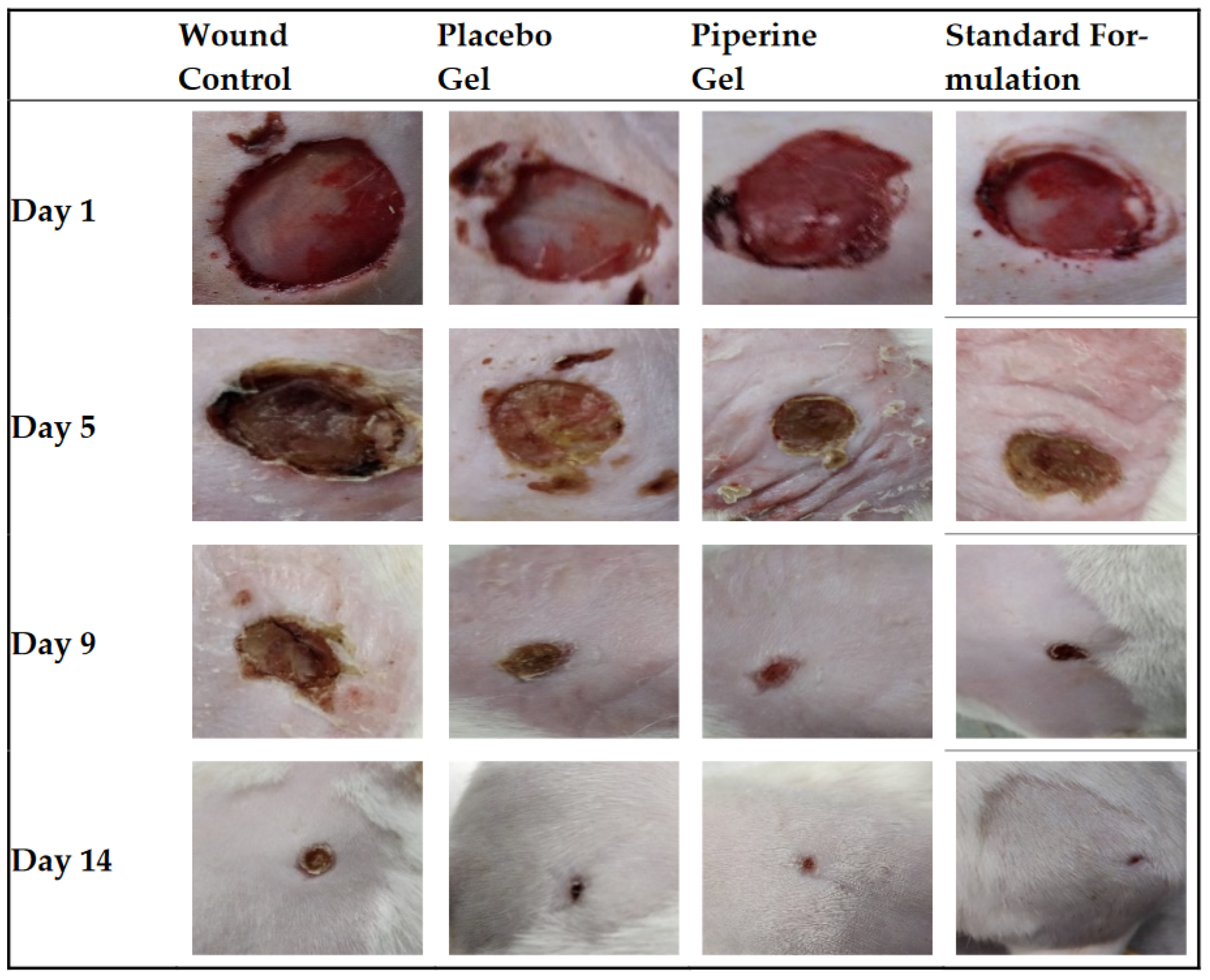
  152. Alsareii, S.A.; Ahmad, J.; Umar, A.; Ahmad, M.Z.; Shaikh, I.A. Enhanced In Vivo Wound Healing Efficacy of a Novel Piperine-Containing Bioactive Hydrogel in Excision Wound Rat Model. Molecules 2023, 28, 545. [Google Scholar] [CrossRef] [PubMed]
  153. Meza-Valle, K.Z.; Saucedo-Acuña, R.A.; Tovar-Carrillo, K.L.; Cuevas-González, J.C.; Zaragoza-Contreras, E.A.; Melgoza-Lozano, J. Characterization and Topical Study of Aloe vera Hydrogel on Wound-Healing Process. Polymers 2021, 13, 3958. [Google Scholar] [CrossRef]
  154. Chelu, M.; Musuc, A.M.; Aricov, L.; Ozon, E.A.; Iosageanu, A.; Stefan, L.M.; Prelipcean, A.-M.; Popa, M.; Moreno, J.C. Antibacterial Aloe vera Based Biocompatible Hydrogel for Use in Dermatological Applications. Int. J. Mol. Sci. 2023, 24, 3893. [Google Scholar] [CrossRef]
  155. Khorasani, G.; Hosseinimehr, S.J.; Azadbakht, M.; Zamani, A.; Mahdavi, M.R. Aloe versus silver sulfadiazine creams for second-degree burns: A randomized controlled study. Surg. Today 2009, 39, 587–591. [Google Scholar] [CrossRef] [PubMed]
  156. Shahzad, M.N.; Ahmed, N. Effectiveness of Aloe vera gel compared with 1% silver sulphadiazine cream as burn wound dressing in second degree burns. J. Pak. Med. Assoc. 2013, 63, 225–230. [Google Scholar]
  157. Panahi, Y.; Beiraghdar, F.; Akbari, H.; Bekhradi, H.; Taghizadeh, M.; Sahebkar, A. A herbal cream consisting of Aloe vera, Lavandula stoechas, and Pelargonium roseum as an alternative for silver sulfadiazine in burn management. Asian Biomed. 2012, 6, 273–278. [Google Scholar] [CrossRef]
  158. Avijgan, M.; Alinaghian, M.; Esfahani, M.H. Aloe vera Gel as a Traditional and Complementary Method for Chronic Skin Burn: A Case Report. Adv. Infect. Dis. 2017, 7, 19–25. [Google Scholar] [CrossRef] [Green Version]
  159. Tungkasamit, T.; Chakrabandhu, S.; Samakgarn, V.; Kunawongkrit, N.; Jirawatwarakul, N.; Chumachote, A.; Chitapanarux, I. Reduction in severity of radiation-induced dermatitis in head and neck cancer patients treated with topical Aloe vera gel: A randomized multicenter double-blind placebo-controlled trial. Eur. J. Oncol. Nurs. 2022, 59, 102164. [Google Scholar] [CrossRef]
  160. Dal’Belo, S.E.; Gaspar, L.R.; Maia Campos, P.M. Moisturizing effect of cosmetic formulations containing Aloe vera extract in different concentrations assessed by skin bioengineering techniques. Ski. Res. Technol. 2006, 12, 241–246. [Google Scholar] [CrossRef]
  161. Zhang, L.; Tizard, I.R. Activation of a mouse macrophage cell line by acemannan: The major carbohydrate fraction from Aloe vera gel. Immunopharmacology 1996, 35, 119–128. [Google Scholar] [CrossRef] [PubMed]
  162. Ma, Y.; Tang, T.; Sheng, L.; Wang, Z.; Tao, H.; Zhang, Q.; Qi, Z. Aloin suppresses lipopolysaccharide-induced inflammation by inhibiting JAK1-STAT1/3 activation and ROS production in RAW264. 7 cells. Int. J. Mol. Med. 2018, 42, 1925–1934. [Google Scholar]
  163. Leng, H.; Pu, L.; Xu, L.; Shi, X.; Ji, J.; Chen, K. Effects of Aloe polysaccharide, a polysaccharide extracted from Aloe vera, on TNF-α-induced HaCaT cell proliferation and the underlying mechanism in psoriasis. Mol. Med. Rep. 2018, 18, 3537–3543. [Google Scholar] [CrossRef] [Green Version]
  164. Jiang, K.; Guo, S.; Yang, C.; Yang, J.; Chen, Y.; Shaukat, A.; Deng, G. Barbaloin protects against lipopolysaccharide (LPS)-induced acute lung injury by inhibiting the ROS-mediated PI3K/AKT/NF-κB pathway. Int. Immunopharm. 2018, 64, 140–150. [Google Scholar] [CrossRef] [PubMed]
  165. Paul, S.; Modak, D.; Chattaraj, S.; Nandi, D.; Sarkar, A.; Roy, J.; Chaudhuri, T.K.; Bhattacharjee, S. Aloe vera gel homogenate shows anti-inflammatory activity through lysosomal membrane stabilization and downregulation of TNF-α and Cox-2 gene expressions in inflammatory arthritic animals. Futur. J. Pharm. Sci. 2021, 7, 12. [Google Scholar] [CrossRef]
  166. Ahluwalia, B.; Magnusson, M.K.; Isaksson, S.; Larsson, F.; Öhman, L. Effects of Aloe barbadensis Mill. extract (AVH200®) on human blood T cell activity in vitro. J. Ethnopharm. 2016, 179, 301–309. [Google Scholar] [CrossRef]
  167. Moriyama, M.; Moriyama, H.; Uda, J.; Kubo, H.; Nakajima, Y.; Goto, A.; Akaki, J.; Yoshida, I.; Matsuoka, N.; Hayakawa, T. Beneficial effects of the genus aloe on wound healing, cell proliferation, and differentiation of epidermal keratinocytes. PLoS ONE 2016, 11, e0164799. [Google Scholar] [CrossRef] [Green Version]
  168. Liu, L.Y.; Chen, X.D.; Wu, B.; Jiang, Q. Influence of Aloe polysaccharide on proliferation and hyaluronic acid and hydroxyproline secretion of human fibroblasts in vitro. J. Chin. Integr. Med. 2010, 8, 256–262. [Google Scholar] [CrossRef]
  169. Hamid, A.A.A.; Soliman, M.F. Effect of topical Aloe vera on the process of healing of full-thickness skin burn: A histological and immunohistochemical study. J. Histol. Histopathol. 2015, 2, 3. [Google Scholar] [CrossRef] [Green Version]
  170. Hashemi, S.A.; Madani, S.A.; Abediankenari, S. The Review on properties of Aloe vera in healing of cutaneous wounds. BioMed Res. Int. 2015, 2015, 714216. [Google Scholar] [CrossRef] [Green Version]
  171. Maenthaisong, R.; Chaiyakunapruk, N.; Niruntraporn, S.; Kongkaew, C. The efficacy of Aloe vera used for burn wound healing: A systematic review. Burns 2007, 33, 713–718. [Google Scholar] [CrossRef]
  172. Moghbel, A.; Ghalambor, A.; Allipanah, S. Wound healing and toxicity evaluation of Aloe vera cream on outpatients with second degree burns. Iran. J. Pharm. Sci. Summer 2007, 3, 157–160. [Google Scholar]
  173. Somboonwong, J.; Thanamittramanee, S.; Jariyapongskul, A.; Patumraj, S. Therapeutic effects of Aloe vera on cutaneous microcirculation and wound healing in second degree burn model in rats. J. Med. Assoc. Thail. 2000, 83, 417–425. [Google Scholar]
  174. Fox, L.T.; du Plessis, J.; Gerber, M.; van Zyl, S.; Boneschans, B.; Hamman, J.H. In Vivo skin hydration and anti-erythema effects of Aloe vera, Aloe ferox and Aloe marlothii gel materials after single and multiple applications. Pharmacogn. Mag. 2014, 10 (Suppl. S2), S392–S403. [Google Scholar] [CrossRef] [Green Version]
  175. Moon, E.-J.; Lee, Y.M.; Lee, O.-H.; Lee, M.-J.; Lee, S.-K.; Chung, M.-H.; Park, Y.-I.; Sung, C.-K.; Choi, J.-S.; Kim, K.-W. A ncovel angiogenic factor derived from Aloe vera gel: β-sitosterol, a plant sterol. Angiogenesis 1999, 3, 117–123. [Google Scholar] [CrossRef]
  176. Choi, S.; Kim, K.-W.; Choi, J.-S.; Han, S.-T.; Park, Y.-I.; Lee, S.-K.; Kim, J.-S.; Chung, M.-H. Angiogenic activity of β-sitosterol in the ischaemia/reperfusion-damaged brain of mongolian gerbil. Planta Med. 2002, 68, 330–335. [Google Scholar] [CrossRef]
  177. Liu, F.-W.; Liu, F.C.; Wang, Y.-R.; Tsai, H.-I.; Yu, H.-P. Aloin protects skin fibroblasts from heat stress-induced oxidative stress damage by regulating the oxidative defense system. PLoS ONE 2015, 10, e0143528. [Google Scholar] [CrossRef] [PubMed]
  178. Shanmugam, D.K.; Madhavan, Y.; Manimaran, A.; Kaliaraj, G.S.; Mohanraj, K.G.; Kandhasamy, N.; Mosas, K.K.A. Efficacy of Graphene-Based Nanocomposite Gels as a Promising Wound Healing Biomaterial. Gels 2023, 9, 22. [Google Scholar] [CrossRef] [PubMed]
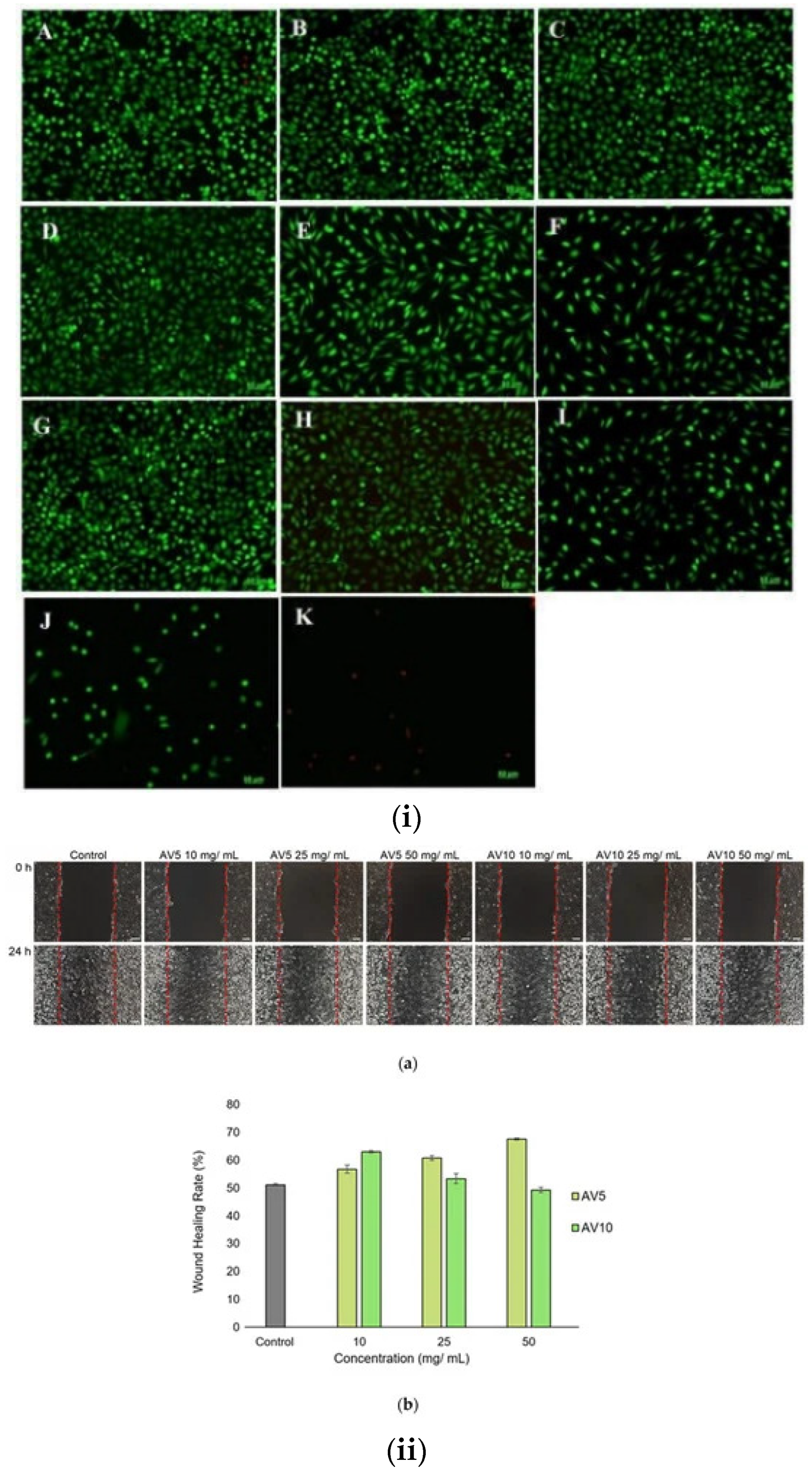
  179. Teplicki, E.; Ma, Q.; Castillo, D.E.; Zarei, M.; Hustad, A.P.; Chen, J.; Li, J. The Effects of Aloe vera on Wound Healing in Cell Proliferation, Migration, and Viability. Wounds 2018, 30, 263–268. [Google Scholar] [PubMed]
  180. Negahdari, S.; Galehdari, H.; Kesmati, M.; Rezaie, A.; Shariati, G. Wound healing activity of extracts and formulations of Aloe vera, henna, adiantum capillus-veneris, and myrrh on mouse dermal fibroblast cells. Int. J. Prev. Med. 2017, 8, 18. [Google Scholar] [CrossRef]
  181. Wahedi, H.M.; Jeong, M.; Chae, J.K.; Gil Do, S.; Yoon, H.; Kim, S.Y. Aloesin from Aloe vera accelerates skin wound healing by modulating MAPK/Rho and Smad signaling pathways in vitro and in vivo. Phytomedicine 2017, 28, 19–26. [Google Scholar] [CrossRef]
  182. Müller-Heupt, L.K.; Wiesmann, N.; Schröder, S.; Korkmaz, Y.; Vierengel, N.; Groß, J.; Dahm, R.; Deschner, J.; Opatz, T.; Brieger, J.; et al. Extracts of Rheum palmatum and Aloe vera Show Beneficial Properties for the Synergistic Improvement of Oral Wound Healing. Pharmaceutics 2022, 14, 2060. [Google Scholar] [CrossRef]
  183. Hamman, J.H. Composition and Applications of Aloe vera leaf gel. Molecules 2008, 13, 1599–1616. [Google Scholar] [CrossRef] [PubMed] [Green Version]
  184. Anilakumar, K.R.; Sudarshanakrishnam, K.R.; Chandramohan, G.; Ilaiyaraja, N.; Khanum, F.; Bawa, A.S. Effect of Aloe vera gel extract on antioxidant enzymes and azoxymethane-induced oxidative stress in rats. Indian J. Exp. Biol. 2010, 48, 837–842. [Google Scholar]
  185. Hassanpour, H. Effect of Aloe vera gel coating on antioxidant capacity, antioxidant enzyme activities and decay in raspberry fruit. LWT Food Sci. Technol. 2015, 60, 495–501. [Google Scholar] [CrossRef]
  186. Hormozi, M.; Assaei, R.; Boroujeni, M.B. The effect of Aloe vera on the expression of wound healing factors (TGFβ1 and bFGF) in mouse embryonic fibroblast cell: In vitro study. Biomed. Pharmacother. 2017, 88, 610–616. [Google Scholar] [CrossRef]
  187. Ali, J.; Khan, A.W.; Kotta, S.; Ansari, S.H.; Sharma, R.K.; Kumar, A. Formulation development, optimization and evaluation of Aloe vera gel for wound healing. Pharmacogn. Mag. 2013, 9, 6–10. [Google Scholar] [CrossRef] [Green Version]
  188. Curto, E.M.; Labelle, A.; Chandler, H.L. Aloe vera: An in vitro study of effects on corneal wound closure and collagenase activity. Vet. Ophthamol. 2014, 17, 403–410. [Google Scholar] [CrossRef]
  189. De Oliveira, A.C.L.; Tabrez, S.; Shakil, S.; Khan, M.I.; Asghar, M.N.; Matias, B.D.; de Carvalho, R.M. Mutagenic, antioxidant and wound healing properties of Aloe vera. J. Ethnopharmacol. 2018, 227, 191–197. [Google Scholar] [CrossRef]
  190. Zeng, W.M.; Barnes, C.W.; Hiro, M.E.; Parus, A.; Robson, M.C.; Payne, W.G. Aloe vera—Mechanisms of Action, Uses, and Potential Uses in Plastic Surgery and Wound Healing. Surg. Sci. 2020, 11, 312–328. [Google Scholar] [CrossRef]
  191. Na, H.S.; Song, Y.R.; Kim, S.; Heo, J.Y.; Chung, H.Y.; Chung, J. Aloin Inhibits Interleukin (IL)-1β− Stimulated IL-8 Production in KB Cells. J. Periodontol. 2016, 87, 108–115. [Google Scholar] [CrossRef] [PubMed]
  192. Reuter, J.; Jocher, A.; Stump, J.; Grossjohann, B.; Franke, G.; Schempp, C. Investigation of the anti-inflammatory potential of Aloe vera gel (97.5%) in the ultraviolet erythema test. Ski. Pharmacol. Physiol. 2008, 21, 106–110. [Google Scholar] [CrossRef] [PubMed]
  193. Li, C.-Y.; Suzuki, K.; Hung, Y.-L.; Yang, M.-S.; Yu, C.-P.; Lin, S.-P.; Hou, Y.-C.; Fang, S.-H. Aloe metabolites prevent lps-induced sepsis and inflammatory response by inhibiting mitogen-activated protein kinase activation. Am. J. Chin. Med. 2017, 45, 847–861. [Google Scholar] [CrossRef] [PubMed]
  194. Pulliero, A.; Profumo, A.; Izzotti, A.; Saccà, S.C. Release of Aloe vera Extracts from Therapeutic Lenses. Appl. Sci. 2020, 10, 9055. [Google Scholar] [CrossRef]
  195. Chelu, M.; Moreno, J.C.; Atkinson, I.; Cusu, J.P.; Rusu, A.; Bratan, V.; Aricov, L.; Anastasescu, M.; Seciu-Grama, A.-M.; Musuc, A.M. Green synthesis of bioinspired chitosan-ZnO-based polysaccharide gums hydrogels with propolis extract as novel functional natural biomaterials. Int. J. Biol. Macromol. 2022, 211, 410–424. [Google Scholar] [CrossRef]
  196. Chelu, M.; Musuc, A.M. Polymer Gels: Classification and Recent Developments in Biomedical Applications. Gels 2023, 9, 161. [Google Scholar] [CrossRef]
  197. Miguel, S.P.; Ribeiro, M.P.; Coutinho, P.; Correia, I.J. Electrospun Polycaprolactone/Aloe Vera_Chitosan Nanofibrous Asymmetric Membranes Aimed for Wound Healing Applications. Polymers 2017, 9, 183. [Google Scholar] [CrossRef] [Green Version]
  198. International Agency for Research on Cancer. Aloe vera. In Some Drugs and Herbal Products (Volume 108); International Agency for research on Cancer: Lyon, France, 2015. Available online: http://monographs.iarc.fr/ENG/Monographs/vol108/mono108.pdf (accessed on 20 May 2023).
  199. Cosmetic Ingredient Review Expert Panel (CIR). Final report on the safety assessment of aloe andongensis extract, aloe an-dongensis leaf juice, aloe arborescens leaf extract, aloe arborescens leaf juice, aloe arborescens leaf protoplasts, aloe barbadensis flower extract, aloe barbadensis leaf, aloe barbadensis leaf extract, aloe barbadensis leaf juice, aloe barbadensis leaf polysac-charides, aloe barbadensis leaf water, aloe ferox leaf extract, aloe ferox leaf juice, and aloe ferox leaf juice extract. Int. J. Toxicol. 2007, 26 (Suppl. S2), 1–50. [Google Scholar]
  200. Boudreau, M.D.; Beland, F.A. An evaluation of the biological and toxicological properties of Aloe barbadensis (miller), Aloe vera. J. Environ. Sci. Health C Environ. Carcinog. Ecotoxicol. Rev. 2006, 24, 103–154. [Google Scholar] [CrossRef] [Green Version]
  201. Boudreau, M.D.; Beland, F.A.; Nichols, J.A.; Pogribna, M. Toxicology and carcinogenesis studies of a nondecolorized whole leaf extract of Aloe barbadensis Miller (Aloe vera) in F344/N rats and B6C3F1 mice (drinking water study). Natl. Toxicol. Program Tech. Rep. Ser. 2013, 577, 1–266. [Google Scholar]
  202. Guo, X.; Mei, N. Aloe vera: A review of toxicity and adverse clinical effects. J. Environ. Sci. Health Part C 2016, 34, 77–96. [Google Scholar] [CrossRef] [PubMed]
  203. Ryall, C.; Duarah, S.; Chen, S.; Yu, H.; Wen, J. Advancements in Skin Delivery of Natural Bioactive Products for Wound Management: A Brief Review of Two Decades. Pharmaceutics 2022, 14, 1072. [Google Scholar] [CrossRef] [PubMed]
  204. Ferreira, A.S.; Macedo, C.; Silva, A.M.; Delerue-Matos, C.; Costa, P.; Rodrigues, F. Natural Products for the Prevention and Treatment of Oral Mucositis—A Review. Int. J. Mol. Sci. 2022, 23, 4385. [Google Scholar] [CrossRef] [PubMed]
Figure 1. Raw morphology of the Aloe vera plant.
Gels 09 00539 g001
Figure 2. Chemical structure of the main biocomponents isolated from Aloe vera [68].
Gels 09 00539 g002
Figure 3. Scheme of the preparation of Aloe vera gel.
Gels 09 00539 g003
Figure 4. Graphical representation of Aloe vera hydrogel network preparation [86].
Gels 09 00539 g004
Figure 5. Synthesis, optical imaging of three composite hydrogels with different concentrations of Aloe vera (5%, 10%, and 20% w/v), and evaluation of their properties [94].
Gels 09 00539 g005
Figure 6. Graphical representation of the correlation between properties and composition of Aloe vera.
Gels 09 00539 g006
Figure 7. Structure of human skin: the epidermis (which contains keratinocytes, melanocytes, and Langerhans cells) and dermis (which includes fibroblasts, neutrophils, mast cells, and dermal dendritic cells), as well as subcutaneous hypodermis (which contains mesenchymal stem cells) [145].
Gels 09 00539 g007
Figure 8. The phases of the wound-healing process [145].
Gels 09 00539 g008
Figure 9. The retention of injected treatments with hydrogel and SUSD2 + mCherry + eMSC (A) Hyd T; (B) Hyd/eMSC T (yellow dotted lines); (C,D) red arrows—zoom area of hydrogel and black arrows—zoom area of collagen; (E) SUSD2, (F) mCherry, and (G) merge image of SUSD2 + mCherry in rat vaginal sections after 1 week. Reprinted with permission from ref. [151] Copyright 2023, Elsevier.
Gels 09 00539 g009
Figure 10. Images of different types of prepared hydrogel systems. (a) Hydrogel based only on Carbopol 934. (b) Placebo bioactive hydrogel (except piperine). (c) The bioactive hydrogel contains Carbopol 934, Aloe vera, and piperine [152].
Gels 09 00539 g010
Figure 11. Phases of the wound-healing process in the untreated (control), placebo gel, piperine gel, and marketed standard formulation groups of rats. Photo for day 1, day 5, day 9, and day 14 of treatment [152].
Gels 09 00539 g011
Figure 12. Images during the wound-healing process in female Wistar rats [153].
Gels 09 00539 g012
Figure 13. Aloe vera-based hydrogel for wound healing dressing: (a) dry hydrogel (b) wet hydrogel. The healing process (c) initial time; (d) after 5 min; (e) after 20 days; (f,g) inverted vial method [154].
Gels 09 00539 g013
Figure 14. (i) Live/dead fluorescent images of L929, control, (A)—untreated and treated with AV5 (BF) and AV10 (GK) hydrogels at different concentrations for 48 h. (B,G)—10 mg/mL; (C,H)—25 mg/mL; (D,I)—50 mg/mL; (E,J)—75 mg/mL; (F,K)—100 mg/mL. (ii) Light microscope images (a) after in vitro generation of a wound for 24 h. (b) ImageJ analysis of wound closure percentage [154].
Gels 09 00539 g014
Figure 15. Agar assay (a) hydrogel; (b) hydrogel + GO; (c) hydrogel + rGO; (d) Aloe vera gel; (e) Aloe vera gel + GO; (f) Aloe vera gel + rGO against (A) Pseudomonas aeruginosa, (B) Bacillus subtilis, (C) Staphylococcus aureus, and (D) E. coli. [178].
Gels 09 00539 g015
Figure 16. Actin staining of fibroblasts with phalloidin (A); the average length of the fibroblasts (B); fluorescent actin staining (C) [182].
Gels 09 00539 g016
Table 1. Compounds found in Aloe vera [64].
TypeCompounds
Anthraquinones/anthronesAloe-emodin, aloetic-acid, anthranol, aloin A and B (collectively known as barbaloin) isobarbaloin, emodin, ester of cinnamic acid
CarbohydratesPure mannan, acetylated mannan, acetylated glucomannan, glucogalactomannan, galactan, pectic substance, arabinogalactan, galactoglucoarabinomannan, galactogalacturan, xylan, cellulose, acemannan
EnzymesAlkaline phosphatase, amylase, carboxypeptidase, carboxylase, catalase, cyclooxidase, phosphoenolpyruvate, cyclooxygenase, superoxide dismutase, lipase, oxidase
Inorganic compoundsCalcium, chlorine, phosphorous, chromium, copper, magnesium, iron, manganese, potassium, sodium, zinc
Non-essential and essential amino acidsAlanine, arginine, aspartic acid, glutamic acid, glycine, histidine, hydroxyproline, isoleucine, leucine, lysine, methionine, proline, threonine, tyrosine, valine, phenylalanine
ProteinsLectins, lectin-like substance
SaccharidesMannose, glucose, L-rhamnose, aldopentose,
VitaminsB1, B2, B6, C, β-carotene, choline, folic acid, α-tocopherol
MiscellaneousArachidonic acid, γ-linolenic acid, potassium sorbate, steroids (campesterol, cholesterol, β-sitosterol), triglycerides, triterpenoid, gibberellin, lignins, salicylic acid, uric acid
Table 2. Beneficial effects for wound healing of AV gels.
Enhanced ReportedReferences
Cell viabilitySholehvar et al. [115], Liu et al. [177]
Epitelial cell proliferationMoriyama et al. [167], Hashemi et al. [170], Shanmugan et al. [178], Teplicki et al. [179]
Epitelial cell midrationTeplicki et al. [179], Negahdari et al. [180], Wahedi et al. [181], Muller et al. [182]
Moisture retentionDal’Belo et al. [160], Hamman et al. [183]
Keratinocyte proliferationMoriyama et al. [167]
Collagen quantityHekmatpou et al. [21], Rahman et al. [64], Nabipour et al. [121], Abdel Hamid et al. [169], Hashemi et al. [170], Shanmugan et al. [178]
Collagen cross-linkingHekmatpou et al. [21], Rahman et al. [64], Abdel Hamid et al. [169], Shanmugan et al. [178]
GSH activityLiu et al. [177]
SOD activityLiu et al. [177]
Antioxidant enzyme activityAnilakumar et al. [184], Hassanpour et al. [185]
Accelerated wound healingMoriyama et al. [167], Maenthaisong et al. [171], Somboonwong et al. [173], Shanmugan et al. [178], Negahdari et al. [180], Wahedi et al. [178,179,180,181], Hormozi et al. [186], Ali et al. [187]
Growth factors productionHashemi et al. [170], Wahedi et al. [181]
Wound closureCurto et al. [188]
Lysosomal stabilizationPaul et al. [165], DeOliveira et al. [189]
Stimulate fibrotic cytokinesWahedi et al. [181], Zeng et al. [190]
AngiogenesisMoon et al. [175], Choi et al. [176]
Block the signaling of JAK1-STAT1/3Sánchez et al. [68]
Thromboxane reductionZeng et al. [21], Hekmatpou et al. [189]
Hindering IL-6Ma et al. [162], Jiang et al. [164]
Hindering IL-8Leng et al. [163], Na et al. [191]
Hindering IL-12Ahluwalia et al. [163], Leng et al. [166]
TNF alpha levels reducedLeng et al. [163], Jiang et al. [164], Paul et al. [165], Ahluwalia et al. [166]
Erythema reductionFox et al. [174], Reuter et al. [192]
Pain reductionHekmatpou et al. [21], Rompicherla et al. [119]
T cell proliferation suppressedLi et al. [193]
Lipid peroxidation reducedLiu et al. [177]
Proinflammatory cytokines reducedMa et al. [162], Leng et al. [163], Jiang et al. [164], Paul et al. [165], Ahluwalia et al. [166]
Type IV collagen degradationCurto et al. [188]
ROS production hinderedMa et al. [162], Jiang et al. [164]
Inflammation reductionHekmatpou et al. [21], Paul et al. [165]
Disclaimer/Publisher’s Note: The statements, opinions and data contained in all publications are solely those of the individual author(s) and contributor(s) and not of MDPI and/or the editor(s). MDPI and/or the editor(s) disclaim responsibility for any injury to people or property resulting from any ideas, methods, instructions or products referred to in the content.

