Autofluorescence Imaging of Living Yeast Cells with Deep-Ultraviolet Surface Plasmon Resonance

Abstract
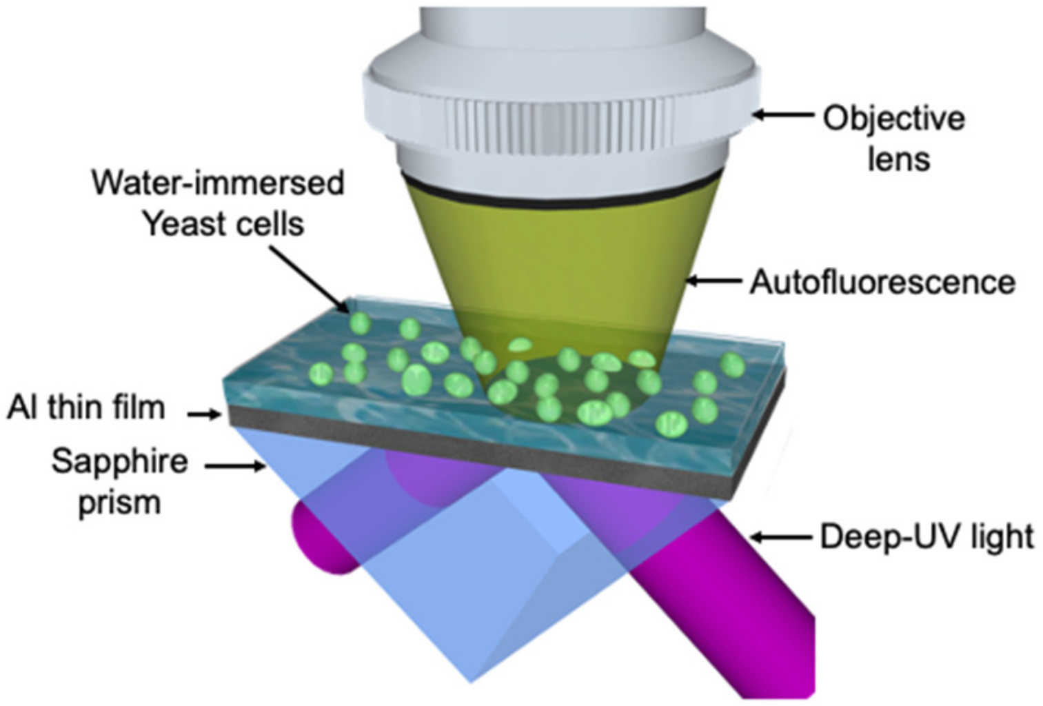
:1. Introduction
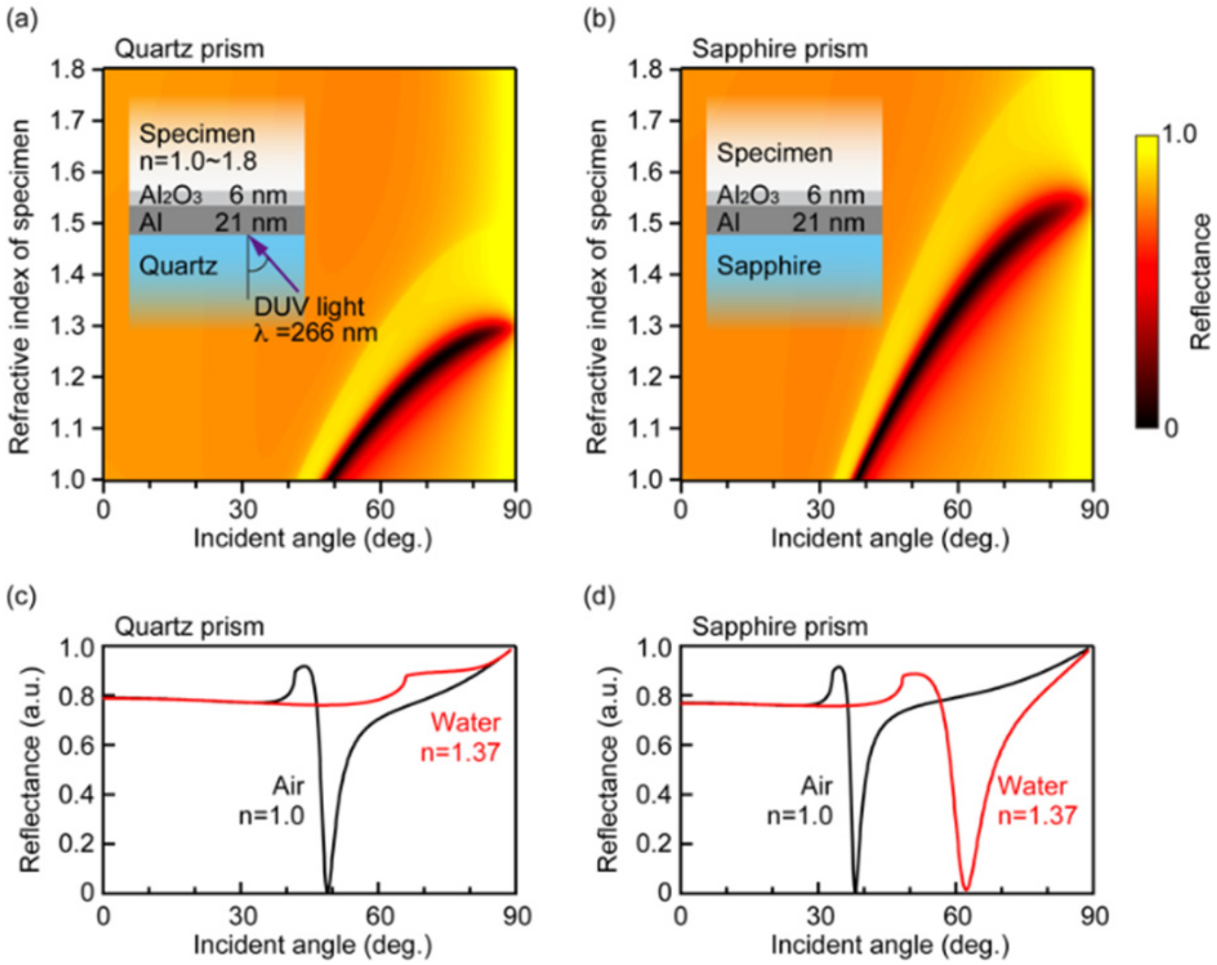
2. Theoretical Analysis
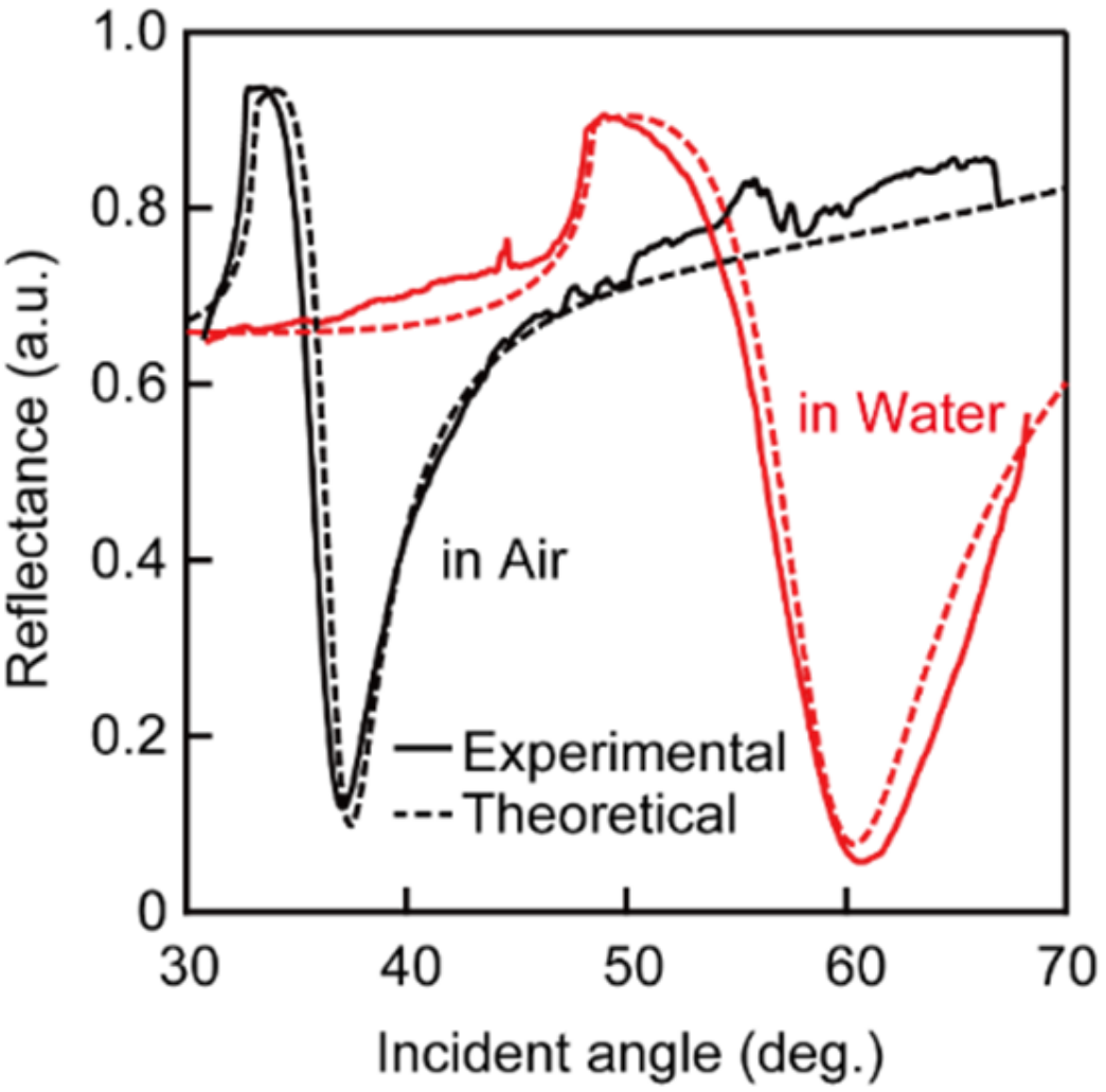
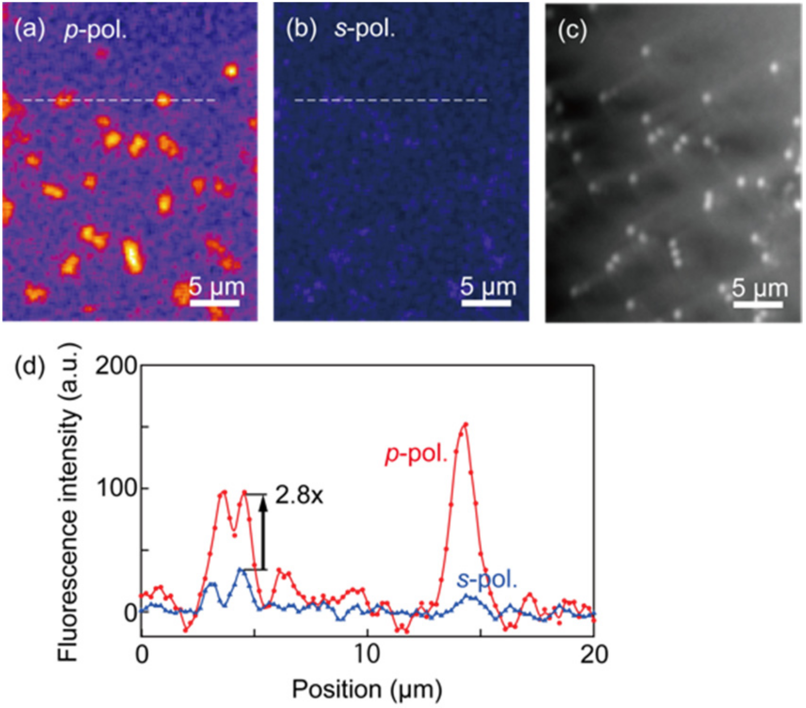
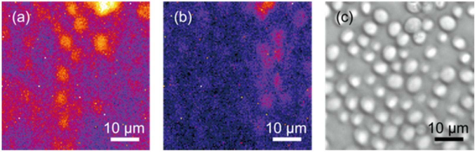
3. Experimental Methods and Results
4. Discussion
5. Conclusions
Author Contributions
Funding
Institutional Review Board Statement
Informed Consent Statement
Data Availability Statement
Conflicts of Interest
References
- Hu, S.; Ren, Y.; Wang, Y.; Li, J.; Qu, J.; Liu, L.; Ma, H.; Tang, Y. Surface plasmon resonance enhancement of photoluminescence intensity and bioimaging application of gold nanorod@CdSe/ZnS quantum dots. Beilstein J. Nanotechnol. 2019, 10, 22–31. [Google Scholar] [CrossRef] [PubMed] [Green Version]
- Uludag, Y.; Tothill, I.E. Cancer Biomarker Detection in Serum Samples Using Surface Plasmon Resonance and Quartz Crystal Microbalance Sensors with Nanoparticle Signal Amplification. Anal. Chem. 2012, 84, 5898–5904. [Google Scholar] [CrossRef] [PubMed]
- Maltais, J.-S.; Denault, J.-B.; Gendron, L.; Grandbois, M. Label-free monitoring of apoptosis by surface plasmon resonance detection of morphological changes. Apoptosis 2012, 17, 916–925. [Google Scholar] [CrossRef] [PubMed]
- Li, D.; Li, L.; Li, P.; Li, Y.; Chen, X. Apoptosis of HeLa cells induced by a new targeting photosensitizer-based PDT via a mitochondrial pathway and ER stress. OncoTargets Ther. 2015, 8, 703–711. [Google Scholar] [CrossRef] [Green Version]
- Zhang, R.; Niu, G.; Liu, Z.; Chau, J.H.; Su, H.; Lee, M.S.; Gu, Y.; Kwok, R.T.; Lam, J.W.; Tang, B.Z. Single AIEgen for multiple tasks: Imaging of dual organelles and evaluation of cell viability. Biomaterials 2020, 242, 119924. [Google Scholar] [CrossRef]
- Hanulia, T.; Inami, W.; Ono, A.; Kawata, Y. Fluorescence lifetime measurement excited with ultraviolet surface plasmon resonance. Opt. Commun. 2018, 427, 266–270. [Google Scholar] [CrossRef]
- Jamme, F.; Kascakova, S.; Villette, S.; Allouche, F.; Pallu, S.; Rouam, V.; Réfrégiers, M. Deep UV autofluorescence microscopy for cell biology and tissue histology. Biol. Cell 2013, 105, 277–288. [Google Scholar] [CrossRef]
- Monici, M. Cell and tissue autofluorescence research and diagnostic applications. Biotechnol. Annu. Rev. 2005, 11, 227–256. [Google Scholar] [CrossRef]
- Cideciyan, A.V.; Swider, M.; Jacobson, S.G. Autofluorescence imaging with near-infrared excitation: Normal-ization by reflectance to reduce signal from choroidal fluorophores. Investig. Ophthalmol. Vis. Sci. 2015, 56, 3393–3406. [Google Scholar] [CrossRef] [Green Version]
- Miljanić, S.; Frkanec, L.; Biljan, T.; Žini, Z.M.M. Recent Advances in linear and non-linear Raman spectroscopy I. J. Raman Spectrosc. 2007, 38, 1538–1553. [Google Scholar]
- Jha, S.K.; Ahmed, Z.; Agio, M.; Ekinci, Y.; Löffler, J.F. Deep-UV Surface-Enhanced Resonance Raman Scattering of Adenine on Aluminum Nanoparticle Arrays. J. Am. Chem. Soc. 2012, 134, 1966–1969. [Google Scholar] [CrossRef]
- Lenzi, E.; De Aberasturi, D.J.; Liz-Marzán, L.M. Surface-Enhanced Raman Scattering Tags for Three-Dimensional Bioimaging and Biomarker Detection. ACS Sens. 2019, 4, 1126–1137. [Google Scholar] [CrossRef]
- Shi, K.; Edwards, P.S.; Hu, J.; Xu, Q.; Wang, Y.; Psaltis, D.; Liu, Z. Holographic coherent anti-Stokes Raman scattering bio-imaging. Biomed. Opt. Express 2012, 3, 1744–1749. [Google Scholar] [CrossRef] [Green Version]
- Day, J.P.R.; Domke, K.F.; Rago, G.; Kano, H.; Hamaguchi, H.O.; Vartiainen, E.M.; Bonn, M. Quantitative coherent anti-stokes raman scattering (CARS) microscopy. J. Phys. Chem. B 2011, 115, 7713–7725. [Google Scholar] [CrossRef]
- Steuwe, C.; Kaminski, C.F.; Baumberg, J.J.; Mahajan, S. Surface Enhanced Coherent Anti-Stokes Raman Scattering on Nanostructured Gold Surfaces. Nano Lett. 2011, 11, 5339–5343. [Google Scholar] [CrossRef]
- Kikawada, M.; Ono, A.; Inami, W.; Kawata, Y. Plasmon-Enhanced Autofluorescence Imaging of Organelles in Label-Free Cells by Deep-Ultraviolet Excitation. Anal. Chem. 2016, 88, 1407–1411. [Google Scholar] [CrossRef]
- Ono, A.; Kikawada, M.; Akimoto, R.; Inami, W.; Kawata, Y. Fluorescence enhancement with deep-ultraviolet surface plasmon excitation. Opt. Express 2013, 21, 17447–17453. [Google Scholar] [CrossRef]
- Ray, K.; Chowdhury, M.H.; Lakowicz, J.R. Aluminum nanostructured films as substrates for enhanced fluo-rescence in the ultraviolet-blue spectral region. Anal. Chem. 2007, 79, 6480–6487. [Google Scholar] [CrossRef] [Green Version]
- Szmacinski, H.; Ray, K.; Lakowicz, J.R. Metal-enhanced fluorescence of tryptophan residues in proteins: Application toward label-free bioassays. Anal. Biochem. 2009, 385, 358–364. [Google Scholar] [CrossRef] [Green Version]
- Gryczynski, I.; Malicka, J.; Gryczynski, Z.; Nowaczyk, A.K.; Lakowicz, J.R. Ultraviolet Surface Plasmon-Coupled Emission Using Thin Aluminum Films. Anal. Chem. 2004, 76, 4076–4081. [Google Scholar] [CrossRef] [Green Version]
- Ono, A.; Shiroshita, N.; Kikawada, M.; Inami, W.; Kawata, Y. Enhanced photoelectron emission from aluminum thin film by surface plasmon resonance under deep-ultraviolet excitation. J. Phys. D Appl. Phys. 2014, 48, 184005. [Google Scholar] [CrossRef]
- Watanabe, Y.; Inami, W.; Kawata, Y. Deep-ultraviolet light excites surface plasmon for the enhancement of photoelectron emission. J. Appl. Phys. 2011, 109, 023112. [Google Scholar] [CrossRef] [Green Version]
- Block, I.D.; Mathias, P.C.; Ganesh, N.; Jones, S.I.; Dorvel, B.R.; Chaudhery, V.; Vodkin, L.O.; Bashir, R.; Cunningham, B.T. A detection instrument for enhanced-fluorescence and label-free imaging on photonic crystal surfaces. Opt. Express 2009, 17, 13222–13235. [Google Scholar] [CrossRef] [Green Version]
- Kikawada, M.; Ono, A.; Inami, W.; Kawata, Y. Enhanced multicolor fluorescence in bioimaging using deep-ultraviolet surface plasmon resonance. Appl. Phys. Lett. 2014, 104, 223703. [Google Scholar] [CrossRef]
- Kikawada, M.; Ono, A.; Inami, W.; Kawata, Y. Surface plasmon-enhanced fluorescence cell imaging in deep-UV region. Appl. Phys. Express 2015, 8, 072401. [Google Scholar] [CrossRef]
- Taguchi, A.; Hayazawa, N.; Furusawa, K.; Ishitobi, H.; Kawata, S. Deep-UV tip-enhanced Raman scattering. J. Raman Spectrosc. 2009, 40, 1324–1330. [Google Scholar] [CrossRef]
- Kumamoto, Y.; Taguchi, A.; Honda, M.; Watanabe, K.; Saito, Y.; Kawata, S. Indium for Deep-Ultraviolet Surface-Enhanced Resonance Raman Scattering. ACS Photonics 2014, 1, 598–603. [Google Scholar] [CrossRef] [Green Version]
- Born, M.; Wolf, E. Principle of Optics; Cambridge University Press: Cambridge, UK, 1997; Volume 7, pp. 360–409. [Google Scholar]
- Daimon, M.; Masumura, A. Measurement of the refractive index of distilled water from the near-infrared region to the ultraviolet region. Appl. Opt. 2007, 46, 3811–3820. [Google Scholar] [CrossRef]


| Material | Refractive Index@266nm |
|---|---|
| Aluminum | 0.21 + i3.14 |
| Alumina | 1.83 |
| Quartz prism | 1.52 |
| Sapphire prism | 1.83 |
| Water | 1.37 |
Publisher’s Note: MDPI stays neutral with regard to jurisdictional claims in published maps and institutional affiliations. |
© 2022 by the authors. Licensee MDPI, Basel, Switzerland. This article is an open access article distributed under the terms and conditions of the Creative Commons Attribution (CC BY) license (https://creativecommons.org/licenses/by/4.0/).
Share and Cite
Che Lah, C.N.H.; Morisawa, H.; Kobayashi, K.; Ono, A.; Inami, W.; Kawata, Y. Autofluorescence Imaging of Living Yeast Cells with Deep-Ultraviolet Surface Plasmon Resonance. Photonics 2022, 9, 424. https://doi.org/10.3390/photonics9060424
Che Lah CNH, Morisawa H, Kobayashi K, Ono A, Inami W, Kawata Y. Autofluorescence Imaging of Living Yeast Cells with Deep-Ultraviolet Surface Plasmon Resonance. Photonics. 2022; 9(6):424. https://doi.org/10.3390/photonics9060424
Chicago/Turabian StyleChe Lah, Che Nur Hamizah, Hirofumi Morisawa, Keita Kobayashi, Atsushi Ono, Wataru Inami, and Yoshimasa Kawata. 2022. "Autofluorescence Imaging of Living Yeast Cells with Deep-Ultraviolet Surface Plasmon Resonance" Photonics 9, no. 6: 424. https://doi.org/10.3390/photonics9060424