Next Issue
Volume 9, October
Previous Issue
Volume 9, August


Biomedicines, Volume 9, Issue 9 (September 2021) – 209 articles

Cover Story (view full-size image): 
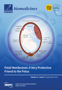
Pregnancy could be considered the paradigm of adaptation. Indeed, the mother undergoes many physiological changes to give life to a new living being. Until term, the fetus has to be protected from the outside world. This is possible partially thanks to its transitory friend: the fetal membranes. They constitute a biological barrier electing what can or cannot go through across them. Since friendship is also allowed some space, they let the fetus leave to discover something else than in utero life. However, even fetal membranes can fail, so it is important to more understand how they function. View this paper.
  • Issues are regarded as officially published after their release is announced to the table of contents alert mailing list.
  • You may sign up for e-mail alerts to receive table of contents of newly released issues.
  • PDF is the official format for papers published in both, html and pdf forms. To view the papers in pdf format, click on the "PDF Full-text" link, and use the free Adobe Reader to open them.
Order results
Result details
Section
Select all
Export citation of selected articles as:
12 pages, 1845 KiB  
Article
Nephroprotective Role of Chrysophanol in Hypoxia/Reoxygenation-Induced Renal Cell Damage via Apoptosis, ER Stress, and Ferroptosis
by Chih-Hung Lin, Han-Fang Tseng, Po-Chun Hsieh, Valeria Chiu, Ting-Yun Lin, Chou-Chin Lan, I-Shiang Tzeng, Huan-Nung Chao, Chia-Chen Hsu and Chan-Yen Kuo
Biomedicines 2021, 9(9), 1283; https://doi.org/10.3390/biomedicines9091283 - 21 Sep 2021
Cited by 17 | Viewed by 3011
Abstract
Acute kidney injury (AKI) is caused by hypoxia-reoxygenation (H/R), which is a kidney injury produced by a variety of causes, resulting in the remaining portion of the kidney function being unable to maintain the balance for performing the tasks of waste excretion metabolism, [...] Read more.
Acute kidney injury (AKI) is caused by hypoxia-reoxygenation (H/R), which is a kidney injury produced by a variety of causes, resulting in the remaining portion of the kidney function being unable to maintain the balance for performing the tasks of waste excretion metabolism, and electrolyte and acid-base balance. Many studies have reported the use of Chinese medicine to slow down the progression and alleviate the complications of chronic renal failure. Chrysophanol is a component of Rheum officinale Baill, a traditional Chinese medicine that has been clinically used to treat renal disease. We aimed to study the nephroprotective effect of chrysophanol on hypoxia/ reoxygenation (H/R)-induced cell damage. The results showed that chrysophanol prevented H/R-induced apoptosis via downregulation of cleaved Caspase-3, p-JNK, and Bax but upregulation of Bcl-2 expression. In contrast, chrysophanol attenuated H/R-induced endoplasmic reticulum (ER) stress via the downregulation of CHOP and p-IRE1α expression. Our data demonstrated that chrysophanol alleviated H/R-induced lipid ROS accumulation and ferroptosis. Therefore, we propose that chrysophanol may have a protective effect against AKI by regulating apoptosis, ER stress, and ferroptosis. Full article
(This article belongs to the Special Issue Mitochondrial Dysfunction and Oxidative Stress in Aging and Disease)
Show Figures

Figure 1

13 pages, 1783 KiB  
Systematic Review
In Vivo Experimental Endovascular Uses of Cyanoacrylate in Non-Modified Arteries: A Systematic Review
by Kévin Guillen, Pierre-Olivier Comby, Olivier Chevallier, Anne-Virginie Salsac and Romaric Loffroy
Biomedicines 2021, 9(9), 1282; https://doi.org/10.3390/biomedicines9091282 - 21 Sep 2021
Cited by 5 | Viewed by 1894
Abstract
Cyanoacrylates were first used for medical purposes during World War II to close skin wounds. Over time, medical applications were developed, specifically in the vascular field. Uses now range from extravascular instillation in vascular grafting to intravascular injection for embolization. These applications were [...] Read more.
Cyanoacrylates were first used for medical purposes during World War II to close skin wounds. Over time, medical applications were developed, specifically in the vascular field. Uses now range from extravascular instillation in vascular grafting to intravascular injection for embolization. These applications were made possible by the conduct of numerous preclinical studies involving a variety of tests and outcome measures, including angiographic and histological criteria. Cyanoacrylates were first harshly criticized by vascular surgeons, chiefly due to their fast and irreversible polymerization. Over the past five years, however, cyanoacrylates have earned an established place in endovascular interventional radiology. Given the irreversible effects of cyanoacrylates, studies in animal models are ethically acceptable only if supported by reliable preliminary data. Many animal studies of cyanoacrylates involved the experimental creation of aneurysms or arteriovenous fistulas, whose treatment by endovascular embolization was then assessed. In clinical practice, however, injection into non-modified arteries may be desirable, for instance, to deprive a tumor of its vascular supply. To help investigators in this field select the animal models and procedures that are most appropriate for their objectives, we have reviewed all published in vivo animal studies that involved the injection of cyanoacrylates into non-modified arteries to discuss their main characteristics and endpoints. Full article
Show Figures

Figure 1

18 pages, 1076 KiB  
Systematic Review
The Impact of Monoclonal Antibodies on Airway Smooth Muscle Contractility in Asthma: A Systematic Review
by Luigino Calzetta, Marina Aiello, Annalisa Frizzelli, Giuseppina Bertorelli, Beatrice Ludovica Ritondo, Paola Rogliani and Alfredo Chetta
Biomedicines 2021, 9(9), 1281; https://doi.org/10.3390/biomedicines9091281 - 21 Sep 2021
Cited by 15 | Viewed by 3491
Abstract
Airway hyperresponsiveness (AHR) represents a central pathophysiological hallmark of asthma, with airway smooth muscle (ASM) being the effector tissue implicated in the onset of AHR. ASM also exerts pro-inflammatory and immunomodulatory actions, by secreting a wide range of cytokines and chemokines. In asthma [...] Read more.
Airway hyperresponsiveness (AHR) represents a central pathophysiological hallmark of asthma, with airway smooth muscle (ASM) being the effector tissue implicated in the onset of AHR. ASM also exerts pro-inflammatory and immunomodulatory actions, by secreting a wide range of cytokines and chemokines. In asthma pathogenesis, the overexpression of several type 2 inflammatory mediators including IgE, IL-4, IL-5, IL-13, and TSLP has been associated with ASM hyperreactivity, all of which can be targeted by humanized monoclonal antibodies (mAbs). Therefore, the aim of this review was to systematically assess evidence across the literature on mAbs for the treatment of asthma with respect to their impact on the ASM contractile tone. Omalizumab, mepolizumab, benralizumab, dupilumab, and tezepelumab were found to be effective in modulating the contractility of the ASM and preventing the AHR, but no available studies concerning the impact of reslizumab on the ASM were identified from the literature search. Omalizumab, dupilumab, and tezepelumab can directly modulate the ASM in asthma, by specifically blocking the interaction between IgE, IL-4, and TSLP, and their receptors are located on the surface of ASM cells. Conversely, mepolizumab and benralizumab have prevalently indirect impacts against AHR by targeting eosinophils and other immunomodulatory effector cells promoting inflammatory processes. AHR has been suggested as the main treatable trait towards precision medicine in patients suffering from eosinophilic asthma, therefore, well-designed head-to-head trials are needed to compare the efficacy of those mAbs that directly target ASM contractility specifically against the AHR in severe asthma, namely omalizumab, dupilumab, and tezepelumab. Full article
(This article belongs to the Special Issue Molecular Targets for Biological Therapies of Severe Asthma)
Show Figures

Figure 1

12 pages, 1121 KiB  
Article
Reinitiation and Subsequent Discontinuation of Antiplatelet Treatment in Nonpersistent Older Patients with Peripheral Arterial Disease
by Martin Wawruch, Jan Murin, Tomas Tesar, Martina Paduchova, Miriam Petrova, Denisa Celovska, Beata Havelkova, Michal Trnka and Emma Aarnio
Biomedicines 2021, 9(9), 1280; https://doi.org/10.3390/biomedicines9091280 - 21 Sep 2021
Cited by 3 | Viewed by 2038
Abstract
The successful treatment of peripheral arterial disease (PAD) depends on adequate adherence to medications including antiplatelet agents. The aims of this study were (a) to identify the proportion of nonpersistent patients who reinitiated antiplatelet therapy and how many of them discontinued therapy after [...] Read more.
The successful treatment of peripheral arterial disease (PAD) depends on adequate adherence to medications including antiplatelet agents. The aims of this study were (a) to identify the proportion of nonpersistent patients who reinitiated antiplatelet therapy and how many of them discontinued therapy after reinitiation, and (b) to identify patient- and medication-related characteristics associated with the likelihood of reinitiation and discontinuation among reinitiators. The analysis of reinitiation was conducted on 3032 nonpersistent users of antiplatelet agents aged ≥65 years, with PAD newly diagnosed in 2012. Discontinuation (i.e., a treatment gap of ≥6 months without antiplatelet medication prescription) was analysed in 2006 reinitiating patients. To identify factors associated with the likelihood of reinitiation and discontinuation, Cox regression with time-dependent covariates was used. Reinitiation was recorded in 2006 (66.2%) of 3032 patients who had discontinued antiplatelet medication. Among these 2006 reinitiators, 1078 (53.7%) patients discontinued antiplatelet therapy again. Ischemic stroke and myocardial infarction during nonpersistence and bronchial asthma/chronic obstructive pulmonary disease were associated with an increased likelihood of reinitiation. University education was associated with discontinuation among reinitiators. Factors associated with the probability of reinitiation and discontinuation in reinitiators make it possible to identify older PAD patients in whom “stop-starting” behaviour may be expected. Full article
Show Figures

Figure 1

10 pages, 258 KiB  
Review
Relevance and Recommendations for the Application of Cardioplegic Solutions in Cardiopulmonary Bypass Surgery in Pigs
by Anna Glöckner, Susann Ossmann, Andre Ginther, Jagdip Kang, Michael A. Borger, Alexandro Hoyer and Maja-Theresa Dieterlen
Biomedicines 2021, 9(9), 1279; https://doi.org/10.3390/biomedicines9091279 - 21 Sep 2021
Cited by 2 | Viewed by 1830
Abstract
Cardioplegic solutions play a major role in cardiac surgery due to the fact that they create a silent operating field and protect the myocardium against ischemia and reperfusion injury. For studies on cardioplegic solutions, it is important to compare their effects and to [...] Read more.
Cardioplegic solutions play a major role in cardiac surgery due to the fact that they create a silent operating field and protect the myocardium against ischemia and reperfusion injury. For studies on cardioplegic solutions, it is important to compare their effects and to have a valid platform for preclinical testing of new cardioplegic solutions and their additives. Due to the strong anatomical and physiological cardiovascular similarities between pigs and humans, porcine models are suitable for investigating the effects of cardioplegic solutions. This review provides an overview of the results of the application of cardioplegic solutions in adult or pediatric pig models over the past 25 years. The advantages, disadvantages, limitations, and refinement strategies of these models are discussed. Full article
(This article belongs to the Special Issue Animal Models of Human Pathology: Revision, Relevance and Refinements)
11 pages, 1127 KiB  
Review
NF-κB-Dependent and -Independent (Moonlighting) IκBα Functions in Differentiation and Cancer
by Lluís Espinosa and Laura Marruecos
Biomedicines 2021, 9(9), 1278; https://doi.org/10.3390/biomedicines9091278 - 21 Sep 2021
Cited by 4 | Viewed by 2322
Abstract
IκBα is considered to play an almost exclusive role as inhibitor of the NF-κB signaling pathway. However, previous results have demonstrated that SUMOylation imposes a distinct subcellular distribution, regulation, NF-κB-binding affinity and function to the IκBα protein. In this review we discuss the [...] Read more.
IκBα is considered to play an almost exclusive role as inhibitor of the NF-κB signaling pathway. However, previous results have demonstrated that SUMOylation imposes a distinct subcellular distribution, regulation, NF-κB-binding affinity and function to the IκBα protein. In this review we discuss the main alterations of IκBα found in cancer and whether they are (most likely) associated with NF-κB-dependent or NF-κB-independent (moonlighting) activities of the protein. Full article
(This article belongs to the Special Issue Roles of NF-κB in Cancer and Their Therapeutic Approaches 2.0)
Show Figures

Figure 1

16 pages, 550 KiB  
Review
Targeting TIGIT for Immunotherapy of Cancer: Update on Clinical Development
by Anand Rotte, Srikumar Sahasranaman and Nageshwar Budha
Biomedicines 2021, 9(9), 1277; https://doi.org/10.3390/biomedicines9091277 - 21 Sep 2021
Cited by 35 | Viewed by 6385
Abstract
Immune checkpoint blockers have dramatically improved the chances of survival in patients with metastatic cancer, but only a subset of the patients respond to treatment. Search for novel targets that can improve the responder rates and overcome the limitations of adverse events commonly [...] Read more.
Immune checkpoint blockers have dramatically improved the chances of survival in patients with metastatic cancer, but only a subset of the patients respond to treatment. Search for novel targets that can improve the responder rates and overcome the limitations of adverse events commonly seen with combination therapies, like PD-1 plus CTLA-4 blockade and PD-1/PD-L1 plus chemotherapy, led to the development of monoclonal antibodies blocking T-cell immunoglobulin and ITIM domain (TIGIT), a inhibitory checkpoint receptor expressed on activated T cells and NK cells. The strategy showed potential in pre-clinical and early clinical studies, and 5 molecules are now in advanced stages of evaluation (phase II and above). This review aims to provide an overview of clinical development of anti-TIGIT antibodies and describes the factors considered and thought process during early clinical development. Critical aspects that can decide the fate of clinical programs, such as origin of the antibody, Ig isotype, FCγR binding, and the dose as well as dosing schedule, are discussed along with the summary of available efficacy and safety data from clinical studies and the challenges in the development of anti-TIGIT antibodies, such as identifying patients who can benefit from therapy and getting payer coverage. Full article
(This article belongs to the Special Issue Cancer: Immunology and Immunotherapy)
Show Figures

Figure 1

17 pages, 4727 KiB  
Article
The Effect of Acute and Chronic Thermotherapy on Type 2 Diabetic Skeletal Muscle Gene Expression and Inflammatory Markers
by Louay Bachnak, Jean Sparks, Daniel E. Newmire, Xavier F. Gonzales and Felix O. Omoruyi
Biomedicines 2021, 9(9), 1276; https://doi.org/10.3390/biomedicines9091276 - 21 Sep 2021
Viewed by 2036
Abstract
Background: Type 2 diabetes (T2D) is a chronic illness associated with resistance to or defective insulin secretion. This study investigates the effects of thermotherapy on cell viability, gene expression and inflammation in skeletal muscle cell lines. Methods: Healthy and T2D human skeletal muscle [...] Read more.
Background: Type 2 diabetes (T2D) is a chronic illness associated with resistance to or defective insulin secretion. This study investigates the effects of thermotherapy on cell viability, gene expression and inflammation in skeletal muscle cell lines. Methods: Healthy and T2D human skeletal muscle cell lines (HSMM and D-HSMM, respectively) were subjected to acute or chronic thermo-therapy (AT or CT, respectively). CT consisted of a 30 min exposure to 40 °C, three times a week for three weeks; AT was a one-time exposure. Results: A significant decrease in D-HSMM cell viability percentage followed AT; however, no significant change occurred in CT. HSMM yielded the highest elevations of genes following CT. In D-HSMM, both treatments yielded gene upregulation. Both treatments significantly down-regulated IL-1β, IL-6, IL-10 and TNF-α in HSMM. AT significantly decreased IL-1β, IL-6 and upregulated IL-10 and TNF-α levels in D-HSMM, while CT yielded a reduction in IL-4, TNF-α and an upregulation of IL-6 and IL-10. Conclusions: An increase in gene expression indicates actin activity and cellular responses, suggesting an increase in transcriptional regulation. The upregulation of IL-6 and IL-10 in D-HSMM negatively correlated with a decrease in TNF-α and IL-1β, indicating improved adverse inflammatory effects associated with the disease. Full article
(This article belongs to the Section Molecular and Translational Medicine)
Show Figures

Figure 1

17 pages, 1970 KiB  
Article
Targeting Cancer Cells Overexpressing Folate Receptors with New Terpolymer-Based Nanocapsules: Toward a Novel Targeted DNA Delivery System for Cancer Therapy
by Elena Bellotti, Maria Grazia Cascone, Niccoletta Barbani, Daniela Rossin, Raffaella Rastaldo, Claudia Giachino and Caterina Cristallini
Biomedicines 2021, 9(9), 1275; https://doi.org/10.3390/biomedicines9091275 - 21 Sep 2021
Cited by 24 | Viewed by 3126
Abstract
Chemotherapeutics represent the standard treatment for a wide range of cancers. However, these agents also affect healthy cells, thus leading to severe off-target effects. Given the non-selectivity of the commonly used drugs, any increase in the selective tumor tissue uptake would represent a [...] Read more.
Chemotherapeutics represent the standard treatment for a wide range of cancers. However, these agents also affect healthy cells, thus leading to severe off-target effects. Given the non-selectivity of the commonly used drugs, any increase in the selective tumor tissue uptake would represent a significant improvement in cancer therapy. Recently, the use of gene therapy to completely remove the lesion and avoid the toxicity of chemotherapeutics has become a tendency in oncotherapy. Ideally, the genetic material must be safely transferred from the site of administration to the target cells, without involving healthy tissues. This can be achieved by encapsulating genes into non-viral carriers and modifying their surface with ligands with high selectivity and affinity for a relevant receptor on the target cells. Hence, in this work we evaluate the use of terpolymer-based nanocapsules for the targeted delivery of DNA toward cancer cells. The surface of the nanocapsules is decorated with folic acid to actively target the folate receptors overexpressed on a variety of cancer cells. The nanocapsules demonstrate a good ability of encapsulating and releasing DNA. Moreover, the presence of the targeting moieties on the surface of the nanocapsules favors cell uptake, opening up the possibility of more effective therapies. Full article
(This article belongs to the Special Issue Feature Papers in "Biomedical Materials and Nanomedicine")
Show Figures

Figure 1

15 pages, 994 KiB  
Review
B Cells and Autoantibodies in AIRE Deficiency
by Anette S. B. Wolff, Sarah Braun, Eystein S. Husebye and Bergithe E. Oftedal
Biomedicines 2021, 9(9), 1274; https://doi.org/10.3390/biomedicines9091274 - 21 Sep 2021
Cited by 4 | Viewed by 3217
Abstract
Autoimmune polyendocrine syndrome type 1 (APS-1) is a rare but severe monogenetic autoimmune endocrine disease caused by failure of the Autoimmune Regulator (AIRE). AIRE regulates the negative selection of T cells in the thymus, and the main pathogenic mechanisms are believed to be [...] Read more.
Autoimmune polyendocrine syndrome type 1 (APS-1) is a rare but severe monogenetic autoimmune endocrine disease caused by failure of the Autoimmune Regulator (AIRE). AIRE regulates the negative selection of T cells in the thymus, and the main pathogenic mechanisms are believed to be T cell-mediated, but little is known about the role of B cells. Here, we give an overview of the role of B cells in thymic and peripheral tolerance in APS-1 patients and different AIRE-deficient mouse models. We also look closely into which autoantibodies have been described for this disorder, and their implications. Based on what is known about B cell therapy in other autoimmune disorders, we outline the potential of B cell therapies in APS-1 and highlight the unresolved research questions to be answered. Full article
Show Figures

Figure 1

14 pages, 1925 KiB  
Article
Hydrogen Sulfide Inhibits TMPRSS2 in Human Airway Epithelial Cells: Implications for SARS-CoV-2 Infection
by Giulia Pozzi, Elena Masselli, Giuliana Gobbi, Prisco Mirandola, Luis Taborda-Barata, Luca Ampollini, Paolo Carbognani, Cristina Micheloni, Francesco Corazza, Daniela Galli, Cecilia Carubbi and Marco Vitale
Biomedicines 2021, 9(9), 1273; https://doi.org/10.3390/biomedicines9091273 - 20 Sep 2021
Cited by 22 | Viewed by 3210
Abstract
The COVID-19 pandemic has now affected around 190 million people worldwide, accounting for more than 4 million confirmed deaths. Besides ongoing global vaccination, finding protective and therapeutic strategies is an urgent clinical need. SARS-CoV-2 mostly infects the host organism via the respiratory system, [...] Read more.
The COVID-19 pandemic has now affected around 190 million people worldwide, accounting for more than 4 million confirmed deaths. Besides ongoing global vaccination, finding protective and therapeutic strategies is an urgent clinical need. SARS-CoV-2 mostly infects the host organism via the respiratory system, requiring angiotensin-converting enzyme 2 (ACE2) and transmembrane protease serine 2 (TMPRSS2) to enter target cells. Therefore, these surface proteins are considered potential druggable targets. Hydrogen sulfide (H2S) is a gasotransmitter produced by several cell types and is also part of natural compounds, such as sulfurous waters that are often inhaled as low-intensity therapy and prevention in different respiratory conditions. H2S is a potent biological mediator, with anti-oxidant, anti-inflammatory, and, as more recently shown, also anti-viral activities. Considering that respiratory epithelial cells can be directly exposed to H2S by inhalation, here we tested the in vitro effects of H2S-donors on TMPRSS2 and ACE2 expression in human upper and lower airway epithelial cells. We showed that H2S significantly reduces the expression of TMPRSS2 without modifying ACE2 expression both in respiratory cell lines and primary human upper and lower airway epithelial cells. Results suggest that inhalational exposure of respiratory epithelial cells to natural H2S sources may hinder SARS-CoV-2 entry into airway epithelial cells and, consequently, potentially prevent the virus from spreading into the lower respiratory tract and the lung. Full article
Show Figures

Figure 1

19 pages, 2150 KiB  
Article
Platelet miRNA Biosignature Discriminates between Dementia with Lewy Bodies and Alzheimer’s Disease
by Ana Gámez-Valero, Jaume Campdelacreu, Dolores Vilas, Lourdes Ispierto, Jordi Gascón-Bayarri, Ramón Reñé, Ramiro Álvarez, Maria P. Armengol, Francesc E. Borràs and Katrin Beyer
Biomedicines 2021, 9(9), 1272; https://doi.org/10.3390/biomedicines9091272 - 20 Sep 2021
Cited by 9 | Viewed by 2880
Abstract
Dementia with Lewy bodies (DLB) is one of the most common causes of degenerative dementia, after Alzheimer’s disease (AD), and presents pathological and clinical overlap with both AD and Parkinson’s disease (PD). Consequently, only one in three DLB cases is diagnosed correctly. Platelets, [...] Read more.
Dementia with Lewy bodies (DLB) is one of the most common causes of degenerative dementia, after Alzheimer’s disease (AD), and presents pathological and clinical overlap with both AD and Parkinson’s disease (PD). Consequently, only one in three DLB cases is diagnosed correctly. Platelets, previously related to neurodegeneration, contain microRNAs (miRNAs) whose analysis may provide disease biomarkers. Here, we profiled the whole platelet miRNA transcriptome from DLB patients and healthy controls. Differentially expressed miRNAs were further validated in three consecutive studies from 2017 to 2019 enrolling 162 individuals, including DLB, AD, and PD patients, and healthy controls. Results comprised a seven-miRNA biosignature, showing the highest diagnostic potential for the differentiation between DLB and AD. Additionally, compared to controls, two miRNAs were down-regulated in DLB, four miRNAs were up-regulated in AD, and two miRNAs were down-regulated in PD. Predictive target analysis identified three disease-specific clusters of pathways as a result of platelet-miRNA deregulation. Our cross-sectional study assesses the identification of a novel, highly specific and sensitive platelet-associated miRNA-based biosignature, which distinguishes DLB from AD. Full article
Show Figures

Figure 1

11 pages, 602 KiB  
Review
Modern Approaches to Lower Lipoprotein(a) Concentrations and Consequences for Cardiovascular Diseases
by Victoria A. Korneva, Tatjana Yurjevna Kuznetsova and Ulrich Julius
Biomedicines 2021, 9(9), 1271; https://doi.org/10.3390/biomedicines9091271 - 20 Sep 2021
Cited by 19 | Viewed by 6677
Abstract
Lipoprotein(a) (Lp(a)) is a low density lipoprotein particle that is associated with poor cardiovascular prognosis due to pro-atherogenic, pro-thrombotic, pro-inflammatory and pro-oxidative properties. Traditional lipid-lowering therapy does not provide a sufficient Lp(a) reduction. For PCSK9 inhibitors a small reduction of Lp(a) levels could [...] Read more.
Lipoprotein(a) (Lp(a)) is a low density lipoprotein particle that is associated with poor cardiovascular prognosis due to pro-atherogenic, pro-thrombotic, pro-inflammatory and pro-oxidative properties. Traditional lipid-lowering therapy does not provide a sufficient Lp(a) reduction. For PCSK9 inhibitors a small reduction of Lp(a) levels could be shown, which was associated with a reduction in cardiovascular events, independently of the effect on LDL cholesterol. Another option is inclisiran, for which no outcome data are available yet. Lipoprotein apheresis acutely and in the long run decreases Lp(a) levels and effectively improves cardiovascular prognosis in high-risk patients who cannot be satisfactorily treated with drugs. New drugs inhibiting the synthesis of apolipoprotein(a) (an antisense oligonucleotide (Pelacarsen) and two siRNA drugs) are studied. Unlike LDL-cholesterol, for Lp(a) no target value has been defined up to now. This overview presents data of modern capabilities of cardiovascular risk reduction by lowering Lp(a) level. Full article
Show Figures

Figure 1

22 pages, 4163 KiB  
Article
WIN55,212-2 Attenuates Cognitive Impairments in AlCl3 + d-Galactose-Induced Alzheimer’s Disease Rats by Enhancing Neurogenesis and Reversing Oxidative Stress
by Onesimus Mahdi, Samaila Musa Chiroma, Mohamad Taufik Hidayat Baharuldin, Nurul Huda Mohd Nor, Che Norma Mat Taib, Saravanan Jagadeesan, Shamala Devi and Mohamad Aris Mohd Moklas
Biomedicines 2021, 9(9), 1270; https://doi.org/10.3390/biomedicines9091270 - 19 Sep 2021
Cited by 12 | Viewed by 4005
Abstract
Neurotransmission and cognitive dysfunctions have been linked to old age disorders including Alzheimer’s disease (AD). Aluminium is a known neurotoxic metal, whereas d-galactose (d-gal) has been established as a senescence agent. WIN55,212-2 (WIN), is a potent cannabinoid agonist which partially [...] Read more.
Neurotransmission and cognitive dysfunctions have been linked to old age disorders including Alzheimer’s disease (AD). Aluminium is a known neurotoxic metal, whereas d-galactose (d-gal) has been established as a senescence agent. WIN55,212-2 (WIN), is a potent cannabinoid agonist which partially restores neurogenesis in aged rats. The current study aimed to explore the therapeutic potentials of WIN on Aluminium chloride (AlCl3) and d-gal-induced rat models with cognitive dysfunction. Healthy male albino Wistar rats weighing between 200–250 g were injected with d-gal 60 mg/kg intra peritoneally (i.p), while AlCl3 (200 mg/kg) was orally administered once daily for 10 consecutive weeks. Subsequently, from weeks 8–11 rats were co-administered with WIN (0.5, 1 and 2 mg/kg/day) and donepezil 1 mg/kg. The cognitive functions of the rats were assessed with a Morris water maze (MWM). Furthermore, oxidative stress biomarkers; malondialdehyde (MDA), superoxide dismutase (SOD), glutathione (GSH) and neurogenesis markers: Nestin and glial fibrillary acidic protein (GFAP) were also evaluated, as well as the histology of the hippocampus. The results revealed that rats exposed to AlCl3 and d-gal alone showed cognitive impairments and marked neuronal loss (p < 0.05) in their hippocampal conus ammonis 1 (CA1). Additionally, a significant decrease in the expressions of GFAP and Nestin was also observed, including increased levels of MDA and decreased levels of SOD and GSH. However, administration of WIN irrespective of the doses given reversed the cognitive impairments and the associated biochemical derangements. As there were increases in the levels SOD, GSH, Nestin and GFAP (p < 0.05), while a significant decrease in the levels of MDA was observed, besides attenuation of the aberrant cytoarchitecture of the rat’s hippocampi. The biochemical profiles of the WIN-treated rats were normal. Thus, these findings offer possible scientific evidence of WIN being an effective candidate in the treatment of AD-related cognitive deficits. Full article
(This article belongs to the Special Issue The Therapeutic Effects of Cannabinoids in Neurodegenerative Diseases)
Show Figures

Figure 1

10 pages, 845 KiB  
Article
Involvement of Angiogenesis in the Pathogenesis of Coronary Aneurysms
by Sylwia Iwańczyk, Tomasz Lehmann, Artur Cieślewicz, Artur Radziemski, Katarzyna Malesza, Michał Wrotyński, Paweł Piotr Jagodziński, Marek Grygier, Maciej Lesiak and Aleksander Araszkiewicz
Biomedicines 2021, 9(9), 1269; https://doi.org/10.3390/biomedicines9091269 - 19 Sep 2021
Cited by 6 | Viewed by 1970
Abstract
The present study aimed to evaluate the plasma concentration of pro and antiangiogenic factors and their role in the pathogenesis of coronary artery abnormal dilation (CAAD). We measured the plasma concentration of matrix metalloproteinase-8 (MMP-8), transforming growth factor beta 1 (TGF-β1), Angiopoietin-2, vascular [...] Read more.
The present study aimed to evaluate the plasma concentration of pro and antiangiogenic factors and their role in the pathogenesis of coronary artery abnormal dilation (CAAD). We measured the plasma concentration of matrix metalloproteinase-8 (MMP-8), transforming growth factor beta 1 (TGF-β1), Angiopoietin-2, vascular endothelial growth factor (VEGF), and fibroblast growth factor (FGF) using a sandwich ELISA technique in the plasma of patients with coronary artery abnormal dilation (CAAD, Group 1), coronary artery disease (CAD, Group 2), and normal coronary arteries (NCA, Group 3). Patients suffering from CAAD showed significantly higher plasma concentrations of VEGF (p = 0.002) than those from the control group. Both pathological angiogenesis and inflammation appear to be crucial in the pathogenesis of aneurysmal dilatation of the coronary arteries. Full article
(This article belongs to the Topic Compounds with Medicinal Value)
Show Figures

Figure 1

21 pages, 2902 KiB  
Review
Splice and Dice: Intronic microRNAs, Splicing and Cancer
by Alex C. H. Wong and John E. J. Rasko
Biomedicines 2021, 9(9), 1268; https://doi.org/10.3390/biomedicines9091268 - 19 Sep 2021
Cited by 8 | Viewed by 3702
Abstract
Introns span only a quarter of the human genome, yet they host around 60% of all known microRNAs. Emerging evidence indicates the adaptive advantage of microRNAs residing within introns is attributed to their complex co-regulation with transcription and alternative splicing of their host [...] Read more.
Introns span only a quarter of the human genome, yet they host around 60% of all known microRNAs. Emerging evidence indicates the adaptive advantage of microRNAs residing within introns is attributed to their complex co-regulation with transcription and alternative splicing of their host genes. Intronic microRNAs are often co-expressed with their host genes, thereby providing functional synergism or antagonism that is exploited or decoupled in cancer. Additionally, intronic microRNA biogenesis and the alternative splicing of host transcript are co-regulated and intertwined. The importance of intronic microRNAs is under-recognized in relation to the pathogenesis of cancer. Full article
(This article belongs to the Special Issue MicroRNA in Solid Tumor and Hematological Diseases 2.0)
Show Figures

Figure 1

14 pages, 18676 KiB  
Article
Secondary Cerebellar Cortex Injury in Albino Male Rats after MCAO: A Histological and Biochemical Study
by Aziza R. Alrafiah
Biomedicines 2021, 9(9), 1267; https://doi.org/10.3390/biomedicines9091267 - 18 Sep 2021
Cited by 4 | Viewed by 2984
Abstract
The present study focused on secondary injury following the middle cerebral artery (MCA) occlusion in rats not linked to the MCA’s feeding zone. This entity has been very rarely studied. Additionally, this study investigated the rates of expression of five fundamental angiogenic biomarkers [...] Read more.
The present study focused on secondary injury following the middle cerebral artery (MCA) occlusion in rats not linked to the MCA’s feeding zone. This entity has been very rarely studied. Additionally, this study investigated the rates of expression of five fundamental angiogenic biomarkers called endoglin, vascular endothelial growth factors-A (VEGF-A), endothelin-1 (ET-1), 2granulocyte colony-stimulating factor (G-CSF), and angiopoietin-using the MCA occlusion (MCAO) model. The random allocation of twelve adult male albino rats was in two groups. As a sham control group, six rats were used. This group was subjected to a sham operation without MCAO. The MCAO group consisted of six rats that were subjected to MCAO operation. After three days, the rats were sacrificed. The cerebellar specimens were immediately processed for light microscopic examination. An angiogenic biomarkers multiplex assay from multiplex was used to assess endoglin levels, VEGF-A, ET-1, angiopoietin-2, and G-CSF in serum samples. Hematoxylin and eosin-stained sections showed that the cerebellar cortex of rats of the MCAO group was more affected than the sham control group. Furthermore, Nissl stain and immunohistochemical analysis revealed an apparent increase in the number of positive immunoreactive in the cerebellar cortex and an evident decrease in Nissl granules in Purkinje cells of the MCAO rats, in contrast to the control rats. In addition, there was a significant increase in angiogenic factors VEGF-A, ET-1, angiopoietin-2, and endoglin. Interestingly, there was an increase in the G-CSF but a non-significant in the MCAO rats compared to the control rats. Furthermore, there was a significant correlation between the angiopoietin-2 and ET-1, and between G-CSF and ET-1. VEGF-A also exhibited significant positive correlations with the G-CSF serum level parameter, Endoglin, and ET-1. Rats subjected to MCAO are a suitable model to study secondary injury away from MCA’s feeding zone. Additionally, valuable insights into the association and interaction between altered angiogenic factors and acute ischemic stroke induced by MCAO in rats. Full article
Show Figures

Figure 1

27 pages, 7393 KiB  
Review
Nutraceuticals and Herbs in Reducing the Risk and Improving the Treatment of COVID-19 by Targeting SARS-CoV-2
by Priti Tagde, Sandeep Tagde, Pooja Tagde, Tanima Bhattacharya, Shams Minhaz Monzur, Md. Habibur Rahman, Pavel Otrisal, Tapan Behl, Syed Shams ul Hassan, Mohamed M. Abdel-Daim, Lotfi Aleya and Simona Bungau
Biomedicines 2021, 9(9), 1266; https://doi.org/10.3390/biomedicines9091266 - 18 Sep 2021
Cited by 24 | Viewed by 6742
Abstract
The worldwide transmission of acute respiratory syndrome coronavirus 2 (SARS-CoV-2) as a deadly or devastating disease is known to affect thousands of people every day, many of them dying all over the planet. The main reason for the massive effect of COVID-19 on [...] Read more.
The worldwide transmission of acute respiratory syndrome coronavirus 2 (SARS-CoV-2) as a deadly or devastating disease is known to affect thousands of people every day, many of them dying all over the planet. The main reason for the massive effect of COVID-19 on society is its unpredictable spread, which does not allow for proper planning or management of this disease. Antibiotics, antivirals, and other prescription drugs, necessary and used in therapy, obviously have side effects (minor or significant) on the affected person, there are still not clear enough studies to elucidate their combined effect in this specific treatment, and existing protocols are sometimes unclear and uncertain. In contrast, it has been found that nutraceuticals, supplements, and various herbs can be effective in reducing the chances of SARS-CoV-2 infection, but also in alleviating COVID-19 symptoms. However, not enough specific details are yet available, and precise scientific studies to validate the approved benefits of natural food additives, probiotics, herbs, and nutraceuticals will need to be standardized according to current regulations. These alternative treatments may not have a direct effect on the virus or reduce the risk of infection with it, but these products certainly stimulate the human immune system so that the body is better prepared to fight the disease. This paper aims at a specialized literary foray precisely in the field of these “cures” that can provide real revelations in the therapy of coronavirus infection Full article
(This article belongs to the Section Drug Discovery, Development and Delivery)
Show Figures

Graphical abstract

28 pages, 6289 KiB  
Review
Stationed or Relocating: The Seesawing EMT/MET Determinants from Embryonic Development to Cancer Metastasis
by Chien-Hsiu Li, Tai-I Hsu, Yu-Chan Chang, Ming-Hsien Chan, Pei-Jung Lu and Michael Hsiao
Biomedicines 2021, 9(9), 1265; https://doi.org/10.3390/biomedicines9091265 - 18 Sep 2021
Cited by 11 | Viewed by 3746
Abstract
Epithelial and mesenchymal transition mechanisms continue to occur during the cell cycle and throughout human development from the embryo stage to death. In embryo development, epithelial-mesenchymal transition (EMT) can be divided into three essential steps. First, endoderm, mesoderm, and neural crest cells form, [...] Read more.
Epithelial and mesenchymal transition mechanisms continue to occur during the cell cycle and throughout human development from the embryo stage to death. In embryo development, epithelial-mesenchymal transition (EMT) can be divided into three essential steps. First, endoderm, mesoderm, and neural crest cells form, then the cells are subdivided, and finally, cardiac valve formation occurs. After the embryonic period, the human body will be subjected to ongoing mechanical stress or injury. The formation of a wound requires EMT to recruit fibroblasts to generate granulation tissues, repair the wound and re-create an intact skin barrier. However, once cells transform into a malignant tumor, the tumor cells acquire the characteristic of immortality. Local cell growth with no growth inhibition creates a solid tumor. If the tumor cannot obtain enough nutrition in situ, the tumor cells will undergo EMT and invade the basal membrane of nearby blood vessels. The tumor cells are transported through the bloodstream to secondary sites and then begin to form colonies and undergo reverse EMT, the so-called “mesenchymal-epithelial transition (MET).” This dynamic change involves cell morphology, environmental conditions, and external stimuli. Therefore, in this manuscript, the similarities and differences between EMT and MET will be dissected from embryonic development to the stage of cancer metastasis. Full article
(This article belongs to the Special Issue Epithelial-to-Mesenchymal Transition (EMT) in Cancer)
Show Figures

Figure 1

14 pages, 3753 KiB  
Article
Yes-Associated Protein Is Required for ZO-1-Mediated Tight-Junction Integrity and Cell Migration in E-Cadherin-Restored AGS Gastric Cancer Cells
by Seon-Young Kim, Song-Yi Park, Hwan-Seok Jang, Yong-Doo Park and Sun-Ho Kee
Biomedicines 2021, 9(9), 1264; https://doi.org/10.3390/biomedicines9091264 - 18 Sep 2021
Cited by 8 | Viewed by 3284
Abstract
Yes-associated protein (YAP) regulates numerous cellular homeostasis processes and malignant transformation. We found that YAP influences ZO-1-mediated cell migration using E-cadherin-restored EC96 cells derived from gastric malignant AGS cells. Ectopic expression of E-cadherin enhanced straightforward migration of cells, in comparison to the meandering [...] Read more.
Yes-associated protein (YAP) regulates numerous cellular homeostasis processes and malignant transformation. We found that YAP influences ZO-1-mediated cell migration using E-cadherin-restored EC96 cells derived from gastric malignant AGS cells. Ectopic expression of E-cadherin enhanced straightforward migration of cells, in comparison to the meandering movement of parental AGS cells. In EC96 cells, YAP and ZO-1 expression increased but nuclear YAP levels and activity were reduced. Nuclear factor-κB (NF-κB) mediated the increase in ZO-1 expression, possibly stabilizing cytoplasmic YAP post-translationally. Downregulation of YAP expression using siYAP RNA or stable knock-down inhibited straightforward cell migration by fragmenting ZO-1 containing tight junctions (TJs) but not adherens junctions, implying involvement of YAP in ZO-1-mediated cell migration. The association of YAP with ZO-1 was mediated by angiomotin (AMOT) because downregulation of AMOT dissociated YAP from ZO-1 and reduced cell migration. E-cadherin restoration in malignant cancer cells induced NF-κB signaling to enhance ZO-1 expression and subsequently stabilize YAP. At high expression levels, YAP associates with ZO-1 via AMOT at TJs, influencing ZO-1-mediated cell migration and maintaining TJ integrity. Full article
(This article belongs to the Special Issue Advanced Research in Cell Motility)
Show Figures

Figure 1

16 pages, 4081 KiB  
Article
Pan-Cancer Analysis Reveals Common and Specific Relationships between Intragenic miRNAs and Their Host Genes
by Baohong Liu, Yu Shyr and Qi Liu
Biomedicines 2021, 9(9), 1263; https://doi.org/10.3390/biomedicines9091263 - 18 Sep 2021
Cited by 2 | Viewed by 1973
Abstract
MicroRNAs (miRNAs) are small endogenous non-coding RNAs that play important roles in regulating gene expression. Most miRNAs are located within or close to genes (host). miRNAs and their host genes have either coordinated or independent transcription. We performed a comprehensive investigation on co-transcriptional [...] Read more.
MicroRNAs (miRNAs) are small endogenous non-coding RNAs that play important roles in regulating gene expression. Most miRNAs are located within or close to genes (host). miRNAs and their host genes have either coordinated or independent transcription. We performed a comprehensive investigation on co-transcriptional patterns of miRNAs and host genes based on 4707 patients across 21 cancer types. We found that only 11.6% of miRNA-host pairs were co-transcribed consistently and strongly across cancer types. Most miRNA-host pairs showed a strong coexpression only in some specific cancer types, demonstrating a high heterogenous pattern. For two particular types of intergenic miRNAs, readthrough and divergent miRNAs, readthrough miRNAs showed higher coexpression with their host genes than divergent ones. miRNAs located within non-coding genes had tighter co-transcription with their hosts than those located within protein-coding genes, especially exonic and junction miRNAs. A few precursor miRNAs changed their dominate form between 5′ and 3′ strands in different cancer types, including miR-486, miR-99b, let-7e, miR-125a, let-7g, miR-339, miR-26a, miR-16, and miR-218, whereas only two miRNAs with multiple host genes switched their co-transcriptional partner in different cancer types (miR-219a-1 with SLC39A7/HSD17B8 and miR-3615 with RAB37/SLC9A3R1). miRNAs generated from distinct precursors (such as miR-125b from miR-125b-1 or miR-125b-2) were more likely to have cancer-dependent main contributors. miRNAs and hosts were less co-expressed in KIRC than other cancer types, possibly due to its frequent VHL mutations. Our findings shed new light on miRNA biogenesis and cancer diagnosis and treatments. Full article
(This article belongs to the Special Issue MicroRNA in Solid Tumor and Hematological Diseases 2.0)
Show Figures

Figure 1

14 pages, 1780 KiB  
Article
Role of UPR Sensor Activation in Cell Death–Survival Decision of Colon Cancer Cells Stressed by DPE Treatment
by Rossella Benedetti, Maria Saveria Gilardini Montani, Maria Anele Romeo, Andrea Arena, Roberta Santarelli, Gabriella D’Orazi and Mara Cirone
Biomedicines 2021, 9(9), 1262; https://doi.org/10.3390/biomedicines9091262 - 18 Sep 2021
Cited by 11 | Viewed by 2146
Abstract
Polyphenols have been shown to possess several beneficial properties, including properties involved in the prevention or treatment of cancer. Among these polyphenols, a leading role is played by dihydroxyphenylethanol (DPE), the most powerful antioxidant compound contained in the olive oil. DPE has been [...] Read more.
Polyphenols have been shown to possess several beneficial properties, including properties involved in the prevention or treatment of cancer. Among these polyphenols, a leading role is played by dihydroxyphenylethanol (DPE), the most powerful antioxidant compound contained in the olive oil. DPE has been previously reported to induce endoplasmic reticulum (ER) stress and to reduce cell survival in colon cancer, one of the most common and aggressive cancers in developed countries. In this study, we further investigated the activation of UPR by DPE and explored the roles of the three UPR sensors, inositol-requiring enzyme (IRE) 1 alpha, protein kinase RNA-like endoplasmic reticulum kinase (PERK), and activating transcription factor (ATF6), in the cell death–survival decision of wt and mutp53 colon cancer cells and the underlying mechanisms involved. We also unveiled a new interplay between ATF6 and wt, as well as mutp53, which may have important implications in cancer therapy. Full article
(This article belongs to the Section Biomedical Materials and Nanomedicine)
Show Figures

Figure 1

14 pages, 3587 KiB  
Article
Complex Electromagnetic Fields Reduce Candida albicans Planktonic Growth and Its Adhesion to Titanium Surfaces
by Simonetta D’Ercole, Silvia Di Lodovico, Giovanna Iezzi, Tania Vanessa Pierfelice, Emira D’Amico, Alessandro Cipollina, Adriano Piattelli, Luigina Cellini and Morena Petrini
Biomedicines 2021, 9(9), 1261; https://doi.org/10.3390/biomedicines9091261 - 18 Sep 2021
Cited by 9 | Viewed by 2282
Abstract
This study evaluates the effects of different programs of complex electromagnetic fields (C.M.F.s) on Candida albicans, in planktonic and sessile phase and on human gingival fibroblasts (HGF cells). In vitro cultures of C. albicans ATCC 10231 and HGF cells were exposed to [...] Read more.
This study evaluates the effects of different programs of complex electromagnetic fields (C.M.F.s) on Candida albicans, in planktonic and sessile phase and on human gingival fibroblasts (HGF cells). In vitro cultures of C. albicans ATCC 10231 and HGF cells were exposed to different cycles of C.M.F.s defined as: oxidative stress, oxidative stress/antibacterial, antibacterial, antibacterial/oxidative stress. Colony forming units (CFUs), metabolic activity, cells viability (live/dead), cell morphology, filamentation analysis, and cytotoxicity assay were performed. The broth cultures, exposed to the different C.M.F.s, were grown on titanium discs for 48 h. The quantity comparisons of adhered C. albicans on surfaces were determined by CFUs and scanning electron microscopy. The C. albicans growth could be readily controlled with C.M.F.s reducing the number of cultivable planktonic cells vs. controls, independently by the treatment applied. In particular, the antibacterial program was associated with lower levels of CFUs. The quantification of the metabolic activity was significantly lower by using the oxidative stress program. Live/dead images showed that C.M.F.s significantly decreased the viability of C. albicans. C.M.F.s inhibited C. albicans virulence traits reducing hyphal morphogenesis, adhesion, and biofilm formation on titanium discs. The MTS assay showed no negative effects on the viability of HGF. Independent of the adopted protocol, C.M.F.s exert antifungal and anti-virulence action against C. albicans, no cytotoxicity effects on HGF and can be useful in the prevention and treatment of yeast biofilm infections. Full article
(This article belongs to the Special Issue Approaches to Fight Microbial Infection-Related Biofilms)
Show Figures

Figure 1

22 pages, 6130 KiB  
Article
Tumor Necrosis Receptor Superfamily Interact with Fusion and Fission of Mitochondria of Adipose Tissue in Obese Patients without Type 2 Diabetes
by Daria Shunkina (Skuratovskaia), Alexandra Komar, Maria Vulf, Hung Vu Quang, Egor Shunkin, Elena Kirienkova, Anastasiia Dakchnevich, Danil Malkov, Pavel Zatolokin and Larisa Litvinova
Biomedicines 2021, 9(9), 1260; https://doi.org/10.3390/biomedicines9091260 - 18 Sep 2021
Cited by 5 | Viewed by 1972
Abstract
Interactions between receptors and ligands of the tumor necrosis factor superfamily (TNFSF) provide costimulatory signals that control the survival, proliferation, differentiation, and effector function of immune cells. All components of the TNF superfamily are associated with NF-kB functions that are not limited to [...] Read more.
Interactions between receptors and ligands of the tumor necrosis factor superfamily (TNFSF) provide costimulatory signals that control the survival, proliferation, differentiation, and effector function of immune cells. All components of the TNF superfamily are associated with NF-kB functions that are not limited to cell death and may promote survival in the face of adipose tissue inflammation in obesity. Inflammation dysfunction of mitochondria is a key factor associated with insulin resistance in obesity. The aim of the study was to analyze the relationship of soluble forms of receptors and ligands of the TNF superfamily in blood plasma with mitochondrial dynamics in adipose tissue (greater omentum (GO) and subcutaneous adipose tissue (Sat)) of obese patients with and without type 2 diabetes mellitus (T2DM). Increased plasma sTNF-R1, sTNF-R2, sTNFRSF8 receptors, and ligands TNFSF12, TNFSF13, TNFSF13B are characteristic of obese patients without T2DM. The TNF-a levels in blood plasma were associated with a decrease in MFN2 gene expression in GO and IL-10 in blood plasma. The TNFSF12 levels contributed to a decrease in glucose levels, a decrease in BMI, and an increase in IL-10 levels by influencing the MFN2 gene expression in GO, which supports mitochondrial fusion. Full article
(This article belongs to the Section Molecular and Translational Medicine)
Show Figures

Figure 1

21 pages, 4407 KiB  
Review
Multi-Modal Biological Destruction by Cold Atmospheric Plasma: Capability and Mechanism
by Dayun Yan, Alisa Malyavko, Qihui Wang, Kostya (Ken) Ostrikov, Jonathan H. Sherman and Michael Keidar
Biomedicines 2021, 9(9), 1259; https://doi.org/10.3390/biomedicines9091259 - 18 Sep 2021
Cited by 21 | Viewed by 4329
Abstract
Cold atmospheric plasma (CAP) is a near-room-temperature, partially ionized gas composed of reactive neutral and charged species. CAP also generates physical factors, including ultraviolet (UV) radiation and thermal and electromagnetic (EM) effects. Studies over the past decade demonstrated that CAP could effectively induce [...] Read more.
Cold atmospheric plasma (CAP) is a near-room-temperature, partially ionized gas composed of reactive neutral and charged species. CAP also generates physical factors, including ultraviolet (UV) radiation and thermal and electromagnetic (EM) effects. Studies over the past decade demonstrated that CAP could effectively induce death in a wide range of cell types, from mammalian to bacterial cells. Viruses can also be inactivated by a CAP treatment. The CAP-triggered cell-death types mainly include apoptosis, necrosis, and autophagy-associated cell death. Cell death and virus inactivation triggered by CAP are the foundation of the emerging medical applications of CAP, including cancer therapy, sterilization, and wound healing. Here, we systematically analyze the entire picture of multi-modal biological destruction by CAP treatment and their underlying mechanisms based on the latest discoveries particularly the physical effects on cancer cells. Full article
(This article belongs to the Special Issue The Advances of Cold Plasma in the Biomedicines)
Show Figures

Figure 1

14 pages, 4465 KiB  
Article
Master Regulators of Epithelial-Mesenchymal Transition and WNT Signaling Pathways in Juvenile Nasopharyngeal Angiofibromas
by Naiade Calanca, Sara Martoreli Silveira Binato, Sabrina Daniela da Silva, Helena Paula Brentani, Luiz Ubirajara Sennes, Clóvis Antonio Lopes Pinto, Maria Aparecida Custódio Domingues, Carlos Eduardo Fonseca-Alves, Claudia Aparecida Rainho and Silvia Regina Rogatto
Biomedicines 2021, 9(9), 1258; https://doi.org/10.3390/biomedicines9091258 - 18 Sep 2021
Cited by 3 | Viewed by 2607
Abstract
Juvenile nasopharyngeal angiofibroma (JNA) is a rare fibrovascular benign tumor showing an invasive growth pattern and affecting mainly male adolescents. We investigated the role of epithelial–mesenchymal transition (EMT) and WNT signaling pathways in JNA. Gene expression profiles using nine JNA paired with four [...] Read more.
Juvenile nasopharyngeal angiofibroma (JNA) is a rare fibrovascular benign tumor showing an invasive growth pattern and affecting mainly male adolescents. We investigated the role of epithelial–mesenchymal transition (EMT) and WNT signaling pathways in JNA. Gene expression profiles using nine JNA paired with four inferior nasal turbinate samples were interrogated using a customized 2.3K microarray platform containing genes mainly involved in EMT and WNT/PI3K pathways. The expression of selected genes (BCL2, CAV1, CD74, COL4A2, FZD7, ING1, LAMB1, and RAC2) and proteins (BCL2, CAV1, CD74, FZD7, RAF1, WNT5A, and WNT5B) was investigated by RT-qPCR (28 cases) and immunohistochemistry (40 cases), respectively. Among 104 differentially expressed genes, we found a significantly increased expression of COL4A2 and LAMB1 and a decreased expression of BCL2 and RAC2 by RT-qPCR. The immunohistochemistry analysis revealed a low expression of BCL2 and a negative to moderate expression of FZD7 in most samples, while increased CAV1 and RAF1 expression were detected. Moderate to strong CD74 protein expression was observed in endothelial and inflammatory cells. A significant number of JNAs (78%) presented reduced WNT5A and increased WNT5B expression. Overall, the transcript and protein profile indicated the involvement of EMT and WNT pathways in JNA. These candidates are promising druggable targets for treating JNA. Full article
(This article belongs to the Special Issue Head and Neck Tumors)
Show Figures

Graphical abstract

17 pages, 1696 KiB  
Review
The Role and Clinical Interest of Extracellular Vesicles in Pregnancy and Ovarian Cancer
by Nazanin Yeganeh Kazemi, Benoìt Gendrot, Ekaterine Berishvili, Svetomir N. Markovic and Marie Cohen
Biomedicines 2021, 9(9), 1257; https://doi.org/10.3390/biomedicines9091257 - 18 Sep 2021
Cited by 11 | Viewed by 3323
Abstract
Ovarian cancer and pregnancy are two states in which the host immune system is exposed to novel antigens. Indeed, both the tumor and placenta must invade tissues, remodel vasculature to establish a robust blood supply, and evade detection by the immune system. Interestingly, [...] Read more.
Ovarian cancer and pregnancy are two states in which the host immune system is exposed to novel antigens. Indeed, both the tumor and placenta must invade tissues, remodel vasculature to establish a robust blood supply, and evade detection by the immune system. Interestingly, tumor and placenta tissue use similar mechanisms to induce these necessary changes. One mediator is emerging as a key player in invasion, vascular remodeling, and immune evasion: extracellular vesicles (EVs). Many studies have identified EVs as a key mediator of cell-to-cell communication. Specifically, the cargo carried by EVs, which includes proteins, nucleic acids, and lipids, can interact with cells to induce changes in the target cell ranging from gene expression to migration and metabolism. EVs can promote cell division and tissue invasion, immunosuppression, and angiogenesis which are essential for both cancer and pregnancy. In this review, we examine the role of EVs in ovarian cancer metastasis, chemoresistance, and immune modulation. We then focus on the role of EVs in pregnancy with special attention on the vascular remodeling and regulation of the maternal immune system. Lastly, we discuss the clinical utility of EVs as markers and therapeutics for ovarian cancer and pre-eclampsia. Full article
(This article belongs to the Special Issue Gynecological Tumor and Placenta Development)
Show Figures

Figure 1

18 pages, 706 KiB  
Review
Dietary Patterns Influence Target Gene Expression through Emerging Epigenetic Mechanisms in Nonalcoholic Fatty Liver Disease
by Mohamed Zaiou, Rim Amrani, Bertrand Rihn and Tahar Hajri
Biomedicines 2021, 9(9), 1256; https://doi.org/10.3390/biomedicines9091256 - 18 Sep 2021
Cited by 14 | Viewed by 3450
Abstract
Nonalcoholic fatty liver disease (NAFLD) refers to the pathologic buildup of extra fat in the form of triglycerides in liver cells without excessive alcohol intake. NAFLD became the most common cause of chronic liver disease that is tightly associated with key aspects of [...] Read more.
Nonalcoholic fatty liver disease (NAFLD) refers to the pathologic buildup of extra fat in the form of triglycerides in liver cells without excessive alcohol intake. NAFLD became the most common cause of chronic liver disease that is tightly associated with key aspects of metabolic disorders, including insulin resistance, obesity, diabetes, and metabolic syndrome. It is generally accepted that multiple mechanisms and pathways are involved in the pathogenesis of NAFLD. Heredity, sedentary lifestyle, westernized high sugar saturated fat diet, metabolic derangements, and gut microbiota, all may interact on a on genetically susceptible individual to cause the disease initiation and progression. While there is an unquestionable role for gene-diet interaction in the etiopathogenesis of NAFLD, it is increasingly apparent that epigenetic processes can orchestrate many aspects of this interaction and provide additional mechanistic insight. Exciting research demonstrated that epigenetic alterations in chromatin can influence gene expression chiefly at the transcriptional level in response to unbalanced diet, and therefore predispose an individual to NAFLD. Thus, further discoveries into molecular epigenetic mechanisms underlying the link between nutrition and aberrant hepatic gene expression can yield new insights into the pathogenesis of NAFLD, and allow innovative epigenetic-based strategies for its early prevention and targeted therapies. Herein, we outline the current knowledge of the interactive role of a high-fat high-calories diet and gene expression through DNA methylation and histone modifications on the pathogenesis of NAFLD. We also provide perspectives on the advancement of the epigenomics in the field and possible shortcomings and limitations ahead. Full article
(This article belongs to the Special Issue Epigenetic Mechanisms of Environmental Diseases)
Show Figures

Graphical abstract

14 pages, 3560 KiB  
Article
CD44v6 High Membranous Expression Is a Predictive Marker of Therapy Response in Gastric Cancer Patients
by Gabriela M Almeida, Carla Pereira, Ji-Hyeon Park, Carolina Lemos, Sofia Campelos, Irene Gullo, Diana Martins, Gilza Gonçalves, Dina Leitão, João Luís Neto, Ana André, Clara Borges, Daniela Almeida, Hyuk-Joon Lee, Seong-Ho Kong, Woo Ho Kim, Fátima Carneiro, Raquel Almeida, Han-Kwang Yang and Carla Oliveira
Biomedicines 2021, 9(9), 1249; https://doi.org/10.3390/biomedicines9091249 - 18 Sep 2021
Cited by 4 | Viewed by 2022
Abstract
In gastric cancer (GC), biomarkers that define prognosis and predict treatment response remain scarce. We hypothesized that the extent of CD44v6 membranous tumor expression could predict prognosis and therapy response in GC patients. Two GC surgical cohorts, from Portugal and South Korea ( [...] Read more.
In gastric cancer (GC), biomarkers that define prognosis and predict treatment response remain scarce. We hypothesized that the extent of CD44v6 membranous tumor expression could predict prognosis and therapy response in GC patients. Two GC surgical cohorts, from Portugal and South Korea (n = 964), were characterized for the extension of CD44v6 membranous immuno-expression, clinicopathological features, patient survival, and therapy response. The value of CD44v6 expression in predicting response to treatment and its impact on prognosis was determined. High CD44v6 expression was associated with invasive features (perineural invasion and depth of invasion) in both cohorts and with worse survival in the Portuguese GC cohort (HR 1.461; 95% confidence interval 1.002–2.131). Patients with high CD44v6 tumor expression benefited from conventional chemotherapy in addition to surgery (p < 0.05), particularly those with heterogeneous CD44v6-positive and -negative populations (CD44v6_3+) (p < 0.007 and p < 0.009). Our study is the first to identify CD44v6 high membranous expression as a potential predictive marker of response to conventional treatment, but it does not clarify CD44v6 prognostic value in GC. Importantly, our data support selection of GC patients with high CD44v6-expressing tumors for conventional chemotherapy in addition to surgery. These findings will allow better stratification of GC patients for treatment, potentially improving their overall survival. Full article
(This article belongs to the Special Issue Gastrointestinal Cancers: Molecular Mechanisms and Therapies)
Show Figures

Figure 1

17 pages, 3018 KiB  
Article
When Activator and Inhibitor of PPARα Do the Same: Consequence for Differentiation of Human Intestinal Cells
by Katerina Cizkova, Tereza Foltynkova, Jiri Hanyk, Zbynek Kamencak and Zdenek Tauber
Biomedicines 2021, 9(9), 1255; https://doi.org/10.3390/biomedicines9091255 - 17 Sep 2021
Cited by 3 | Viewed by 2326
Abstract
Peroxisome proliferator-activated receptor α (PPARα) is a ligand-dependent transcription factor that plays a role in various processes including differentiation of several cell types. We investigated the role of PPARα in the differentiation of intestinal cells using HT-29 and Caco2 cell lines as a [...] Read more.
Peroxisome proliferator-activated receptor α (PPARα) is a ligand-dependent transcription factor that plays a role in various processes including differentiation of several cell types. We investigated the role of PPARα in the differentiation of intestinal cells using HT-29 and Caco2 cell lines as a model as well as human normal colon and colorectal carcinoma tissues. We detected a significant increase in PPARα expression in differentiated HT-29 cells as well as in normal surface colon epithelium where differentiated cells are localised. Thus, it seems that PPARα may play a role in differentiation of intestinal cells. Interestingly, we found that both PPARα activators (fenofibrate and WY-14643) as well as its inhibitor (GW6471) regulated proliferation and differentiation of HT-29 cells in vitro in the same way. Both compounds led to a decrease in proliferation accompanied by a significant increase in expression of villin, intestinal alkaline phosphatase (differentiation markers). Moreover, the same trend in villin expression was observed in Caco2 cells. Furthermore, villin expression was independent of subcellular localisation of PPARα. In addition, we found similar levels of PPARα expression in colorectal carcinomas in comparison to adjacent normal epithelium. All these findings support the hypothesis that differentiation of intestinal epithelium is PPARα-independent. Full article
Show Figures

Figure 1

Previous Issue
Next Issue
Back to TopTop