
Water Is an Active Element: A Randomized Double-Blind Controlled Clinical Trial Comparing Cutaneous Lipidomics in Consumers Drinking Two Different Bicarbonate-Calcic Waters (Medium-Mineral vs. Oligo-Mineral)


Abstract
:1. Introduction
2. Materials and Methods
2.1. Study Design
2.1.1. Mineral Water Evaluation
2.2. Inclusion and Exclusion Criteria
2.3. Dermatological Evaluation
2.4. Sample Processing
2.5. High-Resolution Mass Spectrometry Analysis (UHPLC-HRMS)
Chromatography of the Skin Strips
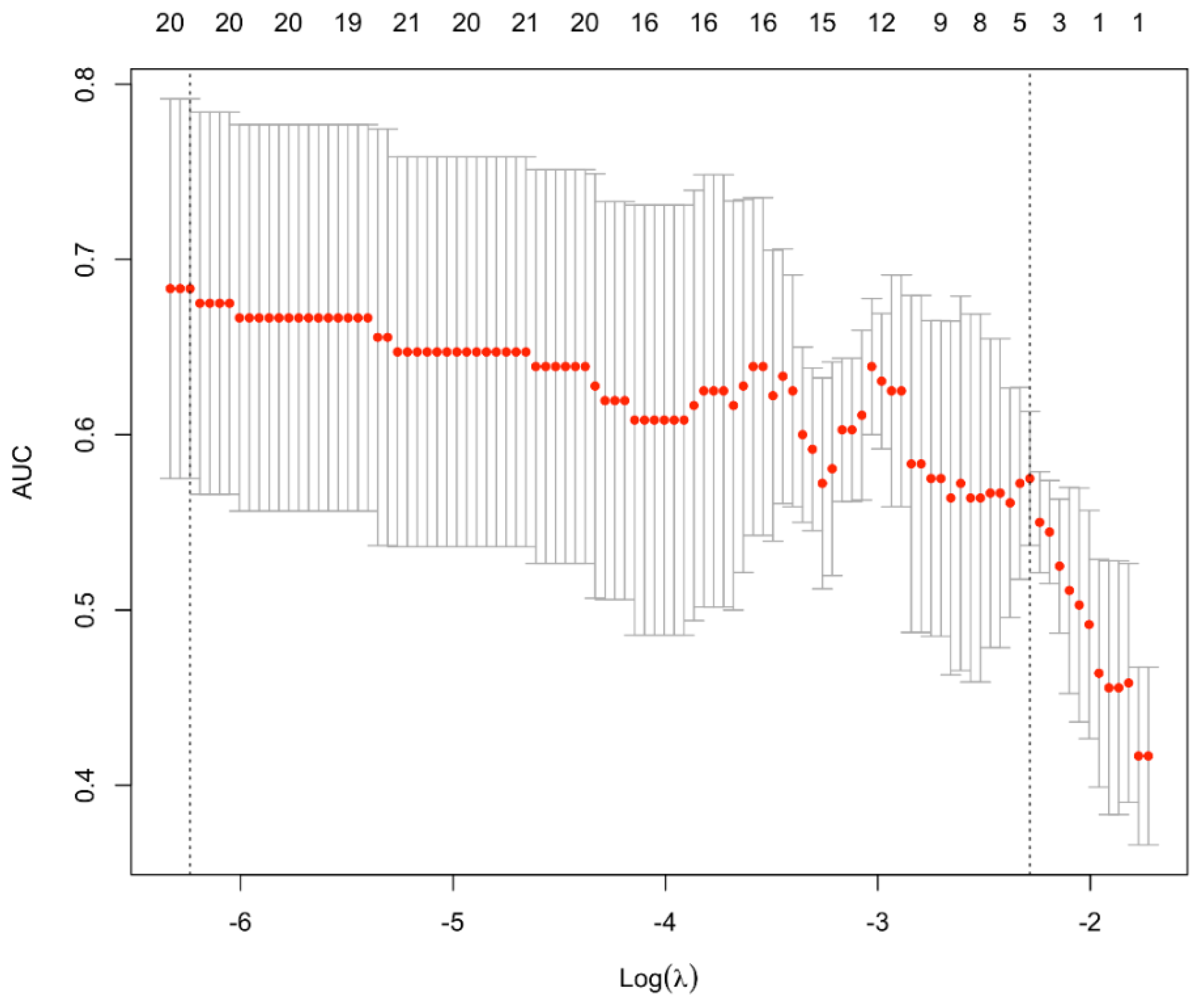
2.6. Data Analysis
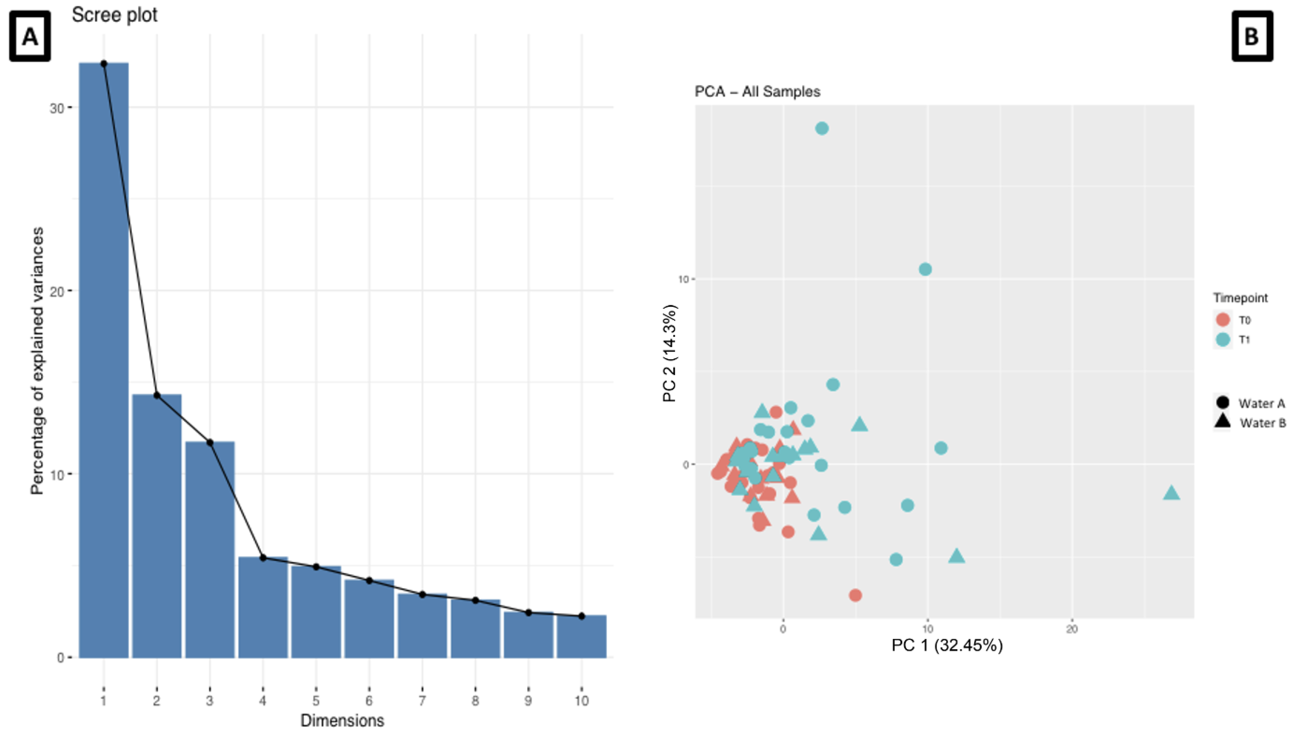
3. Results
3.1. Clinical Data
3.2. Differences in Cutaneous Lipidomics between the Two Groups Drinking Different Waters
4. Discussion
5. Conclusions
Supplementary Materials
Author Contributions
Funding
Institutional Review Board Statement
Informed Consent Statement
Data Availability Statement
Acknowledgments
Conflicts of Interest
References
- Fikri-Benbrahim, K.; Houti, A.; Lalami, A.E.O.; Flouchi, R.; El Hachlafi, N.; Houti, M.; Rachiq, S. Main Therapeutic Uses of Some Moroccan Hot Springs’ Waters. Evid.-Based Complement. Altern. Med. 2021, 2021, 5599269. [Google Scholar] [CrossRef] [PubMed]
- Silva, A.; Oliveira, A.S.; Vaz, C.V.; Correia, S.; Ferreira, R.; Breitenfeld, L.; Martinez-De-Oliveira, J.; Palmeira-De-Oliveira, R.; Pereira, C.M.F.; Cruz, M.T. Anti-inflammatory potential of Portuguese thermal waters. Sci. Rep. 2020, 10, 22313. [Google Scholar] [CrossRef]
- Fillimonova, E.; Kharitonova, N.; Baranovskaya, E.; Maslov, A.; Aseeva, A. Geochemistry and therapeutic properties of Cau-casian mineral waters: A review. Environ. Geochem. Health 2022, 44, 2281–2299. [Google Scholar] [CrossRef]
- Khalilzadeh, S.; Shirbeigi, L.; Naghizadeh, A.; Mehriardestani, M.; Shamohammadi, S.; Tabarrai, M. Use of mineral waters in the treatment of psoriasis: Perspectives of Persian and conventional medicine. Dermatol. Ther. 2019, 32, e12969. [Google Scholar] [CrossRef] [PubMed]
- Kimata, H.; Tai, H.; Nakagawa, K.; Yokoyama, Y.; Nakajima, H.; Ikegami, Y. Improvement of skin symptoms and mineral im-balance by drinking deep sea water in patients with atopic eczema/dermatitis syndrome (AEDS). Acta Med. 2002, 45, 83–84. [Google Scholar]
- Vaz, C.; Oliveira, A.; Silva, A.; Cortes, L.; Correia, S.; Ferreira, R.; Breitenfeld, L.; Martinez-De-Oliveira, J.; Palmeira-De-Oliveira, R.; Pereira, C.; et al. Protective role of Portuguese natural mineral waters on skin aging: In vitro evaluation of anti-senescence and anti-oxidant properties. Int. J. Biometeorol. 2022, 66, 2117–2131. [Google Scholar] [CrossRef]
- Karagülle, M.Z.; Karagülle, M.; Kılıç, S.; Sevinç, H.; Dündar, C.; Türkoğlu, M. In vitro evaluation of natural thermal mineral wa-ters in human keratinocyte cells: A preliminary study. Int. J. Biometeorol. 2018, 62, 1657–1661. [Google Scholar] [CrossRef]
- Lopez, D.J.; Singh, A.; Waidyatillake, N.T.; Su, J.C.; Bui, D.S.; Dharmage, S.C.; Lodge, C.J.; Lowe, A.J. The association between domestic hard water and eczema in adults from the UK Biobank cohort study. Br. J. Dermatol. 2022, 187, 704–712. [Google Scholar] [CrossRef] [PubMed]
- Caiazzo, G.; Parisi, M.; Luciano, M.A.; Di Caprio, R.; Gallo, L.; Cacciapuoti, S.; Quaranta, M.; Fabbrocini, G. Beneficial effects of Roc-chetta® oligomineral water in HaCaT keratinocytes after ultraviolet-B irradiation. Ital. J. Dermatol. Venerol. 2022, 157, 335–341. [Google Scholar]
- Tanaka, A.; Jung, K.; Matsuda, A.; Jang, H.; Kajiwara, N.; Amagai, Y.; Oida, K.; Ahn, G.; Ohmori, K.; Kang, K.G.; et al. Daily in-take of Jeju groundwater improves the skin condition of the model mouse for human atopic dermatitis. J. Dermatol. 2013, 40, 193–200. [Google Scholar] [CrossRef]
- Cacciapuoti, S.; Luciano, M.A.; Megna, M.; Annunziata, M.C.; Napolitano, M.; Patruno, C.; Scala, E.; Colicchio, R.; Pagliuca, C.; Salvatore, P.; et al. The Role of Thermal Water in Chronic Skin Diseases Management: A Review of the Literature. J. Clin. Med. 2020, 9, 3047. [Google Scholar] [CrossRef] [PubMed]
- Fujii, N.; Kataoka, Y.; Lai, Y.-F.; Shirai, N.; Hashimoto, H.; Nishiyasu, T. Ingestion of carbonated water increases middle cerebral artery blood velocity and improves mood states in resting humans exposed to ambient heat stress. Physiol. Behav. 2022, 255, 113942. [Google Scholar] [CrossRef] [PubMed]
- Hataguchi, Y.; Tai, H.; Nakajima, H.; Kimata, H. Drinking deep-sea water restores mineral imbalance in atopic eczema/dermatitis syndrome. Eur. J. Clin. Nutr. 2005, 59, 1093–1096. [Google Scholar] [CrossRef] [PubMed] [Green Version]
- Williams, S.; Krueger, N.; Davids, M.; Kraus, D.; Kerscher, M. Effect of fluid intake on skin physiology: Distinct differences be-tween drinking mineral water and tap water. Int. J. Cosmet. Sci. 2007, 29, 131–138. [Google Scholar] [CrossRef]
- Mac-Mary, S.; Creidi, P.; Marsaut, D.; Courderot-Masuyer, C.; Cochet, V.; Gharbi, T.; Guidicelli-Arranz, D.; Tondu, F.; Humbert, P. Assessment of effects of an additional dietary natural mineral water uptake on skin hydration in healthy subjects by dy-namic barrier function measurements and clinic scoring. Skin Res. Technol. 2006, 12, 199–205. [Google Scholar] [CrossRef]
- Silva, F.; Rodrigues Amorim Adegboye, A.; Lachat, C.; Curioni, C.; Gomes, F.; Collins, G.S.; Kac, G.; de Beyer, J.A.; Cook, J.; Ismail, L.C.; et al. Completeness of Reporting in Diet- and Nutrition-Related Randomized Controlled Trials and Systematic Reviews with Meta-Analysis: Protocol for 2 Independent Meta-Research Studies. JMIR Res. Protoc. 2023, 12, e43537. [Google Scholar] [CrossRef] [PubMed]
- Marotta, D.; Sica, C. Composition and classification of Italian mineral waters Nota II. Ann. Chim. Appl. 1933, 23, 245–257. [Google Scholar]
- Pacifico, A.; Conic, R.R.Z.; Cristaudo, A.; Garbarino, S.; Ardigò, M.; Morrone, A.; Iacovelli, P.; di Gregorio, S.; Pigatto, P.D.M.; Grada, A.; et al. Diet-Related Phototoxic Reactions in Psoriatic Patients Undergoing Photo-therapy: Results from a Multicenter Prospective Study. Nutrients 2021, 13, 2934. [Google Scholar] [CrossRef]
- Bragazzi, N.L.; Trabelsi, K.; Garbarino, S.; Ammar, A.; Chtourou, H.; Pacifico, A.; Malagoli, P.; Kocic, H.; Conic, R.R.Z.; Young Derma-Tologists Italian Network; et al. Can intermittent, time-restricted circadian fasting modulate cutaneous severity of dermatological disorders? Insights from a multicenter, observational, prospective study. Dermatol. Ther. 2021, 34, e14912. [Google Scholar] [CrossRef]
- Damiani, G.; Mahroum, N.; Pigatto, P.D.M.; Pacifico, A.; Malagoli, P.; Tiodorovic, D.; Conic, R.R.; Amital, H.; Bragazzi, N.L.; Watad, A.; et al. The Safety and Impact of a Model of Intermittent, Time-Restricted Circadian Fasting (“Ramadan Fasting”) on Hidradenitis Suppurativa: Insights from a Multicenter, Observational, Cross-Over, Pilot, Exploratory Study. Nutrients 2019, 11, 1781. [Google Scholar] [CrossRef] [PubMed] [Green Version]
- Adawi, M.; Damiani, G.; Bragazzi, N.L.; Bridgewood, C.; Pacifico, A.; Conic, R.R.Z.; Morrone, A.; Malagoli, P.; Pigatto, P.D.M.; Amital, H.; et al. The Impact of Intermittent Fasting (Ramadan Fasting) on Psoriatic Arthritis Disease Activity, Enthesitis, and Dactylitis: A Multicentre Study. Nutrients 2019, 11, 601. [Google Scholar] [CrossRef] [PubMed] [Green Version]
- Damiani, G.; Watad, A.; Bridgewood, C.; Pigatto, P.D.M.; Pacifico, A.; Malagoli, P.; Bragazzi, N.L.; Adawi, M. The Impact of Ramadan Fasting on the Reduction of PASI Score, in Moderate-To-Severe Psoriatic Patients: A Real-Life Multicenter Study. Nutrients 2019, 11, 277. [Google Scholar] [CrossRef] [Green Version]
- Bragazzi, N.L.; Sellami, M.; Salem, I.; Conic, R.Z.; Kimak, M.; Pigatto, P.D.M.; Damiani, G. Fasting and Its Impact on Skin Anatomy, Physiology, and Physiopathology: A Comprehensive Review of the Literature. Nutrients 2019, 11, 249. [Google Scholar] [CrossRef] [PubMed] [Green Version]
- Available online: https://ginasthma.org/wp-content/uploads/2022/07/GINA-Main-Report-2022-FINAL-22-07-01-WMS.pdf (accessed on 15 March 2023).
- Damiani, G.; Calzavara-Pinton, P.; Stingeni, L.; Hansel, K.; Cusano, F.; “Skin Allergy” Group of SIDeMaST; “ADOI” (Associazione Dermatologi Ospedalieri Italiani); “SIDAPA” (Società Italiana di Dermatologia Allergologica, Professionale e Ambientale); Pigatto, P.D.M. Italian guidelines for therapy of atopic dermatitis-Adapted from consensus-based European guidelines for treatment of atopic eczema (atopic dermatitis). Dermatol. Ther. 2019, 32, e13121. [Google Scholar] [CrossRef]
- Stingeni, L.; Bianchi, L.; Hansel, K.; Corazza, M.; Gallo, R.; Guarneri, F.; Patruno, C.; Rigano, L.; Romita, P.; Pigatto, P.D.; et al. Italian Guidelines in Patch Testing—Adapted from the European Society of Contact Dermatitis (ESCD). G. Ital. Dermatol. Venereol. 2019, 154, 227–253. [Google Scholar] [CrossRef]
- Damiani, G.; Alessandrini, M.; Caccamo, D.; Cormano, A.; Guzzi, G.; Mazzatenta, A.; Micarelli, A.; Migliore, A.; Piroli, A.; Bianca, M.; et al. Italian Expert Consensus on Clinical and Therapeutic Management of Multiple Chemical Sensitivity (MCS). Int. J. Environ. Res. Public Health 2021, 18, 11294. [Google Scholar] [CrossRef]
- Grolle, M.; Kupfer, J.; Brosig, B.; Niemeier, V.; Hennighausen, L.; Gieler, U. The Skin Satisfaction Questionnaire—An Instrument to Assess Attitudes toward the Skin in Healthy Persons and Patients. Dermatol. Psychosom. 2003, 4, 14–20. [Google Scholar] [CrossRef]
- Duchatelet, S.; Miskinyte, S.; Delage, M.; Ungeheuer, M.N.; Lam, T.; Benhadou, F.; Del Marmol, V.; Vossen, A.R.J.V.; Prens, E.P.; Cogrel, O.; et al. Low Prevalence of GSC Gene Mu-tations in a Large Cohort of Predominantly Caucasian Patients with Hidradenitis Suppurativa. J. Investig. Dermatol. 2020, 140, 2085–2088.e14. [Google Scholar] [CrossRef]
- Tawfik, N.Z.; Abdallah, H.Y.; Hassan, R.; Hosny, A.; Ghanem, D.E.; Adel, A.; Atwa, M.A. PSORS1 Locus Genotyping Profile in Psoriasis: A Pilot Case-Control Study. Diagnostics 2022, 12, 1035. [Google Scholar] [CrossRef]
- Nguyen, T.V.; Damiani, G.; Orenstein, L.A.; Hamzavi, I.; Jemec, G. Hidradenitis suppurativa: An update on epidemiology, phenotypes, diagnosis, pathogenesis, comorbidities and quality of life. J. Eur. Acad. Dermatol. Venereol. 2021, 35, 50–61. [Google Scholar] [CrossRef] [PubMed]
- Damiani, G.; Bragazzi, N.L.; Aksut, C.K.; Wu, D.; Alicandro, G.; McGonagle, D.; Guo, C.; Dellavalle, R.; Grada, A.; Wong, P.; et al. The Global, Regional, and National Burden of Psoriasis: Results and Insights from the Global Burden of Disease 2019 Study. Front. Med. 2021, 8, 743180. [Google Scholar] [CrossRef] [PubMed]
- Radhakrishna, U.; Ratnamala, U.; Jhala, D.; Vadsaria, N.; Patel, M.; Uppala, L.; Vishweswaraiah, S.; Vedangi, A.; Saiyed, N.; Damiani, G.; et al. Methylated miRNAs may serve as potential biomarkers and therapeutic targets for hidradenitis suppurativa. J. Eur. Acad. Dermatol. Venereol. 2022, 36, 2199–2213. [Google Scholar] [CrossRef] [PubMed]
- Radhakrishna, U.; Ratnamala, U.; Jhala, D.D.; Vadsaria, N.; Patel, M.; Uppala, L.V.; Vedangi, A.; Saiyed, N.; Rawal, R.M.; Damiani, G.; et al. Cytochrome P450 Genes Mediated by DNA Methylation Are Involved in the Resistance to Hidradenitis Suppurativa. J. Investig. Dermatol. 2022, 142, 670–673.e19. [Google Scholar] [CrossRef] [PubMed]


| N. | Time | Flow (μL/min) | %A | %B |
|---|---|---|---|---|
| 1 | 0.00 | Run | ||
| 2 | 0.00 | 400 | 55.0 | 45.0 |
| 3 | 2.00 | 400 | 55.0 | 45.0 |
| 4 | 12.00 | 400 | 3.0 | 97.0 |
| 5 | 17.00 | 400 | 3.0 | 97.0 |
| 6 | 17.10 | 400 | 55.0 | 45.0 |
| 7 | 20.00 | 400 | 55.0 | 45.0 |
| 8 | 20.00 | Stop run |
| Formula | Metabolite Name | Ontology | Adduct Type | Fold Change | p Value |
|---|---|---|---|---|---|
| C26H50NO7P | LPC 18:2 | LPC | [M + H]+ | 1.918 | 0.001 |
| C26H52NO7P | LPC 18:1 | LPC | [M + H]+ | 1.432 | 0.001 |
| C26H54NO7P | LPC 18:0 | LPC | [M + H]+ | 1.853 | 0.001 |
| C31H56O5 | DG 28:2|DG 14:1_14:1 | DG | [M + NH4]+ | 2.180 | 0.001 |
| C31H58O5 | DG 28:1|DG 12:0_16:1 | DG | [M + NH4]+ | 1.620 | 0.001 |
| C31H60O5 | DG 28:0|DG 12:0_16:0 | DG | [M + NH4]+ | 1.652 | 0.001 |
| C33H64O4 | DG O-30:1|DG O-15:0_15:1 | EtherDG | [M + NH4]+ | 14.417 | 0.001 |
| C34H64O4 | DG O-31:2|DG O-17:1_14:1 | EtherDG | [M + NH4]+ | 5.561 | 0.001 |
| C34H66O4 | DG O-31:1|DG O-16:0_15:1 | EtherDG | [M + NH4]+ | 11.102 | 0.001 |
| C32H60O6 | TG 29:0|TG 8:0_10:0_11:0 | TG | [M + NH4]+ | 2.867 | 0.001 |
| C33H62O5 | DG 30:1 | DG | [M + Na]+ | 1.212 | 0.001 |
| C34H54O5 | DG 31:6 | DG | [M + Na]+ | 1.878 | 0.001 |
| C33H62O6 | TG 30:0|TG 10:0_10:0_10:0 | TG | [M + Na]+ | 1.940 | 0.001 |
| C36H68O4 | DG O-33:2|DG O-17:1_16:1 | EtherDG | [M + NH4]+ | 6.269 | 0.001 |
| C35H64O5 | DG 32:2 | DG | [M + Na]+ | 9.000 | 0.001 |
| C37H68O4 | DG O-34:3|DG O-17:1_17:2 | EtherDG | [M + NH4]+ | 7.566 | 0.001 |
| C37H70O4 | DG O-34:2|DG O-19:1_15:1 | EtherDG | [M + NH4]+ | 7.996 | 0.001 |
| C36H66O5 | DG 33:2 | DG | [M + Na]+ | 5.208 | 0.001 |
| C38H72O4 | DG O-35:2|DG O-19:1_16:1 | EtherDG | [M + NH4]+ | 3.744 | 0.001 |
| C37H70O5 | DG 34:1 | DG | [M + Na]+ | 1.064 | 0.001 |
| C38H68O5 | DG 35:3|DG 15:0_20:3 | DG | [M + NH4]+ | 2.732 | 0.001 |
| C40H76O4 | DG O-37:2|DG O-19:1_18:1 | EtherDG | [M + NH4]+ | 2.652 | 0.001 |
| C38H70O6 | TG 35:1|TG 8:0_10:0_17:1 | TG | [M + NH4]+ | 3.620 | 0.001 |
| C40H78O4 | DG O-37:1|DG O-15:0_22:1 | EtherDG | [M + NH4]+ | 3.468 | 0.001 |
| C39H70O5 | DG 36:3 | DG | [M + Na]+ | 1.262 | 0.001 |
| C38H72O6 | TG 35:0|TG 8:0_10:0_17:0 | TG | [M + NH4]+ | 3.330 | 0.001 |
| C40H76O6 | TG 37:0|TG 8:0_14:0_15:0 | TG | [M + NH4]+ | 2.478 | 0.001 |
| C42H80O6 | TG 39:0|TG 12:0_13:0_14:0 | TG | [M + NH4]+ | 1.839 | 0.001 |
| C45H80O4 | DG O-42:5|DG O-26:3_16:2 | EtherDG | [M + NH4]+ | 5.756 | 0.001 |
| C43H78O6 | TG 40:2|TG 8:0_16:1_16:1 | TG | [M + NH4]+ | 3.351 | 0.001 |
| C43H82O6 | TG 40:0|TG 12:0_12:0_16:0 | TG | [M + NH4]+ | −2.792 | 0.001 |
| C42H79NO9 | HexCer 36:2;3O|HexCer 18:2;2O/18:0;O | HexCer_HS | [M + H]+ | 2.489 | 0.001 |
| C46H86O6 | TG 43:1|TG 13:0_14:0_16:1 | TG | [M + NH4]+ | 1.356 | 0.001 |
| C46H88O6 | TG 43:0|TG 13:0_14:0_16:0 | TG | [M + NH4]+ | 2.181 | 0.001 |
| C42H78NO8P | PC 34:3 | PC | [M + H]+ | 3.238 | 0.001 |
| C49H90O6 | TG 46:2|TG 14:0_16:1_16:1 | TG | [M + NH4]+ | 1.398 | 0.001 |
| C45H86NO8P | PC 37:2|PC 15:1_22:1 | PC | [M + H]+ | 2.673 | 0.001 |
| C53H84O5 | DG 50:10|DG 24:5_26:5 | DG | [M + NH4]+ | 1.467 | 0.001 |
| C47H86NO8P | PC 39:4 | PC | [M + H]+ | 2.299 | 0.001 |
| C53H96O5 | DG 50:4|DG 16:1_34:3 | DG | [M + NH4]+ | 1.197 | 0.001 |
| C53H100O5 | DG 50:2|DG 16:1_34:1 | DG | [M + H]+ | −1.180 | 0.001 |
| C52H100O6 | TG 49:0|TG 16:0_16:0_17:0 | TG | [M + NH4]+ | 1.112 | 0.001 |
| C53H92O6 | TG 50:5|TG 16:1_16:1_18:3 | TG | [M + NH4]+ | −1.038 | 0.001 |
| C55H96O6 | TG 52:5|TG 16:1_18:1_18:3 | TG | [M + NH4]+ | 1.021 | 0.001 |
| C56H100O6 | TG 53:4|TG 17:1_18:1_18:2 | TG | [M + NH4]+ | −1.114 | 0.001 |
| C18H37NO2 | SPB 18:1;2O—Sphingosine | Sph | [M + H]+ | 1.945 | 0.001 |
| C57H108O6 | TG 54:1|TG 18:0_18:0_18:1 | TG | [M + NH4]+ | −1.389 | 0.001 |
| C18H39NO2 | SPB 18:0;2O—Sphinganine | DHSph | [M + H]+ | 2.398 | 0.001 |
| C58H108O6 | TG 55:2|TG 23:0_16:1_16:1 | TG | [M + NH4]+ | 1.699 | 0.001 |
| C58H112O6 | TG 55:0|TG 15:0_16:0_24:0 | TG | [M + NH4]+ | 1.117 | 0.001 |
| C59H104O6 | TG 56:5|TG 16:1_18:1_22:3 | TG | [M + NH4]+ | 1.001 | 0.001 |
| C59H108O6 | TG 56:3|TG 20:0_18:1_18:2 | TG | [M + NH4]+ | −1.311 | 0.001 |
| C59H112O6 | TG 56:1|TG 16:0_24:0_16:1 | TG | [M + NH4]+ | 1.018 | 0.001 |
| C61H116O6 | TG 58:1|TG 16:0_24:0_18:1 | TG | [M + NH4]+ | 1.032 | 0.001 |
| C63H96O6 | TG 60:13|TG 20:4_20:4_20:5 | TG | [M + NH4]+ | 1.062 | 0.001 |
| C63H98O6 | TG 60:12|TG 20:4_20:4_20:4 | TG | [M + NH4]+ | −1.013 | 0.001 |
| C23H44O5 | DG 20:0 | DG | [M + Na]+ | 2.008 | 0.001 |
| C25H50NO4 | CAR 18:0 | CAR | [M]+ | 2.523 | 0.001 |
| C25H48O5 | DG 22:0|DG 10:0_12:0 | DG | [M + NH4]+ | −1.282 | 0.001 |
| C27H50O5 | DG 24:1|DG 8:0_16:1 | DG | [M + NH4]+ | 4.595 | 0.001 |
| C24H50NO7P | LPC 16:0 | LPC | [M + H]+ | 1.607 | 0.001 |
| C29H54O5 | DG 26:1|DG 8:0_18:1 | DG | [M + NH4]+ | 8.049 | 0.001 |
| C31H58O4 | DG O-28:2|DG O-11:0_17:2 | EtherDG | [M + NH4]+ | 8.626 | 0.001 |
| C30H56O5 | DG 27:1|DG 13:0_14:1 | DG | [M + NH4]+ | 3.118 | 0.001 |
| C31H60O4 | DG O-28:1|DG O-15:0_13:1 | EtherDG | [M + NH4]+ | 4.586 | 0.001 |
| C26H48NO7P | LPC 18:3 | LPC | [M + H]+ | 1.072 | 0.001 |
| Formula | Metabolite Name | Ontology | Adduct Type |
|---|---|---|---|
| C26H52NO7P | LPC 18:1 | LPC | [M + H]+ |
| C31H56O5 | DG 28:2|DG 14:1_14:1 | DG | [M + NH4]+ |
| C31H58O5 | DG 28:1|DG 12:0_16:1 | DG | [M + NH4]+ |
| C31H60O5 | DG 28:0|DG 12:0_16:0 | DG | [M + NH4]+ |
| C33H64O4 | DG O-30:1|DG O-15:0_15:1 | EtherDG | [M + NH4]+ |
| C34H54O5 | DG 31:6 | DG | [M + Na]+ |
| C37H70O5 | DG 34:1 | DG | [M + Na]+ |
| C40H76O4 | DG O-37:2|DG O-19:1_18:1 | EtherDG | [M + NH4]+ |
| C40H78O4 | DG O-37:1|DG O-15:0_22:1 | EtherDG | [M + NH4]+ |
| C39H70O5 | DG 36:3 | DG | [M + Na]+ |
| C43H82O6 | TG 40:0|TG 12:0_12:0_16:0 | TG | [M + NH4]+ |
| C45H86NO8P | PC 37:2|PC 15:1_22:1 | PC | [M + H]+ |
| C53H96O5 | DG 50:4|DG 16:1_34:3 | DG | [M + NH4]+ |
| C52H100O6 | TG 49:0|TG 16:0_16:0_17:0 | TG | [M + NH4]+ |
| C55H96O6 | TG 52:5|TG 16:1_18:1_18:3 | TG | [M + NH4]+ |
| C18H39NO2 | SPB 18:0;2O—Sphinganine | DHSph | [M + H]+ |
| C58H108O6 | TG 55:2|TG 23:0_16:1_16:1 | TG | [M + NH4]+ |
| C63H96O6 | TG 60:13|TG 20:4_20:4_20:5 | TG | [M + NH4]+ |
| C63H98O6 | TG 60:12|TG 20:4_20:4_20:4 | TG | [M + NH4]+ |
| C26H48NO7P | LPC 18:3 | LPC | [M + H]+ |
Disclaimer/Publisher’s Note: The statements, opinions and data contained in all publications are solely those of the individual author(s) and contributor(s) and not of MDPI and/or the editor(s). MDPI and/or the editor(s) disclaim responsibility for any injury to people or property resulting from any ideas, methods, instructions or products referred to in the content. |
© 2023 by the authors. Licensee MDPI, Basel, Switzerland. This article is an open access article distributed under the terms and conditions of the Creative Commons Attribution (CC BY) license (https://creativecommons.org/licenses/by/4.0/).
Share and Cite
Damiani, G.; Controne, I.; Al-Shakhshir, H.; Pigatto, P.D.M. Water Is an Active Element: A Randomized Double-Blind Controlled Clinical Trial Comparing Cutaneous Lipidomics in Consumers Drinking Two Different Bicarbonate-Calcic Waters (Medium-Mineral vs. Oligo-Mineral). Biomedicines 2023, 11, 1036. https://doi.org/10.3390/biomedicines11041036
Damiani G, Controne I, Al-Shakhshir H, Pigatto PDM. Water Is an Active Element: A Randomized Double-Blind Controlled Clinical Trial Comparing Cutaneous Lipidomics in Consumers Drinking Two Different Bicarbonate-Calcic Waters (Medium-Mineral vs. Oligo-Mineral). Biomedicines. 2023; 11(4):1036. https://doi.org/10.3390/biomedicines11041036
Chicago/Turabian StyleDamiani, Giovanni, Ilaria Controne, Hilmi Al-Shakhshir, and Paolo D. M. Pigatto. 2023. "Water Is an Active Element: A Randomized Double-Blind Controlled Clinical Trial Comparing Cutaneous Lipidomics in Consumers Drinking Two Different Bicarbonate-Calcic Waters (Medium-Mineral vs. Oligo-Mineral)" Biomedicines 11, no. 4: 1036. https://doi.org/10.3390/biomedicines11041036