Article

osr1 Maintains Renal Progenitors and Regulates Podocyte Development by Promoting wnt2ba via the Antagonism of hand2

1
Department of Biological Sciences, Center for Stem Cells and Regenerative Medicine, Center for Zebrafish Research, University of Notre Dame, Notre Dame, IN 46556, USA
2
Brigham and Women’s Hospital, Genetics and Gastroenterology Division, Harvard Medical School, Harvard Stem Cell Institute, Boston, MA 02215, USA
*
Author to whom correspondence should be addressed.
Biomedicines 2022, 10(11), 2868; https://doi.org/10.3390/biomedicines10112868
Submission received: 2 October 2022 / Revised: 1 November 2022 / Accepted: 5 November 2022 / Published: 9 November 2022
(This article belongs to the Special Issue Zebrafish Models for Development and Disease 3.0)

Abstract

:
Knowledge about the genetic pathways that control nephron development is essential for better understanding the basis of congenital malformations of the kidney. The transcription factors Osr1 and Hand2 are known to exert antagonistic influences to balance kidney specification. Here, we performed a forward genetic screen to identify nephrogenesis regulators, where whole genome sequencing identified an osr1 lesion in the novel oceanside (ocn) mutant. The characterization of the mutant revealed that osr1 is needed to specify not renal progenitors but rather their maintenance. Additionally, osr1 promotes the expression of wnt2ba in the intermediate mesoderm (IM) and later the podocyte lineage. wnt2ba deficiency reduced podocytes, where overexpression of wnt2ba was sufficient to rescue podocytes and osr1 deficiency. Antagonism between osr1 and hand2 mediates podocyte development specifically by controlling wnt2ba expression. These studies reveal new insights about the roles of Osr1 in promoting renal progenitor survival and lineage choice.

1. Introduction

The kidney is the organ that cleanses our blood and initiates the process of waste excretion. The portions of the kidney that make this possible are the nephrons, which are composed of a blood filter, tubule, and collecting duct. The blood filter itself is composed of several cellular features including a capillary bed with a fenestrated endothelium. Octopus-like epithelial cells known as podocytes are situated in opposition to a specialized glomerular basement membrane (GBM) surrounding the capillaries [1]. Filtration is accomplished due to the layered ultrastructure of fenestrated epithelium, GBM, and podocytes, which keeps large bulky particles from entering the tubule [2,3]. Podocytes form elaborate cellular extensions and are connected to adjacent podocytes through cell membrane-based protein interactions that create a specialized barrier known as the slit diaphragm [2]. The slit diaphragm allows small or appropriately charged molecules to pass, which initiates a filtrate product that flows into the tubule and is subsequently augmented by specialized solute transporters arranged in a segmental pattern to create a concentrated waste product [1,4]. Within the nephron tubule, the proximal segments perform the bulk of reabsorption, particularly of organic molecules, while the distal segments fine tune the amount of water within the filtrate [5]. Human kidneys, each composed of around a million nephrons, filter all of the blood in our body almost 30 times daily to produce 1–2 quarts of urine (NIDDK). Damage to the specialized cells of the kidney is nearly permanent, as the human kidney has limited regenerative capacity. Therapeutic interventions that can reverse renal damage for patients with acquired kidney diseases and birth defects are urgently needed [6,7,8]. Expanding our understanding about renal development pathways proffers a valuable avenue for addressing these needs.
Early in embryogenesis, the intermediate mesoderm (IM) gives rise to the earliest form of the kidney, or pronephros. The paraxial mesoderm (PM) and lateral plate mesoderm (LPM) flank the developing IM [9,10]. While functional in lower vertebrates, the pronephros is vestigial in mammals [11]. This structure degenerates to give rise to the mesonephros, which is the terminal kidney in fish and amphibians, but in mammals, it is followed by the metanephros [12]. While humans are born with a static number of nephrons in their metanephros [13,14], teleost fishes such as the zebrafish (Danio rerio) continue to form nephrons throughout their lifetime and upon injury [15,16].
Despite these differences, zebrafish do exhibit fundamental genetic and morphological similarities in kidney organogenesis to mammals. For example, the mammalian renal progenitor markers Lim homeobox 1 (LHX1) and paired box gene 2 (PAX2) are orthologous to LIM homeobox 1a (lhx1a) and paired box 2a (pax2a), which are also renal progenitor markers in zebrafish [17,18]. Zebrafish podocytes resemble their mammalian counterparts and express markers including Wilms tumor 1a, Wilms tumor 1b, nephrosis 1, congenital Finnish type (nephrin), nephrosis 2, idiopathic, and steroid-resistant (podocin) (wt1a/b, nphs1, and nphs2) that closely correspond to the human homologs WT1, NPHS1, and NPHS2, respectfully [19,20,21,22]. Further, the zebrafish nephron exhibits a conserved collection of solute transporter genes that are arranged into two proximal and two distal segments similar to other vertebrates including mammals (Figure 1A) [23,24,25]. The genetic conservation combined with the simplicity of the two-nephron and single blood filter pronephros makes the embryonic zebrafish kidney an accessible and powerful genetic model for gaining insight into the many puzzles and complexities of kidney development.
A critical regulator of kidney development in both zebrafish and mammals is the zinc-finger transcription factor odd skipped-related 1 (osr1); in mice, Osr1 is one of the earliest markers of the IM. Fate-mapping studies have shown that Osr1+ cells differentiate into renal progenitors and renal-associated vasculature [26]. Osr1−/− mice fail to express renal progenitors or develop metanephric kidneys, which contributes to embryonic lethality [27,28]. Similar to mouse studies, zebrafish osr1 is an initial marker of the IM [26]. Further, knockdown of osr1 causes edema, disrupts glomerular morphogenesis, and reduces proximal tubules in both zebrafish and Xenopus [29]. Subsequent studies have confirmed these findings [10,30,31,32,33], though the intriguing observation that osr1 knockdown causes the kidney structure to be lost in the region abutting somites 3–5 is not fully understood. In humans, mutations in OSR1 have been clinically linked to hypomorphic kidneys, making the continued study of this factor and its genetic regulatory network a necessity [34].
Here, we report the zebrafish oceanside (ocn) mutation, which was identified from a forward genetic screen for defects in kidney development based on its striking reduction in podocytes and anterior pronephros tubules [35,36], a phenotype resembling other podocyte mutants [37]. Whole-genome sequencing revealed a novel causative lesion: a premature stop codon in exon 2 of osr1. In addition to ocn−/− recapitulating previously observed alterations in mesoderm-derived tissues, reductions in nephron tubules and podocytes were rescued with ectopic osr1 cRNA. Interestingly, osr1 was not needed to establish the renal progenitor field but was needed to maintain the renal progenitors, as they underwent apoptosis in the absence of Osr1. We also found that developing podocytes expressed wnt2ba and that this expression was significantly decreased in ocn−/−. A loss of wnt2ba led to a reduction in podocytes, and ectopic wnt2ba partially restored these cells in ocn−/−. Further, we placed wnt2ba downstream of the antagonistic influences exerted by osr1 and hand2 during renal progenitor ontogeny. Together, these data illuminate novel functions of Osr1 that are essential for forwarding our understanding of kidney development and may have important implications for congenital renal defects and diseases.

2. Materials and Methods

2.1. Creation and Maintenance of Zebrafish Lines

Zebrafish were housed in the Center for Zebrafish Research in the Freimann Life Science Center at the University of Notre Dame. All experiments and protocols used in this study were approved by the Institutional Animal Care and Use Committee (IACUC) with protocol numbers 16-07-325 and 19-06-5412. We performed an ENU haploid genetic screen as described [35,36].

2.2. Live Imaging and Dextran Injections

Embryos were grown in E3 medium at approximately 28 °C. For live imaging, embryos were placed in a solution of 2% methylcellulose/E3 and 0.02% tricaine and placed in a glass depression slide. For dextran injection experiments, embryos were also incubated with 0.0003% phenylthiourea (PTU) in E3 to inhibit pigment development. At 3 dpf, ocn::cdh17::GFP animals were anesthetized and injected with 40 kDA rhodamine-dextran. Embryos were then examined and imaged 24 h after injection.

2.3. WISH, FISH, IF, Sectioning and Image Acquisition

WISH was performed as described in previous studies [23,24,38,39,40]. For each marker, embryos from at least 3 sets of adult parents (e.g., 3 biological replicates) were assessed, and a minimum 5 mutants and 5 siblings were imaged for each experiment. FISH was performed as described [41,42]. Immunofluorescence (IF) was performed as previously described [37,42]. Embryos from WISH experiments were embedded in JB-4 plastic blocks and cut to obtain 4 μm sections that were counterstained with methylene blue (0.5%). Alcian blue staining was performed as described [36].

2.4. Genotyping

Direct genotyping on ocn fin clips and embryos was carried out by PCR amplification of exon 2 of osr1: forward primer: CCCCATTCACTTTGCCACGCTGCACCTTTTC, reverse primer: CTGTGGTCTCTCAGGTGGTCCTGCCTCCTAAA). Dilutions of purified PCR products were then subjected to Sanger sequencing by the Genomics Core at University of Notre Dame using the forward primer.

2.5. Morpholinos and RT-PCR

osr1 morpholino (ATCTCATCCTTACCTGTGGTCTCTC) was first described in [30] and was designed to block the splice donor site of exon 2. We used the primers GTGACTGTATCTGAATCCTCTTATTTTGGATCGTCTCGCTTCACAAAGAACTG and CTGTAGGCTATGGAAGTTTGCCTTTTCAGGAAGCTCTTTGGTCAG to perform RT-PCR as described in [37] to confirm the interruption of exon 2 splicing activity. wnt2ba splice-blocking morpholino (CTGCAGAAACAAACAGACAATTAAG) was previously utilized in [32], and the following primers were used to amplify the entire transcript by RT-PCR: forward primer: ATGCCAGAGTGTGATGGAGTTGGGTGCGCGTCGCCGGCGC, reverse primer: GCTGGAGCGAGACCACACTGTGTTCGGCCGC, and additionally looked for the presence of intronic sequence with intronic forward primer: ATCACAGGGGTATCATTATCACAAAAAATTGTAAATAAATG. While this splice-blocking morpholino was the primary method of wnt2ba knockdown, a wnt2ba ATG morpholino, ACCCAACTCCATCACACTCTGGCAT [43], was used to confirm phenotypes seen with the splicing morpholino. The hand2 ATG morpholino (CCTCCAACTAAACTCATGGCGACAG) was used as described in [44].

2.6. Statistics and Measurements

Absolute domain lengths and area measurements were taken from five representative embryos per control or experimental treatment group, performed in triplicate using Fiji ImageJ. Averages, standard deviations, and unpaired Student’s t-tests were then calculated in Microsoft Excel and GraphPad Prism. In experiments where osr1 cRNA was used, body axis measurements were taken for injected and uninjected embryos. The tubule measurements for each group were divided by the body length to discern what percentage of the body length was occupied by the kidney. To normalize the data, these percentages were subjected to arcsine degree transformation and then run through a Student’s t-test to determine significance.

3. Results

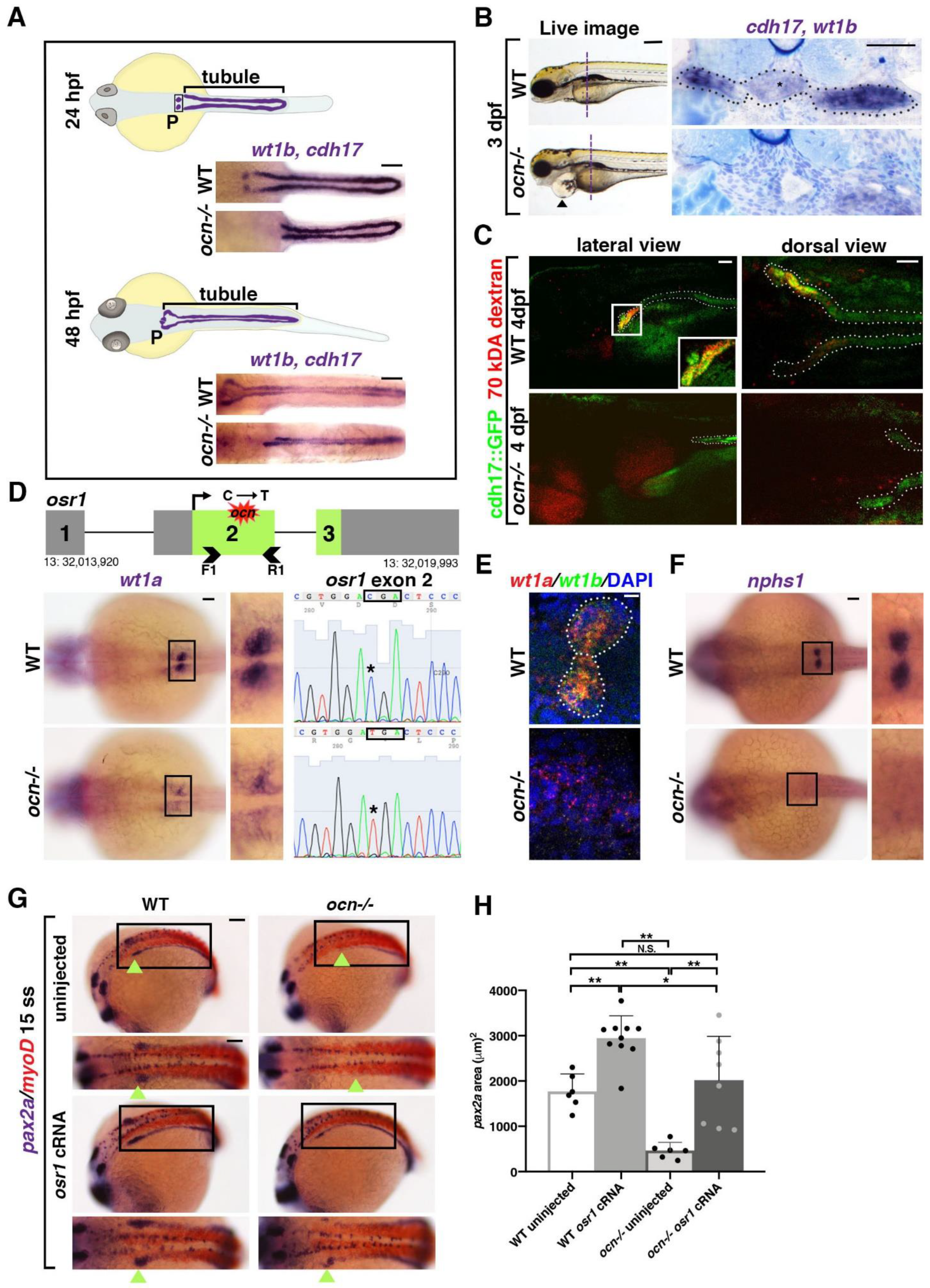
3.1. ocn Encodes a Premature Stop Codon in osr1 and Mutants Exhibits Defective Podocyte and Pronephric Tubule Development

A forward genetic haploid screen was performed to identify regulators of nephrogenesis using the zebrafish pronephros model [35,36]. The ocn mutant was isolated due to its loss of podocytes and abrogation of the anterior pronephros (Figure 1A). Whole-mount in situ hybridization (WISH) was performed to delineate the two pronephros tubules based on the expression of transcripts encoding cadherin 17 (cdh17) and the podocytes based on wt1b expression at 24 and 48 h post fertilization (hpf) (Figure 1A). Both tubule length and podocyte area were significantly reduced in ocn mutants at these time points compared with wild-type (WT) embryos (Supplemental Figure S1A). By 72 hpf, ocn mutants exhibited dramatic pericardial edema that progressed in severity through 120 hpf and was ultimately lethal (Figure 1B and Figure S1B). Since the kidneys play a major role in fluid homeostasis, this phenotype was a probable indicator of renal dysfunction.
To explore this further, WISH staining to assess tubule and podocyte morphology was conducted on 72 hpf ocn and WT embryos. The animals were embedded in JB-4 plastic resin and serially sectioned. In WT embryos, the blood filter could be detected as a mass of dense capillaries containing glomerular podocytes (wt1b+) that were flanked by cdh17+ tubules (Figure 1B). In ocn mutants, however, both the midline glomerulus structure and the flanking proximal tubules were abrogated (Figure 1B). Instead, a dilated dorsal aorta was identified in this region (Figure 1B). While it was clear that the proximal pronephros was absent in ocn mutants, it was uncertain if this truncated kidney retained any functionality. Therefore, kidney functionality was assessed using an endocytosis assay whereby 70 kDA rhodamine-dextran was injected into the vasculature of ocn::cdh17::GFP embryos, which exhibited a pronephric truncation that phenocopied WISH experiments at 3 days post fertilization (dpf) onwards (Supplemental Figure S1C). Transgenic animals were injected with rhodamine-dextran at 48 hpf and then assessed at 48 h post injection (hpi). While dextran was endocytosed in the proximal tubules of non-edemic WT siblings, there was no dextran uptake observed within the truncated tubules of the edemic ocn mutant embryos (Figure 1C). Additionally, we assessed epithelial polarity through the immunofluorescence (IF) staining of Na-K-ATPase, which marks these transporters localized along the basolateral sides of kidney epithelial cells, and aPKC, which marks the apical epithelial side [45]. This experiment revealed a similar reduction in tubule and podocytes in ocn as seen with our WISH experiments using the markers cdh17/wt1b (Supplemental Figure S1D). Together, this provided strong evidence that the stunted pronephros in ocn−/− was indeed functionally defective.
Next, to identify the causative lesion in ocn, whole-genome sequencing was conducted on pools of genomic DNA collected from 24 hpf WISH-identified putative mutants and WT siblings [46,47]. The analysis of the sequencing was performed using SNPtrack software, whereby we detected a strong candidate SNP that was centrally located on chromosome 13 (Supplemental Figure S2A). Specifically, the putative SNP encoded a missense C to T mutation and was predicted to result in an amino acid substitution from an arginine to a premature stop codon in exon 2 of osr1 (Figure 1D and Figure S2A).
To further assess if the predicted stop codon in exon 2 of osr1 was linked with the ocn phenotype, we performed additional genotyping analysis. For this, genomic DNA was isolated from individual embryos that had been identified as ocn mutants or WTs, based on WISH with the podocyte marker wt1a at 24 hpf, and PCR was performed to amplify exon 2 of osr1 followed by direct Sanger sequencing (Figure 1D). Out of 20 ocn embryos with reduced wt1a staining, all 20 were homozygous for the C to T mutation in exon 2 of osr1 (Figure 1D). Protein alignment showed that zebrafish and human OSR1 protein are 264 amino acids (aa) and 266 aa in length, respectively (Supplemental Figure S2B). While they exhibit 77% conservation in overall aa sequence, the three zinc-finger domains responsible for DNA binding activity are 100% conserved across humans, mice, and zebrafish (Supplemental Figure S2B). The osr1 genetic lesion in ocn−/− placed a premature stop codon at residue 165 before all three zinc-finger domains (Supplemental Figure S2B). This suggested that the truncated Osr1 protein produced in ocn−/− would not contain any functional domains and would thus be unable to act as a targeted transcription factor. Next, we verified the effectiveness of a splice-blocking osr1 morpholino with RT-PCR (Supplemental Figure S3). The osr1 morphants had a decrease in podocytes and proximal tubule length that phenocopied ocn−/− and was consistent with previously reported phenotypes [10,30,31,32] (Supplemental Figure S3).
Previous literature has indicated that osr1 acts to restrict venous development in order to promote other mesodermal fates such as the kidney and the pectoral fins [10,30,32]. At 4 dpf, Alcian blue staining indicated that ocn−/− possessed shorter, malformed pectoral fins (Supplemental Figure S1E). The fin bud area, which gives rise to pectoral fins, was significantly reduced in ocn−/− mutants compared with siblings as seen by the marker MDS1 and EVI1 complex locus (mecom) at 24 hpf (Supplemental Figure S1F). Additionally, Alcian blue staining revealed altered craniofacial cartilage formation in mutants, which fits with previous literature placing osr1 as a regulator of palatogenesis in zebrafish and mice [48,49] (Supplemental Figure S1E). In sum, these mesodermal phenotypes were consistent with osr1 deficiency.
Next, we evaluated other aspects of pronephros development. As wt1a expression appeared to be severely diminished and also disorganized in ocn−/−, we evaluated additional markers to better understand the features of podocyte lineage development in mutants. Podocytes were examined at 24 hpf using a wt1a/wt1b double fluorescent in situ (FISH). While clusters of wt1a+/wt1b+ podocytes were visible in siblings, mutants had a scarcity of double-positive cells (Figure 1E). There was also a dearth of nphs1+ cells, which is a marker of the podocyte slit diaphragm and suggested that podocyte differentiation was also disrupted (Figure 1F). Additionally, ocn−/− embryos displayed diminished pax2a expression at the 15 ss compared to siblings (Figure 1G), a characteristic of osr1 morphants in previous studies as well [10,30,32].
To test whether the mutation in osr1 was the specific cause of this phenotype, we performed rescue studies. Injection with osr1 capped RNA (cRNA) rescued this domain in ocn mutants and expanded it in WT siblings (Figure 1G,H). In sum, ocn mutants reciprocated a multitude of mesodermal phenotypes seen in osr1 literature in zebrafish and across taxa. Given the ability of osr1 cRNA to rescue key mesodermal phenotypes in ocn−/− and the catastrophic nature of the osr1 mutation, we concluded that Osr1 deficiency is responsible for the ocn phenotype.

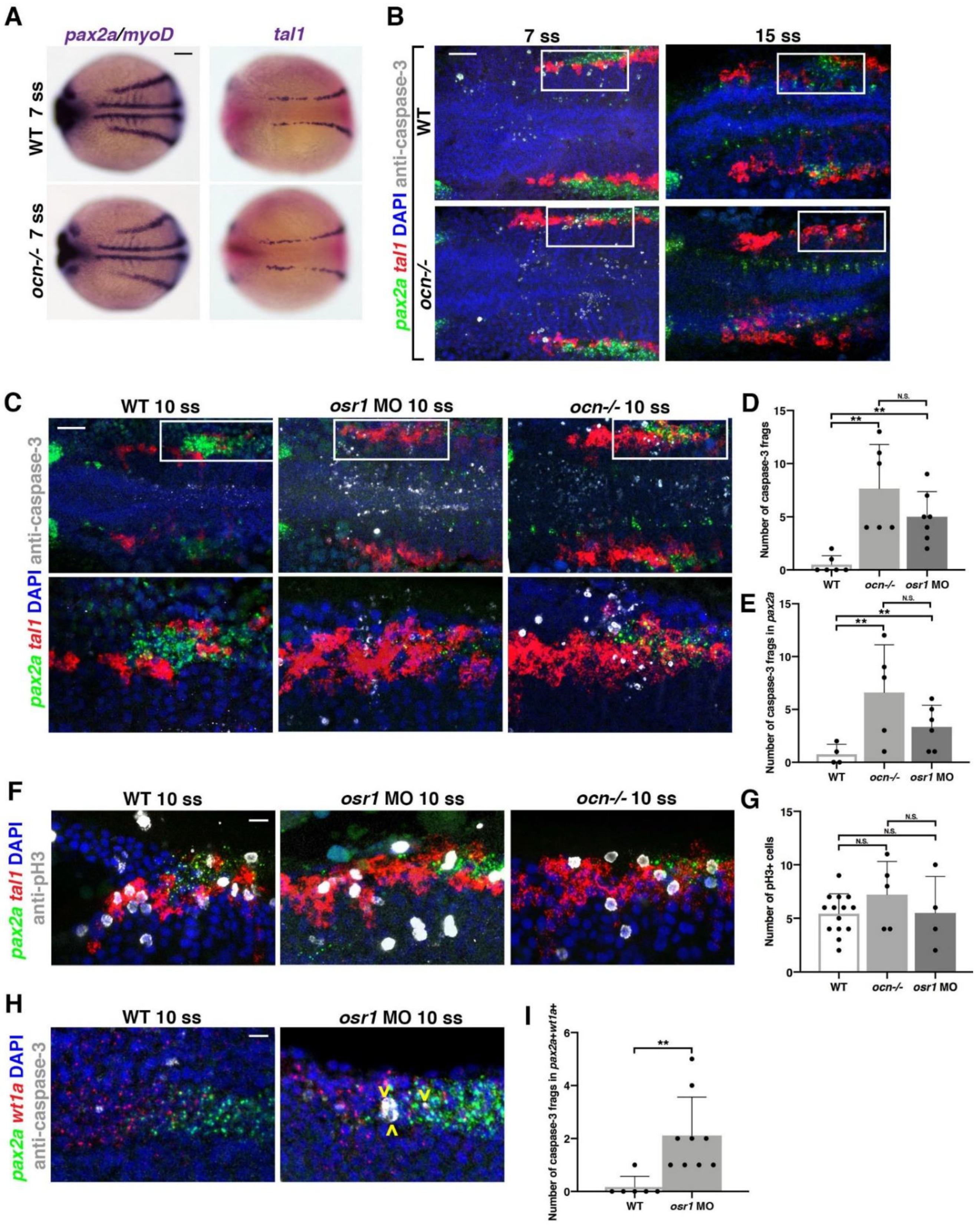
3.2. Kidney Progenitors Are Specified in osr1 Deficient Animals, but Subsequently Undergo Apoptosis

Previous studies suggest that the anterior pronephros abrogation in osr1 zebrafish morphants is due to a fate change where blood/vasculature and endoderm form instead of renal progenitors [30,33,50]. Interestingly, in the Osr1 mouse knockout model, there was an increase in apoptosis that occurred within the kidney tissue [27]. However, in both models, renal progenitors are initially established [27,30]. Thus, we next sought to delineate the cellular dynamics of renal progenitor specification in our ocn mutant model and to address if alterations in proliferation or apoptosis occur during pronephros development in the absence of osr1.
To investigate this, we first performed WISH studies. The LPM is marked by T-cell acute lymphocytic leukemia 1 (tal1), and gives rise to hemangioblasts [51,52]. The IM and hemangioblast domains at the 7 ss were not noticeably different between WT and ocn−/− embryos, as indicated by the markers pax2a, and tal1 (Figure 2A). However, as previously noted, by the 15 ss, there was a decrease in the anterior-most domain of pax2a expression in ocn−/− embryos (Figure 1G). To further assess the anterior pax2a+ cells between the 7 ss and 15 ss, we performed double FISH studies in WT and ocn−/− embryos to assess pax2a and tal1 expression. DAPI staining was also utilized to discern features such as the trunk somites, which allowed for accurate staging. Embryos were flat-mounted and imaged as previously described (Supplemental Figure S2A) [39]. Further, IF was also performed on these samples with anti-caspase-3 antibody to assess the number apoptotic bodies or anti-pH3 to identify proliferating cells. In our analysis, we focused on the changes to these markers within somites 1–5, as the IM adjacent to somite 3 gives rise to podocytes [21].
Beginning at the 7 ss, ocn−/ embryos exhibited a significant increase in the number of caspase-3+ fragments within the combined tal1 and pax2a fields near somites 1–5 (Figure 2B). However, by the 15 ss, few apoptotic fragments were visible in the area of interest, with no significant differences between WT and ocn−/− embryos (Figure 2B). Similar to the 7 ss, we found a significant increase in the number of total caspase-3+ fragments at the 10 ss in ocn−/− mutants and osr1 morphants, while the WT siblings had little to no apoptosis occurring in this area (Figure 2C–E). Interestingly, most of the apoptosis that occurred in mutants and morphants happened within the pax2a kidney field specifically (Figure 2E). Another finding of note was that the caspase-3+ fragment number was not significantly different between osr1 morphants and ocn−/− for either assessment (Figure 2D,E).
To further understand the cell dynamics across this time course, absolute area measurements of pax2a and tal1 were taken at 7, 10, and 15 ss from somites 1–5 in WT and ocn−/−. Surprisingly, the area of the tal1 domain was already expanded at 7 ss and continued to expand through the 15 ss (Supplemental Figure S4). However, across the 3 time points examined, a reduction in pax2a area was only significantly different between WT and ocn−/− at 15 ss (Supplemental Figure S4). Further, although the tal1 field was expanded in ocn−/− embryos at the 10 ss, there was no significant difference in proliferating pH3+ cells between WT and osr1 loss of function models (Figure 2F,G). Additionally, no significant changes in proliferation were seen between mutants and WTs at the 8 ss (Supplemental Figure S4).
To determine if apoptosis was occurring within podocyte progenitors in the pax2a kidney field, we performed an additional FISH with wt1a and pax2a at 10 ss. During this time point, while pax2a expression begans adjacent to somite 3, wt1a was expressed from somites 1–3 (Figure 2H). Similar to the pax2a domain, the wt1a domain did not appear to be reduced at this time point, though it did become restricted and disorganized by 24 hpf (Figure 1D). We found a significant increase in caspase-3+ fragments that were double-positive for wt1a and pax2a in osr1 morphants compared with WTs (Figure 2H,I). These results demonstrated that abnormal apoptosis occurred in podocyte progenitors due to the loss of osr1. In sum, osr1 is not needed to initiate the pax2a progenitor pool, but it is needed to maintain this population, including the podocyte precursors, during pronephros development.

3.3. Ectopic osr1 Is Transiently Sufficient to Rescue Renal Progenitors

Our observation that pax2a+ renal progenitors arise in ocn mutants, but are not maintained, is consistent with previous data that osr1 knockdown leads to a reduced pax2a+ renal progenitor field by the 14 ss [30]. As pax2a expression marks both podocyte and tubule precursors [31], we hypothesized that osr1 is likely needed for podocyte and tubule progenitor maintenance. To determine this, we performed a series of rescue studies in our ocn mutants to test if one or both of these compartments requires osr1 for its maintenance.
First, we tested whether the overexpression of osr1 mRNA was sufficient to rescue podocytes in ocn mutants by assessing wt1b expression, which specifically marks the podocyte lineage [20,21]. The provision of osr1 mRNA robustly rescued the development of wt1b+ podocytes in ocn mutants at the 15 ss (Figure 3A,B). However, by the 22 ss, we were only able to achieve a partial podocyte rescue, though tubules within the same individuals appeared to be WT in length (Figure 3C,D). Consistent with this, we were unable to obtain a podocyte rescue at 24 hpf (data not shown), though again we could achieve a rescue of the truncated tubules (Figure 3E,F). Interestingly, the overexpression of osr1 was sufficient to induce ectopic cdh17+ cells in about 55% of injected embryos (Figure 3G). It should be noted that osr1 cRNA did lead to a decrease in body axis length when compared with uninjected WTs and mutants, which in turn affected pronephros length (Supplemental Figure S5). Despite this, the percentage of kidney length to body length was not significantly different between embryos injected with osr1 cRNA and uninjected animals.
We also conducted a rescue time course by co-injecting osr1 MO and osr1 cRNA and performed WISH using cdh17 to assess the tubules during a number of stages. While 90% of animals injected with both constructs exhibited a WT tubule length at 24–27 ss, by 36 hpf, only 50% showed a rescue (Figure 3H and Figure S5B). At 48 hpf, only 20% of injected embryos had a WT length pronephric tubules while 80% had a unilateral or bilateral reduction (Figure 3H and Figure S5B). Together, this indicated that the pronephros requires a continued presence of osr1 to maintain the tubule population as development progressed.

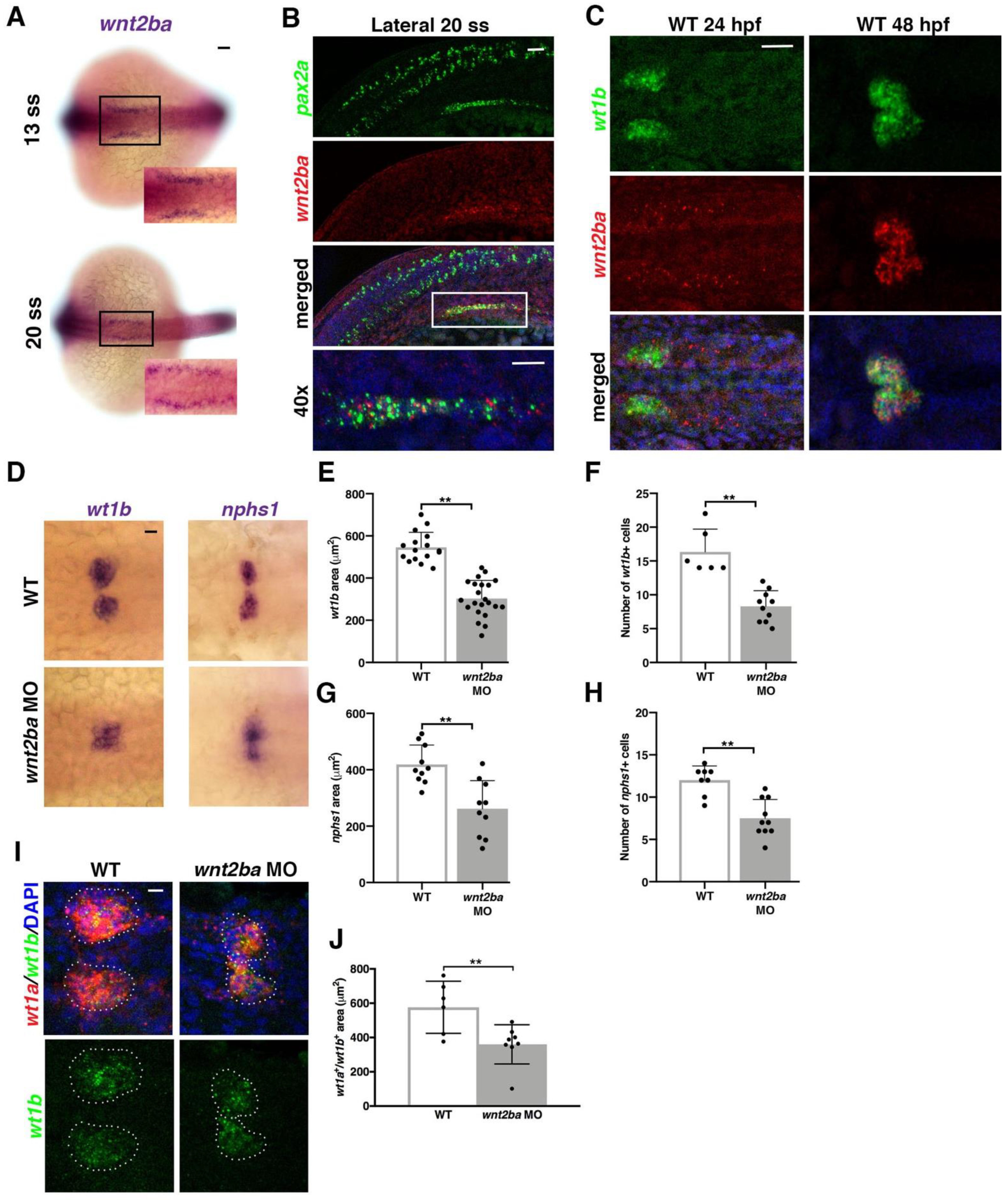
3.4. wnt2ba Is a Novel Podocyte Marker and Regulator

Given the importance of osr1 to podocyte development and maintenance, we wanted to identify downstream factors that promote podocytes. It was previously shown that the canonical Wnt ligand wingless-type MMTV integration site family, member 2Ba (wnt2ba) is expressed in a similar proximal swath of IM as osr1 [32]. We observed a similar expression pattern of wnt2ba in the anterior IM as early as 13 ss (Figure 4A). To specifically determine which cells wnt2ba was expressed in, we conducted FISH studies. At the 20–22 ss, wnt2ba transcripts were colocalized in cells within the anterior most region of pax2a+ and wt1b+ IM (Figure 4B and Figure S6). At 15 ss, wnt2ba transcripts were also colocalized with wt1a/b+ podocyte progenitor cells (Supplemental Figure S6). At 24 hpf, wnt2ba was expressed in both wt1b+ podocyte precursor cells and in the neighboring cells of the IM (Figure 4C). By 48 hpf, wnt2ba was restricted to the podocytes and overlapped precisely with wt1b expression (Figure 4C). Taken together, we conclude that wnt2ba is a novel podocyte marker, thereby extending prior observations [32]. We also examined the expression of the zebrafish wnt2ba paralogue wnt2bb at 24 hpf using FISH and determined that these transcripts by comparison were located anterior to both the podocyte and kidney fields (Supplemental Figure S7).
Given its expression in podocyte progenitors, we hypothesized that Wnt2ba might have roles in podocyte specification or differentiation. To explore whether wnt2ba is needed for proper podocyte formation, we performed wnt2ba loss of function studies. We first verified a morpholino that blocked splicing at exon 1, as well as a morpholino that targeted the start site (Supplemental Figure S8). When wnt2ba morphants were examined at 24 hpf, there was a significant reduction in the expression of wt1b and nphs1 that corresponded to a smaller podocyte area and net cell number (Figure 4D–H). We also found that the area of wt1a+/wt1b+ co-expressing podocytes was decreased in wnt2ba morphants at 24 hpf (Figure 4I,J). Furthermore, the decrease in podocyte number in wnt2ba morphants occurred between the 15 and 22 ss, suggesting wnt2ba is required for maintaining the podocyte lineage (Supplemental Figure S9). In contrast, wnt2ba morphants showed no discernable changes in the development or maintenance of the cdh17+ nephron tubule (Supplemental Figure S8E). Collectively, these data lead us to conclude that wnt2ba is a significant regulator of podocyte ontogeny.

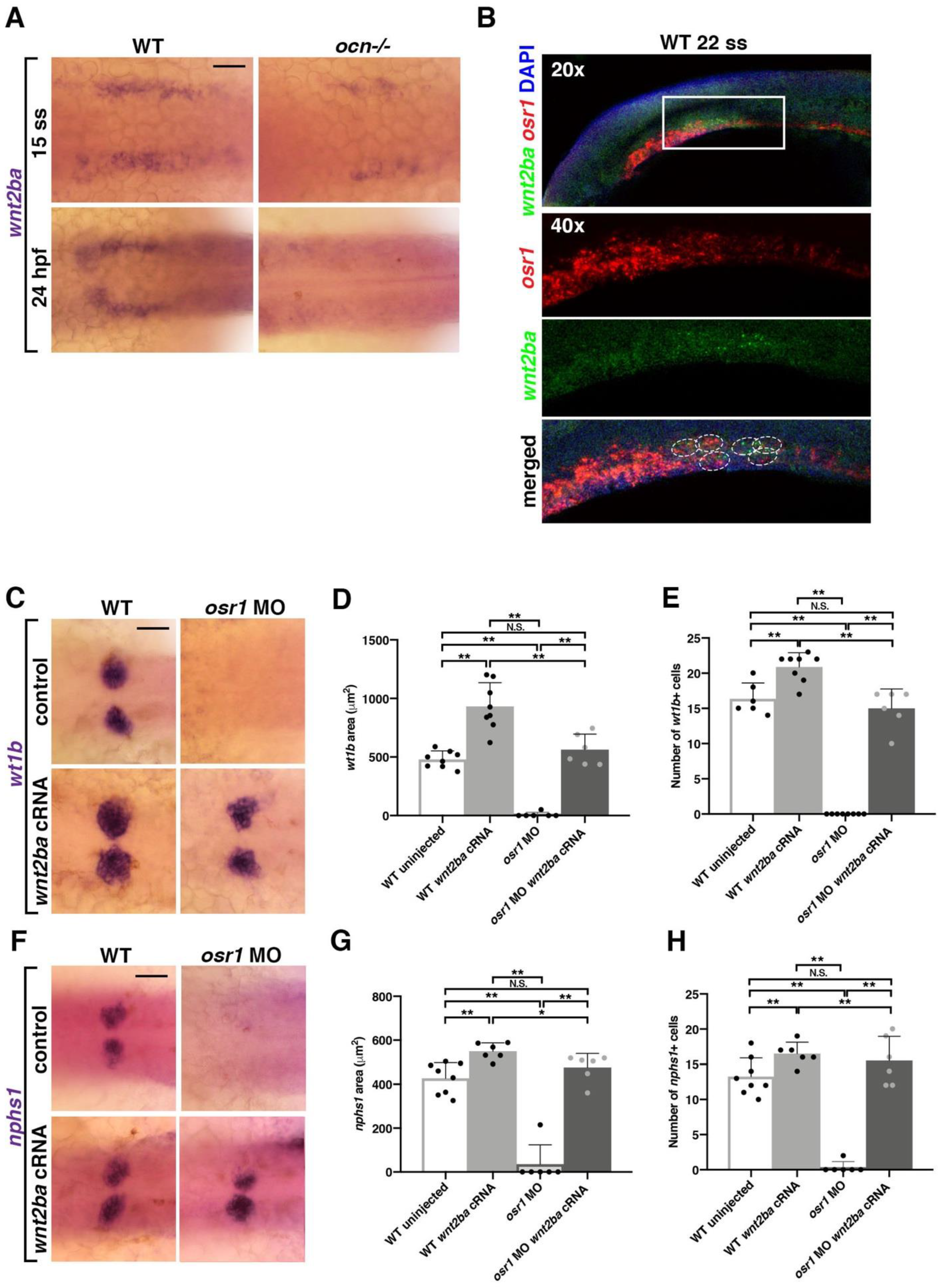
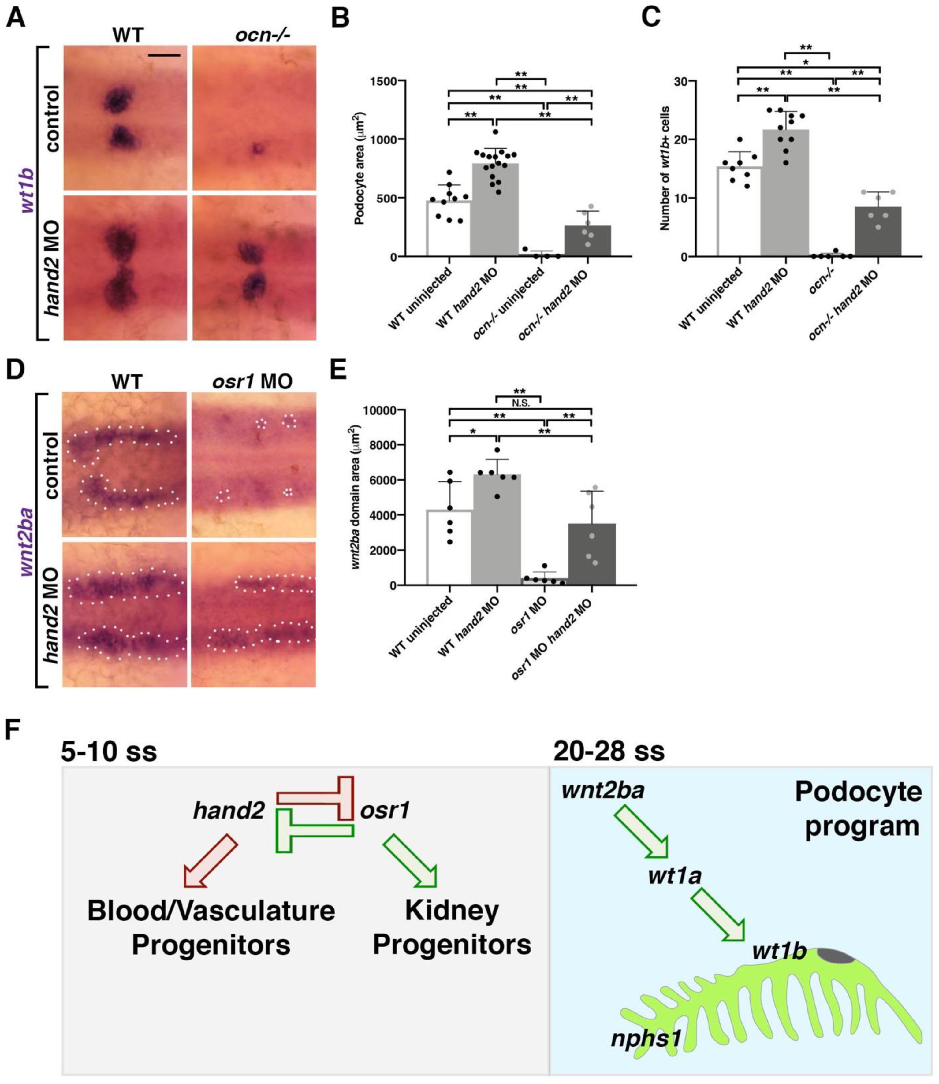
3.5. osr1 Promotes wnt2ba in the Podocyte Developmental Pathway

Previous research has demonstrated that osr1 morphants exhibited a dramatic decrease in the wnt2ba pronephric domain, though wnt2ba morphants had no notable change in osr1 expression [32]. They postulated that osr1 acts to promote wnt2ba in the IM, so which allows for proper pectoral fin development to occur [32]. This led us to hypothesize that this same genetic cascade in the IM promotes the formation of proximal pronephric tissues, such as the podocytes, and is dysfunctional in ocn−/−.
In congruence with this prior study, we found that wnt2ba is significantly reduced in ocn−/− at both 15 ss and is almost completely absent by 24 hpf (Figure 5A). We also observed that wnt2ba and osr1 transcripts were colocalized in a population of presumptive IM cells at 15 ss and 22 ss, putting them in the right place and the right time to interact (Figure 5B and Figure S10). The overexpression of wnt2ba led to an increase in podocyte number and domain area in injected WT embryos, as seen with an increase in the markers wt1b and nphs1 (Figure 5C–H). While injection with osr1 MO alone leads to diminished podocytes, coinjection of wnt2ba cRNA with osr1 MO led to a rescue in podocyte area and cell count (Figure 5C–H). Together, this indicates that wnt2ba is sufficient to drive podocyte development and does so downstream of osr1.

3.6. hand2 Suppresses Podocyte Development by Restricting wnt2ba Expression and Podocyte Development

The bHLH transcription factor heart and neural crest derivatives expressed 2 (hand2) has been shown to be antagonistic to osr1 in early mesoderm development [10]. The loss of osr1 leads to decreases in podocytes and tubules and an increase in hemangioblasts; in contrast, the loss of hand2 results in expansions in renal cells at the expense of vasculature [10]. The concomitant knockdown of osr1 and hand2 rescues tubule development [10] and podocyte development [33].
When we knocked down hand2 using an ATG morpholino, we observed a separation in the myosin light chain 7 (myl7) heart field at 22 ss that matched previously observed phenotypes [44] (Supplemental Figure S11). The knockdown of hand2 also caused a significant increase in wt1b+ podocyte domain area and cell number (Figure 6A–C). While uninjected ocn−/− embryos had few to no podocytes, injecting ocn-/- with hand2 MO resulted in an expansion in podocyte number and area that was significantly different from mutants (Figure 6A–C). Similarly, hand2 morphants had a significantly larger wnt2ba domain, and hand2/osr1 MO coinjection rescued the usually abrogated wnt2ba domain (Figure 6D,E). This indicated that an imbalance of hand2 and osr1 leads to changes in wnt2ba expression, which alters podocyte development.

4. Discussion

While dozens to hundreds of podocyte diseases and maladies have been characterized, the genetic explanations for their origins and progression are lacking. One reason for this is that there are relatively few factors that are known to promote the development of these specialized epithelia. Continuing to identify these factors is critical for future diagnostics and treatments for podocytopathy. In this study, we have both reexamined a previous factor shown to promote podocyte fates, osr1, and also identified a new downstream regulator, wnt2ba.
In this study, ocn was identified as a mutant of interest in a forward genetic screen due to displaying pericardial edema and decreases in podocytes and proximal tubules. Through whole-genome sequencing, we determined that ocn−/− harbors a SNP in exon 2 that leads to a premature stop in osr1. This SNP was confirmed as the causative lesion in ocn−/− when osr1 cRNA could rescue each of these phenotypes. Upon confirmation that ocn was an osr1 mutant line, we next sought to fully assess how osr1 loss of function impacts kidney development in the context of a zebrafish mutant. We found that osr1 is needed to maintain renal progenitors and inhibit the development of hemangioblasts.
Further, we established a genetic pathway controlled by osr1 that regulates podocyte survival by promoting wnt2ba expression. We found that wnt2ba is an IM/podocyte marker that is likewise diminished in ocn−/−. Loss and gain of wnt2ba lead to a decrease and increase in podocyte area, demonstrating that wnt2ba is both necessary and sufficient to drive podocyte development. Notably, wnt2ba can rescue podocytes in an osr1-deficient background, which places this factor downstream of osr1 (Figure 6F). Finally, the osr1/wnt2ba podocyte pathway is negatively regulated by hand2 (Figure 6F).

4.1. Osr1 Acts to Promote Podocytes

The earliest known podocyte marker in zebrafish is wt1a, though the paralogue, wt1b, that appears at 12 ss is expressed in a more specific territory [20,53]. It has also been suggested that wt1a is more dominant than wt1b, as knockdown of wt1a leads to loss of nphs1/2, while knockdown of wt1b causes less dramatic podocyte phenotypes [54]. Zebrafish literature has shown that osr1 morphants exhibit reductions in wt1b, lhx1a, and nphs1/2 at 24 hpf that have also been observed in ocn−/− [29,30,31]. However, the relationship between wt1a and osr1 has yet to be fully understood. Tomar et al. [31] placed wt1a upstream of osr1 due to osr1 being reduced in wt1 morpholino-injected embryos and wt1a expression being interpreted as unchanged in osr1 morpholino-injected animals. However, in our studies, osr1 morphants did exhibit alterations in wt1a+ cell organization and a restriction in domain that phenocopies ocn−/−. Mouse studies have shown that WT1+/−; OSR1+/− mice exhibit smaller kidneys, suggesting that these factors act cooperatively in kidney and podocyte development [55]. If osr1 and wt1a did have a similar synergistic relationship in zebrafish kidney development, this would also explain reports that wt1 morphants exhibit a loss of podocytes and proximal tubules reminiscent of osr1 loss of function models [31]. While there are currently limitations in using anti-Osr1 antibodies in any in vivo model, progress in this area is needed in order to ascertain if wt1a and osr1 are directly interacting during kidney development.

4.2. Osr1 Is Needed for Kidney Cell Maintenance

While ocn−/− exhibit normal patterning of IM early in development, by the time specification to pronephros is beginning to occur around 15 ss, the anterior domain is absent. Our experiments demonstrated that this is due to two events: (1) an expansion of hemangioblasts and (2) the apoptosis of podocyte progenitors in this region. Work in chick and mouse has shown that while mesonephric tissues and markers are present, apoptosis occurs within the nephrogenic mesenchyme that keeps metanephric tissues from forming in Osr1 knockout animals [27,28]. Further, previous studies have shown that Osr1 acts synergistically with factors such as Wt1 and Six2 to renew renal stem cell pools to inhibit premature differentiation and thus cell death [55]. A similar apoptosis event has not been recorded in osr1 loss of function zebrafish models prior to this study, and we hypothesize that osr1 plays a similar role in progenitor self-renewal in zebrafish.
The expansion in the hemangioblast domain in osr1 morphants has been documented by other groups, where it was suggested that pax2a+ cells were converting to tal+ cells [30]. Additionally, the expansion in vessel progenitors has been reported in an osr1 TALEN mutant [33]. However, our results add one further element to these early events, as we have captured cell apoptosis in pax2a+ cells of osr1 mutant embryos. Further, our studies have revealed that the timing of the pax2a domain decrease and hemangioblast domain increase is not equivalent. The hemagioblasts expand hours prior to the loss of the anterior IM domain. We postulate that osr1 may inhibit hemangioblast formation either indirectly or in an independent mechanism than it uses to promote IM and podocytes.

4.3. Wnt2ba Is a Novel Regulator of Podocyte Development

Finally, wnt2ba is a ligand that functions in the canonical Wnt/beta-catenin pathway. As a member of this pathway, wnt2ba acts to promote cell growth, differentiation, and migration during development. In regard to kidney development, Wnt2b can be detected in the kidney stroma in mice as early as E11.5 [56,57] and in humans WNT2B is expressed in fetal kidney stroma [58]. In addition, cells expressing Wnt2b promote ureteric branching in culture [56]. Wnt2/2b is also paramount to normal lung and pectoral fin development in both aquatic and mammalian species [32,59]. Interestingly, osr1 has been shown to act downstream of retinoic acid signaling yet upstream of wnt2b in both pectoral fin development in zebrafish [32] and in lung progenitor specification in foregut endoderm in Xenopus [60]. However, our study has both evaluated the role of wnt2ba as a regulator of kidney development and placed its function downstream of osr1 to specifically promote the podocyte lineage. Further, we show that osr1 promotes wnt2ba expression during podocyte development through a mechanism involving the inhibition of hand2. In synchrony with our data, a recent report similarly concluded that the reciprocal antagonism between osr1 and hand2 is essential for the normal emergence of wt1b+ podocyte precursors [33]. Future work to assess whether Osr1 directly binds the wnt2ba promotor, and the identity of other regulatory factor(s), will be absolutely critical in order to decipher the underlying molecular mechanisms of the genetic relationships reported in the present work. Furthermore, the identification of other candidate Osr1 targets will be crucial in expanding our knowledge about the roles of this critical gene.
We show in the present study that wnt2ba is a regulator of podocyte development but that loss of wnt2ba does not cause compelling changes in either the PCT segment or the nephron tubule length. Another study by the team of Lyons et al. [61] showed that the broad inhibition of Wnt signaling through the heat-shock activation of dkk1 led to an abrogation in the zebrafish pronephros that resembles osr1 loss of function models. Wnt ligands are highly regionalized to allow for precise regulation during tissue development [57,62]. Our findings that wnt2ba is restricted to the podocytes by 48 hpf could reflect regional specificity. This suggests that there are other Wnt ligands and receptors that act to regulate certain kidney lineages in zebrafish development. The loss of one or more of these factors in combination with wnt2ba could lead to an anterior truncation of the pronephros that resembles the experiments from Lyons et al. [61]. Future studies are needed to discern these factors and additional downstream targets of both wnt2ba and osr1.
Taken together, these results have allowed us to garner new insights into podocyte development in zebrafish. By selecting ocn as a mutant of interest from our ENU screen, we have discovered an osr1 mutant and confirmed its significance in zebrafish pronephros development in an unbiased manner. We have expanded on previous findings by demonstrating that osr1 is required to inhibit apoptosis in specified kidney precursors, and later for nephron cell maintenance. We have also ascertained new roles for osr1 in promoting wnt2ba expression, which it does in part through the antagonism of hand2. Finally, our results show that wnt2ba mitigates podocyte development downstream of the osr1/hand2 interaction. Given how little is known about CAKUT and kidney agenesis, findings from genetics studies such as the present work are crucial to furthering our understanding about the causes and solutions to these disease states.

5. Conclusions

The ocn zebrafish mutant provides a new vertebrate genetic model to expand our understanding about the roles of osr1 during kidney development. During the genesis of the zebrafish pronephros, a deficiency of osr1 causes an abrogation of podocyte and proximal tubule lineages, which are specified but subsequently undergo apoptosis in the absence of osr1. Furthermore, we conclude that osr1 regulates podocyte survival by promoting the expression of wnt2ba, a factor that is both necessary and sufficient for podocyte ontogeny. Finally, the function of wnt2ba in the podocyte developmental program is impacted by the antagonistic interactions between osr1 and the transcription factor hand2.

Supplementary Materials

The following supporting information can be downloaded at: https://www.mdpi.com/article/10.3390/biomedicines10112868/s1, Figure S1: Additional ocn phenotypes; Figure S2: A genetic lesion in the osr1 transcription factor results in truncated osr1 protein in the ocn mutant line; Figure S3: Verification of osr1 splice-blocking morpholino phenotypes; Figure S4: Additional analysis of cell dynamics in osr1-deficient models; Figure S5: osr1 cRNA injection additional analysis; Figure S6: wnt2ba colocalizes with early, developing podocytes; Figure S7: wnt2bb does not colocalize with podocytes; Figure S8: Assessment of wnt2ba MO through RT-PCR analysis; Figure S9: wnt2ba knockdown causes decreased podocytes at 22 ss; Figure S10: Additional stages of wnt2ba and osr1 co-localization; Figure S11: hand2 MO replicates previous studies.

Author Contributions

Conceptualization, R.A.W. and B.E.D.; methodology, R.A.W. and B.E.D.; validation, R.A.W. and B.E.D.; formal analysis, R.A.W. and B.E.D.; investigation, B.E.D., B.E.C., H.M.W., S.G., L.A., M.N.U., G.F.G., P.T.K., I.L. and W.G.; writing—original draft preparation, R.A.W. and B.E.D.; writing—review and editing, R.A.W., B.E.D., B.E.C., H.M.W. and S.G.; supervision, R.A.W.; project administration, R.A.W.; funding acquisition, R.A.W. and B.E.D. All authors have read and agreed to the published version of the manuscript.

Funding

This research was funded by NIH Grant R01DK100237 to R.A.W., start-up funds from the University of Notre Dame College of Science to R.A.W., a National Science Foundation Graduate Research Fellowship DGE-1313583 awarded to B.E.D. We also thank Elizabeth and Michael Gallagher for their generous gift to the University of Notre Dame to support stem cell research. The funders had no role in the study design, data collection and analysis, decision to publish, or manuscript preparation.

Institutional Review Board Statement

The study was approved and conducted according to the guidelines of the University of Notre Dame Institutional Animal Care and Use Committee under protocol numbers 16-07-325 and 19-06-5412.

Informed Consent Statement

Not applicable.

Data Availability Statement

All data are contained in this article and the Supplementary Materials.

Acknowledgments

We thank the staffs of the Department of Biological Sciences and Center for Stem Cells and Regenerative Medicine and particularly express our gratitude to staff of the Center for Zebrafish Research at the University of Notre Dame for their dedication to and care for our zebrafish aquarium. R.A.W. thanks G.R.W. for unwavering support and encouragement and B.C., K.P., and M.M. for their support and advice. Finally, we thank all the past and current members of our lab for their support, discussions, and insights about the fascinating topic of kidney development.

Conflicts of Interest

The authors declare no conflict of interest.

Abbreviations

cadherin 17 (cdh17); centrin 4 (cetn4); chronic kidney disease (CKD); chloride channel K (clcnk); congenital anomalies of the kidney and urinary tract (CAKUT); corpuscle of Stannius (CS); days post fertilization (dpf); distal early (DE) segment; distal late (DL) segment; fluorescent in situ hybridization (FISH); hours post fertilization (hpf); intermediate mesoderm (IM); interrenal gland (IR); LIM homeobox 1a (lhx1a); multiciliated cells (MCCs); N-ethyl-N-nitrosourea (ENU); nephrosis 1, congenital Finnish type (nephrin); nephrosis 2 (nphs1), idiopathic, steroid-resistant (podocin)(nphs2), odd-skipped related transcription factor 1 (osr1); odd-skipped related transcription factor 2 (osr2); outer dense fiber of sperm tails 3B (odf3b); paired box 2a (pax2a); proximal convoluted tubule (PCT); proximal straight segment (PST); renal outer medullary potassium, channel 2 (romk2); solute carrier family 4 (sodium bicarbonate cotransporter), member 4a (slc4a4a); solute carrier family 9, subfamily A (slc9a3); solute carrier family 12 (slc12a1); solute carrier family 12 sodium/chloride transporter, member 3 (slc12a3); solute carrier family 20, member 1a (slc20a1a); transient receptor potential cation channel, subfamily M, member 7 (trpm7); somite stage (ss); whole mount in situ hybridization (WISH); Wilms tumor 1a (wt1a); Wilms tumor 1b (wt1b); wingless-type MMTV integration site family, member 2Ba (wnt2ba); wingless-type MMTV integration site family, member 2Bb (wnt2bb); Zebrafish International Research Center (ZIRC).

References

  1. Ichimura, K.; Kakuta, S.; Kawasaki, Y.; Miyaki, T.; Nonami, T.; Miyazaki, N.; Nakao, T.; Enomoto, S.; Arai, S.; Koike, M.; et al. Morphological process of podocyte development revealed by block-face scanning electron microscopy. J. Cell Sci. 2017, 130, 132–142. [Google Scholar] [CrossRef] [PubMed] [Green Version]
  2. Grahammer, F. New structural insights into podocyte biology. Cell Tissue Res. 2017, 369, 5–10. [Google Scholar] [CrossRef] [PubMed] [Green Version]
  3. Pavenstädt, H.; Kriz, W.; Kretzler, M. Cell biology of the glomerular podocyte. Physiol Rev. 2003, 83, 253–307. [Google Scholar] [CrossRef] [Green Version]
  4. Garg, P. A review of podocyte biology. Am. J. Nephrol. 2018, 47, 3–13. [Google Scholar] [CrossRef] [PubMed]
  5. Zhuo, J.L.; Li, X.C. 2013. Proximal nephron. Compr. Physiol. 2013, 3, 1079. [Google Scholar] [PubMed] [Green Version]
  6. Wiggins, R.C. The spectrum of podocytopathies: A unifying view of glomerular diseases. Kidney Int. 2007, 71, 1205–1214. [Google Scholar] [CrossRef] [Green Version]
  7. Romagnani, P.; Remuzzi, G.; Glassock, R.; Levin, A.; Jager, K.J.; Tonelli, M.; Massy, Z.; Wanner, C.; Anders, H. Chronic kidney disease. Nat. Rev. Dis. Primers 2017, 3, 17088. [Google Scholar] [CrossRef]
  8. Reiser, J.; Sever, S. Podocyte biology and pathogenesis of kidney disease. Annu. Rev. Med. 2013, 64, 357–366. [Google Scholar] [CrossRef] [Green Version]
  9. Gerlach, G.F.; Wingert, R.A. Kidney organogenesis in the zebrafish: Insights into vertebrate nephrogenesis and regeneration. Wiley Interdiscip. Rev. Dev. Biol. 2013, 2, 559–585. [Google Scholar] [CrossRef] [Green Version]
  10. Perens, E.A.; Garavito-Aguilar, Z.V.; Guio-Vega, G.P.; Peña, K.T.; Schindler, Y.L.; Yelon, D. Hand2 inhibits kidney specification while promoting vein formation within the posterior mesoderm. eLife 2016, 5, e19941. [Google Scholar] [CrossRef]
  11. Little, M.H.; McMahon, A.P. Mammalian kidney development: Principles, progress, and projections. Cold Spring Harb. Perspect. Biol. 2012, 4, a008300. [Google Scholar] [CrossRef] [PubMed]
  12. McMahon, A.P. Development of the Mammalian Kidney. Curr. Top. Dev. Biol. 2016, 117, 31. [Google Scholar] [PubMed] [Green Version]
  13. Luyckx, V.A.; Shukha, K.; Brenner, B.M. Low nephron number and its clinical consequences. Rambam Maimonides Med. J. 2011, 2, e0061. [Google Scholar] [CrossRef] [PubMed]
  14. Little, M.H. Growing kidney tissue from stem cells: How far from "party trick" to medical application? Cell Stem Cell 2016, 18, 695–698. [Google Scholar] [CrossRef] [PubMed] [Green Version]
  15. Drummond, B.E.; Wingert, R.A. Insights into kidney stem cell development and regeneration using zebrafish. World J. Stem Cells 2016, 8, 22–31. [Google Scholar] [CrossRef]
  16. Drummond, I.A.; Davidson, A.J. Zebrafish kidney development. Methods Cell Biol. 2016, 134, 391–429. [Google Scholar]
  17. Diep, C.Q.; Ma, D.; Deo, R.C.; Holm, T.M.; Naylor, R.W.; Arora, N.; Wingert, R.A.; Bollig, F.; Djordjevic, G.; Lichman, B.; et al. Identification of adult nephron progenitors capable of kidney regeneration in zebrafish. Nature 2011, 470, 95–100. [Google Scholar] [CrossRef] [Green Version]
  18. Naylor, R.W.; Przepiorski, A.; Ren, Q. HNF1B is essential for nephron segmentation during nephrogenesis. J. Am. Soc. Nephrol. 2013, 24, 77–87. [Google Scholar] [CrossRef] [Green Version]
  19. Hsu, H.; Lin, G.; Chung, B. Parallel early development of zebrafish interrenal glands and pronephros: Differential control by Wt1 and Ff1b. Development 2003, 130, 2107–2116. [Google Scholar] [CrossRef] [Green Version]
  20. Bollig, F.; Mehringer, R.; Perner, B.; Hartung, C.; Schäfer, M.; Schartl, M.; Volff, J.; Winkler, C.; Englert, C. Identification and comparative expression analysis of a second Wt1 gene in zebrafish. Dev. Dyn. 2006, 235, 554. [Google Scholar] [CrossRef]
  21. O’Brien, L.L.; Grimaldi, M.; Kostun, Z.; Wingert, R.A.; Selleck, R.; Davidson, A.J. Wt1a, Foxc1a, and the Notch Mediator Rbpj physically interact and regulate the formation of podocytes in zebrafish. Dev. Biol. 2011, 358, 318–330. [Google Scholar] [CrossRef] [PubMed]
  22. Zhu, X.; Chen, Z.; Zeng, C.; Wang, L.; Xu, F.; Hou, Q.; Liu, Z. Ultrastructural characterization of the pronephric glomerulus development in zebrafish. J. Morphol. 2016, 277, 1104–1112. [Google Scholar] [CrossRef] [PubMed]
  23. Wingert, R.A.; Selleck, R.; Yu, J.; Song, H.; Chen, Z.; Song, A.; Zhou, Y.; Thisse, B.; Thisse, C.; McMahon, A.P.; et al. The cdx genes and retinoic acid control the positioning and segmentation of the zebrafish pronephros. PLoS Genet. 2007, 3, 1922–1938. [Google Scholar] [CrossRef] [PubMed] [Green Version]
  24. Wingert, R.A.; Davidson, A.J. Zebrafish nephrogenesis involves dynamic spatiotemporal expression changes in renal progenitors and essential signals from retinoic acid and irx3b. Dev. Dyn. 2011, 240, 2011–2027. [Google Scholar] [CrossRef] [PubMed] [Green Version]
  25. Desgrange, A.; Cereghini, S. Nephron patterning: Lessons from Xenopus, zebrafish, and mouse studies. Cells 2015, 4, 483–499. [Google Scholar] [CrossRef] [Green Version]
  26. Mugford, J.W.; Sipilä, P.; McMahon, J.A.; McMahon, A.P. Osr1 expression demarcates a multi-potent population of intermediate mesoderm that undergoes progressive restriction to an Osr1-dependent nephron progenitor compartment within the mammalian kidney. Dev. Biol. 2008, 324, 88–98. [Google Scholar] [CrossRef] [Green Version]
  27. James, R.G.; Kamei, C.N.; Wang, Q.; Jiang, R.; Schultheiss, T.M. Odd-Skipped related 1 is required for development of the metanephric kidney and regulates formation and differentiation of kidney precursor cells. Development 2006, 133, 2995–3004. [Google Scholar] [CrossRef] [Green Version]
  28. Wang, Q.; Lan, Y.; Cho, E.; Maltby, K.M.; Jiang, R. Odd-Skipped Related 1 (Odd 1) is an essential regulator of heart and urogenital development. Dev. Biol. 2005, 288, 582–594. [Google Scholar] [CrossRef] [Green Version]
  29. Tena, J.J.; Neto, A.; de la Calle-Mustienes, E.; Bras-Pereira, C.; Casares, F.; Gómez-Skarmeta, J.L. Odd-Skipped genes encode repressors that control kidney development. Dev. Biol. 2007, 301, 518–531. [Google Scholar] [CrossRef]
  30. Mudumana, S.P.; Hentschel, D.; Liu, Y.; Vasilyev, A.; Drummond, I.A. Odd skipped related1 reveals a novel role for endoderm in regulating kidney versus vascular cell fate. Development 2008, 135, 3355–3367. [Google Scholar] [CrossRef] [Green Version]
  31. Tomar, R.; Mudumana, S.P.; Pathak, N.; Hukriede, N.A.; Drummond, I.A. Osr1 is Required for Podocyte Development Downstream of Wt1a. J. Am. Soc. Neph. 2014, 25, 2539–2545. [Google Scholar] [CrossRef] [PubMed]
  32. Neto, A.; Mercader, N.; Gómez-Skarmeta, J.L. The Osr1 and Osr2 genes act in the pronephric anlage downstream of retinoic acid signaling and upstream of Wnt2b to maintain pectoral fin development. Development 2012, 139, 301–311. [Google Scholar] [CrossRef] [PubMed] [Green Version]
  33. Perens, E.A.; Diaz, J.T.; Quesnel, A.; Crump, J.G.; Yelon, D. osr1 couples intermediate mesoderm cell fate with temporal dynamics of vessel progenitor cell differentiation. Development 2021, 148, dev198408. [Google Scholar] [CrossRef] [PubMed]
  34. Zhang, Z.; Iglesias, D.; Eliopoulos, N.; El Kares, R.; Chu, L.; Romagnani, P.; Goodyer, P. A variant OSR1 allele which disturbs OSR1 mRNA expression in renal progenitor cells is associated with reduction of newborn kidney size and function. Hum. Mol. Genet. 2011, 20, 4167–4174. [Google Scholar] [CrossRef] [PubMed]
  35. Kroeger, P.T.; Poureetezadi, S.J.; McKee, R.; Jou, J.; Miceli, R.; Wingert, R.A. Production of haploid zebrafish embryos by in vitro fertilization. J. Vis. Exp. 2014, 89, 51708. [Google Scholar] [CrossRef] [Green Version]
  36. Chambers, B.E.; Gerlach, G.F.; Clark, E.G.; Chen, K.H.; Levesque, A.E.; Leshchiner, I.; Goessling, W.; Wingert, R.A. Tfap2a is a novel gatekeeper of nephron differentiation during kidney development. Development 2019, 146, dev172387. [Google Scholar] [CrossRef] [Green Version]
  37. Kroeger, P.T.; Drummond, B.E.; Miceli, R.; McKernan, M.; Gerlach, G.F.; Marra, A.N.; Fox, A.; McCampbell, K.K.; Leshchiner, I.; Rodriguez-Mari, A.; et al. The zebrafish kidney mutant zeppelin reveals that brca2/fancd1 is essential for pronephros development. Dev. Biol. 2017, 428, 148–163. [Google Scholar] [CrossRef]
  38. Li, Y.; Cheng, C.N.; Verdun, V.A.; Wingert, R.A. Zebrafish nephrogenesis is regulated by interactions between retinoic acid, mecom, and Notch signaling. Dev. Biol. 2014, 386, 111–122. [Google Scholar] [CrossRef] [Green Version]
  39. Cheng, C.N.; Li, Y.; Marra, A.N.; Verdun, V.; Wingert, R.A. Flat mount preparation for observation and analysis of zebrafish embryo specimens stained by whole mount in situ hybridization. J. Vis. Exp. 2014, 89, 51604. [Google Scholar] [CrossRef] [Green Version]
  40. Cheng, C.N.; Wingert, R.A. Nephron proximal tubule patterning and corpuscles of Stannius formation are regulated by the sim1a transcription factor and retinoic acid in zebrafish. Dev. Biol. 2015, 399, 100–116. [Google Scholar] [CrossRef] [Green Version]
  41. Marra, A.N.; Ulrich, M.; White, A.; Springer, M.; Wingert, R.A. Visualizing multiciliated cells in the zebrafish through a combined protocol of whole mount fluorescent in situ hybridization and immunofluorescence. J. Vis. Exp. 2017, 129, 56261. [Google Scholar] [CrossRef] [PubMed]
  42. Marra, A.N.; Chambers, B.E.; Chambers, J.M.; Drummond, B.E.; Adeeb, B.D.; Wesselman, H.M.; Morales, E.E.; Handa, N.; Pettini, T.; Ronshaugen, M.; et al. Visualizing gene expression during zebrafish pronephros development and regeneration. Methods Cell. Biol. 2019, 154, 183–215. [Google Scholar] [PubMed]
  43. Wakahara, T.; Kusu, N.; Yamauchi, H.; Kimura, I.; Konishi, M.; Miyake, A.; Itoh, N. Fibin, a novel secreted lateral plate mesoderm signal, is essential for pectoral fin bud initiation in zebrafish. Dev. Biol. 2007, 303, 527–535. [Google Scholar] [CrossRef] [PubMed] [Green Version]
  44. Maves, L.; Tyler, A.; Moens, C.B.; Tapscott, S.J. Pbx acts with Hand2 in early myocardial differentiation. Dev. Biol. 2009, 333, 409–418. [Google Scholar] [CrossRef] [Green Version]
  45. Gerlach, G.F.; Wingert, R.A. Zebrafish pronephros tubulogenesis and epithelial identity maintenance are reliant on the polarity proteins prkc iota and zeta. Dev. Biol. 2014, 396, 183–200. [Google Scholar] [CrossRef] [Green Version]
  46. Leshchiner, I.; Alexa, K.; Kelsey, P.; Adzhubei, I.; Austin-Tse, C.A.; Cooney, J.D.; Anderson, H.; King, M.J.; Stottmann, R.W.; Garnaas, M.K.; et al. Mutation mapping and identification by whole-genome sequencing. Genome Res. 2012, 22, 1541–1548. [Google Scholar] [CrossRef] [Green Version]
  47. Ryan, S.; Willer, J.; Marjoram, L.; Bagwell, J.; Mankiewicz, J.; Leshchiner, I.; Goessling, W.; Bagnat, M.; Katsanis, N. Rapid identification of kidney cyst mutations by whole exome sequencing in zebrafish. Development 2013, 140, 4445–4451. [Google Scholar] [CrossRef] [Green Version]
  48. Swartz, M.E.; Sheehan-Rooney, K.; Dixon, M.J.; Eberhart, J.K. Examination of a palatogenic gene program in zebrafish. Dev. Dyn. 2011, 240, 2204–2220. [Google Scholar] [CrossRef] [Green Version]
  49. Liu, H.; Lan, Y.; Xu, J.; Chang, C.; Brugmann, S.A.; Jiang, R. Odd-Skipped related-1 controls neural crest chondrogenesis during tongue development. Proc. Natl. Acad. Sci. USA 2013, 110, 18555–18560. [Google Scholar] [CrossRef] [Green Version]
  50. Terashima, A.V.; Mudumana, S.P.; Drummond, I.A. Odd skipped related 1 is a negative feedback regulator of nodal-induced endoderm development. Dev. Dyn. 2014, 243, 1571–1580. [Google Scholar] [CrossRef] [Green Version]
  51. Gering, M.; Rodaway, A.R.; Göttgens, B.; Patient, R.K.; Green, A.R. The SCL gene specifies haemangioblast development from early mesoderm. EMBO J. 1998, 17, 4029–4045. [Google Scholar] [CrossRef] [PubMed]
  52. Liao, E.C.; Paw, B.H.; Oates, A.C.; Pratt, S.J.; Postlethwait, J.H.; Zon, L.I. SCL/Tal-1 transcription factor acts downstream of cloche to specify hematopoietic and vascular progenitors in zebrafish. Genes Dev. 1998, 12, 621–626. [Google Scholar] [CrossRef] [PubMed] [Green Version]
  53. Kroeger, P.T.; Wingert, R.A. Using zebrafish to study podocyte genesis during kidney development and regeneration. Genesis 2014, 52, 771–792. [Google Scholar] [CrossRef] [PubMed] [Green Version]
  54. Perner, B.; Englert, C.; Bollig, F. The Wilms tumor genes Wt1a and Wt1b control different steps during formation of the zebrafish pronephros. Dev. Biol. 2007, 309, 87–96. [Google Scholar] [CrossRef] [PubMed] [Green Version]
  55. Xu, J.; Liu, H.; Chai, H.; Lan, Y.; Jiang, R. Osr1 interacts synergistically with Wt1 to regulate kidney organogenesis. PLoS ONE 2016, 11, e0159597. [Google Scholar] [CrossRef] [Green Version]
  56. Lin, Y.; Liu, A.; Zhang, S.; Ruusunen, T.; Kreidberg, J.A.; Peltoketo, H.; Drummond, I.; Vainio, S. Induction of ureter branching as a response to Wnt-2b signaling during early kidney organogenesis. Dev. Dyn. 2001, 222, 26–39. [Google Scholar] [CrossRef]
  57. Iglesias, D.M.; Hueber, P.; Chu, L.; Campbell, R.; Patenaude, A.; Dziarmaga, A.J.; Quinlan, J.; Mohamed, O.; Dufort, D.; Goodyer, P.R. canonical WNT signaling during kidney development. Am. J. Physiol. Ren. Physiol. 2007, 293, 494. [Google Scholar] [CrossRef] [Green Version]
  58. Combes, A.N.; Combes, A.N.; Zappia, L.; Er, P.X.; Oshlack, A.; Little, M.H. Single-cell analysis reveals congruence between kidney organoids and human fetal kidney. Genome Med. 2019, 11, 3. [Google Scholar] [CrossRef] [Green Version]
  59. Goss, A.M.; Tian, Y.; Tsukiyama, T.; Cohen, E.D.; Zhou, D.; Lu, M.M.; Yamaguchi, T.P.; Morrisey, E.E. Wnt2/2b and Β-Catenin signaling are necessary and sufficient to specify lung progenitors in the foregut. Dev. Cell 2009, 17, 290–298. [Google Scholar] [CrossRef] [Green Version]
  60. Rankin, S.A.; Gallas, A.L.; Neto, A.; Gómez-Skarmeta, J.L.; Zorn, A.M. Suppression of Bmp4 signaling by the zinc-finger repressors Osr1 and Osr2 is required for Wnt/Β-Catenin-mediated lung specification in Xenopus. Development 2012, 139, 3010–3020. [Google Scholar] [CrossRef] [Green Version]
  61. Lyons, J.P.; Miller, R.K.; Zhou, X.; Weidinger, G.; Deroo, T.; Denayer, T.; Park, J.; Ji, H.; Hong, J.Y.; Li, A.; et al. Requirement of Wnt/Β-Catenin signaling in pronephric kidney development. Mech. Dev. 2009, 126, 142–159. [Google Scholar] [CrossRef] [PubMed]
  62. Verkade, H.; Heath, J.K. Wnt signaling mediates diverse developmental processes in zebrafish. Methods Mol. Biol. 2008, 469, 225. [Google Scholar] [PubMed]
Figure 1. The ENU mutant ocn has a proximally abrogated pronephros due to a lesion in the gene osr1. (A) At 24 hpf, the zebrafish pronephros contains two clusters of podocytes (P) and two nephron tubules. By 48 hpf, the pronephros is functional as the podocyte progenitors have migrated to the midline and fused. In ocn−/− mutants, podocyte progenitors (wt1b) are reduced at both stages. The pronephric tubules (cdh17) were truncated at 24 hpf, which became more dramatic at 48 hpf. Scale bar is 50 μm. (B) A live time course of ocn revealed pericardial edema beginning at 72 hpf, as indicated by black arrow heads. This fluid imbalance was symptomatic of organ dysfunction. Scale bar is 70 μm. WISH experiments to view podocytes and tubules (wt1b, cdh17) were also conducted at 72 hpf. JB-4 serial sectioning was conducted on three WT and three ocn−/− embryos to examine the anterior pronephros; the location is marked by the dashed vertical line. WT siblings had an intact pronephros (dotted outline), including a glomerulus (asterisk) with two tubules. Mutant sections of this same region had no discernable blood filter or tubule structure. Scale bar is 50 μm. (C) At 48 hpf, ocn::cdh17::GFP embryos were injected with 70 kDA rhodamine dextran (red). These embryos were assessed at 96 hpf. Nephron tubules are shown by the dotted outline. WT siblings exhibited no edema and appeared to uptake the dextran in the proximal region, as indicated by yellow coloration (inset). However, in mutants with pericardial edema and truncated tubules, there was no evidence of dextran within the tubule, suggesting that active uptake was not occurring in these mutants. Scale bar is 15 μm for lateral images and 50 μm for dorsal views. (D) After assessment of the genetic candidates obtained via whole genome sequencing, osr1 appeared to be an attractive possibility due to a C to T SNP that was predicted to cause a premature stop codon. The predicted lesion (red shape) is located in exon 2 of osr1. We designed primers that flanked exon 2 (arrow heads) for Sanger sequencing. Embryos with reduced wt1a WISH staining exhibited a “TGA” codon within exon 2 of osr1 that is normally a “CGA” codon in WT embryos. Scale bar is 30 μm. (E) To confirm that wt1a+ podocytes were reduced in ocn−/−, FISH with wt1a and wt1b was performed at 24 hpf. There were little to no double-positive cells seen in genotype-confirmed mutants, whereas both clusters of wt1a/b+ podocytes were evident in WT embryos. Scale bar is 10 μm. (F) The slit diaphragm marker nphs1 was similarly reduced in 24 hpf ocn mutants. Scale bar is 50 μm. (G,H) At the 15 ss, pax2a marks the developing IM, the beginning of which is shown with green arrowheads. In ocn−/−, the anterior region of pax2a is decreased. When ocn−/− was injected with osr1 cRNA, pax2a expression was restored. Interestingly, pax2a was significantly expanded in WT embryos injected with osr1. Absolute area measurements of pax2a were taken from somites 1–5. p-values: ** p < 0.001, * p < 0.05, N.S. = not significant. Scale bar is 50 μm.
Biomedicines 10 02868 g001
Figure 2. osr1 is required to maintain and promote kidney development at the expense of hemangioblasts. (A) Although pax2a is restricted in 15 ss mutants, at 7 ss, ocn−/− embryos had a pax2a domain that appeared to occupy the same domain as WT siblings. Similarly, the hemangioblast marker tal1 appeared mostly WT in ocn−/− at 7 ss. Scale bar is 50 μm (B) FISH with probes for pax2a (green) and tal1 (red) and ICC using anti-caspase-3 (white) to mark apoptotic cells was conducted at 7 ss and 15 ss. The number of fragments in the combined pax2a and tal1 fields from somites 1–5 were increased in mutants at 7 ss but not at 15 ss. Scale bar is 35 μm (CE) At 10 ss, little to no caspase-3 fragments were seen in pax2a or tal1 domains from somites 1–5, but a significant number were seen in osr1 morphants and ocn−/−. The tal1 domain was also expanded in both loss-of-function models. Scale bar is 35 μm. (F,G) ICC with the proliferative cell marker anti-pH3 was also conducted. Despite the expansion in tal1 in osr1-deficient models, there was no significant change in the number of proliferating cells. Scale bar is 10 μm. (H,I) FISH experiments were conducted to assess changes in apoptosis in wt1a+/pax2a+ podocyte progenitors. There was a significant increase in the number of apoptotic fragments within this field in mutants compared with WT siblings. Scale bar is 10 μm. A minimum of three individuals were assessed for each group across experiments. Photos are max intensity projections from z-stacks, and each side of mesoderm was quantified individually. p-values: ** p < 0.001, N.S. = not significant.
Biomedicines 10 02868 g002
Figure 3. osr1 is required for the continued development of kidney lineages. (A,B) Embryos from ocn incrosses were injected at the 1-cell stage with 50 pgs of osr1 cRNA and examined. At 15 ss, podocytes (wt1b) were robustly rescued in ocn−/−. (C,D) However, by 22 ss, podocytes were only partially rescued in ocn−/− injected embryos, while tubule lengths in the same animals were not significantly different from WTs, as shown by the green arrowheads. (EG) Truncated tubule was still able to be rescued in mutants at 24 hpf. Further, overexpression of osr1 induced ectopic tubule formation (green arrowheads). (H) A rescue time course was conducted with osr1 MO and osr1 cRNA to determine when the osr1 cRNA dosage became insufficient to rescue. While there was a 90% rescue rate at 24 hpf, by 48 hpf, this rate had dropped to 20%. This indicated that continued osr1 is needed for normal tubule development. p-values: ** p < 0.001, N.S. = not significant. For tubule rescue at 24 hpf, p-values were obtained from arcsin transformed kidney to body percentage calculations for each group. Scale bar is 50 μm for all images.
Biomedicines 10 02868 g003
Figure 4. wnt2ba is a podocyte marker and regulator. (A) wnt2ba is expressed in bilateral stripes as early as 13 ss. Scale bar is 30 μm. (B) wnt2ba (red) is expressed within the anteriormost region of the IM, as shown by colocalization with pax2a (green). White box denotes area of colocalization, which is magnified in bottom panel. DAPI (blue) marks nuclei. Scale bar is 15 μm. (C) wnt2ba (red) also colocalized with the podocyte marker wt1b at 24 hpf, though at this time point it was also expressed in the putative neck segment domain. By 48 hpf, the wnt2ba domain was specified to the podocytes. Scale bar is 30 μm. (DH) Podocyte area and cell number was assessed in wnt2ba morpholino-injected animals and determined to be reduced compared with WT controls. Both wt1b and nphs1 showed a significant decrease in domain area in wnt2ba morphants compared with WT embryos. (I,J) FISH with wt1a and wt1b was performed at 24 hpf. There was a significant area reduction in the podocyte domain seen in wnt2ba morphants compared to WTs. Scale bar is 10 μm. p-values: ** p < 0.001. Scale bar is 30 μm.
Biomedicines 10 02868 g004
Figure 5. wnt2ba is sufficient for podocyte development downstream of osr1. (A) When wnt2ba was assessed at 15 ss, in ocn−/− and WT siblings, it was evident that staining was reduced in mutants. By 24 hpf, wnt2ba staining was almost completely absent, tracking with the loss of other podocyte markers in ocn−/−. Scale bar is 30 μm. (B) FISH experiments showed that osr1 and wnt2ba colocalized in cells at 22 ss. (CH) Embryos were injected with osr1 MO and/or wnt2ba cRNA at the one-cell stage, and podocytes and the developing slit diaphragm were visualized at 24 hpf using wt1b and nphs1, respectively. Embryos injected with osr1 MO alone showed few podocyte or slit diaphragm cells, while embryos injected with wnt2ba cRNA alone had an increased podocyte area. Coinjected embryos had a partial rescue of podocytes, indicating that wnt2ba is a downstream factor in the podocyte pathway. A minimum of 5 individuals were imaged for quantification. p-values: ** p < 0.001, * p < 0.05, N.S. = not significant. Scale bar is 30 μm.
Biomedicines 10 02868 g005
Figure 6. Acting in opposition to osr1, hand2 inhibits wnt2ba-driven podocyte development (AC) Embryos from ocn incrosses were injected with hand2 MO. Podocyte area and cell number were partially rescued in ocn−/− injected embryos and were expanded in WT injected siblings. This signified that hand2 suppresses podocyte formation. (D,E) Similarly, wnt2ba expression was rescued in osr1/hand2 morphants compared with uninjected WT controls. Injection of hand2 MO alone led to a significant increase in the wnt2ba domain. All images were at 24 hpf, scale bar is 30 μm. A minimum of 5 individuals were imaged for quantification. p-values: ** p < 0.001, * p < 0.05, N.S. = not significant. (F) osr1 promotes podocyte and kidney lineages and suppress blood and vasculature, while hand2 acts in opposition. Imbalance of either of these factors leads to changes in mesodermal fates. Podocyte development is one example of a mesodermal fate that is altered by imbalance of osr1/hand2. This is because the downstream factor wnt2ba is decreased without osr1 yet increased in the absence of hand2. wnt2ba endorses the podocyte factor wt1a/b, which has been shown to be required for formation of podocytes and the slit diaphragm (nphs1).
Biomedicines 10 02868 g006
Publisher’s Note: MDPI stays neutral with regard to jurisdictional claims in published maps and institutional affiliations.

