Article

Genome-Wide Analysis of the Wall-Associated Kinase (WAK) Genes in Medicago truncatula and Functional Characterization of MtWAK24 in Response to Pathogen Infection

1
College of Grassland Science, Nanjing Agricultural University, Nanjing 210095, China
2
Jiangsu Key Laboratory for the Research and Utilization of Plant Resources, Institute of Botany, Jiangsu Province and Chinese Academy of Sciences (Nanjing Botanical Garden Mem. Sun Yat-Sen), Nanjing 210014, China
*
Authors to whom correspondence should be addressed.
Plants 2023, 12(9), 1849; https://doi.org/10.3390/plants12091849
Submission received: 13 March 2023 / Revised: 27 April 2023 / Accepted: 27 April 2023 / Published: 30 April 2023
(This article belongs to the Section Plant Genetics, Genomics and Biotechnology)

Abstract

:
The wall-associated kinases (WAKs) can perceive and transmit extracellular signals as one kind of unique receptor-like kinases (RLKs) involved in the regulation of cell expansion, pathogen resistance and abiotic stress tolerance. To understand their potential roles and screen some key candidates in Medicago truncatula (M. truncatula), genome-wide identification and characterization of MtWAKs were conducted in this study. A total of 54 MtWAK genes were identified and classified into four groups based on their protein domains. They were distributed on all chromosomes, while most of them were clustered on chromosome 1 and 3. The synteny analysis showed that 11 orthologous pairs were identified between M. truncatula and Arabidopsis thaliana (A. thaliana) and 31 pairs between M. truncatula and Glycine max (G. max). The phylogenetic analysis showed that WAK-RLKs were classified into five clades, and they exhibited a species-specific expansion. Most MtWAK-RLKs had similar exon–intron organization and motif distribution. Multiple cis-acting elements responsive to phytohormones, stresses, growth and development were observed in the promoter regions of MtWAK-RLKs. In addition, the expression patterns of MtWAK-RLKs varied with different plant tissues, developmental stages and biotic and abiotic stresses. Interestingly, plasm membrane localized MtWAK24 significantly inhibited Phytophthora infection in tobacco. The study provides valuable information for characterizing the molecular functions of MtWAKs in regulation of plant growth, development and stress tolerance in legume plants.

1. Introduction

Plants perceive and process various signals on cell surface to modulate biological processes through members of the receptor-like kinase family [1]. WAK is a unique class of RLKs that link cell wall to cytoplasm physically, consisting of extracellular epidermal growth factor (EGF) domain and/or galacturonan-binding (GUB) domain, transmembrane domain and intercellular Ser/Thr kinase domain [2]. Based on the organization of the conserved domains, WAKs are divided into four types: the RLK type, which contains both extracellular domain and kinase domain; the receptor-like cytoplasmic kinase (RLCK), which contains kinase domain alone; the receptor-like protein (RLP), which contains only extracellular domain; and the short protein type, which harbors no domain but shows high similarity in the emerging sequence.
WAKs participate in regulation on plant growth and development as well as biotic and abiotic stress tolerance. Down-regulation of AtWAK4 or AtWAK2 expression leads to impaired cell expansion in Arabidopsis [3,4]. OsWAK11 regulates both stem and seed elongation in rice through monitoring cell wall pectin changes that fine-tune brassinosteroid signaling [5]. OsiWAK1 positively regulates male sterility by binding its extracellular domain to pectic polysaccharides and plays a pivotal role in regulating programmed cell death process [6]. Several WAK genes, such as AtWAK1 [7], Htn1 (ZmWAK) [8,9], Stb6 [10] and Xa4 [11], are involved in plant immunity against fungal and bacterial pathogens. OsWAK14, OsWAK91 and OsWAK92 expression is up-regulated by inoculation with Magnaporthe oryzae and chitin treatment for regulating blast resistance in rice positively. On the contrary, OsWAK112d expression is down-regulated by M. oryzae and negatively regulates blast resistance [12]. The AtWAK1 transcript level is induced by oligogalacturonides (OGs) that act as damage-associated molecular patterns (DAMPs) for activating plant immune responses [13], and the cytoplasmic kinase domain is activated by OGs [3]. AtWAK1 [14], AtWAKL4 [15] and OsWAK11 [16] are involved in metal tolerance. AtWAKL4 expression is induced by Na+, K+, Cu2+, Ni2+ and Zn2+, and the T-DNA insertion of AtWAKL4 resulted in hypersensitivity to excessive K+, Na+, Cu2+, and Zn2+ [15]. OsWAK112 negatively regulates plant salt responses by binding with OsSAMS1/2/3 for inhibiting ethylene production [17].
M. truncatula is a diploid plant with a small genome size. It is a model legume species for functional genomics research. However, there are no investigations on WAKs in M. truncatula. Given the important role of WAKs in plants, the genome-wide analysis of WAK family genes was conducted to provide valuable information for understanding the roles of MtWAKs in the model legume in the present study. The chromosomal location, gene duplication and coding protein properties of MtWAK genes were analyzed. To highlight the putative functions of the classical RLK type (MtWAK-RLKs), their phylogenetic relationship, gene structure and conserved domains were characterized. Furthermore, their cis-acting elements in the promoter and expression profiles in response to diverse stresses were examined.

2. Results

2.1. Identification and Characterization of WAKs in M. truncatula

To comprehensively identify WAK genes in the genome of M. truncatula, the data harvested from BLASTP and HMMER searches analysis were integrated. The putative candidates were further verified for the presence of the EGF_CA (PF07645), WAK_assoc (PF14380), GUB_WAK_bind (PF13947) and Pkinase_Tyr (PF07714) domain. A total of 54 non-redundant and full-length MtWAKs were obtained (Table S1). According to the classification of OsWAKs [18], MtWAKs were divided into 4 groups that contained 26 MtWAK-RLKs, 9 MtWAK-RLCKs, 10 MtWAK-RLPs and 9 MtWAK short proteins (Table S1). The numbers of amino acid (aa) ranged from 52 (MtWAK32) to 771 (MtWAK29), while the molecular weight (MW) varied from 5.55 kDa (MtWAK32) to 85.6 kDa (MtWAK29). The isoelectric point (pI) of MtWAKs ranged from 4.47 (MtWAK54) to 10.17 (MtWAK32), with an average of 6.94 (Table S1).

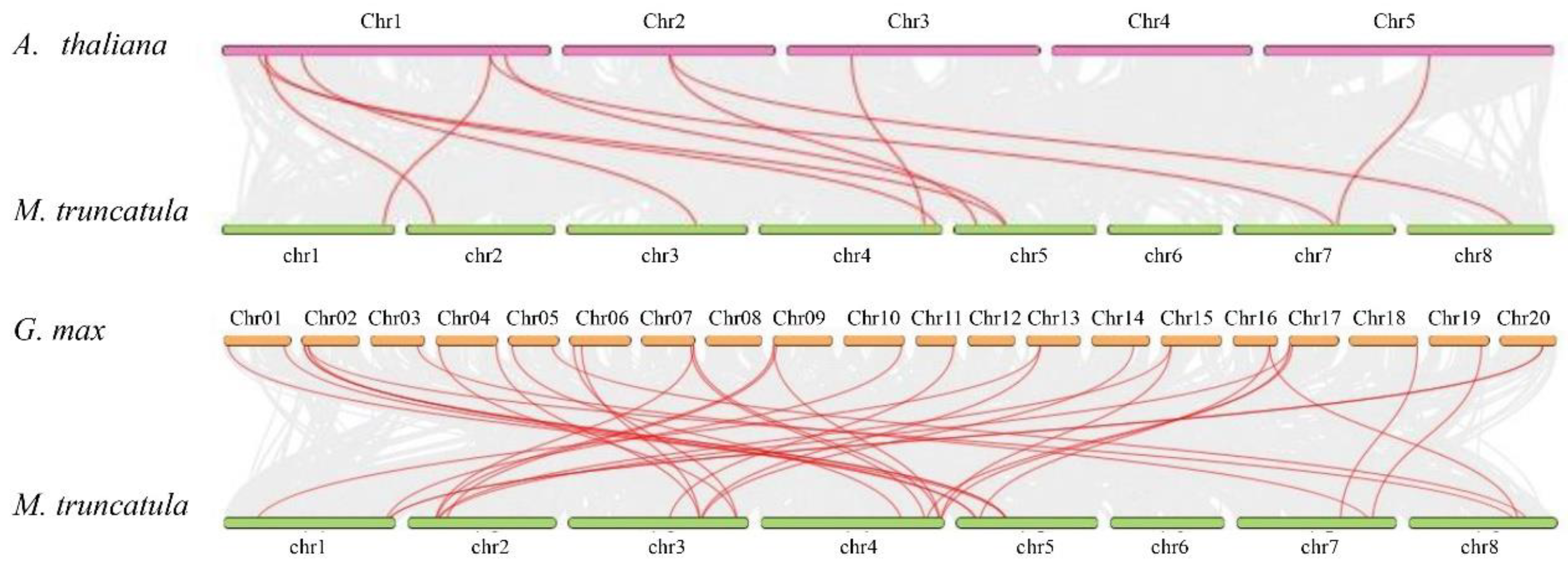
2.2. Chromosomal Location and Expansion Analysis of MtWAKs

Fifty-four MtWAKs were unevenly distributed on eight chromosomes (Figure 1). Chromosome 1 had 17 MtWAKs, while the other chromosomes had 2 to 9 MtWAKs. Interestingly, 20 MtWAK-RLKs and 7 MtWAK-RLCKs were found in clusters with 10 pairs of tandem duplicated gene pairs, while most MtWAK-RLPs and MtWAK short proteins were separately distributed on 8 chromosomes (Figure 1, Table S2).
Two pairs of paralogous genes, MtWAK19-MtWAK35 and MtWAK36-MtWAK53, belong to segmental duplications (Table S2). The synteny analysis showed that there were 11 pairs of orthologous genes between M. truncatula and A. thaliana and 31 pairs between M. truncatula and G. max (Figure 2, Table S2).

2.3. Phylogeny, Gene Structure, Protein Domain and Motif Analysis of MtWAK-RLK Members

Phylogenetic analysis of WAK-RLKs from M. truncatula, A. thaliana, G. max and O. sativa showed that the WAK-RLKs could be divided into five clades. Clade Ⅰ and Ⅴ contained only OsWAK-RLKs. Clade Ⅱ contained 5 AtWAKs, 14 GmWAK-RLKs, 16 MtWAK-RLKs and 2 OsWAK-RLKs. Clade Ⅲ contained 14 AtWAKL-RLKs, 6 GmWAK-RLKs and 2 MtWAK-RLKs. Clade Ⅳ was comprised of 2 AtWAKL-RLKs, 35 GmWAK-RLKs, 4 OsWAK-RLKs and 8 MtWAK-RLKs (Figure 3).
The gene structure, protein domain and motif of MtWAK-RLKs were further analyzed (Figure 4 and Figure S1). Most members showed similar gene structure, harboring two long exons at both ends and a short one in the middle, except MtWAK3 and MtWAK51 with two exons only and MtWAK4, 24, 42, 43 and 50 with four exons (Figure 4B). One or two transmembrane domains were shown in the majority of MtWAK-RLKs except MtWAK3 and MtWAK45. Both Gub_WAK_bind and EGF domains existed in all members of clade Ⅱ and MtWAK36, and there were 51 in clade Ⅳ (Figure 4C). Moreover, 10 conserved motifs were predicted using MEME program. Motifs 1 to 7 corresponded to the conserved kinase domain in all MtWAK-RLKs (Figure 4D and Figure S1). A conserved arginine (R) residue was present in front of the motif DxxxxN; thus, all MtWAK-RLKs were classified as RD kinase. In addition, motif 8 was absent in the members of clade Ⅳ.

2.4. Analysis of Cis-Acting Elements in the Promoter Region of MtWAK-RLKs

To understand the potential function of MtWAK-RLKs, the putative cis-acting elements were analyzed using PlantCARE software. Abundant phytohormone and stress responsive elements were observed in the promoter regions (Figure 5). Twenty-four MtWAK-RLKs possessed a larger number of ABA-responsive elements, ethylene-responsive elements (ERE) and MeJA responsive elements (CGTCA-motif, TGACG-motif). Twelve MtWAK-RLKs had salicylic-acid-responsive elements (TCA-element), while nine MtWAK-RLKs had auxin-responsive elements (TGA-element). Most MtWAK-RLKs had anaerobic induction elements (ARE) and stress-responsive elements (STRE) in the promoter region (Figure 5). A series of growth and development related cis-elements, such as meristem-expression element (CAT-box) and CCGTCC motif were observed in MtWAK-RLKs. The results suggested that MtWAK-RLKs might be involved in phytohormone regulation and stress response in M. truncatula.

2.5. Expression Analysis of MtWAK-RLKs across Different Tissues and Developmental Stages

To obtain insights into their temporal and spatial expression patterns, 12 MtWAK-RLKs having corresponding probesets in the gene expression database were prioritized for analysis (Table S3). MtWAK1, 4, 7, 8 and 18 were almost equally expressed in all tissues (Figure 6A). MtWAK10, 24 and 50 were exclusively and highly expressed in roots. MtWAK36 and 45 were highly expressed in roots, whereas MtWAK3 was highly expressed in stem. From base to apex of stem, MtWAK24 and MtWAK53 expression was increased gradually, while MtWAK3 exhibited the opposite trend with the lowest expression level in the top internode (Figure 6B). Interestingly, MtWAK24 and 53 were significantly up-regulated along with seed development (Figure 6C). In addition, most MtWAK-RLKs were down-regulated during nodulation, except that MtWAK1 was up-regulated greatly at 6 dpi and 20 dpi (Figure 6D).

2.6. Expression Analysis of MtWAK-RLKs in Response to Biotic and Abiotic Stresses

To understand the expression of MtWAK-RLKs in response to pathogens, datasets of ‘Cell suspension_Yeast elicitor’, ‘Root_Macrophomina infected’ and ‘Root Tip_A17_Ralstonia’ from the MtGEA were used for analysis (Table S3). Transcripts of nine MtWAK-RLKs were induced greatly after 2 h of treatment with yeast elicitor (Figure 7A). Although most MtWAK-RLKs were not responsive to infection by Macrophomina or Ralstonia, MtWAK7 transcript was induced greatly in roots at 36 and 48 hpi with Macrophomina (Figure 7B), and MtWAK3, 50 and 53 transcripts were up-regulated at 72 hpi with Ralstonia (Figure 7C). In addition, seven MtWAK-RLKs were up-regulated by infection with Erysiphe pisi (Figure 7D).
MtWAK-RLKs were responsive to abiotic stress (Figure 8, Table S3). MtWAK8, 21 and 43 were up-regulated at 2 h after drought stress, while MtWAK27 was down-regulated. MtWAK2, 5, 45 and 50 were continuously up-regulated after 2 h of drought stress, and MtWAK24 was not responsive to drought (Figure 8A). MtWAK21, 24, 43 and 53 were down-regulated in response to cold stress, while MtWAK10 was not responsive to cold. MtWAK3, 4, 8, 27, 36, 45 and 50 were up-regulated after cold treatment (Figure 8B). MtWAK8, 21 and 27 were up-regulated after 12 h of salt treatment, while MtWAK3, 10, 43 and 54 were down-regulated (Figure 8C).

2.7. Gene Expression Validation of MtWAK-RLKs by qRT-PCR

To verify the expression profiles obtained from microarray data, transcripts of five MtWAK-RLKs (MtWAK1, 3, 10, 24, and 53) in different tissues, including root, stem, leaf, flower, pod and seed, were detected using qRT-PCR. The data showed the expression patterns in different tissues were in consistence with the microarray data. For example, MtWAK10, 24 and 53 were highly expressed in roots (Figure 9A). The response of transcript levels in five MtWAK-RLKs (MtWAK3, 8, 10, 21, and 24) were also detected using qRT-PCR. MtWAK3, 8 and 21 were up-regulated after cold treatment, while MtWAK10 was down-regulated (Figure 9B), which was consistent with the microarray data.

2.8. Plasma Membrane Localized MtWAK24 Inhibited Phytophthora Infection in Tobacco

To explore the functions of some candidate MtWAKs in modulating plant immunity, we performed P. parasitica infection experiments in N. benthamiana. The lesion diameter, as well as relative biomass of P. parasitica, in leaves expressing MtWAK24, but not MtWAK36, was significantly decreased compared to the control leaves expressing empty vector, indicating that MtWAK24 could inhibit the pathogen infection by P. parasitica (Figure 10A,B).
Subcellular localization of MtWAK24 was further analyzed. The data showed that GFP protein was located in the cytoplasm and nucleus, while MtWAK24 protein was located in plasma membrane because the fluorescence was overlapped with that of AtAKT1, the plasma membrane marker protein (Figure 10C).

3. Discussion

The WAK gene family consists of a large number of members. The numbers of WAK proteins identified by the iTAK (http://bioinfo.bti.cornell.edu/tool/itak) program [19] enlarged from moss to dicot and monocot species, indicating a large expansion of WAK family genes during the evolutionary process (Table S4). There are 26, 29, 29, 96, 91, 125, 175, 27 and 320 WAK genes in the genomes of Araidopsis [20], cotton [21], tomato [22], Chinese cabbage [23], barley [24], rice [25], Populus [26], Juglans regia [27] and Triticum aestivum [28], respectively. Through BLASTP and conserved domain searches, 54 MtWAK genes were identified in M. truncatula, including 26 MtWAK-RLK, 9 MtWAK-RLCK, 10 MtWAK-RLP and 9 short protein types (Table S1). The majority of MtWAK-RLK and MtWAK-RLCK genes were clustered in chromosome 1 and 3, attributed mainly to tandem duplications, and the rest were distributed separately among chromosomes (Figure 1, Table S2). Compared to 11 pairs of orthologous WAKs between Arabidopsis and M. truncatula, 31 pairs between M. truncatula and G. max were found, indicating that the expansion of MtWAK family might appear before the separation of G. max (Figure 2). Phylogenetic analysis showed that MtWAK25 and MtWAK26 were close to AtWAKs (Figure 3), and AtWAK4-MtWAK25 and MtWAK25-MtWAK26 were orthologous and paralogous pairs, respectively (Table S2), indicating that MtWAK25 and MtWAK26 were probably involved in cell expansion like AtWAK4 [3].
Multiple cis-acting elements responsive to phytohormones, stresses, growth and development were observed in the promoter regions of MtWAK-RLKs, indicating their potential roles in these processes. The GhWAKs/WAKLs having the above cis-elements in the promoter were responsive to multiple phytohormones and abiotic stresses [12]. Transcript levels of five TaWAKs in wheat (Triticum aestivum) were altered by treatments with GA, BR, IAA, JA and ABA [29]. Gene function is associated with its tissue specific expression pattern. MtWAK10, 24 and 50 showed a root-specific expression with extremely low expression in other organs. MtWAK36 and MtWAK45 were also mainly expressed in roots, whereas MtWAK3 and MtWAK53 were highly expressed in stem and petiole, respectively. MtWAK1, 4, 7, 8 and 18 were evenly expressed in each organ, but the transcript levels were lower than that of other genes (Figure 6A, Table S3). Diverse tissue expression patterns of MtWAK-RLKs implied that they might function broadly in plant tissues.
Plant cell expansion and elongation depend on turgor maintenance and cell wall modification, which is associated with the rigidity and elasticity of the cell wall [30]. WAK genes can monitor pectin and participate in both turgor maintenance and cell wall modification [31]. AtWAK4 and AtWAK2 positively regulate cell expansion [3,4], HvWAK1 positively regulates root growth [32], while OsWAK11 regulates both stem and seed elongation [5]. Transcript levels of MtWAK24 and MtWAK53 were increased gradually from bottom to top internodes and along with seed growth, while expressions of MtWAK3 and MtWAK45 increased, followed by a decrease from internode 1 to internode 8 and along with seed development (Figure 6B,C, Table S3), indicating that they are probably involved in the regulation of cell elongation.
Cell wall modification is involved in symbiosis between rhizobia and legume plants. Passing of an infection thread from cell to cell requires local cell wall degradation [33]. Modifications in the localization of high- and low-methylated homogalacturonans were detected in nodules [34]. The majority of MtWAK-RLKs were greatly down-regulated in the process of nodulation, while MtWAK1 transcript was increased dramatically, implying that MtWAKs are probably associated with nodulation in leguminous plants (Figure 6D, Table S3).
A large body of investigations revealed that WAK genes regulate pathogen resistance. AtWAK1 [7], AtWAKL22 [35], Htn1 (ZmWAK) [8,9], Stb6 [10], Xa4 [11], OsWAK1 [36], OsWAK90-92 [37], OsWAK25 [38], SiWAK1 [39], TaWAK6 [40], GhWAK7A [41], CsWAKL08 [42] and Rlm4/7/9 [43] positively regulate resistance to various pathogens, and OsWAK112d and Sbs1/2 negatively regulate the defense against pathogenic fungus [12,44]. MtWAK3, 18, 50, 10, 24, 45, 36, 7 and 53 transcripts were rapidly induced by yeast elicitor treatment (Figure 7A). Among them, MtWAK7 was up-regulated by Macrophomina infection other than Ralstonia, and MtWAK3, 50 and 53 were up-regulated by Ralstonia other than Macrophomina infection (Figure 7B, C), indicating that these genes had specific roles in resistant to Macrophomina or Ralstonia. Powdery mildews caused by Erysiphe pisi are a serious disease that leads to great decreases in crop production worldwide [45]. Ten MtWAK-RLK genes were up-regulated greatly after incubation with Erysiphe pisi, indicating that they were probably involved in powdery mildew resistance (Figure 7D). To explore the functions of MtWAK-RLKs in plant immunity, we selected two yeast elicitor induced MtWAKs, MtWAK24 and 36, for P. parasitica infection experiments in N. benthamiana. It was revealed that expressing MtWAK24, but not MtWAK36, inhibited infection by P. parasitica (Figure 10).
WAK genes are involved in metal, salt, drought and cold tolerance, although the mechanisms remain unknown. OsWAK112 negatively regulates plant salt tolerance, possibly via direct binding with OsSAMS1/2/3 [17]. The Slwak1 null mutant exhibited disturbed osmotic homeostasis and source-sink balance under long term salinity and thereby reduced fruit yield [46]. The CpGRP1-CpWAK1 complex regulates dehydration-induced morphological changes in Craterostigma plantagineum [47]. Twelve MtWAK-RLK genes were quickly responsive to drought, salt and cold (Figure 8). Ten MtWAK-RLKs transcripts were induced after 2 h of drought (Figure 8A). Seven and four MtWAK-RLKs were up-regulated or down-regulated by cold stress, respectively (Figure 8B), and three and four MtWAK-RLKs were up-regulated or down-regulated by salt stress, respectively (Figure 8C). The results indicate that MtWAK expression is probably involved in abiotic stress resistance.

4. Materials and Methods

4.1. Identification of MtWAK Genes

The genome sequences were obtained from M. truncatula genome database (http://www.medicagogenome.org/, Mt4.0v2). The gene information of WAK family in Arabidopsis and Rice were retrieved from the previous studies. The hidden Markov model (HMM) profiles of the WAKs were down-loaded from the Pfam database (http://pfam.xfam.org/, accessed on 1 August 2020). EGF_CA(PF07645), WAK_assoc (PF14380), GUB_WAK_bind (PF13947) and Pkinase_Tyr (PF07714) were used to identify MtWAKs. Firstly, BLASTP search was performed at M. truncatula genome database with an e-value of 1e-5 using previously reported sequences of AtWAKs and OsWAKs as query. Then, we searched EGF_CA(PF07645), WAK_assoc (PF14380), GUB_WAK_bind (PF13947) and Pkinase_Tyr (PF07714) domain from putative MtWAKs with e-value cut-off at 1.0 by using HMMER v3.1b2 software. The integrity of the four domains was verified by using the online program SMART (http://smart.emblheidelberg.de/, accessed on 1 August 2020) with an e-value < 0.1. Lastly, each candidate gene was assessed for its sequence similarity to other putative MtWAKs. Only genes that fit into one of the four MtWAK types (see Table S1) according to Zhang et al. [18] were defined as MtWAKs. Protein length, molecular weight (MW) and isoelectric point (PI) were predicted by ExPasy program (http://www.expasy.org/tools/, accessed on 10 August 2021).

4.2. Chromosomal Location and Synteny Correlation Analysis

The physical position of the MtWAK genes on the chromosome was mapped using TBtools software (Version 1.108). The synteny correlation analysis of WAK genes between the homologs in M. truncatula and A. thaliana or G. max were verified and visualized using TBtools software.

4.3. Phylogenetic Analysis and Gene Structure, Motif and Conserved Domain Analysis

Multiple alignments of protein sequences were performed using CLUSTALX software (Version 1.81). The phylogenetic tree was constructed by using MEGAX with the neighbor-joining method and 1000 bootstrap replications. The gene structure, conserved domain and conserved motifs were displayed using TBtools software. The conserved domains and conserved motifs of the 26 MtWAK-RLKs were analyzed by SMART program and MEME program (Version 5.4.1) (http://meme-suite.org/tools/meme, accessed on 20 July 2022).

4.4. Analysis of cis-Acting Regulatory Elements

The 2000 bp promoter sequences upstream from the initiation codon of MtWAK-RLKs were extracted from the genome of M. truncatula and analyzed using PlantCARE software (http://bioinformatics.psb.ugent.be/webtools/plantcare/html/, accessed on 1 August 2020). However, less than 300 bp promoter sequence of MtWAK10 was available.

4.5. Expression Pattern Analysis

The genome-wide microarray data obtained from MtGEA (https://mtgea.noble.org/v3/, accessed on 1 August 2020) was used to analyze the expression of MtWAK genes in different tissues and developing stages. The expression data were gene-wise normalized. The clustered heatmap of expression pattern profile on log2 scale was portrayed using TBtools software. Analyses of WAK expression in response to biotic and abiotic stress were conducted on datasets: (ⅰ) ‘Cell suspension_Yeast elicitor’, ‘Root_Macrophomina infected’ and ‘Root Tip_A17_Ralstonia’ (microarray data obtained from MtGEA); (ⅱ) powdery mildew Erysiphe pisi treatment [48] RNA-seq data were retrieved from NCBI Database (SRR7589436, SRR7589435, SRR7589438, and SRR7589437); (ⅲ) drought, salt and cold treatment, date from NCBI GEO (dataset accession: GSE136739). The expression abundance was presented by the reads per kilobase per million (Table S3). The relative transcript level after treatments was calculated compared with the untreated control or before treatment (0 h). The clustered heatmap of relative expression pattern on log2 scale was analyzed by the TBtools.

4.6. Tissue Samples Collection and Cold and Salt Treatment

M. truncatula plants were grown in a growth chamber at 25 °C with 16 h of light. Root, stem, mature leave, flower, pod and seed were sampled from three-month-old plants. Six-week-old plants were exposed to low temperature (5 °C) as a cold stress treatment. Leaves were collected at time intervals of 0, 1, 2, 6 and 12 h.

4.7. RNA Extraction and qRT-PCR

Total RNA was extracted using the RNAprep pure Plant Kit (Tiangen, Beijing, China). cDNA synthesis was performed with HiScript III RT SuperMix for qPCR (+gDNA wiper) reagent kit with gDNA Eraser (Vazyme, Nanjing, China). qRT-PCR was performed following the instructions of ChamQ Universal SYBR qPCR Master Mix (Vazyme, Nanjing, China). The MtActin7 (Medtr3g095530) gene was used as the controls. All primer sequences are shown in Supplementary Table S5. The relative gene expression level was calculated with 2−ΔCt method.

4.8. Subcellular Localization

The full CDSs of MtWAK24 without the terminator were cloned and ligated to pCAMBIA1305-GFP vector driven by CaMV 35S promoter. Primer sequences for constructions are shown in Supplementary Table S5. Positive Agrobacterium colonies were cultured in LB medium containing 50 μg/mL rifampicin and kanamycin at 28 °C overnight; the cells were harvested by centrifugation at 2500 × g at room temperature for 3–5 min and resuspended in infiltration buffer (10 mM MgCl2, 10 mM MES pH 5.6, and 100–200 µM acetosyringone). The cell density was adjusted to OD600 = 0.2 before the cell suspensions were infiltrated into one-month-old Nicotiana benthamiana (N. benthamiana) leaves. After 48–72 h, fluorescence was observed using confocal laser scanning microscopy (Zeiss LSM800, Jena, Germany).

4.9. Phytophthora Infection in N. benthamiana

Vectors expressing selected MtWAK-RLKs under control of CaMV 35S promoter were constructed. The infection experiments were performed as described [49]. Briefly, the infiltrated leaves were inoculated with P. parasitica mycelium at 12 hpi, and leaf lesions were determined using the Evans blue method at 48 hpi. P. parasitica strain was obtained from Yuanchao Wang‘s lab (Nanjing Agricultural University, Nanjing, China). Relative biomass of P. parasitica was determined by qPCR of P. parasitica genome DNA normalized to tobacco genome DNA at 48 hpi. All primer sequences are shown in Supplementary Table S5.

5. Conclusions

In summary, a comprehensive genome-wide analysis of WAK family was performed. A total of 54 MtWAKs were identified in M. truncatula, including 26 MtWAK-RLK, 9 MtWAK-RLCK, 10 MtWAK-RLP and 9 short protein type genes. MtRLKs and MtRLCKs were largely tandem duplicated. Most MtWAK-RLKs had similar exon–intron organization and motif distribution. Multiple cis-acting elements responsible for phytohormones, stresses, growth and development were observed in the promoter regions. The expression patterns of MtWAK-RLKs varied in different plant tissues and developmental stages and biotic and abiotic stress conditions. The results suggest that MtWAKs might have multiple functions in M. truncatula. Furthermore, plasma-membrane-localized MtWAK24 significantly inhibited Phytophthora infection in tobacco, indicating its role in pathogen resistance, which is worthy to be investigated in the future.

Supplementary Materials

The following supporting information can be downloaded at: www.mdpi.com/article/10.3390/plants12091849/s1, Figure S1: The motif of MtWAK-RLKs; Table S1: The predicated information of MtWAK family genes in M. truncatula. Table S2: The synteny analysis of WAK homologs between M. truncatula and A. thaliana or G. max; Table S3: The gene expression profiles of MtWAK-RLKs in different tissues and developmental stages as well as biotic and abiotic stress; Table S4: The number of plant protein kinases and WAKs predicted via iTAK program; Table S5: Primers.

Author Contributions

Z.G., H.Z. and W.K. designed and wrote the article; H.Z., W.K., J.S., S.Y. and P.Z. collected data and performed most of the data analysis. B.Y. provided instructions in the Phytophthora infection experiments. All authors approved the submitted version. All authors have read and agreed to the published version of the manuscript.

Funding

This work was supported by the National Natural Science Foundation of China (Grant numbers 32101439) and the Natural Science Foundation of Jiangsu Province (BK20190527).

Data Availability Statement

The data that support the findings of this study are available from the corresponding author upon reasonable request.

Conflicts of Interest

The authors declare no conflict of interest.

References

  1. Gish, L.A.; Clark, S.E. The RLK/Pelle family of kinases. Plant J. 2011, 66, 117–127. [Google Scholar] [CrossRef] [PubMed]
  2. He, Z.H.; Cheeseman, I.; He, D.; Kohorn, B.D. A cluster of five cell wall-associated receptor kinase genes, Wak1-5, are expressed in specific organs of Arabidopsis. Plant Mol. Biol. 1999, 39, 1189–1196. [Google Scholar] [CrossRef] [PubMed]
  3. Lally, D.; Ingmire, P.; Tong, H.Y.; He, Z.H. Antisense expression of a cell wall-associated protein kinase, WAK4, inhibits cell elongation and alters morphology. Plant Cell 2001, 13, 1317–1331. [Google Scholar] [CrossRef] [PubMed]
  4. Kohorn, B.D.; Kobayashi, M.; Johansen, S.; Riese, J.; Huang, L.F.; Koch, K.; Fu, S.; Dotson, A.; Byers, N. An Arabidopsis cell wall-associated kinase required for invertase activity and cell growth. Plant J. 2006, 46, 307–316. [Google Scholar] [CrossRef]
  5. Yue, Z.L.; Liu, N.; Deng, Z.P.; Zhang, Y.; Wu, Z.M.; Zhao, J.L.; Sun, Y.; Wang, Z.Y.; Zhang, S.W. The receptor kinase OsWAK11 monitors cell wall pectin changes to fine-tune brassinosteroid signaling and regulate cell elongation in rice. Curr. Biol. 2022, 32, 2454–2466.e7. [Google Scholar] [CrossRef]
  6. Yin, W.; Yang, H.; Wang, Y.; Feng, P.; Deng, Y.; Zhang, L.; He, G.; Wang, N. Oryza sativa PECTIN DEFECTIVE TAPETUM1 affects anther development through a pectin-mediated signaling pathway in rice. Plant Physiol. 2022, 189, 1570–1586. [Google Scholar] [CrossRef]
  7. He, Z.H.; He, D.Z.; Kohorn, B.D. Requirement for the induced expression of a cell wall associated receptor kinase for survival during the pathogen response. Plant J. 1998, 14, 55–63. [Google Scholar] [CrossRef]
  8. Hurni, S.; Scheuermann, D.; Krattinger, S.G.; Kessel, B.; Wicker, T.; Herren, G.; Fitze, M.N.; Breen, J.; Presterl, T.; Ouzunova, M.; et al. The maize disease resistance gene Htn1 against northern corn leaf blight encodes a wall-associated receptor-like kinase. Proc. Natl. Acad. Sci. USA 2015, 112, 8780–8785. [Google Scholar] [CrossRef] [PubMed]
  9. Zuo, W.L.; Chao, Q.; Zhang, N.; Ye, J.R.; Tan, G.Q.; Li, B.L.; Xing, Y.X.; Zhang, B.Q.; Liu, H.J.; Fengler, K.A.; et al. A maize wall-associated kinase confers quantitative resistance to head smut. Nat. Genet. 2015, 47, 151–157. [Google Scholar] [CrossRef]
  10. Saintenac, C.; Lee, W.S.; Cambon, F.; Rudd, J.J.; King, R.C.; Marande, W.; Powers, S.J.; Berges, H.; Phillips, A.L.; Uauy, C.; et al. Wheat receptor-kinase-like protein Stb6 controls gene-for-gene resistance to fungal pathogen Zymoseptoria tritici. Nat. Genet. 2018, 50, 368–374. [Google Scholar] [CrossRef]
  11. Hu, K.; Cao, J.; Zhang, J.; Xia, F.; Ke, Y.; Zhang, H.; Xie, W.; Liu, H.; Cui, Y.; Cao, Y.; et al. Improvement of multiple agronomic traits by a disease resistance gene via cell wall reinforcement. Nat. Plants 2017, 3, 17009. [Google Scholar] [CrossRef] [PubMed]
  12. Delteil, A.; Gobbato, E.; Cayrol, B.; Estevan, J.; Michel-Romiti, C.; Dievart, A.; Kroj, T.; Morel, J.B. Several wall-associated kinases participate positively and negatively in basal defense against rice blast fungus. Bmc Plant Biol. 2016, 16, 17. [Google Scholar] [CrossRef] [PubMed]
  13. Denoux, C.; Galletti, R.; Mammarella, N.; Gopalan, S.; Werck, D.; De Lorenzo, G.; Ferrari, S.; Ausubel, F.M.; Dewdney, J. Activation of defense response pathways by OGs and Flg22 elicitors in Arabidopsis seedlings. Mol. Plant. 2008, 1, 423–445. [Google Scholar] [CrossRef]
  14. Sivaguru, M.; Ezaki, B.; He, Z.H.; Tong, H.; Osawa, H.; Baluska, F.; Volkmann, D.; Matsumoto, H. Aluminum-induced gene expression and protein localization of a cell wall-associated receptor kinase in Arabidopsis. Plant Physiol. 2003, 132, 2256–2266. [Google Scholar] [CrossRef] [PubMed]
  15. Hou, X.; Tong, H.; Selby, J.; Dewitt, J.; Peng, X.; He, Z.H. Involvement of a cell wall-associated kinase, WAKL4, in Arabidopsis mineral responses. Plant Physiol. 2005, 139, 1704–1716. [Google Scholar] [CrossRef]
  16. Zhang, H.; Lian, C.; Shen, Z. Proteomic identification of small, copper-responsive proteins in germinating embryos of Oryza sativa. Ann. Bot. 2009, 103, 923–930. [Google Scholar] [CrossRef] [PubMed]
  17. Lin, W.; Wang, Y.H.; Liu, X.Y.; Shang, J.X.; Zhao, L.Q. OsWAK112, a wall-associated kinase, negatively regulates salt stress responses by inhibiting ethylene production. Front. Plant Sci. 2021, 12, 751965. [Google Scholar] [CrossRef] [PubMed]
  18. Zhang, S.; Chen, C.; Li, L.; Meng, L.; Singh, J.; Jiang, N.; Deng, X.W.; He, Z.H.; Lemaux, P.G. Evolutionary expansion, gene structure, and expression of the rice wall-associated kinase gene family. Plant Physiol. 2005, 139, 1107–1124. [Google Scholar] [CrossRef] [PubMed]
  19. Zheng, Y.; Jiao, C.; Sun, H.H.; Rosli, H.G.; Pombo, M.A.; Zhang, P.F.; Banf, M.; Dai, X.B.; Martin, G.B.; Giovannoni, J.J.; et al. iTAK: A program for genome-wide prediction and classification of plant transcription factors, transcriptional regulators, and protein kinases. Mol. Plant 2016, 9, 1667–1670. [Google Scholar] [CrossRef]
  20. Verica, J.A.; Chae, L.; Tong, H.Y.; Ingmire, P.; He, Z.H. Tissue-specific and developmentally regulated expression of a cluster of tandemly arrayed cell wall-associated kinase-like kinase genes in Arabidopsis. Plant Physiol. 2003, 133, 1732–1746. [Google Scholar] [CrossRef]
  21. Dou, L.; Li, Z.; Shen, Q.; Shi, H.; Li, H.; Wang, W.; Zou, C.; Shang, H.; Li, H.; Xiao, G. Genome-wide characterization of the WAK gene family and expression analysis under plant hormone treatment in cotton. BMC Genom. 2021, 22, 85. [Google Scholar] [CrossRef] [PubMed]
  22. Sun, Z.Y.; Song, Y.P.; Chen, D.; Zang, Y.D.; Zhang, Q.L.; Yi, Y.T.; Qu, G.Q. Genome-wide identification, classification, characterization, and expression analysis of the wall-associated kinase family during fruit development and under wound stress in tomato (Solanum lycopersicum L.). Genes 2020, 11, 1186. [Google Scholar] [CrossRef] [PubMed]
  23. Zhang, B.; Li, P.; Su, T.B.; Li, P.R.; Xin, X.Y.; Wang, W.H.; Zhao, X.Y.; Yu, Y.J.; Zhang, D.S.; Yu, S.C.; et al. Comprehensive analysis of wall-associated kinase genes and their expression under abiotic and biotic stress in chinese cabbage (Brassica rapa ssp. pekinensis). J. Plant Growth Regul. 2020, 39, 72–86. [Google Scholar] [CrossRef]
  24. Tripathi, R.K.; Aguirre, J.A.; Singh, J. Genome-wide analysis of wall associated kinase (WAK) gene family in barley. Genomics 2021, 113, 523–530. [Google Scholar] [CrossRef] [PubMed]
  25. Hu, W.; Lv, Y.Y.; Lei, W.R.; Li, X.; Chen, Y.H.; Zheng, L.Q.; Xia, Y.; Shen, Z.G. Cloning and characterization of the Oryza sativa wall-associated kinase gene OsWAK11 and its transcriptional response to abiotic stresses. Plant Soil 2014, 384, 335–346. [Google Scholar] [CrossRef]
  26. Tocquard, K.; Lafon-Placette, C.; Auguin, D.; Muries, B.; Bronner, G.; Lopez, D.; Fumanal, B.; Franchel, J.; Bourgerie, S.; Maury, S.; et al. In silico study of wall-associated kinase family reveals large-scale genomic expansion potentially connected with functional diversification in Populus. Tree Genet. Genom. 2014, 10, 1135–1147. [Google Scholar] [CrossRef]
  27. Li, M.; Ma, J.; Liu, H.; Ou, M.; Ye, H.; Zhao, P. Identification and characterization of wall-associated kinase (WAK) and WAK-like (WAKL) gene family in Juglans regia and its wild related species Juglans mandshurica. Genes 2022, 13, 134. [Google Scholar] [CrossRef]
  28. Xia, X.; Zhang, X.; Zhang, Y.; Wang, L.; An, Q.; Tu, Q.; Wu, L.; Jiang, P.; Zhang, P.; Yu, L.; et al. Characterization of the WAK gene family reveals genes for FHB resistance in bread wheat (Triticum aestivum L.). Int. J. Mol. Sci. 2022, 23, 7157. [Google Scholar] [CrossRef]
  29. Liu, Y.; Liu, D.C.; Zhang, H.Y.; Gao, H.B.; Guo, X.L.; Fu, X.D.; Zhang, A.M. Isolation and characterisation of six putative wheat cell wall-associated kinases. Funct. Plant Biol. 2006, 33, 811–821. [Google Scholar] [CrossRef]
  30. Cosgrove, D.J. Growth of the plant cell wall. Nat. Rev. Mol. Cell Biol. 2005, 6, 850–861. [Google Scholar] [CrossRef]
  31. Cosgrove, D.J. Plant cell walls: Wall-associated kinases and cell expansion. Curr. Biol. 2001, 11, R558–R559. [Google Scholar] [CrossRef]
  32. Kaur, R.; Singh, K.; Singh, J. A root-specific wall-associated kinase gene, HvWAK1, regulates root growth and is highly divergent in barley and other cereals. Funct. Integr. Genom. 2013, 13, 167–177. [Google Scholar] [CrossRef] [PubMed]
  33. Van Spronsen, P.C.; Bakhuizen, R.; van Brussel, A.A.; Kijne, J.W. Cell wall degradation during infection thread formation by the root nodule bacterium Rhizobium leguminosarum is a two-step process. Eur. J. Cell Biol. 1994, 64, 88–94. [Google Scholar] [PubMed]
  34. Wilmowicz, E.; Kucko, A.; Alche, J.D.; Czeszewska-Rosiak, G.; Florkiewicz, A.B.; Kapusta, M.; Karwaszewski, J. Remodeling of cell wall components in root nodules and flower abscission zone under drought in yellow lupine. Int. J. Mol. Sci. 2022, 23, 1680. [Google Scholar] [CrossRef] [PubMed]
  35. Diener, A.C.; Ausubel, F.M. RESISTANCE TO FUSARIUM OXYSPORUM 1, a dominant Arabidopsis disease-resistance gene, is not race specific. Genetics 2005, 171, 305–321. [Google Scholar] [CrossRef]
  36. Li, H.; Zhou, S.Y.; Zhao, W.S.; Su, S.C.; Peng, Y.L. A novel wall-associated receptor-like protein kinase gene, OsWAK1, plays important roles in rice blast disease resistance. Plant Mol. Biol. 2009, 69, 337–346. [Google Scholar] [CrossRef]
  37. Cayrol, B.; Delteil, A.; Gobbato, E.; Kroj, T.; Morel, J.B. Three wall-associated kinases required for rice basal immunity form protein complexes in the plasma membrane. Plant Signal. Behav. 2016, 11, e1149676. [Google Scholar] [CrossRef]
  38. Harkenrider, M.; Sharma, R.; De Vleesschauwer, D.; Tsao, L.; Zhang, X.T.; Chern, M.; Canlas, P.; Zuo, S.M.; Ronald, P.C. Overexpression of rice wall-associated kinase 25 (OsWAK25) alters resistance to bacterial and fungal pathogens. PLoS ONE 2016, 11, e0147310. [Google Scholar] [CrossRef]
  39. Rosli, H.G.; Zheng, Y.; Pombo, M.A.; Zhong, S.; Bombarely, A.; Fei, Z.; Collmer, A.; Martin, G.B. Transcriptomics-based screen for genes induced by flagellin and repressed by pathogen effectors identifies a cell wall-associated kinase involved in plant immunity. Genom. Biol. 2013, 14, R139. [Google Scholar] [CrossRef]
  40. Dmochowska-Boguta, M.; Kloc, Y.; Zielezinski, A.; Werecki, P.; Nadolska-Orczyk, A.; Karlowski, W.M.; Orczyk, W. TaWAK6 encoding wall-associated kinase is involved in wheat resistance to leaf rust similar to adult plant resistance. PLoS ONE 2020, 15, e0227713. [Google Scholar] [CrossRef]
  41. Wang, P.; Zhou, L.; Jamieson, P.; Zhang, L.; Zhao, Z.X.; Babilonia, K.; Shao, W.Y.; Wu, L.Z.; Mustafa, R.; Amin, I.; et al. The Cotton Wall-associated kinase GhWAK7A mediates responses to fungal wilt pathogens by complexing with the chitin sensory receptors. Plant Cell 2020, 32, 3978–4001. [Google Scholar] [CrossRef] [PubMed]
  42. Li, Q.; Hu, A.H.; Qi, J.J.; Dou, W.F.; Qin, X.J.; Zou, X.P.; Xu, L.Z.; Chen, S.C.; He, Y.R. CsWAKL08, a pathogen-induced wall-associated receptor-like kinase in sweet orange, confers resistance to citrus bacterial canker via ROS control and JA signaling. Hortic. Res. 2020, 7, 42. [Google Scholar] [CrossRef] [PubMed]
  43. Haddadi, P.; Larkan, N.J.; Van DeWouw, A.; Zhang, Y.Q.; Neik, T.X.; Beynon, E.; Bayer, P.; Edwards, D.; Batley, J.; Borhan, M.H. Brassica napus genes Rlm4 and Rlm7, conferring resistance to Leptosphaeria maculans, are alleles of the Rlm9 wall-associated kinase-like resistance locus. Plant Biotechnol. J. 2022, 20, 1229–1231. [Google Scholar] [CrossRef]
  44. Ameen, G.; Solanki, S.; Drader, T.; Sager-Bittara, L.; Steffenson, B.; Kleinhofs, A.; Vogiatzis, C.; Brueggeman, R.S. rcs5-mediated spot blotch resistance in barley is conferred by wall-associated kinases that resist pathogen manipulation. BioRxiv 2020. [Google Scholar] [CrossRef]
  45. Glawe, D.A. The powdery mildews: A review of the world’s most familiar (yet poorly known) plant pathogens. Annu. Rev. Phytopathol. 2008, 46, 27–51. [Google Scholar] [CrossRef] [PubMed]
  46. Meco, V.; Egea, I.; Ortiz-Atienza, A.; Drevensek, S.; Esch, E.; Yuste-Lisbona, F.J.; Barneche, F.; Vriezen, W.; Bolarin, M.C.; Lozano, R.; et al. The salt sensitivity induced by disruption of cell wall-associated kinase 1 (SlWAK1) tomato gene is linked to altered osmotic and metabolic homeostasis. Int. J. Mol. Sci. 2020, 21, 6308. [Google Scholar] [CrossRef] [PubMed]
  47. Giarola, V.; Krey, S.; von den Driesch, B.; Bartels, D. The Craterostigma plantagineum glycine-rich protein CpGRP1 interacts with a cell wall-associated protein kinase 1 (CpWAK1) and accumulates in leaf cell walls during dehydration. New Phytol. 2016, 210, 535–550. [Google Scholar] [CrossRef] [PubMed]
  48. Gupta, M.; Sharma, G.; Saxena, D.; Budhwar, R.; Vasudevan, M.; Gupta, V.; Gupta, A.; Gupta, R.; Chandran, D. Dual RNA-Seq analysis of Medicago truncatula and the pea powdery mildew Erysiphe pisi uncovers distinct host transcriptional signatures during incompatible and compatible interactions and pathogen effector candidates. Genomics 2020, 112, 2130–2145. [Google Scholar] [CrossRef]
  49. Qiu, X.; Kong, L.; Chen, H.; Lin, Y.; Tu, S.; Wang, L.; Chen, Z.; Zeng, M.; Xiao, J.; Yuan, P.; et al. The Phytophthora sojae nuclear effector PsAvh110 targets a host transcriptional complex to modulate plant immunity. Plant Cell 2022, 35, 574–597. [Google Scholar] [CrossRef]
Figure 1. Chromosomal location of MtWAKs. Grey bars represent the chromosomes. The chromosome numbers are shown in yellow. The scale bar is shown on the left. WAK gene names are highlighted with different colors. MtWAK-RLKs, MtWAK-RLCKs, MtWAK-RLPs and MtWAK short proteins are indicated using red, green, blue, and pink, respectively. Tandem duplicated gene pairs are linked using black lines.
Plants 12 01849 g001
Figure 2. The synteny analysis of MtWAKs between M. truncatula and A. thaliana or G. max. Gray lines indicate all collinear blocks within M. truncatula and A. thaliana or G. max, while red lines indicate the synteny of WAK genes between M. truncatula and A. thaliana or G. max.
Plants 12 01849 g002
Figure 3. Phylogenetic analysis of WAK-RLKs from M. truncatula, A. thaliana, G. max and O. sativa: 26 MtWAK-RLKs (red square), 21 AtWAK-RLKs (blue triangle), 56 GmWAK-RLKs (orange circle) and 71 OsWAK-RLKs (green circle) were aligned using the CLUSTALX software (Version 1.81). The phylogenetic tree was constructed with MEGAX software (version 10.0.5) by the neighbor-joining methodwith 1000 bootstrap replicates.
Plants 12 01849 g003
Figure 4. The gene structure, conserved domain and motif analysis of MtWAK-RLKs. (A) The phylogenetic tree of MtWAK-RLKs. (B) The gene structure of MtWAK-RLKs. (C) The conserved domain of MtWAK-RLKs. (D) The top ten conserved motifs of MtWAK-RLKs.
Plants 12 01849 g004
Figure 5. Cis-acting elements in the promoters of MtWAK-RLKs. The numbers and the depth of red represent the frequency of the elements. ABRE, ABA-responsive element; AuxRR-core and TGA-element, auxin-responsive elements; ERE, ethylene-responsive element; CGTCA and TGACG motif, MeJA-responsive elements; SARE and TCA-element, salicylic-acid-responsive elements; GARE-motif and P-box, GA-responsive elements; ARE and GC-motif, anaerobic induction elements; TC-rich repeats and STRE, stress-responsive elements; MBS, drought-induced element; DRE-core, dehydration-responsive element; LTR, low-temperature-responsive element; WRE3 and WUN-motif, wound-responsive element; CAT-box, meristem expression element; CCGTCC motif and NON-box, meristem-specific activation elements; circadian, circadian control element; GCN4_motif, endosperm-expression element; HD-Zip 1, palisade mesophyll cell-expression element; MBS, flavonoid biosynthetic gene regulation element; O2-site, zein metabolism regulation element.
Plants 12 01849 g005
Figure 6. Clustered heatmap of the relative expression levels of MtWAK-RLKs in different tissues and developmental stages. (A) Different organs. (B) Developing seeds. (C) Developing internodes. (D) Developing nodules. Microarray data are normalized by the mean expression value of each gene in tissues. The clustered heatmap on log2 scale is drawn using TBtools software (Version 1.108). DAP, days after pollination. DPI, days post-incubation.
Plants 12 01849 g006
Figure 7. Clustered heatmap of the relative expression levels of MtWAK-RLKs involved in biotic stress response. (A) Yeast eliciter (YE) treatment. (B) Macrophomina infection. (C) Ralstonia infection. (D) Erysiphe pisi infection. The relative gene expression levels after treatments were calculated and compared with the untreated control or before treatment. The clustered heatmap on log2 scale was drawn using TBtools software (Version 1.108).
Plants 12 01849 g007
Figure 8. Clustered heatmap of the relative expression levels of MtWAK-RLKs involved in abiotic stress response. (A) Drought stress. (B). Cold stress (C) Salt stress. The relative gene expression levels after treatments were calculated and compared with the control untreated or before treatment. The clustered heatmap on log2 scale was drawn using TBtools software (Version 1.108).
Plants 12 01849 g008
Figure 9. Expression analysis of selected MtWAK-RLK genes in different tissues and under cold stress using qRT-PCR. (A) Different tissues. (B) Cold stress. The error bars were obtained from three measurements. nd indicates not detected. Significant differences are indicated as different lowercase letters (P ≤ 0.05, by one-way ANOVA).
Plants 12 01849 g009
Figure 10. Plasma membrane localized MtWAK24 inhibited Phytophthora infection in tobacco. N. benthamiana leaves individually expressing 35S::MtWAK36, 35S::MtWAK24 or 35S::GFP were inoculated with P. parasitica mycelial plugs at 24 h after Agrobacterium infiltration. At 48 hpi, infected leaves were stained using Evans blue staining for lesion determination (A). Relative biomass of P. parasitica was determined by qPCR of P. parasitica genome DNA normalized to tobacco genome DNA (B). The results are indicated with means ± SE, n = 6. Asterisks indicate significant differences (* p < 0.05; Student’s t test). The subcellular localization of MtWAK24-GFP proteins and a free GFP protein in tobacco (C). P. M-marker: a plasma membrane localization protein AtAKT1. Bars = 20 µm.
Plants 12 01849 g010
Disclaimer/Publisher’s Note: The statements, opinions and data contained in all publications are solely those of the individual author(s) and contributor(s) and not of MDPI and/or the editor(s). MDPI and/or the editor(s) disclaim responsibility for any injury to people or property resulting from any ideas, methods, instructions or products referred to in the content.

