Review

Light and Plant Growth Regulators on In Vitro Proliferation

by
Valeria Cavallaro
1,*,
Alessandra Pellegrino
1,
Rosario Muleo
2,* and
Ivano Forgione
2
1
Institute of BioEconomy (IBE), National Research Council of Italy, 95126 Catania, Italy
2
Tree Physiology and Fruit Crop Biotechnology Laboratory, Department of Agriculture and Forest Sciences (DAFNE), University of Tuscia, 01100 Viterbo, Italy
*
Authors to whom correspondence should be addressed.
Plants 2022, 11(7), 844; https://doi.org/10.3390/plants11070844
Submission received: 14 February 2022 / Revised: 9 March 2022 / Accepted: 17 March 2022 / Published: 22 March 2022
(This article belongs to the Special Issue The Effects of LED Light Spectra and Intensities on Plant Growth)

Abstract

:
Plant tissue cultures depend entirely upon artificial light sources for illumination. The illumination should provide light in the appropriate regions of the electromagnetic spectrum for photomorphogenic responses and photosynthetic metabolism. Controlling light quality, irradiances and photoperiod enables the production of plants with desired characteristics. Moreover, significant money savings may be achieved using both more appropriate and less consuming energy lamps. In this review, the attention will be focused on the effects of light characteristics and plant growth regulators on shoot proliferation, the main process in in vitro propagation. The effects of the light spectrum on the balance of endogenous growth regulators will also be presented. For each light spectrum, the effects on proliferation but also on plantlet quality, i.e., shoot length, fresh and dry weight and photosynthesis, have been also analyzed. Even if a huge amount of literature is available on the effects of light on in vitro proliferation, the results are often conflicting. In fact, a lot of exogenous and endogenous factors, but also the lack of a common protocol, make it difficult to choose the most effective light spectrum for each of the large number of species. However, some general issues derived from the analysis of the literature are discussed.

1. Introduction

Plants, like any other living organisms on planet Earth, are strongly influenced by environmental cues. Unlike animals, plants are sessile and at the mercy of their surrounding environment. Consequently, they have evolved mechanisms that perceive and respond to environmental changes and adapt their development and growth accordingly. Light plays a pivotal role in a plant’s life, not only for photosynthetic energy production but also for its regulative role of molecular, biochemical and morphological processes that underlie plant growth and development [1,2,3]. Fluence rate, regions of wavelength electromagnetic spectrum, duration and direction are the key attributes of light that drive photosynthesis and photomorphogenesis through mechanisms including the selective activation of various light receptors [4,5,6,7,8,9]. Plant light photoreceptors have evolved in articulated biochemistry structure that capture photons and detect many of the light physical properties. Subsequently, through interactive pathways the photoreceptors interpret information from incoming light and traduce them in biochemical and biological responses able to regulate plant growth and development. A discrete number of photosensor families have evolved in plants. The phytochrome (PHY) family receptors monitor the red (R, 600–700 nm) and far red (FR, 700–750 nm) light regions [10,11,12]. PHY can be present in two states and the active state (Pfr) is formed due to absorption of red light by the inactive state (Pr) [13]. The wavelength region of light from UV-A to blue (B, 320–500 nm) is perceived by three small families of photoreceptors [14] that mediate plant responses. All three photoreceptor families contain flavin adenine dinucleotide (FAD) as a chromophore: three cryptochromes (CRY) with CRY1 and CRY2 acting in the nucleus, whereas CRY3 is probably acting in the mitochondrion and chloroplast [15,16], two phototropins (PHOT) [9,11,17] and the members of the Zeitlupe family (ztl, fkf1 and lkp2) [18]. In addition, PHY has also been found to mediate various blue responses [19]. The UV Resistance Locus 8, monitoring ultraviolet B wavelengths (UV-B, 280–315 nm), regulates both developmental and UV-protective outcomes [20,21,22].
PHYs act in detecting mutual plant shading through the change in the R:FR ratio and appropriately redirect growth and development through the modulation of apical dominance and of axillary meristems formation according to survival [23,24,25]. CRY1 is thought to be the CRY responsible for the B high-irradiance response, inhibiting stem plant growth and reducing internode elongation, whereas CRY2 is likely responsible for the inhibition because of the B low-irradiance response [19]; collectively, in plants they perform important traits such as flowering and plant stem elongation [26]. PHOT1 and PHOT2 are involved in auxin polar transport, modulation of auxin sensing and phototropism [27,28,29].
Micropropagation is considered an effective large-scale in vitro plant multiplication of selected insect/disease/virus-free plants in a short time, all year round, and is a reliable method for in vitro preservation of threatened plant species. The micropropagation technology differs strongly from all other agamic propagation methods since the plants, cultured frequently as microcuttings, can remain under constant environmental conditions for a long time. The habitat of an in vitro culture is strongly restricted, and plants switch from an ontogenetic processing that starts from similar juvenility traits to a much deeper juvenility state [30]. Photoperiod, light intensity, light quality, temperature and relative humidity are factors that in the in vitro habitat are subjected to scarce fluctuations that alter the periodic and oscillator systems upon which plants depend; therefore, plants remain under largely invariable conditions. Although, currently, we cannot establish whether the mutations that are detected in the genomes of in vitro growing plants appear during in vitro culture, however, we could hypothesize that under pressure of these unnatural conditions, plants develop adaptive mechanisms to survive in limited spaces. These adaptive mechanisms involve epigenetic modifications that are programmed to confer plasticity to in vitro plants [31].
Tissue culture is also used in genetic improvement procedures with the aim of selecting subjects under the conditions of selected stress pressure, although in most cases the conditions do not reproduce the real ones. Evolution, in fact, diversifies and adapts species to better achieve suitability to the environmental conditions prevailing at a given time and habitat; a chain of genetic adjustments is selected at the same time as the periodic physiological events that generally occur during plant’s life [32].
In vitro propagation proved to be particularly valuable for vegetatively propagated plants such as Solanum tuberosum L., Allium sativum L., Musa acuminata, Saccharum officinarum L., different ornamentals, orchids and fruit trees and energy crops [33,34]. Currently, micropropagation has also attracted growing attention from researchers as an efficient alternative way for rapid and controlled production of bioactive phyto-chemicals or food ingredients from medicinal and aromatic plants.
However, the effectiveness of a micropropagation protocol depends on the proliferation rate and stability, i.e., the number of explants, such as microshoots and single nodes, obtained from a single donor plant [35]. In addition, adventitious roots induction and the subsequent extra vitro acclimation of plantlets determine the success of a commercial propagation protocol [2]. The multiplication of shoots is based on the concomitance of two iterative processes: the induction and formation of phytomer, which includes lateral meristems formation (axillary buds) from the apical meristem (apex) and the subsequent outgrowth of the axillary buds into new shoots [36]. In this contest, artificial light plays a crucial role in successful in vitro plant production, together with other factors such as medium composition, gas exchange in the culture vessel, temperature and specific physiological outcomes of plant explant, i.e., the species-specific physiologic adaptation to the in vitro conditions previously described. Illumination should provide light in the appropriate spectral regions for promoting photosynthetic metabolism and photomorphogenic responses [37,38]. Controlling light quality (wavelength ranges), irradiances (photon flux) and light regime (photoperiod) enables the production of plants with desired characteristics [35,39].
From the outset, the lighting systems used in in vitro plant growth had been fluorescent tubes (Fls), high pressure sodium (HPS), metal halide (MH) and incandescent lamps (IL) with varying wavelengths from 400 to 700 nm. Among these, Fls have been the most popular in tissue culture rooms and consume approximately 65% of total electricity in tissue culture labs [40]. The Fls have high amounts of photons in the infrared and red ranges, gradually dropping toward blue. Due to the presence of phosphor coating, white FLs also have a continuous visible spectrum with peaks near 400–450 nm (violet-blue), 540–560 nm (green-yellow) and 620–630 nm (orange-red). The main inconveniences tied to the use of these lamps are: (i) a significant portion of the spectral output emitted (from 350 to 750 nm) [41] is not utilized by the plant cultures since they are abundant in green (G) and yellow (Y) light, which are less efficient for plants and usually lack FR light [35,41], (ii) light irradiation may cause photo-inhibition of growth and differentiation [42] and photooxidative damage in plants [43] and (iii) the dissipation of a large amount of energy as heat [44].
In recent years, light-emitting diodes (LEDs) have attracted increasing attention as potential light sources for various applications of plant tissue culture [40]. The advantages of LED lights over conventional lighting systems mainly consist in the higher photosynthetic photon efficacy (PPE) as compared to the previously used HPS or Fls. The maximum PAR efficiency of LED lamps ranges between 80 and 100%, while Fls provide only 20–30% [45,46]. The precision in converting electrical energy to photons of specific wavelengths at the desired photosynthetic photon flux density (PPFD) with negligible heat loss makes LEDs more energy-efficient than all other available artificial lighting sources. Based on the manufacturers’ specifications, the LED lamps require about 32% less energy than the Fls per μmol m2 s−1 of photons delivered to the plants [34] and 10–25% total energy saving can be realized when considering climate modification by the transition from HPS to LED [47]. Moreover, LED lamps possess a longer operating lifetime (>50,000 h), negligible heat emissions and, consequently, an indirect reduction in refrigeration costs, a more robust and easy-to-handle plastic body, no emissions of greenhouse gases (CO2) for their production and they produce no mercury pollution [46,48].
The narrow waveband emission and dynamic control of light intensity in LED-based illumination systems allow the choice of spectral quality to match the absorption range of a specific photoreceptor and thus to regulate the photosynthetically and photomorphogenic responses required for the cultivation of each species in vitro [41]. For these reasons, the use of LED lamps in the in vitro culture systems is a useful tool for photobiological studies since they allow the control of irradiance and the emission of specific spectral patterns [41]. With the rapid advancement of the technology, the reduction of LED prices and the diverse studies that show more vigorous in vitro plants cultivated under these lighting conditions, the replacement of Fls with LED lamps has attracted considerable attention around the globe [9].
Numerous studies reported the applications of LEDs, as compared to white Fls, in promoting in vitro organogenesis, growth and morphogenesis from various plant species such as Gossypium hirsutum, Anthurium andreanum, Brassica napus, Musa acuminata and so on [49,50,51,52]. The impact of LED lighting on somatic embryogenesis has also been explored for a few plant species [53,54,55,56,57,58].
Although there are a discrete number of studies, many tissue culture laboratories hesitate to replace conventional lighting systems with LEDs out of apprehension of an unpredictable and aberrant in vitro, which may damage consolidated production protocols [59].
Moreover, light quality influences the biological effectiveness of the growth regulators added to the culture substrate, as well as the endogenous hormonal balance in the tissues [60], which must be readdressed after the substitution of the old ones with LED lamps.
Keeping this in mind, in this review, the attention will focus on the literature on the effects of light on shoot proliferation, a main process of in vitro propagation. The effects of the light spectrum on the balance of endogenous growth regulators will be also presented.

2. Effects of Spectral Quality of Light on In Vitro Proliferation

The spectral quality of light significantly influences the shoot biological response. Since plant photoreceptors responsible for plant development and photosynthesis are known to be primarily and most significantly stimulated by red (RL) and blue (BL) regions of the light spectrum, most of the studies evaluated the impact of monochromatic RL (660 nm), BL (460 nm) and combined BL (440–480 nm) with RL (630–665 nm) lights. Scarce is the information available on the effects of the far-red (FRL), green (GL) and yellow (YL) regions of the spectrum [44]. For each light spectrum, the evaluated effects concern the proliferation rate and characters related to development, morphology and plantlet quality, i.e., shoot length, fresh and dry weight and photosynthetic pigment accumulation. In fact, the light treatments yielding higher chlorophyll and carotenoid contents (relevant components of the light-harvesting antenna of photosystems) are generally linked with improved fresh and dry matter accumulation and shoot growth [50,61,62,63,64,65,66]. The main results obtained on flowering plant species are presented in Table 1 and Table 2.

2.1. Red Light Effects

2.1.1. Red Light Effects on Shoot Proliferation

Some authors agree on the positive role of RL [123], and high-ratio RL:FRL [35] on shoot proliferation [135]. RL significantly enhanced the adventitious bud formation and development in Gerbera jamesonii [136], in Lactuca sativa. [137], in Spathiphyllum cannifolium [83], in Stevia rebaudiana [114] and in Mirtus communis [120]. RL was effective for bud formation and outgrowth in Pseudotsuga menziesii embryo cultures [122]. In contrast, as compared to the cultivation under WL or combined RL with BL, under monochromatic RL or BL, Bello-Bello et al. [106] observed a decrease in the proliferation ratio in Vanilla planifolia Andrews and Estrada et coll. [111] and Lotfi et al. [59] found the same decrease in Anthurium andreanum and in Pyrus communis L., respectively. Somatic embryo germination and conversion of three southern pine species [53] and Cydonia oblonga [126] were positively affected by application of RL.
Positive effects of RL illumination have been ascertained in many orchids. In Cymbidium Waltz ‘cv Idol’, the highest protocorm-like bodies (PLBs) formation rate (100%) was found in the culture media containing 0.01 and 0.1 mg L−1 N- acetylglucosamine (NAG) under RL, although a promotive role was observed under GL, but at 1 mg L−1 NAG [100]. In a study of Mengxi et al. [90], the highest PLBs induction rate, propagation coefficient and fresh weight of Oncidium Gower Ramsey were observed under RL treatment, which agrees with observations on the Cattleya hybrid [138]. However, in this last species, monochromatic RL resulted in an impaired leaf growth and chlorophyll content. Moreover, in Oncidium Gower Ramsey, even if R-LEDs promoted PLB induction, it was observed that BL emitted by LEDs promoted a differentiation of PLBs [90]. Hamada et al. [88] found that R fluorescent lamps increased the PLB proliferation of Cymbidium finlaysonianum, even if used only during the early stage of the culture. The R spectrum was effective for Cymbidium callus proliferation [80] but not for the successive propagation. The combination of RL and FRL wavelengths determined the highest number of somatic embryos in Doritaenopsis ‘Happy Valentine’ [54].
The action mechanisms promoted by RL has been investigated by different authors. In Vitis vinifera, the axillary shoot development could be due to the release of apical dominance caused by BL, as suggested by Chée [68] and Chée and Pool [70]. Similarly, Burritt and Leung [79] observed that the inhibitory influence of FRL on shoot proliferation is reversible, whereas exposure to BL permanently reduces explant’s competence for new shoot formation. They suggested that PHY and an independent BL photoreceptor, probably CRY, regulate shoot production from Begonia × erythrophylla petiole explants. RL has been shown to exert effects on plants proliferation through the PHY, which, in the active form, would alter the endogenous hormonal balance increasing in the quantity of cytokinin (CK) in tissue, counteracting the action of auxin and thus determining an increase in the development of lateral shoots [139,140].
Moreover, research on the effects of PHY on in vitro multiplication of shoots of the Prunus domestica rootstock GF655-2 [141] demonstrated that the actions of WL, BL and FRL on shoot proliferation were fluence-rate dependent, while RL was effective both at 37 μmol m−2 s−1 and at 9 μmol m−2 s−1. The increase in light intensity had, instead, a positive effect on the production of axillary shoots in a Prunus domestica Mr.S.2/5 shoot exposed to RL and BL. However, if the number of shoots produced was expressed as a percentage of the total number of axillary buds, the rate of bud outgrowth for each shoot under RL was significantly higher than that detected under BL [142].
The effects of RL on proliferation are also largely dependent on the growth regulators, mainly cytokinins (CKs) applied to the culture medium, and they were found to be indispensable in the outgrowth of lateral buds in Prunus domestica rootstock shoots [142]. The same was true for Spiraea nipponica where the interaction between CKs and RL resulted in an enhancement of the shoot proliferation rate [123]. Plantlets of this species exposed to RL and FRL resulted in more marked growth than under WL [123]. Interesting interactions resulted from the growth of this species under low RL:FRL photon fluence followed by high-fluence WL and the benzyl aminopurine (BA) levels [123]. More detailed information on the interactions between light and growth regulators will be provided in paragraph 5.

2.1.2. Red Light Effects on Shoot Morphology

Stem elongation, leaf growth and chlorophyll reduction are frequently observed under RL and are all supposed to be associated with shade-avoidance syndrome (SAS) [8].
Shoot and internode elongation: It is mostly reported that RL enhances the elongation of primary and axillary shoots when there is an actively growing apex [74,75], and it determines changes in the plant anatomies [143] of multiple species [36]. The RL effect on stem elongation is species dependent. RL increases shoots and internode lengths in Pelargonium × hortorum [144], Vitis vinifera [85,145], Rehmannia glutinosa [65,146], Gerbera jamesonii [118], Abeliophyllum distichum [98], Vaccinium ashei reade [110,147], Ficus benjamina [94], Cymbidium spp. [148], Plectranthus amboinicus [48] and Fragaria × ananassa plantlets [149]. The promotive effect of RL was also found on the elongation of secondary and tertiary shoots of Malus domestica rootstock MM106 [128], and on in vitro zygotic embryo germination and seedling growth in chestnut, whereas BL suppresses them [150]. In Populus americana, cultivar ‘I-476′, shoot length and leaf area of in vitro plants were greatest when exposed to RL, whereas on the other poplar cultivar, ‘Dorskamp’, BL plus RL were more effective [131]. An increase in the shoot elongation caused by internode elongation under red LEDs may result in stem fragility because of excessive elongation of the internode, as occurred in the third internode from the apex of Dendranthema grandiflorum Kitam cv.Cheonsu [42] and in Rehmannia glutinosa [146]. Following these results, it is required to adjust the ratio of RL when mixed with BL or Fl. In Fragaria × ananassa under R-LEDs, leaf petioles were elongated but the leaves turned yellowish green, revealing an irregular in vitro growth [149].
RL also caused thin elongated shoots and the formation of small leaves in Solanum tuberosum cv. Miranda, while BL produced short shoots with regular leaf development and many micro-tubers. The micro-tuber development was reversed when the IAA was added to the medium [71]. According to Kim et al. [42], synergistic interactions among CRYs and PHYs may promote or inhibit stem elongation in various ways in different species.
Differences in the response of the different species in the response to the RL:FRL ratios may be explained by the different habitats in which the species evolved. It has been proposed from studies on the elongation of shoots of Vitis vinifera [70], Disanthus cercidifolius and Crataegus oxyacantha axillary shoots [75] that this enhancement is PHY-mediated through the control of enzyme-affected auxin degradation, such that the extremely photolabile auxin would be conserved in cultures illuminated with RL and degraded in cultures under BL. In addition, other plant hormones may be modulated by light and by PHY directly (see paragraph 5).
Fresh and dry weight: The greatest mean fresh and dry weight of each cluster of the Malus domestica rootstock M9 was observed under RL and it was 83% greater than that observed under WL [135]. Gains in fresh weight were observed in Vaccinium ashei [110] and cattleya [138]. Dry weight was positively affected by RL in Myrtus communis L. [120], in Euphorbia milii and Spathiphyllum cannifolium [83] and in Plectranthus amboinicus [48]. Furthermore, increased growth of in vitro cultured plants provided by RL was also shown in Scrophularia takesimensis [102], Lippia gracilis [119] and Vitis vinifera [145]. Likewise, dry weight increased under RL, probably by the promotion of starch accumulation [50].
Chlorophyll content: R-LED increases chlorophyll content in Musa acuminata [52], Passiflora edulis [151] and Rehmannia glutinosa, although less than B-LED [65]. Most authors agree that RL, as compared to other light spectra, promoted leaf growth [74,131,152] but decreased the chlorophyll and carotenoids content of in vitro plantlets [83,90,148,153,154]. On the contrary, Cybularz-Urban et al. [138] found that in Cattleya plantlets grown in vitro RL caused the collapse of some of the mesophyll cells and a reduction of leaf blades, meaning that, in the absence of BL and/or WL/GL, the regular development of cells and leaf tissues is blocked. Similar results were found in cultures of birch [154] where the total content of chlorophyll under BL was twice that detected under RL. Smaller amounts of chlorophyll a and carotenoids were also detected in cultures of Azorina vidalii [74] under RL, FRL and RL:FRL. Other authors wrote that prolonged RL illumination may result in the ‘RL syndrome’, which is characterized by low photosynthetic capacity, low maximum quantum yield of chlorophyll fluorescence (Fv/Fm), carbohydrate accumulation and impaired growth. It was observed, also, that thylakoid disarrangement in the chloroplast is proportional to the increasing incidence of RL [155]. This damage may be reduced by adding BL to the light spectrum [156]. Regulation of carbohydrate metabolism by light quality has been well documented [41,157]. RL emitted by LED seemed to promote the accumulation of soluble sugar, starch and carbohydrate in upland Gossypium hirsutum L. and Brassica napus [50,51,158] and in Oncidium [16,87]. RL probably may inhibit the translocation of photosynthetic products, thereby increasing the accumulation of starch [50,154]. Moreover, Li et al. [50] suggested that plantlets with lower chlorophyll content utilize the chlorophyll more efficiently than plantlets with higher chlorophyll content under R-LEDs.

2.2. Blue Light Effects

2.2.1. Blue Light Effects on Shoot Proliferation

The effects of BL are often reported to be antagonistic of RL ones, although the studies reported in literature concerning the role played by BL on new meristem formation are not always consistent. The positive effects of BL on the stimulation of shoot production and growth of Nicotiana tabacum during in vitro culture were reported, but at a higher light intensity [67], and the authors hypothesized photoinactivation of IAA. Five weeks of exposure to BL induced the highest shoot production from Nicotiana tabacum callus [159]. Monochromatic BL increased shoot number in Ficus benjamina [94], the number of shoots and nodes in Vitis vinifera L. hybrid [68,70], the number of adventitious buds in Hyacinthus orientalis L. [160] and the percentage of organogenesis and the mean number of buds per explant in Curculigo orchioides [103]. Higher percentages of BL in the light spectrum were also effective on in vitro shoot induction and proliferation of Anthurium andreanum [49], Gerbera jamesonii ‘Rosalin’ [107], Remnania glutinosa [65] and Saintpaulia ionantha [69]. In various species, positive results on proliferation from adding different ratios of B to the R spectrum have been described and will be widely discussed in sub-paragraph 2.3.1. The proliferation rate was greater in Brassica napus plantlets when cultured under monocromatic BL and BL plus RL [51]. In lavandin, on a BA-free medium, shoot number was enhanced under BL, WL and RL at low photon fluence rates [72]. In Oryza sativa [121] under B-LED illumination, the time required for callus proliferation, differentiation and regeneration was the shortest and the frequency of plantlet initiation, differentiation and regeneration was the highest. Concerning orchids, in Dendrobium officinale, the monochromatic BL and RL:BL (1:2) emitted by LEDs determined a higher percentage of protocorm-like bodies (PLBs) producing a higher number (1.5 fold) of shoots [92], in Cattleya intermedia × C. aurantiaca the number of shoots regenerated from PLBs was enhanced by BL [161]. In Oncidium, RL promoted PLB induction from shoot apex and the higher content of carbohydrate but the lowest differentiation rate, while the highest differentiation rate and protein content were observed under B-LED [87]. BL increased node and total shoot number as compared to RL, FRL and dark in Prunus avium cv ‘Hedelfinger’ and one of its somaclones [127]. In contrast, on Begonia erythrophylla petiole explants, RL played a role in meristem initiation and BL and FRL were antagonistic to meristem formation, but BL was important for primordia development [79]. In Gerbera jamesonii [118], inhibition of shoot multiplication and a reduced plant height was observed under BL compared to what resulted from all other light treatments, and a decrease of lateral shoots number was observed on Malus domestica [135] as compared to RL. The same study demonstrated that BL inhibited the rate of proliferation, increasing the apical dominance. Inhibition of meristematic tissue proliferation by BL has also been observed for the embryogenic tissue of Norway spruce [162]. The conflicting reports found in the literature might not only be attributed to species effects, but also to the different types of explants and to the stage of the organogenic process. Hunter and Burritt [81], working on different Lactuca sativa L. genotypes, observed a significant decrease under monochromatic BL in shoot proliferation as compared to RL or WL. They argued that RL is required for the formation of shoot primordia, whereas BL is inhibitory to primordia initiation. The effects of RL and BL on this species depended on the stage of the organogenic process in which Lactuca sativa plantlets were exposed to the different lights. Exposure to BL during the critical first few days of culture, when meristems are being initiated, results in a significant reduction in the number of shoots produced as compared to exposure to RL and WL. Furthermore, this suppression of meristem initiation is permanent and not reversible afterward by culturing plants under RL. Observations with a scanning electron microscope (SEM) clarified that the lowest shoot development under BL was attributable to the production of much more callus as compared to those cultured under WL or RL, demonstrating that rapid cell division occurred, although the organized center of cell division required for primordia formation was reduced. Moreover, the same authors observed that explants exposed to continuous RL developed numerous small shoot primordia, which occurred more slowly than those detected on tissue exposed to WL. Based on the literature, they stated that the stimulatory effects of RL as compared to WL is genotype dependent, but the inhibitory effect of BL is more widely diffused. Callus formation as affected by continuous BL illumination was observed also in Pyrus communis, where callus weight doubled as compared to BL plus RL and BL plus FRL [59]. In Ficus benjamina, BL induced a huge formation of callus at the basal section of shoots [94]. Other studies have shown that the timing of exposure to different light regimes is also critical for shoot development in vitro. For example, at least 2 wks under RL were required to improve shoot numbers from Pseudotsuga menziesii callus, and the length of time in which RL promoted shoot production lasted only 2–3 wks [122]. It was suggested that PHY plays an inductive role in organogenesis of Lactuca sativa L., as suggested by Kadkade and Seibert [137], in contrast to antagonistic role of BL, probably via CRYs.
In a series of research projects carried out with different rootstocks of Malus domestica, Prunus domestica and Prunus persica, M9, MM106, Mr.S.2/5, and GF677, respectively [125,128,142,163], it was demonstrated that BL induced, in the starting explant and in the developed shoots, a greater number of nodes with shorter internodes than those observed in RL and in dark. It should be noted that the percentage of nodes that formed lateral shoots was higher in the presence of RL as compared to the BL one. In the Malus domestica M9 rootstock, the percentage of sprouted buds under RL was double that under BL [135].
Based on these results, shoot multiplication can be defined as the result of two events: the induction and formation of new buds from the apical meristem and their sprouting through the reduction or the suppression of apical dominance [2,36]. BL would increase the number of axillary buds but, in contrast, it exerts an inhibitory action on buds sprouting (increase in apical dominance). RL, on the other hand, would reduce the apical dominance even though it reduces the formation of new axillary buds. The lower outgrowth of buds in the presence of BL compared to RL would indicate a role in a specific photoreceptor(s) of BL, which would act as an antagonist of the PHY. Photomorphogenetic events detected in the presence of RL and BL would agree with an antagonistic model of stem branching, modulated by light through the PHYs and the photoreceptors of BL, which would interact with each other according to a dynamic model. Moreover, Muleo et al. [142] also showed that the internode extension inhibition under BL exposure and the concomitant positive effect of BL in enhancing axillary bud formation (neoformed nodes) was dependent on the photon fluence rate, but not on PHY photoequilibrium or on concomitant exposure to RL. A quantitative BL threshold was found near 30 µmol m−2 s−1 (400–500 nm); up to this value, internode extension decreased [142].
Plants, thus, possess a complex and dynamic light response and memory system that involves reactive oxygen species and hormonal signaling, which are used to optimize light acclimation and immune defenses [164]. Thus, regulating the spectral quality, particularly by the B-LED, improves the antioxidant defense line and is directly correlated with the enhancement of phytochemicals in Rehmannia glutinosa [65]. Mengxi et al. [90] found higher values of superoxide dismutase (SOD), peroxidase (POD) and catalase (CAT) activities in leaves under B-spectrum irradiance and concluded that B-LED may be more satisfactory for activating different defensive systems to reduce excessive amounts of reactive oxygen species. However, in two important Dianthus caryophyllus cultivars, ‘Green Beauty’ and ‘Purple Beauty’, RL treatment also increased the activities of antioxidant enzymes and nutrient contents [165]. The B-LED illumination also significantly increased the antioxidant enzyme activities in leaves and roots in Amaranthus tricolor and Brassica rapa L. subsp. oleifera [166]. In the in vitro cultured Pyrus communis plantlets, it was detected that the gene encoding the pathogenesis-related protein PR10 is regulated daily by the body clock of a plant, while PR1 was expressed without clear evidence of circadian regulation [167]. In the same studies, a specific function was played by PHYB and CRY1 photoreceptors, considering that in transgenic plants the first photoreceptor enhanced the gene expression of PR1 5- to 15-fold, and CRY1 enhanced plant resistance to the Erwinia amylovora bacterial infection [167]. Prunus avium rootstock plantlets, overexpressing the PHYA gene and grown in vitro, displayed a strong resistance to bacterial canker (Pseudomonas syringae pv. morsprunorum), highlighting a role of light quality and quantity in the regulation of plant resistance to bacterial disease [168]. Therefore, light quality through the regulative network of photoreceptors plays a relevant role in the endogenous rhythms of gene expression and pathogen attacks.

2.2.2. Blue Light Effects on Plantlet Morphology

BL is mostly considered to be able to increase leaf growth, photosynthetic pigment synthesis, chloroplast development and stomatal opening, soluble proteins and carbohydrates and dry matter content and to inhibit stem and root elongation, while RL enhances stem growth and carbohydrate accumulation [41,50,87,158]. In Scrophularia kakudensis, BL imposed a stressful environment that resulted in the activation of several proteins related to stress tolerance, photosynthesis, gene regulation, post-translational modification and secondary metabolism [169]. The improvement in the leaf characteristics induced by the addition of BL to RL seem to indicate a better quality of micropropagated plantlets, which in turn may also improve acclimation [2,170].
Plant height: A few papers report positive effects of BL on shoot length, while most studies agree on its inhibition of plantlet elongation. The blue spectrum was recognized to inhibit stem growth in Oncidium [90], in Pelargonium × hortorum [144], in Dendranthema grandiflorum [42] and in Zantedeschia jucunda [171], especially as compared to RL or RL:FRL. In different tree species, Prunus domestica Mr.S.2/5 and Malus domestica MM106 and M9, inhibition of internode elongation was also detected [128,135,142]. In contrast, BL (470 nm) and RL (660 nm) illumination were found effective for increasing shoot length in Achillea millefolium [172] and Dendrobium Sonia, where, however, BL significantly reduced multiplication as compared to YL [116].
In some cases, BL is necessary to contrast the excessive effects of RL on shoot length assuring good plantlet development. Nhut et al. [149] observed that Fragaria x ananassa plantlet growth was inhibited under BL, whereas an irregular plantlet growth and development was observed in the absence of BL. In the experiment of Jao et al. [171], a shorter stem of plant and a higher chlorophyll content was found in the RL plus BL treatment, highlighting that BL may be involved in the regulation of both plant height and chlorophyll development.
BL induces the production of short shoots with good leaf development and many micro-tubers in Solanum tuberosum. Under BL, kinetin not only strongly stimulated tuber formation, but also increased the total fresh weight and root(+stolons)/shoot ratio [71].
Fresh and dry weight: In Dendrobium officinale, compared to other light treatments (dark, Fl and R-LEDs), B-LEDs, alone or with R-LEDs (1:2), induced higher dry matter accumulations of PLBs and shoots [92]. Increased biomass production in cultures of A. millefolium [172] was noted under monochromatic B-LED or R + B-LEDs. Monochromatic BL determined higher fresh and dry weight and leaf number per plantlets in Euphorbia milii, Spathiphyllum cannifolium [83] and Rehmannia glutinosa [146].
It is noteworthy that monochromatic BL had a negative effect on the dry matter production of Lippia gracilis [119], Plectranthus amboinicus [48], Gossypium hirsutum [50] and Vanilla planifolia [106], as well as in the sensitive cv Dopey of Rhododendron where it also reduced leaf chlorophyll content [75]. In most cases, however, RL was the most effective in all these species.
Many authors, however, agree on the most positive effects obtained on fresh and/or dry weight of plantlets by adding different ratios of BL to RL as compared to only monochromatic BL (see the next chapter) [62,65,90,173,174]. Moreover, Kurilčik et al. [174] demonstrated that the influence on shoot length and weight of the BL component of a mixed light is tied to the photon flux density (PFD) of the FRL component. Once more, these results indicate the species-specific effects of BL on in vitro plantlet growth [51]. Cioć et al. [120] evidenced the relationship of BL and growth regulators. B-LED illumination and a high BA content in the substrates stimulated the growth of a greater number of Mirtus communis L. leaves (BL and RL plus BL) and increased the fresh weight as compared to Fls, but did not affect the dry weight, whereas RL with low amount of BA enhanced both proliferation and shoot growth. Moreover, in Oncidium, the amounts of soluble protein in the PLBs and leaves were the highest in the BL treatment, which suggests that the B spectrum was advantageous for protein synthesis [87,90].
Leaf morphology and functionality: BL is considered an important regulator of leaf expansion; however, differences have been ascertained among the different species. BL induced the largest number of leaves per plant, and the largest leaf thickness and area in Altenanthera brasiliana [175] and Platycodon grandiflorum [158] and a similar response on leaf area was demonstrated in Gossypium hirsutum [50] and Brassica napus [51]. BL enhanced leaf chloroplast area and the translocation of carbohydrates from chloroplasts in Betula pendula [154]. In contrast, less leaf area was observed in Pyrus communis under monochromatic BL, as compared to RL, RL plus FRL and RL plus BL [59] and in Azorina vidalii [74], as compared to RL plus FRL. Furthermore, CRYs are known to regulate chloroplast development in response to BL [176].
Photosynthetic pigments accumulation: Several studies have reported that B irradiation resulted in higher chlorophyll contents and carotenoids in the in vitro plantlets as compared to RL and FL. Cultures of Euphorbia milii [61], Doritaenopsis [63], Oncidium [16,87], Stevia rebaudiana [114], Dendrobium officinale [92], Prunus avium cv ‘Hedelfinger’ and in its somatoclone [127], Zantedeschia jucunda [171], Tripterospermum japonicum [62], Chrysanthemum [174], Anthurium andreanum [111], Phalaenopsisis [177], Brassica napus [51] and Vaccinium ashei reade [147] exhibited higher total chlorophyll content under monochromatic B-LEDs or combinations of R- plus B-LEDs as compared to cultures exposed to R-LED or Fls treatments. The chlorophyll content, leaf and stomata number per explant were also highest on plants cultured under BL in Vitis vinifera [85] and in Gossypium hirsutum [50].
BL and UV irradiation enhanced chlorophyll content in Hyacinthus orientalis L. [160] and chlorophyll a+b content, but not the carotenoid content, in leaves of Pyrus communis [59]. Photosynthetic capacity was highest in Betula pendula Roth [154] and in chrysanthemum (Dendranthema grandiflorum) [42] when the plantlets were exposed to BL as compared to RL. In Dendrobium kingianum, the average number of PLBs and the chlorophyll content were highest under B-LEDs, in contrast to the explants cultured under R-LEDs where the highest shoot formation and fresh weight were observed [99]. Likewise, a study of Oncidium PLBs by Mengxi et al. [90] showed that chlorophyll a and b and carotenoid levels and the greatest growth were detected under B-LEDs. On the contrary, a reduction in chlorophyll levels in plants grown under BL was observed in Vanilla planifolia [106]. Thus, according to Li et al. [51], the chlorophyll content of in vitro plantlets grown under different light qualities varies within plant species or cultivars. Moreover, even if BL, as compared to RL or different RL:BL ratios, reduced leaf expansion and hence leaf area in Azorina vitalii, the chlorophyll and carotenoid content per unit leaf area was higher than RL:FRL [74].
Changes in chlorophyll biosynthesis induced by changes in spectral quality may provide advantages regarding plant growth [178]. The species-specific responses to the B spectrum, in terms of photosynthetic pigments, are probably tied to the different environments in which the different species developed and to the type of explant used for in vitro initiation. In Lippia gracilis, plantlets that originated from apical explants had higher pigment production under the BL spectrum, whereas those from nodal explants showed higher production under WL, followed by the BL conditions [119]. These studies indicate that BL provides important environmental information and mostly promotes higher photosynthetic efficiency.

2.3. Combined Blue and Red Light Effects

2.3.1. Blue and Red Light Effects on Shoot Proliferation

Many studies have been carried out on the effects of combining BL and RL. A mixture of photon quantity of BL plus RL may combine the advantages of monochromic RL and BL and may overcome the individual disadvantages of these lights. However, a large amount of research regarded the assessment of the best proportion of photon quantity of BL and RL, since different behaviors have been ascertained between species and varieties [50]. In some cases, the same ratio between RL and BL is effective (RL:BL = 1:1); in other cases, higher percentages of RL as compared to BL or vice versa are effective.
A large number of studies demonstrated the promoting role of R- plus B-LEDs in various combinations on shoot regeneration and the growth of the regenerated plants: BL:RL = 1:1 in Lilium oriental [78], RL:BL = 9:1 in the recovery of Solanum tuberosum plantlets after cryoconservation [97], RL:BL = 9:1 [104] and RL:BL = 7:3 in Fragaria x ananassa [149], RL:BL = 7:3 in Saccharum officinarum [101] and RL:BL = 1:1 in upland Gossypium hirsutum L. [50] and Abeliophyllum distichum [98]. In Gerbera jamesonii [118], the highest shoot multiplication rate (40% higher proliferation as compared to plantlets grown under Fls) was observed under RL:BL = 50:50 and RL:BL = 70:30. In Anthurium andreanum, shoot propagation was promoted by exposure to RL:BL illumination and higher growth under BL [111]. In the same species, following Budiarto [49], the number of regenerated shoots was greater when exposed to higher percentages of B than R-LEDs (RL:BL = 25:75). In Brassica napus L. as well, proliferation was greater under higher percentages of BL (BL:RL = 3:1 light, [51]. Good results on shoot proliferation have been also reported in Azorina vidalii using high RL and BL combinations (2,3; BL:RL, [74] or high RL:FRL ratios (1,1)). For Panax vietnamensis [105], the most effective plant formation was obtained when embryogenic calli were cultured under the combination of 60% RL and 40% BL and was reported to be two times higher than under Fl [105]. Concerning woody species, better results on proliferation were obtained on Phoenix dactylifera with an RL:BL ratio equal to 18:2 [133], on Pyrus communis with an RL:BL ratio equal to 1:1 [59] and on Populus x euramericana with an RL:BL combination of both 70:30 and 50:50 [131] as compared to monochromic lights and Fl.
Concerning orchids, it seems that higher RL percentages as compared to BL ones are effective. A combination of R:B = 9:1 gave the highest shoot proliferation in Phalaenopsis protocorms [86]. In Cymbidium, 100% R-LED was the most effective for callus induction, but callus proliferation was best under 75% R-LED plus 25% B-LED treatment. PLB formation from callus was obtained in 25% R-LED plus 75% B-LED [80].
The composite light of R- and FR-abundant G2 LEDs (8% BL, 2% GL, 65% RL and 25% FRL-Valoya Oy, Helsinki, Finland) resulted effective in C. grandiflorum, G. jamesonii, H. hybrida and Lamprocapnos spectabilis giving similar or higher propagation of the Fls. However, in this case, the influence of FRL and GL must be considered and will be discussed in the following chapters [35].

2.3.2. Blue and Red Light Effects on Plantlet Morphology

Many studies confirmed the effectiveness of R- and B-LEDs in enhancing growth and photosynthesis in many plant species. B- and R-LEDs were developed to grow in vitro plants because chlorophyll a and b show a maximum absorption at their respective wavelengths (460 and 660 nm). The same light ratios were effective on proliferation and in promoting the quality of plantlet characteristics.
Plantlet elongation: Various combinations of R- and B-LEDs proved to determine the best results for stem length and leaf growth for Saccharum officinarum [112], Stevia rebaudiana [114], Populus x euramericana cv ‘Dorskamp’ [131], Pyrus communis [59], Fragaria x ananassa [104] and Dendrobium officinale [92]. Sivakumar et al. [179] showed that continuous RL plus BL or intermittent BL significantly stimulated shoot elongation of sweet Solanum tuberosum plantlets in vitro. Hahn et al. [146], on Rehmannia glutinosa, found that shoot lengths under either B- or R-LEDs were greater than under mixed LED or Fls, but the plantlets overgrew and appeared fragile, whereas plantlets under mixed LED or Fls were healthy, with normal shoot lengths. Thus, normal plant growth was clearly related to the presence of monochromatic BL or RL. According to some authors, the synergistic interactions between CRY and PHY could either promote or inhibit the shoot elongation in different plant species.
Plantlet growth: The composite spectra of R- and B-LEDs positively regulated fresh and, in most cases, also dry matter accumulation. As compared to the cultures raised under Fls or monochromatic lights, in most cases LEDs supplying higher RL ratios (from 70–90%) as compared to the BL ones were effective in enhancing the in vitro growth of different species such as banana [180], grape [145], Fragaria x ananassa a [149], Vaccinium corymbosum [147], Tripterospermum japonicum [62], Eucalyptus citriodora [181], Phoenix dactylifera [133] and Lippia alba [66]. Highest growth was observed under Fl and under a mixture of BL and RL in Withania somnifera plantlets [182]. Highest fresh and dry weights were obtained when plantlets were cultured under an equal BL and RL combination (50:50) in different species such as Chrysanthemum [42], Lilium [78], Doritaenopsis [63], Pyrus communis [59], Saccharum officinarum ([112], upland Gossypium hirsutum L. [50], Vanilla planifolia [106] and Solanum tuberosum [183]. As for proliferation, higher BL rates as compared to the other species are necessary to obtain the best growth in Brassica napus [51]. Similarly, to proliferation, higher RL ratios enhanced plant growth and the development of different orchids: Cymbidium [148] and Phalaenopsis [86]. RL plus BL and FRL or RL plus FRL light significantly enhanced the fresh and dry weights of Oncidium plantlets [89].
Differently from other cultures in which the same lights resulted in optimal proliferation and plantlet growth, according to Mengxi et al. [90], in Oncidium, the highest induction rate, propagation and fresh weight appeared in the RL treatment, whereas the largest dry weight per plantlet were obtained under B:R = 20%:80% and B:R = 30%:70%, respectively. Differently from other orchids, the in vitro growth of plantlets of the Calanthe hybrid was efficiently enhanced under a mixture of BL plus RL (0.7:1) and inhibited by RL plus FRL [184].
Leaf number and area: In Gerbera jamesonii [118], monochromatic RL and BL treatments resulted in a reduced leaf area, whereas leaf number was enhanced by exposure to RL:BL = 1:1.
R and B mixed LED treatments in various combinations improved leaf number and sometimes length of in vitro cultures of Fragaria x ananassa [149] and Doritaenopsis [63], leaf area of Populus x euramericana [131] and leaf growth of Stevia rebaudiana [114].
Photosynthetic pigment levels: Many studies showed that optimizing the RL:BL ratio may improve photosynthesis. The positive effect of the appropriate B-:R-LEDs combination on the synthesis of photosynthetic pigments was reported in several studies [51,92]. An appropriate mixture of B- and R-LEDs, compared with solely monochromatic BL or RL, is more effective to increase the chlorophyll a/b ratio and/or carotenoids content of the in vitro grown plants of Tripterospermum. japonicum [62], Lippia alba [66] and Staphylea pinnata [113]. On Fragaria x ananassa mixotrophic cultures, the chlorophyll content was the greatest under RL:BL = 70:30 and the least under 100% RL [149].
Plant growth and development caused by increasing the net photosynthetic rate was also observed in Chrysanthemum (Dendranthema grandiflorum) under mixed R-:B-LED treatments and has been attributed to the adjustment of the spectral energy distribution of RL:BL to chlorophyll absorption [42]. RL or BL plus RL treatments were found more effective in grape for net photosynthetic rates [145] as compared to BL alone. Differences in chlorophyll content in Artemisia and Nicotiana tabacum plants were ascertained. In plants grown under WL, significantly less chlorophyll content than plants growing in RL:BL (3:1) or RL:BL (1:1) was determined [34]. In Gossypium hirsutum L., chlorophyll content, leaf thickness and leaf and stomata area were higher in plantlets cultured under BL; however, the best growth was provided by BL:RL = 1:1 [50]. In addition, in the Colt rootstock of Prunus avium exposed to BL and BL plus RL dichromatic light, the leaves had a greater accumulation of chlorophyll [170].
A ratio of BL:RL = 1:1 emitted by LED light facilitated the growth and produced the highest chlorophyll, carotenoid contents and photosynthetic rates in Oryza sativa seedlings, but not callus proliferation, differentiation and regeneration, which were enhanced by BL [121].
Different from the other species, higher BL rates as compared to RL (3:1) are necessary in Brassica napus L. (cv Westar) to increase chlorophyll concentrations compared to the other LED treatments and Fl. Therefore, the response of chlorophyll content of in vitro plantlets to different light qualities may vary among plant species or cultivars [51].
In different orchid species, BL plus RL was reported as the most efficient treatment on the synthesis of photosynthetic pigments. Shin et al. [63], in Doritaenopsis, showed that mixtures of RL plus BL stimulated photosynthesis and chlorophyll accumulation. In Dendrobium officinale, chlorophyll a and b and carotenoid contents were the highest in protocorm-like bodies incubated under RL:BL LEDs = 66.6:33.3 [92]. Moreover, in Oncidium plantlets, it was demonstrated that the RL and BL combined with FRL or RL plus FRL radiation significantly enhanced chlorophyll content [89].

2.4. White Light Effects

2.4.1. White Light Effects on Shoot Proliferation

The use of monochromatic or combined R- or B- LEDs may determine a mismatch with the photosynthetic spectrum. The application of the broad band WL may overcome this problem [44].
Shoot number: The best proliferation in Vanilla planifolia Andrews [106] was obtained under WL and RL plus BL. Fls and WL increased the Gerbera jamesonii ‘Rosalin’ propagation ratio [107]. Similarly, W-LEDs (NS1 lamps of Valoya Oy, Helsinki, Finland) determined by the combination of 20% BL, 39% GL, 35% RL, 5% FRL and G2 LED lamps, enriched in RL and FRL, were as effective as Fls on shoot propagation of Gerbera jamesonii, Heuchera × hybrida, and Lamprocapnos spectabilis. In the same study, the propagation ratio for Ficus benjamina was significantly higher under Fls as compared to all tested LEDs. These positive results were attributed to the absence of UV or cool light in the LEDs [35]. Similarly, the most positive effects of Fls on propagation were observed in Saccharum officinarum [112] and in Spathiphyllum cannifolium, where, however, high citokinins (3 mg L−1 BA) were applied [83]. White LED exposure improved the shoot proliferation as compared to Fls but also to RL or RL plus BL lamps in Musa spp. [130], Bacopa monnieri [109] and Malus domestica genotype MM106 [128]. An exposure to low-level WL after 10 days in the dark (to induce organogenesis) determined the regeneration of well-proportioned shoots within 3–4 weeks in transgenic Petunia x atkinsiana [77]. In Prunus domestica subsp. insititia, however, the effect of the light differed in relation to the concentration of CK applied. At the optimal BA concentration (2.7 mM), WL (66 μmol m−2 s−1) provided better responses on proliferation than RL, BL and FRL, if the CK concentration was below the optimal level, the production of axillary shoots was greater in the RL. The highest BA concentration (13.3 mM) decreased proliferation in monochromatic lights, as BL, RL and FRL, but not in WL [141].
The regeneration of buds from cotyledons of Lycopersicon esculentum was high under continuous RL and WL [69]. In Anthurium [111], proliferation obtained in WL was similar to Fl. Muleo and Thomas [125] working on Prunus cerasifera, obtained better effects on shoot proliferation in intact microcuttings (with apical bud) under WL. Although apical dominance was weakest in the RL and FRL treatments, the highest proliferation of new shoots was detected under WL because of the shorter internodes and high number of new nodes in that treatment as compared to RL, FRL and dark [125].
In contrast, WL, which establishes a similar Pfr/Ptot ratio to RL, did not reduce apical dominance compared with dark. WL would also excite blue-absorbing photoreceptors and the effects of BL on apical dominance were similar to those of WL. It seems, therefore, that the cytokine ratio may be enhanced in woody species under WL to obtain higher proliferation; however, in some species, after a long cultivation time under WL the rate of newly formed sprouts was reduced regardless of the cytokinin concentration but increased when plantlets were exposed to RL [2]. Moreover, under a low BA addition to the substrate (0.5 mg L−1), after one month permanence under an R-enriched light (12% BL, 19% GL, 61% RL and 8% FRL), significant enhancement in shoot proliferation in Ananas comosus was observed after it was transferred under WL (Cavallaro et al. unpublished data). More than one cycle permanence under the enriched RL, however, determined callus formation on the basis of the shoots, the loss of leaves and impaired growth in Euphorbia milii and in Ceratonia siliqua L. [185].

2.4.2. White Light Effects on Plantlet Morphology

In Phalaenopsisis and Anthurium andreanum, treatments with Fls, W-LEDs (460 and 560 nm) and the combination of B- and R-LEDs showed the greatest plantlet length and number of leaves [177]. Shoot fresh and dry weight, plant height, number of leaves, number and length of roots were greater under Fls and W-LEDs in Vanilla planifolia [106].
Enhanced chlorophyll biosynthesis was also noted in Vanilla planifolia [106] and in different Saccharum officinarum varieties [101,112] under W-LED illumination. Exposure to WL was also beneficial for the accumulation of carotenoid pigments in Saccharum officinarum [112]. For the apical and nodal segments of Hyptis suaveolens, the best growth parameters were provided by W-LED light and RL:BL combinations [186].

2.5. Green Light Effects on Shoot Proliferation and Plantlet Morphology

GL has received less attention from the scientific community because it is a misconception that GL mainly plays a role in stomatal regulation, driving photosynthesis through chloroplast gene expression and so contributing to carbon gain. GL’s role in plant growth and development was controversial because it was supposed that, in conveying information, physiological responses were scarce. Since photons of the RL and BL spectrum are depleted by the absorption of plant tissues, the light reflected from and transmitted through the tissues is enriched in photons of the GL wavelength region that efficiently penetrate farther into the body of a plant [187]. Under this condition, GL carries signals for acclimation to irradiance on a whole plant, providing information for fine-tuning developmental acclimation to shade and acting as a secondary antagonistic regulator to the well-known RL:FRL and BL responses [188]. Unlike for RL and BL, a green-light-specific photoreceptor has yet to be discovered [189]. The most accredited GL sensor is the CRY-DASH, which reverts the physiological effect of CRY [190] because many physiological responses regulated by CRY are reversible by GL [191]. Tanada [192] hypnotized the existence of the heliochrome, an FRL:GL reversible receptor acting in complement to PHY. Therefore, GL effects share several attributes that are specific to the receptor antagonists of the physiological actions of RL or BL photoreceptors [128,135,193]. Consequently, GL penetration of the plant canopy potentially increases plant growth by increasing photosynthesis of the leaves in the lower canopy more efficiently than either BL or RL [194].
GL positively influenced shoot branching on the first- and second-order branches of Mr.S.2/5 Prunus domestica rootstock and determined a higher internode number and shoot elongation in GF677 Prunus persica rootstock [142]. Based on these results, Morini and Muleo [2] hypothesized that GL had a negative effect on apical dominance, similar to RL and YL.
Kim et al. [195] reported that adding 24% of GL to R- plus B-LEDs illumination increased Lactuca sativa L. biomass by 47%, even if the total PPFD was the same in both lighting treatments. They attributed the growth-stimulation effect of GL on its ability to penetrate deeper into leaves and canopies. In Achillea millefolium, the concentrations of chlorophyll a, chlorophyll b, b/a ratio and carotenoids were higher in plantlets under GL. The highest levels of pigments observed in the GL may indicate plant stress, which can be a way to compensate for the lack of photosynthetically active light [172].
In a study on the Cymbidium insigne orchid, the highest PLB formation, shoot formation rate (90%) and root formation rate (50%) were found among explants cultured in a medium supplemented with 0.1 mg L−1 chitosan H under GL. After 11 weeks of culture, the fresh weight of PLBs was higher in the treatment with hyaluronic acid (0.1 mg/L) under GL [93]. GL and BL also enhanced in vitro PLB production in Cymbidium dayanum and Cymbidium finlaysonianum with the addition of chondroitin sulfate [108]. In Gerbera jamesonii, GL and RL illumination resulted in a highest number of axillary shoots and leaves number in the medium with 5 mg L−1 kinetin. However, in the same medium, a high fresh weight was obtained in WL [136].
On Cymbidium Waltz ‘cv Idol’, the highest shoot formation (80%) was observed in the medium containing 0.1 mg L−1 N- acetylglucosamine (NAG), under RL and 1 mg L−1 under GL; the fresh weight of PLBs was highest at 0.01 mg L−1 NAG under GL [100]. In the same orchid, six times of breaking the weekly light by 1 day of G-lighting during R-LED illumination showed optimal numbers and formation rates of PLBs. Optimal shoot formation was obtained by treatments of Fl+interval lighting of G-LED and B-LED+G interval lighting [95].
In combination with RL and BL, GL also positively affects plant growth, including leaf growth and early stem elongation [196,197], and is involved in the orientation of chloroplasts and in regulation of the stomatal opening [198].
In Solanum tuberosum plantlets in vitro, the addition of GL to the combined RL and BL increased stem diameter and leaf area, and the amounts of chlorophyll, soluble sugar, soluble protein and starch. The addition of GL to the combined RL and BL contributed to the growth and development of Solanum tuberosum plantlets more than the combined of RL and BL without GL [64].
Further research is necessary to understand the role of radiation oscillating around 550 nm, since the studies in this field are very limited and are mainly conducted in combination with other spectral wavelength radiations under in vivo conditions.

2.6. Yellow Light Effects on Shoot Proliferation and Plantlet Morphology

The reduction of apical dominance seems to be the main effect determined by YL and by the GL [128,135]. YL applied to cultures of Prunus domestica rootstocks Mr.S.2/5 and GF677 reduced apical dominance [199]; in Malus domestica rootstock M9, this light induced a production of axillary shoots greater than that detected under BL and FRL but still lower than that detected under RL [135]. Similar to the RL, the YL and GL induced a greater elongation of the internodes and outgrowth axillary shoots than the BL; in particular, the YL stimulated longer internodes in Prunus domestica rootstocks Mr.S.2/5 [142]. YL illumination induced higher proliferation in Populus alba × P. berolinensis [129].
YL irradiation followed by the RL one induced higher shoot proliferation (98%), a higher number of shoots per explants and early PLB formation, differentiation and shoot initiation in Dendrobium sonia [116]. YL elicited response of callus multiplication in Vitis vinifera [200]. YL also determined a higher leaf area and fresh weight and a lower shoot length in Dendrobium sonia [116]. YL showed a smaller increase in mean fresh weight as compared to BL but less than RL [135].
The YL positively affected growth in Lactuca sativa [201]. Based on current knowledge, the behavior of in vitro cultures subjected to YL would not be attributable to the actions of PHYs and BL photoreceptors.

2.7. Far Red Light Effects on Shoot Proliferation

Sunlight emits almost as much FR radiation as R radiation. Leaves absorb most RL but reflect or transmit most FRL [202]. As stated before, plants under a canopy or the lower leaves of plants spaced close together receive a greater proportion of FRL than RL radiation, i.e., a reduced RL:FRL ratio. Plants perceive this filtering of light and, in response, redirect growth and development according to the survival strategies of shade avoidance, increasing apical dominance and typically elongating in an attempt to capture available light [25]. In contrast, once sunlight has been reached, PHY and UVR8 inhibit shade avoidance. Several studies suggest that multiple plant photoreceptors converge on a shared signaling network to regulate responses to shade [203]. PHYs are the receptors of RL and FRL and are mainly involved in this perception, but plants shaded within a canopy also perceive reduced BL and possibly enriched green light through CRYs [190]. The detection of canopy gaps may be further facilitated by BL sensing phototropins and the UV-B photoreceptor, UVR8. Moreover, Zhen and van Iersel [204] reported that adding FRL consistently increased net photosynthesis of Lactuca sativa L. as compared to RL and BL. They attributed this effect to the increased quantum yield of photosystem II (ΦPSII).
The commonly applied Fl but also the R:B LEDs usually lack FRL, which is important for plant development, stem elongation and PHY activity, whereas they are abundant in GL and YL, which are less efficient for plants [35].
PHY in its active form, as may occur under high RL or RL:FR ratio, seems to alter the endogenous hormonal balance, reducing the apical dominance and increasing the shoot proliferation rate through enhancing lateral shoot development. On the contrary, low RL:FRL ratio or FRL alone reduces in vitro proliferation [2,205].
FRL appeared to increase node formation and decrease internode extension (but to a less degree than BL) as compared to the effects of RL. With dichromatic BL plus FRL, the effects on these two variables induced by BL were found to be slightly modified, indicating that the active form of PHY was only partially able to influence CRY-regulated physiological functions. While the effects of RL and BL and the RL:FRL effects during in vitro phases have been extensively examined, the effects of FRL alone have been less studied [59]. A high RL:FRL ratio or a low BL:RL ratio stimulated the sprouting of axillary buds in Azorina vidalii [74] and Vaccinium corymbosum, where, however, the presence of UV in the lighting device influenced shoot length differently in different cultivars [206]. Even in Spirea nipponica, shoot proliferation was greater when explants were exposed to combinations of high-ratio RL and FRL [124]. In a study on Oncidium [89], the best results on PLB formation were obtained under R+B+FR LEDs. This study also indicated that this combined radiation or RL:FRL radiation significantly enhanced leaf expansion, number of leaves and roots, chlorophyll contents and fresh and dry weight. The highest propagation ratios for Chrysanthemum × morifolium, Heuchera × hybrida, Gerbera jamesonii and Lamprocapnos spectabilis were reported under light emitted by RL- and FRL-abundant G2 LEDs [35]. The G2 spectrum was favorable in most of the species tested, probably because of the high GL:BL and RL:FRL ratios, which provide a higher portion of active PHYs [207].
Under a constant fraction of RL and BL, root number, length of roots and stems and fresh weight of the plantlets was related to the FRL component of the total PPFD in the Chrysanthemum morifolium. At the higher intensity of FRL tested (9 μmol m−2 s−1 of the total 43 μmol m−2 s−1 of PPFD), a reduction of the previous morphogenic characters was observed [174].
On the Prunus domestica rootstock GF655-2 cultured in vitro in the presence of BA, at a photon fluence rate of 20 µmol m−1 s−1, FRL irradiation significantly promoted shoot proliferation as compared to the dark [141]. At a lower photon fluence rate of 9 µmol m−1 s−1 the response was lower than the other lights and similar to that detected in the dark. Based on the data obtained in their experiments, the authors concluded that the proliferation rate induced under BL, FRL and WL strongly depended on the photon fluence rate, while no statistically significant differences could be found in the effects of RL irradiation at different photon fluence rates. In Pyrus communis, FRL was advantageous for shoot number, but shoot quality was inferior because of low shoot weight, hyperhydricity and chlorosis as indicated by the low total chlorophyll and carotenoid content [59]. Werbrouck et al. [94] reported the negative effect of FRL on in vitro biomass production of F. benjamina showing a reduction in the total number of shoots and in both shoot cluster and callus weight.
A reduced RL:FRL ratio (1:1.1) had an inhibitory effect on the growth of two Calanthe hybrids [184].
In microcuttings of a Prunus cerasifera rootstock, BL and WL produced a higher number of nodes, with shorter internodes compared to RL or FRL or dark. Differently, the proportion of nodes producing outgrowing of lateral shoots was higher in RL followed by FRL than in WL, BL or dark because of the weakening of apical dominance induced by the former two lights [125]. However, the highest proliferation of new shoots was seen in WL because of the high number of new nodes. Even here, as evidenced also by Baraldi et al. [141], the effectiveness of FRL required prolonged exposures and was dependent on photon fluence rates [125]. On M9 rootstock of Malus domestica, the development of phytomers appeared to be primarily caused by the active form of PHY, with a marginal effect from BL. Shoot growth, which combines internode elongation, development of the phytomer and branching, was highest under RL and the lowest under BL and FRL, showing the largely positive role of PHY photoequilibrium. FRL was the most inhibiting light type, reducing the proliferation rate compared with BL. Under FRL, reduced stem elongation was due to the very small number of phytomers formed [135].

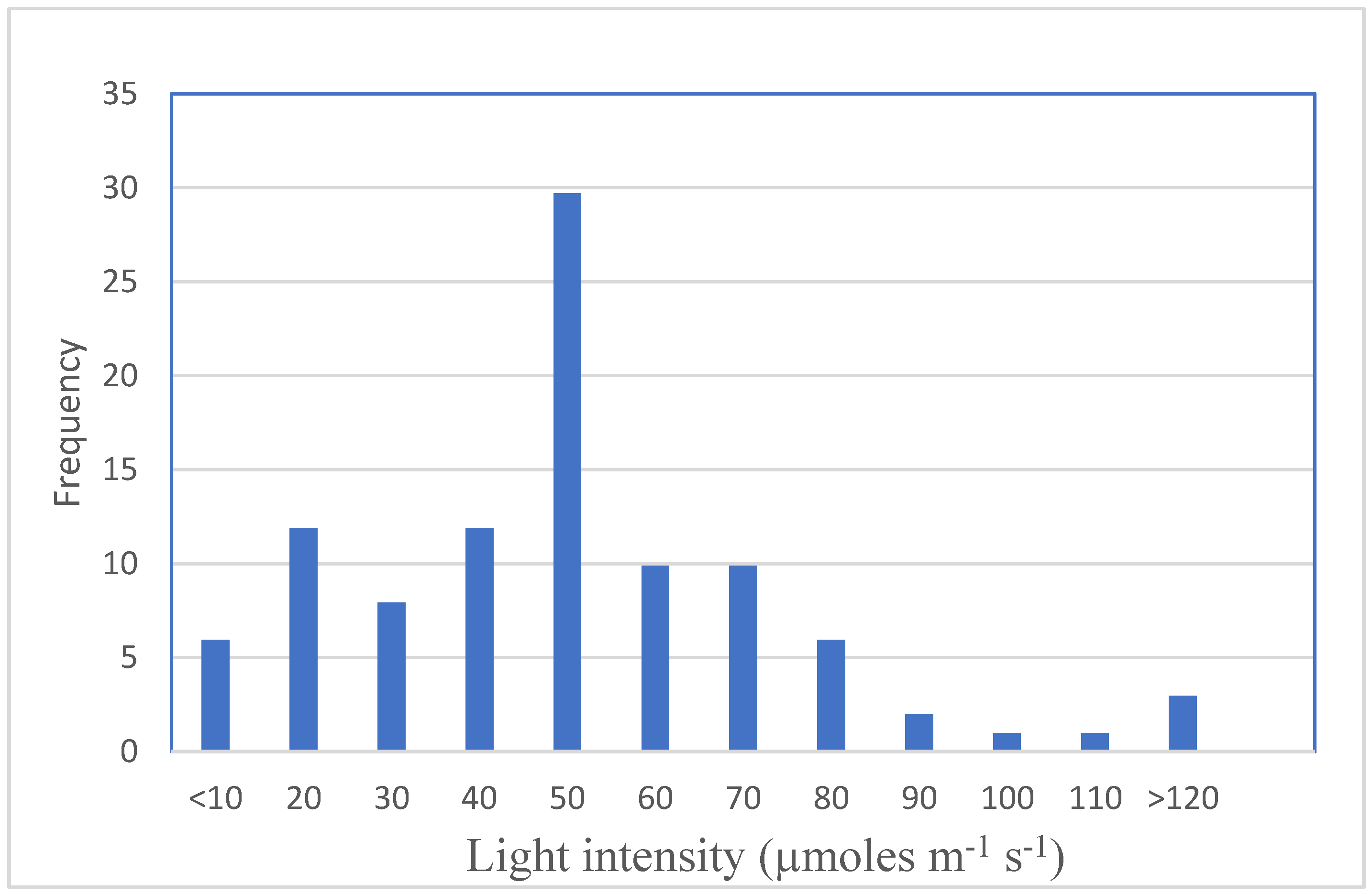
3. Effects of Light Intensity

The selection of the optimal light intensity to support in vitro proliferation and growth is also important for an optimization of the processes. Among others, light intensity regulates the dimension of leaves and stems, as well as their morphogenic pathway, and is involved in pigment formation and hyperhydricity [208].
In vitro cultures are subjected to a much lower light intensity as compared to those grown under open field conditions. The permanent low light conditions in vitro have been considered a limiting factor for photosynthesis and for supporting plant morphogenesis in vitro, so it is necessary, in most cases, to supply sucrose to the medium [209]. In vitro plants are also very susceptible to high light conditions [210] and prone to photoinhibition [211]. Too high irradiation can severely damage the photosynthetic apparatus and photosynthetic pigment synthesis [48,212], leading to the formation of harmful free oxygen radicals and damage to cells [213].
In Table 3, the research that mainly addressed the effects of different light intensities is shown, but only in a few of the studies shoot proliferation is examined.
The optimal value of the PFD for plantlets changes from species to species and the predominant in vivo light conditions may give an indication of the requirements for optimal culture growth in vitro [75]. In Alocasia amazonica [222] and Momordica grosvenori [219], shoot length increased with the reduction in light intensity, an adaptation mechanism indicating that these species can survive in low light-intensity environments. In Lippia gracilis, the weight increase of plantlets grown under high light intensities indicates that this species originates in a semiarid environment where high irradiance (HI) incoming light occurs [119]. Evidence has been previously presented [178] that plants adapted to an environment with incoming HI present better photosynthetic rates and high growth rates under intense light. In an extensive study on the photosynthetic pigments, Lazzarini et al. [119] concluded that the increase in chlorophyll b content under low irradiance (LI) is indicated as an important marker of plant adaptation to shaded environments because this pigment is more efficient for capturing the photons of the higher wavelengths of the spectrum that are mainly present. Furthermore, it is worth noting that the type of explant also influences the amount of photosynthetic pigment: leaves of plantlets generated from apical explants had higher amounts of chlorophyll a, total chlorophyll and carotenoids regardless of light conditions, whereas the amount of chlorophyll b resulted in more plantlets generated from the lateral buds of nodal segments. Moreover, an increase in the synthesis of carotenoids was observed in plants grown under high light intensities and was associated with the photoprotection exerted by these pigments within the photosystems. In Lippia gracilis, this increase led to better efficiency of the photosynthetic activity and, hence, the higher production of dry weight observed under these conditions [119]. In three different species, Disanthus cercidifolius, Rhododendron cultivars and Crataegus oxyacantha, low levels of irradiance (11 µmol m−2 s−1) were optimal for in vitro growth, while higher irradiance determined a decrease in shoot development and leaf chlorophyll content in Disanthus and Rhododendron cultivars, which are shade-tolerant species in their natural habitat. Plantlets of Crataegus generated from in vivo plants adapted to higher levels of irradiance resulted in tolerance to a wide range of irradiances in vitro. Only shoot extension was inhibited at the highest levels tested, whereas leaf chlorophyll content was unaffected. These differences were attributed to a differential adaptation to light determined by the natural habitats of these plants and of the possible direct effect of irradiance upon plant growth regulators in the culture system [75]. Different effects of rising light intensity were observed in Plectranthus amboinicus grown in vitro. In this species, intensities below or above the optimum (69 μmol m−2 s−1) led to the lowest growth. In fact, photosynthesis was inefficient under low light intensity (26 μmol m−2 s−1) but increased light intensities led to reduced concentrations of a, b and total chlorophyll, and carotenoids and thus of growth [48]. In Withania somnifera and Achillea millefolium, the treatments with the highest light intensity (60 and 69 µmol m−2 s−1, respectively) showed the highest levels of photosynthetic pigments but not the highest growth. Alvarenga et al. [172] concluded that the significant increase observed in chlorophyll and carotenoids under high light conditions would indicate that these pigments have the photoprotective function, as assumed by Biswal et al. [223], since they may be inefficient in absorbing light and increasing photosynthetic efficiency. They also attributed the damage of excess light to the photosynthetic apparatus to the production of free radicals, which may degrade these pigments [45,213]. Kurilčik et al. [174] on Chrysanthemum (Chrysanthemum morifolium), noticed that the maximal PFD (85 μmol m−2 s−1) used in their experiment induces light abnormalities on the leaf surface. In ginger [224], the growth was restrained when the light reached 180 μmol m−2 s−1 and the chlorophyll content decreased as the light intensity increased.
However, a different sensibility to light intensity seems to affect proliferation rate and the plantlet growth, and in most cases lower plant intensities are required for proliferation.
Based on the observation of the examined papers for this review, in Figure 1, the light intensities were grouped in ranges and the frequency of their use is shown. From this study, it emerged that whatever the light spectrum, the most used light intensities range from 20 to 80 µmoles m−2 s−1 and the most used intensity for proliferation is 50 (µmoles m−2 s−1).
In Rubus spp, rising WL fluence rates from 0 to 81 µmol m−2 s−1 did not improve the organogenesis from cotyledons [225]. In Vaccinium corymbosum, exposure at rising intensities from 55 up to 210 µmol m−2 s−1 improved proliferation and rooting ratios only with short time applications (7 days). Longer exposure of the leaves (14 and 28 days) determined inhibition of growth and the red color of leaves and sprouts, and less vigorous plants after in vivo transferring [215].
However, a better multiplication under increasing irradiance, from 10 to 80 µmol m−2 s−1, resulted in Pyrus communis [218], in L. gracilis at 94 µmol m−2 s−1 [119] and in Rosa hybrida from 4 to 148 µmol m−2 s−1 [221]. In this last species, higher irradiance (66 and 148 µmol m−2 s−1) showed better effects on shoot proliferation, but leaf chlorosis was observed and better results on shoot growth were obtained at 17 µmol m−2 s−1 [221]. The chlorosis occurring at the higher levels of irradiance may be due to photochemical oxidation, photoinhibition or chloroplast damage [226].
In Castanea sativa, Sáez et al. [227] highlighted a correlation between light intensity and the addition of sugar to the growth medium. They demonstrated that HI (150 µmol m−2 s−1) and high sugar amounts (30 g L−1) produced an increase in photosynthetic activity and chlorophyll content and determined a higher proliferation rate and biomass production. However, a high proliferation rate was obtained even under LI with a higher sugar content in the medium. Thus, HI but also LI may be beneficial during the in vitro culture, but this is only possible in the presence of sucrose added to the culture medium.
Kozai [228], in Cymbidium, doubled in vitro growth by adding CO2 to the culture vessels at high PFD (230 μmol m−2 s−1), demonstrating that CO2 limitation may have a relevant role in enhancing the growth when high PFDs are adopted. The same was also true for Actinidia deliciosa where the proliferation rate and dry and fresh weight increased up to 120 μmol m−2 s−1 but decreased at higher rates. The biomass produced was also affected by light intensity, since both dry and fresh weight increased at the PPFD up to 120 µmol m−2 s−1, while only dry weight increases thereafter up to the highest value of 250 µmol m−2 s−1.
The photosynthetic rate was nearly four times higher when raising CO2 up to 1450 and 4500 μL L−1 compared to the lowest CO2 concentration tested (330 μL L−1) [220].
In fact, it has been shown that, just a few hours after the light was turned on, CO2 underwent a drastic reduction in concentration and sub-optimal CO2 availability has been correlated with reduced photosynthetic ability [229]. Thus, exogenous enrichments of this gas in the culture vessels improves photosynthesis at high PFDs [230,231].
Finally, most studies on the effects of light intensities have been carried out under Fl or W-LED. However, some studies revealed a relationship between the light spectrum and the intensity that affects plant growth and development. In the presence of BA, WL, BL and FRL, action on proliferation was dependent on the fluence rate [141].
Phytochrome has been shown to induce a high-irradiance response and low-irradiance response in Prunus domestica rootstock Mr.S. 2/5 [142]. Similar results were also obtained with the rootstock GF 677 in which the newly formed shoots were fewer but longer under the two intensities of RL (15 and 40 μmol m−2 s−1) than those treated with WL. In addition, the low intensity RL (15 μmol m−2 s−1) induced higher shoot multiplication as compared to the higher irradiance (40 μmol m−2 s−1). The formation of new shoots in the two species was affected differently by the increase in the RL irradiance, and shoot formation was found to increase in the cultures of Mr.S. 2/5 and decrease in those of GF 677. This result could be related to a species-specific response on which would depend different PHY regulation strategies [2].

4. Effects of Photoperiod

An organism’s life has evolved adaptation mechanisms that are related to environmental variations. Some of these variations exhibit regular cyclicality such as light:dark cycles, others fluctuate, such as temperature; however, all of them induce significant changes in the physiology and metabolism of most organisms, occurring in their life trajectory as characterized by the night and day cycle [232,233]. Plants possess the circadian clock, an endogenous time-keeping device that triggers and regulates physiological events in accordance with predicted daily changes in the environment. The input of light into the circadian clock is led by a set of photoreceptors such as the ZTL-type and UVR8 receptors [234]. Photosynthesis and stomatal movements are controlled by the circadian clock [235,236]. Among several physiological processes that include chromatin-regulation, diurnal rhythmic gene expression generates networks of genes that act specifically throughout the day or the night [237,238,239,240]. The circadian clock is an endogenous oscillator with a duration of approximately 24 h, and it is coordinated by external factors such as temperature and light. These external factors are relatively constant during the micropropagation procedure since there is no change in photoperiodism and thermoperiodism. During the shoot multiplication phase of in vitro cultures, photoperiod regimes of 16 h of light and 8h of dark are usually adopted. Plantlets in vitro are mixotrophic organisms, therefore nutrients such as carbohydrates are absorbed from the medium. In plantlets exposed to a 16:8 h photoperiod, the photosynthetic activity is intense at the onset of the light cycle and decreases rapidly thereafter. The block of CO2 assimilation depends on the rapid and progressive lower concentration of CO2 in the culture vessels. The CO2 availably in the culture vessels is largely generated during the respiration of sucrose supplied with the growth medium, since the gas exchange between the inside and the outside the vessel is almost absent (Abbot and [220,230,241,242]. The modification of the photoperiodic regime from a 16 h photoperiod cycle to a 4 h photoperiod cycle promoted the increment of fresh and dry weights of shoot clusters, and the number of neo-formed shoots from initial shoot explants in two Prunus persica rootstocks [243]. An analogous response was found in the Prunus persica cultivars Suncrest, Belle of Georgia and Evergreen when cultured in the presence of 10µM of BA in the medium [244]. However, Morini et al. [245] have found that the photosynthetic activity was only extended until 4 h after the beginning of the illumination, although the concentration of CO2, (under the 16/8 h regime) was not a limiting factor since at the end of the light period its availability was still much higher than that outside the vessel. From the same authors, the reduction of photosynthetic capacity was attributed to a reduced efficiency of the chloroplasts coupled with the lengthening of the light period. The promotive role of the 4 h photoperiod cycle on the shoot proliferation rate was hypothesized to be dependent on the diverse regime of photo-equilibrium of photoreceptors that promoted the reduction in apical dominance and development of axillary buds.
However, in studies carried out on other species, subjected to a 16 h photoperiod, low concentration of CO2 into the vessels was observed: Pfaffia glomerata [246], Solanum tuberosum [247,248], Carica papaya [249], Castanea sativa [227], Vitis vinifera [250,251], Fragaria x ananassa [252], Hyptis marrubioides and Hancornia speciosa [253].

5. Light and Plant Growth Regulators

Some in vitro studies highlighted the effects of light spectra on the effectiveness of endogenous- and exogenous-applied growth regulators.

5.1. Light Effects on Endogenous Growth Regulators

Endogenous auxins and CKs are the most involved growth regulators in regulating apical dominance [254]. Apical dominance and its correlative inhibition are determined by the synthesis of auxins by the apex [255]. In the classical model, it is hypothesized that these hormones are synthesized by the apex and transported downward into axillary buds, with subsequent direct downregulation of outgrowth, or indirect regulation via other mechanisms such as nutrient diversion, expression of genes that control the growth of axillary buds, adjustment of the auxin/cytokinin ratio, including activation of strigolactones capable of modifying the hormonal balance, and the apical dominance [256,257]. On the other side, an increase of CK quantity in tissues leads to a marked growth of axillary buds, counteracting the action of auxins. Studies on transgenic plants have shown that regulation of apical dominance by plant hormones is not determined by the absolute concentration of hormones but by the ratio between them [258]. In vitro shoot proliferation is strongly dependent on the ability of CKs to counteract apical dominance, i.e., the physiological control exerted by the apex over the induction and development of the new lateral meristems in axillary buds along the axis of the growing explant. Light acts mainly as a morphogenic signal in the triggering of bud outgrowth and initial steps in the light signaling pathway induce changes in the levels of cytokinin-like substances [259,260,261]. The effect of light in modulating endogenous CKs levels is well-known and has been demonstrated in several species such as Rosa hybrida and Chlorella minutissima (Chlorophyta: Trenouxiophyceae) [262,263]. In Rosa hybrida, in dark, inhibition of bud outgrowth is suppressed solely by the application of CKs. In contrast, application of sugars has a limited effect. Exposure of plants to WL induces a rapid (after 3–6 h) up-regulation of RhIPT3 and RhIPT5 genes involved in CK synthesis, of the RhLOG8 gene involved in CK activation and of the RhPUP5 gene involved in CK putative transporter and induces the repression of the RhCKX1 gene involved in CK degradation in the node. This leads to the accumulation of CKs in the node and to the triggering of bud outgrowth [263]. In C. minutissima [262], a rise in endogenous auxin and CK and a decrease over time in gibberellin concentrations was observed in the actively growing cultures under light:dark conditions (L:D) and continuous dark+glucose (CD+G) but no increase was determined under continuous dark (CD). The L:D cultures had the largest CK increase.
It has been known for several years [264] that the bands of the light spectrum that have been shown to promote morphogenetic processes through the activation of the various photoreceptors are mainly represented by RL, FRL and BL. As stated in paragraph 2.1, RL increases the quantity of cytokinin in tissue, counteracting the action of auxins and thus determining an increase in the development of lateral shoots [139,140]. RL also regulates the synthesis of carotenoids and strigolactones [265]. Previous studies reported that RL decreased the IAA concentrations in maize epidermal cells [266].
The interaction between CK and PHY would induce, in the latter, an extension of the active form (Pfr) even in conditions of dark and FRL [2]. In addition, other plant hormones may be modulated by light and by phytochrome directly. Among these are gibberellins [65] and brassinosteroids [267], another important category of growth regulators affecting cell elongation and cell division. Thus, RL may promote stem growth by regulating the biosynthesis of gibberellin or induce the expression of an auxin inhibitor gene to promote stem and root lengthening in grape [8]. In contrast, BL seems to affect more the auxin content (indoleacetic acid-IAA in particular). In fact, it was demonstrated that BL induced higher IAA content than RL in the leaves of the balloon flower [158] and thus it is more effective in promoting leaf growth. Significantly higher IAA contents occurred in the leaves under the BL:RL = 3:1 and BL:RL 1:3 and induced larger leaf areas compared to RL. Thus, BL appeared more beneficial for increasing IAA concentrations and for promoting better leaf growth than RL. However, in tobacco, a species in which BL stimulated shoot proliferation, contrasting effects of BL have been reported, since it was hypothesized that at higher intensities it determines the photoinactivation of IAA [67]. These mechanisms, both related to apical dominance and bud dormancy, are masked by WL, a condition under which cryptochrome and phytochrome are activated.

5.2. Effects of Light on Exogenous Applied Growth Regulators

In Prunus domestica subsp. insititia, clone GF655-2, BA, a promotive effect on proliferation was repressed under dark, whereas no proliferation was observed under light conditions without BA. It is noteworthy that at the highest BA supplied, the proliferation rate increased under the broadband WL, whereas it decreased under the monochromatic sources RL, BL and FRL [141]. Light and BA also proved to be indispensable factors in adventitious shoot formation from Pinus radiata cotyledons [268]. In Spirea nipponica, the interaction between CKs (0.25 mg L−1) and RL resulted in an enhancement of the shoot proliferation rate [123]. The same indications on the interaction between light quality and CK content were obtained on multiplication and growth during in vitro culture of Myrtus communis L. [120] and Spirea nipponica [124]. The highest number of shoots was obtained under RL or R:FR-LEDs with the higher CK concentrations tested in the media (5 µg L−1 i.e 1.1 and 0.5 mg L−1, respectively).
At lower BA levels (0.4 mg L−1), 4 weeks of RL:FRL at low fluence followed by 1 week of WL at higher fluence rate produced almost the same proliferation levels and optimal growth [124]. If the CK concentration was below the optimal level, the production of axillary shoots was greater in the RL; at higher CK concentration, the multiplication rate decreased [2]. The effect of light spectrum differs, however, in relation to the concentration of CK applied: at the optimal concentration, WL provided responses better than those obtained with RL and BL. Thus, the quantity of applied CKs may decrease under RL. Analogously, CK incorporation into the culture medium annulled the promoting effect of RL in axillary bud proliferation from azalea apices and adventitious bud regeneration from Petunia spp. leaf segments [269,270]. Probably, light quality and hormone application may affect the morphogenesis of in vitro plants, in part because of changes in sink strength and, as a consequence, to redistribution of active growth [71].
Panizza et al. [72] analyzed the effect of spectral composition on axillary proliferation of lavandin (Lavandula officinalis Chaix • L. latifolia ViUars cv. Grosso) in relation to the application of exogenous BA, putrescine (Put) and endogenous ethylene production. The effect of BA was predominant over the light quality, whereas in BA-free medium, shoot number was enhanced under BL, WL and RL at low photon fluence rates. BA, however, could reduce the inhibiting effect of BL and UVL at high photon fluence rates. Exogenous Put stimulated axillary bud proliferation under some light treatments in the presence of BA, although the short fluence RL treatment was critical to allow the positive effect of Put on shoot formation. A positive correlation between biotic ethylene production and shoot formation was evidenced under FRL at a high photon fluence rate in the presence of BA. In the BA-free medium, further evidence of the correlation between biotic ethylene and the proliferation process was given since the biotic emanation increased under those radiation treatments (RL, BL and WL), which also improved shoot number. The authors conclude that in the evaluation of the responsiveness of a tissue to radiation in vitro, great care should also be devoted to radiation-induced changes in the abiotic environment (e.g., ethylene release) [72].

6. Discussion and Conclusions

Several papers on different species concern the effects of light on in vitro proliferation to assess the light properties capable of enhancing the efficiency of the micropropagation process, also ensuring consistent energy savings, as compared to traditionally used Fls lamps, or the broad range of WL. However, the results are often conflicting. Many authors ascribe these results to the different responses to light of plant species, cultivars or even explant types [119], plant stage development [122], medium composition [143] and micro-environmental characteristics such as PPFD [174] and vessel ventilation [146]. However, a large cause of variability may be tied also to the difficulty in applying uniform intensities along the shelves, and/or the use of the right spectral composition for each light quality.
Moreover, the lack of sufficient in vitro experimental protocols like those available for in vivo study, which would make the effects of light clearer, limits the comparability of the experiments [34]. The issues of major concern, among others, in this regard are (i) the short timescale in which these experiments are carried out (mostly a propagation cycle), (ii) the quality and quantity of exogenous applied growth regulators, (iii) the narrow range of light intensity values within which the efficiency of axillary multiplication of explants occurs and (iv) the mixotrophic state of plantlets. Concerning the first issue, the short-time experiments strongly limit the comprehension of the effects of light spectra on the stability of proliferation and plantlet growth during subsequent multiplication cycles (see particularly the RL effects). Concerning the second one, due to the interaction of light with endogenous growth regulators (particularly CKs), attention must be paid to the doses of the exogenous growth regulators applied. It seems from the examined literature, in fact, that RL effects are visible under low CK concentrations in the medium, whereas WL effects are even visible under high CKs doses [83]. Too high CKs quantities mask the effects of RL or may determine growth alteration. Concerning light intensities, excessive LIs or HIs may determine low growth rates, photoinhibition and may mask light spectra effects. Moreover, information on how the mixotrophic metabolism of a plantlet grown in vitro affects the development and morphology of the microcutting is scarce.
In this review, several research are presented regarding the different response of species and cultivars to different light spectra, intensities and photoperiod and it seems that some general indications arise from the different studies. Concerning the optimal irradiance intensity, it has been hypothesized that the prevailing light conditions under the natural habitats of some species can be used to indicate their requirements for optimal in vitro growth [75]. Evidence have been presented that plants adapted to an environment characterized by high light intensities present better photosynthetic rates and high growth rates under in vitro intense light, whereas shade-tolerant plants are damaged by high intensities. A survey of the tested literature revealed that in most species, whatever the light spectrum, the most used light intensities range from 20 to 80 µmoles m−2 s−1 and the most used intensity for proliferation is 50 µmoles m−2 s−1. Better growth, however, have been registered especially in plants adapted to high intensities (see Saccharum officinarum, Actinidia deliciosa, Lippia gracilis, etc.) at intensities up to or exceeding 80 µmoles m−2 s−1. Significant improvements on in vitro fresh and dry weights of shoot clusters, and the number of neo-formed shoots from initial shoot explants were obtained, also modifying the photoperiodic regime from a 16 h photoperiod to a 4 h photoperiod cycle, thus permitting the plantlets to replace the CO2 [243,244]. In fact, in plantlets exposed to the 16:8 h photoperiod, the photosynthetic activity is intense at the onset of the light cycle and decrease rapidly thereafter because of the rapid and progressive lower concentration of CO2 in the culture vessels. Moreover, the promotive role of the 4 h photoperiod cycle on the shoot proliferation rate was hypothesized to be dependent on the diverse regime of photo-equilibrium of photoreceptors that promoted the reduction in apical dominance and development of axillary buds [243]. In this view, also adding CO2 [220] or aerating the vessels [146] proved to be effective in enhancing in vitro growth.
Concerning light spectra, RL alone or high RL:FRL ratios seem to enhance shoot proliferation, as well as PLB and callus formation, in many species. The main effects of RL are tied to the promotive role of phytochrome in the synthesis of CK in tissue, which counteracts the actions of auxins, increasing the development of lateral shoots. RL also regulates the synthesis of carotenoids and, in particular, strigolactones that seem to regulate apical dominance by modification of auxin fluxes [271]. The stimulatory effects of RL seem to be exerted during the beginning of the multiplication phases. However, different reports indicated that RL alone is not able to activate the pathway of chlorophyll synthesis and may determine excessive stem elongation and leaf disorders, the so-called Red Light Syndrome [36]. In fact, when plants are grown under 100% monochromatic RL a strong decrease in photosynthetic capacity, rates of electron transport, dark-adapted Fv/Fm and leaf thickness, as well as unresponsive stomata and reduced leaf pigmentation occurs [272]. BL is effective in increasing callus formation and the number of axillary buds but exerts an inhibitory action on buds sprouting (increase in apical dominance). It has been demonstrated that this light mostly controls some morphological characteristics such as shoot length and enhances chlorophyll synthesis and chloroplast development. RL, on the other hand, would remove the apical dominance but seem to reduce the formation of new axillary buds. Hence, a minimum threshold of BL is necessary for normal plant growth [146]. Moreover, regulating the spectral quality particularly by the BL improves the antioxidant defense line and is directly correlated with the enhancement of phytochemicals [65,90,166] or with the regulation of gene expression [167]. All these reasons would explain why the RL:BL illumination resulted effectively in a wide range of species. Moreover, more recently, an abundance of evidence has indicated the role of GL in carrying information about the environment to the plants, because it is involved in the shade avoidance response, but also in regulating different biological, morphological and biological processes in vitro and in vivo [189]. The addition of GL to the combined RL and BL contributed to the proliferation, the growth and development of some in vitro cultures. In a few cases, even the addition of YL seems to improve plant proliferation and growth. In addition, the absence of ultraviolet light may determine foliar intumescence and could become a serious limitation for some crops lighted solely by narrow-band LEDs [273]. Thus, the use of monochromatic or combined R- or B-LEDS may determine a mismatch with the photosynthetic spectrum. The application of the broad band WL may overcome this problem [44]. In some species, better results have been obtained under W-LEDs [109,112,130]. Even if WL is not as effective as RL in overcoming apical dominance, high proliferation rates are obtained when CKs are added to the medium. In most cases, the best propagation was obtained at higher CK ratio [141]. It seems that the CK ratio may be enhanced in woody species under WL to obtain high and stable proliferation. However, in some species, after long-time cultivation under WL the rate of newly formed sprouts was reduced regardless of the CK concentration but increased when RL was applied to the crops [2]. Thus, in some cases, an early phase of RL irradiation of at least 2 weeks [122], followed by growth under a WL, may be advisable. The use of an initial stimulatory effect of RL or RL enriched followed by the WL may also improve proliferation and somatogenesis [126] in species that are particularly difficult to regenerate in vitro and/or with an high sensibility to higher concentration of CKs in the medium, such as Euphorbia milii and Ceratonia siliqua L. (Cavallaro et al., unpublished data). Moreover, the exposition to a period of RL:FRL followed by the WL may enable a reduction in exogenous growth regulator concentrations, mainly CKs added to the medium [124], which may be unnaturally high in vitro. This reduction may be favorable for enhancing the following phases of the in vitro process (rooting and acclimation). Finally, currently, lamps with a more optimal spectral composition of WL enriched in the most useful wavelengths (BL, RL and GL) are already available on the market [185,274] for vertical farming systems and could be interesting for in vitro production after appropriate investigation.

Author Contributions

Conceptualization, V.C. and R.M. contributed to the conception and the design of the review; V.C. and A.P., contributed to the drafting of tables; writing—original draft Preparation, V.C. and R.M. contributed to define the original draft; writing—review & editing, V.C., I.F., A.P. and R.M. wrote and edited the final version of the manuscript. All authors have read and agreed to the published version of the manuscript.

Funding

This research received no external funding.

Conflicts of Interest

The authors declare no conflict of interest.

References

  1. Smith, H. Sensing the light environment: The functions of the phytochrome family. In Photomorphogenesis in Plants; Kendrick, R.E., Kronenberg, G.H.M., Eds.; Springer: Dordrecht, The Netherlands, 1994; pp. 377–416. [Google Scholar]
  2. Morini, S.; Muleo, R. Il ruolo della qualità della luce nei processi di sviluppo e differenziazione delle colture in vitro. Italus Hortus Rev. 2012, 19, 37–49. [Google Scholar]
  3. Kozai, T. Why LED lighting for urban agriculture? In LED Lighting for Urban Agriculture; Kozai, T., Fujiwara, K., Runkle, E., Eds.; Springer: Singapore, 2016; pp. 3–18. [Google Scholar] [CrossRef]
  4. Whitelam, G.C.; Halliday, K.J. Photomorphogenesis: Phytochrome takes a partner! Curr. Biol. 1999, 9, 225–227. [Google Scholar] [CrossRef] [Green Version]
  5. Muneer, S.; Kim, E.J.; Park, J.S. Influence of green, red and blue light emitting diodes on multiprotein complex proteins and photosynthetic activity under different light intensities in lettuce leaves (Lactuca sativa L.). Int. J. Mol. Sci. 2014, 15, 4657–4670. [Google Scholar] [CrossRef] [PubMed] [Green Version]
  6. Higuchi, Y.; Hisamatsu, T. Light acts as a signal for regulation of growth and development. In LED Lighting for Urban Agriculture; Kozai, T., Fujiwara, K., Runkle, E., Eds.; Springer: Singapore, 2016; pp. 57–73. [Google Scholar]
  7. Pedmale, U.V.; Huang, S.C.; Zander, M.; Cole, B.J.; Hetzel, J.; Ljung, K.; Chory, J. Cryptochromes interact directly with PIFs to control plant growth in limiting blue light. Cell 2016, 164, 233–245. [Google Scholar] [CrossRef] [Green Version]
  8. Li, C.-X.; Xu, Z.-G.; Dong, R.-Q.; Chang, S.-X.; Wang, L.-Z.; Khalil-Ur-Rehman, M.; Tao, J.-M. An RNA-Seq analysis of grape plantlets grown in vitro reveals different responses to blue, green, red led light, and white fluorescent light. Front. Plant Sci. 2017, 8, 78. [Google Scholar] [CrossRef] [Green Version]
  9. Batista, D.S.; Felipe, S.H.S.; Silva, T.D.; de Castro, K.M.; Mamedes-Rodrigues, T.C.; Miranda, N.A.; Ríos-Ríos, A.M.; Faria, D.V.; Fortini, E.A.; Chagas, K.; et al. Light quality in plant tissue culture: Does it matter? Vitr. Cell. Dev. Biol. Plant 2018, 54, 195–215. [Google Scholar] [CrossRef]
  10. Chen, M.; Chory, J.; Fankhauser, C. Light signal transduction in higher plants. Annu. Rev. Genet. 2004, 38, 87–117. [Google Scholar] [CrossRef] [Green Version]
  11. Banerjee, R.; Batschauer, A. Plant blue-light receptors. Planta 2005, 220, 498–502. [Google Scholar] [CrossRef]
  12. Chen, M.; Chory, J. Phytochrome signaling mechanisms and the control of plant development. Trends Cell Biol. 2011, 21, 664–671. [Google Scholar] [CrossRef] [Green Version]
  13. Vierstra, R.D.; Zhang, J. Phytochrome signaling: Solving the Gordian knot with microbial relatives. Trends Plant Sci. 2011, 16, 417–426. [Google Scholar] [CrossRef]
  14. Galvão, V.C.; Fankhauser, C. Sensing the light environment in plants: Photoreceptors and early signaling steps. Curr. Opin. Neurobiol. 2015, 34, 46–53. [Google Scholar] [CrossRef] [PubMed] [Green Version]
  15. Chaves, I.; Pokorny, R.; Byrdin, M.; Hoang, N.; Ritz, T.; Brettel, K.; Essen, L.-O.; van der Horst, G.T.J.; Batschauer, A.; Ahmad, M. The cryptochromes: Blue light photoreceptors in plants and animals. Annu. Rev. Plant Biol. 2011, 62, 335–364. [Google Scholar] [CrossRef] [PubMed]
  16. Liu, H.; Liu, B.; Zhao, C.; Pepper, M.; Lin, C. The action mechanisms of plant cryptochromes. Trends Plant Sci. 2011, 16, 684–691. [Google Scholar] [CrossRef] [Green Version]
  17. Christie, J.M. Phototropin blue-light receptors. Annu. Rev. Plant Biol. 2007, 58, 21–45. [Google Scholar] [CrossRef] [Green Version]
  18. Suetsugu, N.; Wada, M. Evolution of three LOV blue light receptor families in green plants and photosynthetic stramenopiles: Phototropin, ZTL/FKF1/LKP2 and aureochrome. Plant Cell Physiol. 2013, 54, 8–23. [Google Scholar] [CrossRef] [PubMed] [Green Version]
  19. Lin, C. Plant blue-light receptors. Trends Plant Sci. 2000, 5, 337–342. [Google Scholar] [CrossRef]
  20. Rizzini, L.; Favory, J.-J.; Cloix, C.; Faggionato, D.; O’Hara, A.; Kaiserli, E.; Baumeister, R.; Schäfer, E.; Nagy, F.; Jenkins, G.I.; et al. Perception of UV-B by the Arabidopsis UVR8 protein. Science 2011, 332, 103–106. [Google Scholar] [CrossRef] [Green Version]
  21. Jenkins, G.I. The UV-B Photoreceptor UVR8: From structure to physiology. Plant Cell 2014, 26, 21–37. [Google Scholar] [CrossRef] [Green Version]
  22. Tilbrook, K.; Arongaus, A.B.; Binkert, M.; Heijde, M.; Yin, R.; Ulm, R. The UVR8 UV-B Photoreceptor: Perception, signaling and response. Arab. Book 2013, 11, e0164. [Google Scholar] [CrossRef] [Green Version]
  23. Aphalo, P.J.; Ballare, C.L. On the importance of information-acquiring systems in plant-plant interactions. Funct. Ecol. 1995, 9, 5–14. [Google Scholar] [CrossRef]
  24. Gilbert, I.R.; Jarvis, P.G.; Smith, H. Proximity signal and shade avoidance differences between early and late successional trees. Nature 2001, 411, 792–795. [Google Scholar] [CrossRef] [PubMed]
  25. Ballaré, C.L. Keeping up with the neighbours: Phytochrome sensing and other signalling mechanisms. Trends Plant Sci. 1999, 4, 97–102. [Google Scholar] [CrossRef]
  26. Cao, S.; Luo, X.; Xu, D.; Tian, X.; Song, J.; Xia, X.; Chu, C.; He, Z. Genetic architecture underlying light and temperature mediated flowering in Arabidopsis, rice, and temperate cereals. New Phytol. 2021, 230, 1731–1745. [Google Scholar] [CrossRef]
  27. Zhao, X.; Wang, Y.-L.; Qiao, X.-R.; Wang, J.; Wang, L.-D.; Xu, C.-S.; Zhang, X. Phototropins function in high-intensity blue light-induced hypocotyl phototropism in Arabidopsis by altering cytosolic calcium. Plant Physiol. 2013, 162, 1539–1551. [Google Scholar] [CrossRef] [PubMed] [Green Version]
  28. Kami, C.; Allenbach, L.; Zourelidou, M.; Ljung, K.; Schütz, F.; Isono, E.; Watahiki, M.K.; Yamamoto, K.T.; Schwechheimer, C.; Fankhauser, C. Reduced phototropism in Pks mutants may be due to altered auxin-regulated gene expression or reduced lateral auxin transport. Plant J. 2014, 77, 393–403. [Google Scholar] [CrossRef] [Green Version]
  29. Christie, J.M.; Yang, H.; Richter, G.L.; Sullivan, S.; Thomson, C.E.; Lin, J.; Titapiwatanakun, B.; Ennis, M.; Kaiserli, E.; Lee, O.R.; et al. Phot1 inhibition of ABCB19 primes lateral auxin fluxes in the shoot apex required for phototropism. PLoS Biol. 2011, 9, e1001076. [Google Scholar] [CrossRef]
  30. Sgamma, T.; Pape, J.; Massiah, A.; Jackson, S. Selection of reference genes for diurnal and developmental time-course real-time PCR expression analyses in lettuce. Plant Methods 2016, 12, 21. [Google Scholar] [CrossRef] [Green Version]
  31. Cirilli, M.; Delfino, I.; Caboni, E.; Muleo, R. EpiHRMAssay, in tube and in silico combined approach for the scanning and epityping of heterogeneous DNA methylation. Biol. Methods Protoc. 2017, 2, bpw008. [Google Scholar] [CrossRef]
  32. Hoffmann, A.A.; Sgrò, C.M. Climate change and evolutionary adaptation. Nature 2011, 470, 479–485. [Google Scholar] [CrossRef]
  33. Cavallaro, V.; Tringali, S.; Patanè, C. Large-scale in vitro propagation of giant reed (Arundo donax L.), a promising biomass species. J. Hortic. Sci. Biotechnol. 2011, 86, 452–456. [Google Scholar] [CrossRef]
  34. Shukla, M.R.; Singh, A.S.; Piunno, K.; Saxena, P.K.; Jones, A.M.P. Application of 3D printing to prototype and develop novel plant tissue culture systems. Plant Methods 2017, 13, 6. [Google Scholar] [CrossRef] [PubMed] [Green Version]
  35. Miler, N.; Kulus, D.; Woźny, A.; Rymarz, D.; Hajzer, M.; Wierzbowski, K.; Nelke, R.; Szeffs, L. Application of wide-spectrum light-emitting diodes in micropropagation of popular ornamental plant species: A study on plant quality and cost reduction. Vitr. Cell. Dev. Biol. Plant 2019, 55, 99–108. [Google Scholar] [CrossRef] [Green Version]
  36. Morini, S.; Muleo, R.; Jain, S.; Ishii, K. Micropropagation of woody trees and fruits. In Effect of Light Quality on Micropropagation of Woody Species; Jain, S.M., Ishii, K., Eds.; Springer: Dordrecht, The Netherlands, 2003; pp. 3–35. [Google Scholar] [CrossRef]
  37. Bula, R.J.; Morrow, R.C.; Tibbitts, T.W.; Barta, D.J.; Ignatius, R.W.; Martin, T.S. Light-emitting diodes as a radiation source for plants. HortScience 1991, 26, 203–205. [Google Scholar] [CrossRef] [PubMed] [Green Version]
  38. Seabrook, J.E.A. Light effects on the growth and morphogenesis of potato (Solanum tuberosum) in vitro: A review. Am. J. Potato Res. 2005, 82, 353–367. [Google Scholar] [CrossRef]
  39. Bantis, F.; Ouzounis, T.; Radoglou, K. Artificial LED lighting enhances growth characteristics and total phenolic content of Ocimum basilicum, but variably affects transplant success. Sci. Hortic. 2016, 198, 277–283. [Google Scholar] [CrossRef] [Green Version]
  40. Yeh, N.; Chung, J.-P. High-brightness LEDs energy efficient lighting sources and their potential in indoor plant cultivation. Renew. Sustain. Energy Rev. 2009, 13, 2175–2180. [Google Scholar] [CrossRef]
  41. Dutta Gupta, S.; Jatothu, B. Fundamentals and applications of light-emitting diodes (LEDs) in vitro plant growth and morphogenesis. Plant Biotechnol. Rep. 2013, 7, 211–220. [Google Scholar] [CrossRef]
  42. Kim, S.-J.; Hahn, E.-J.; Heo, J.-W.; Paek, K.-Y. Effects of LEDs on net photosynthetic rate, growth and leaf stomata of chrysanthemum plantlets in vitro. Sci. Hortic. 2004, 101, 143–151. [Google Scholar] [CrossRef]
  43. Kasahara, M.; Kagawa, T.; Sato, Y.; Kiyosue, T.; Wada, M. Phototropins mediate blue and red light-induced chloroplast movements in Physcomitrella patens. Plant Physiol. 2004, 135, 1388–1397. [Google Scholar] [CrossRef] [Green Version]
  44. Dutta Gupta, S.; Agarwal, A. Influence of LED Lighting on in vitro plant regeneration and associated cellular redox balance. In Light Emitting Diodes for Agriculture; Dutta Gupta, S., Ed.; Springer: Singapore, 2017; pp. 273–303. ISBN 978-981-10-5806-6. [Google Scholar]
  45. Darko, E.; Heydarizadeh, P.; Schoefs, B.; Sabzalian, M.R. Photosynthesis under artificial light: The shift in primary and secondary metabolism. Philos. Trans. R. Soc. B-Biol. Sci. 2014, 369, 1640. [Google Scholar] [CrossRef]
  46. Gupta, S.D.; Agarwal, A. Artificial Lighting System for Plant Growth and Development: Chronological Advancement, Working Principles, and Comparative Assessment; Gupta, S.D., Ed.; Springer: Singapore, 2017; pp. 1–26. ISBN 978-981-10-5806-6. [Google Scholar]
  47. Martineau, V.; Lefsrud, M.; Naznin, M.T.; Kopsell, D.A. Comparison of light-emitting diode and high-pressure sodium light treatments for hydroponics growth of Boston lettuce. HortScience 2012, 47, 477–482. [Google Scholar] [CrossRef] [Green Version]
  48. Silva, S.T.; Bertolucci, S.K.V.; da Cunha, S.H.B.; Lazzarini, L.E.S.; Tavares, M.C.; Pinto, J.E.B.P. Effect of light and natural ventilation systems on the growth parameters and carvacrol content in the in vitro cultures of Plectranthus amboinicus (Lour.) Spreng. Plant Cell Tissue Organ Cult. 2017, 129, 501–510. [Google Scholar] [CrossRef]
  49. Budiarto, K. Spectral quality affects morphogenesis on Anthurium plantlet during in vitro culture. AJAS 2010, 32, 234–240. [Google Scholar]
  50. Li, H.; Xu, Z.; Tang, C. Effect of light-emitting diodes on growth and morphogenesis of upland cotton (Gossypium hirsutum L.) Plantlets in vitro. Plant Cell Tissue Organ Cult. 2010, 103, 155–163. [Google Scholar] [CrossRef]
  51. Li, H.; Tang, C.; Xu, Z. The effects of different light qualities on rapeseed (Brassica napus L.) plantlet growth and morphogenesis in vitro. Sci. Hortic. 2013, 150, 117–124. [Google Scholar] [CrossRef]
  52. Do Nascimento Vieira, L.; de Freitas Fraga, H.P.; dos Anjos, K.G.; Puttkammer, C.C.; Scherer, R.F.; da Silva, D.A.; Guerra, M.P. Light-emitting diodes (LED) increase the stomata formation and chlorophyll content in Musa acuminata (AAA)‘Nanicão Corupá’in vitro plantlets. Theor. Exp. Plant Physiol. 2015, 27, 91–98. [Google Scholar] [CrossRef]
  53. Merkle, S.A.; Montello, P.M.; Xia, X.; Upchurch, B.L.; Smith, D.R. Light quality treatments enhance somatic seedling production in three southern pine species. Tree Physiol. 2006, 26, 187–194. [Google Scholar] [CrossRef] [PubMed] [Green Version]
  54. Park, S.Y.; Yeung, E.C.; Paek, K.Y. Endoreduplication in Phalaenopsis is affected by light quality from light-emitting diodes during somatic embryogenesis. Plant Biotechnol. Rep. 2010, 4, 303–309. [Google Scholar] [CrossRef]
  55. Kim, Y.W.; Moon, H.K. Enhancement of somatic embryogenesis and plant regeneration in Japanese red pine (Pinus densiflora). Plant Biotechnol. Rep. 2014, 8, 259–266. [Google Scholar] [CrossRef]
  56. Botero Giraldo, C.; Urrea Trujillo, A.I.; Naranjo Gomez, E.J. Regeneration potential of Psychotria ipecacuanha (Rubiaceae) from thin cell layers. Acta Biol. Colomb. 2015, 20, 181–192. [Google Scholar] [CrossRef]
  57. Chen, C.-C.; Agrawal, D.C.; Lee, M.-R.; Lee, R.-J.; Kuo, C.-L.; Wu, C.-R.; Tsay, H.-S.; Chang, H.-C. Influence of LED light spectra on in vitro somatic embryogenesis and LC–MS analysis of chlorogenic acid and rutin in Peucedanum japonicum Thunb: A medicinal herb. Bot. Stud. 2016, 57, 9. [Google Scholar] [CrossRef] [PubMed] [Green Version]
  58. Mai, N.T.; Binh, P.T.; Khoi, P.H.; Hung, N.K.; Ngoc, P.B.; Ha, C.H.; Binh, H.T.T. Effects of light emitting diodes-LED on regeneration ability of Coffea canephora mediated via somatic embryogenesis. Acad. J. Biol. 2016, 38, 228–235. [Google Scholar] [CrossRef] [Green Version]
  59. Lotfi, M.; Mars, M.; Werbrouck, S. Optimizing pear micropropagation and rooting with light emitting diodes and trans-cinnamic Acid. Plant Growth Regul. 2019, 88, 173–180. [Google Scholar] [CrossRef]
  60. Ding, Z.; Galván-Ampudia, C.S.; Demarsy, E.; Łangowski, Ł.; Kleine-Vehn, J.; Fan, Y.; Morita, M.T.; Tasaka, M.; Fankhauser, C.; Offringa, R.; et al. Light-mediated polarization of the PIN3 auxin transporter for the phototropic response in Arabidopsis. Nat. Cell Biol. 2011, 13, 447–452. [Google Scholar] [CrossRef] [PubMed]
  61. Dewir, Y.; Chakrabarty, D.; Hahn, E.; Paek, K. The effects of paclobutrazol, light emitting diodes (LEDs) and sucrose on flowering of Euphorbia millii plantlets in vitro. Eur. J. Hortic. Sci. 2006, 71, 240. [Google Scholar]
  62. Moon, H.K.; Park, S.-Y.; Kim, Y.W.; Kim, C.S. Growth of Tsuru-rindo (Tripterospermum japonicum) cultured in vitro under various sources of light-emitting diode (LED) irradiation. J. Plant Biol. 2006, 49, 174–179. [Google Scholar] [CrossRef]
  63. Shin, K.S.; Murthy, H.N.; Heo, J.W.; Hahn, E.J.; Paek, K.Y. The effect of light quality on the growth and development of in vitro cultured Doritaenopsis plants. Acta Physiol. Plant. 2008, 30, 339–343. [Google Scholar] [CrossRef]
  64. Ma, X.; Wang, Y.; Liu, M.; Xu, J.; Xu, Z. Effects of green and red lights on the growth and morphogenesis of potato (Solanum tuberosum L.) plantlets in vitro. Sci. Hortic. 2015, 190, 104–109. [Google Scholar] [CrossRef]
  65. Manivannan, A.; Soundararajan, P.; Halimah, N.; Ko, C.H.; Jeong, B.R. Blue LED light enhances growth, phytochemical contents, and antioxidant enzyme activities of Rehmannia glutinosa cultured in vitro. Hortic. Environ. Biotechnol. 2015, 56, 105–113. [Google Scholar] [CrossRef]
  66. Batista, D.S.; de Castro, K.M.; da Silva, A.R.; Teixeira, M.L.; Sales, T.A.; Soares, L.I.; das Graças Cardoso, M.; de Oliveira Santos, M.; Viccini, L.F.; Otoni, W.C. Light quality affects in vitro growth and essential oil profile in Lippia alba (Verbenaceae). Vitr. Cell. Dev. Biol. Plant 2016, 52, 276–282. [Google Scholar] [CrossRef]
  67. Seibert, M.; Wetherbee, P.J.; Job, D.D. The effects of light intensity and spectral quality on growth and shoot initiation in tobacco callus. Plant Physiol. 1975, 56, 130–139. [Google Scholar] [CrossRef] [PubMed] [Green Version]
  68. Chée, R. In vitro culture of Vitis: The effects of light spectrum, manganese sulfate and potassium iodide on morphogenesis. Plant Cell Tissue Organ Cult. 1986, 7, 121–134. [Google Scholar] [CrossRef]
  69. Lercari, B.; Tognoni, F.; Anselmo, G.; Chapel, D. Photocontrol of in vitro bud differentiation in Saintpaulia ionantha leaves and lycopersicon esculentum cotyledons. Physiol. Plant. 1986, 67, 340–344. [Google Scholar] [CrossRef]
  70. Chee, R.; Pool, R. Morphogenic responses to propagule trimming, spectral irradiance, and photoperiod of grapevine shoots recultured in vitro. J. Am. Soc. Hortic. Sci. 1989, 114, 350–354. [Google Scholar]
  71. Aksenova, N.P.; Konstantinova, T.N.; Sergeeva, L.I.; Macháčková, I.; Golyanovskaya, S.A. Morphogenesis of potato plants in vitro. I. Effect of light quality and hormones. Plant Growth Regul. 1994, 13, 143–146. [Google Scholar] [CrossRef]
  72. Panizza, M.; Tognoni, F.; Lercari, B. Axillary bud proliferation and ethylene production as controlled by radiation of different spectral composition and exogenous phytohormones. Biol. Plant. 1994, 36, 553–563. [Google Scholar] [CrossRef]
  73. Castillo, B.; Smith, M.A.L. Direct somatic embryogenesis from Begonia gracilis explants. Plant Cell Rep. 1997, 16, 385–388. [Google Scholar] [CrossRef]
  74. Da Silva, M.; Debergh, P. The effect of light quality on the morphogenesis of in vitro cultures of Azorina vidalii (Wats.) Feer. Plant Cell Tissue Organ Cult. 1997, 51, 187–193. [Google Scholar] [CrossRef]
  75. Marks, T.; Simpson, S. Effect of irradiance on shoot development in vitro. Plant Growth Regul. 1999, 28, 133–142. [Google Scholar] [CrossRef]
  76. Lercari, B.; Moscatelli, S.; Ghirardi, E.; Niceforo, R.; Bertram, L. Photomorphogenic control of shoot regeneration from etiolated and light-grown hypocotyls of tomato. Plant Sci. 1999, 140, 53–62. [Google Scholar] [CrossRef]
  77. Michalczuk, B.; Michalczuk, L. The effect of light quality on regeneration rate and plantlet development in transgenic petunia ‘Revolution’(Surfinia type). Acta Hortic. 2000, 530, 397–402. [Google Scholar] [CrossRef]
  78. Lian, M.-L.; Murthy, H.; Paek, K.-Y. Effects of light emitting diodes (LEDs) on the in vitro induction and growth of bulblets of Lilium oriental hybrid ‘Pesaro’. Sci. Hortic. 2002, 94, 365–370. [Google Scholar] [CrossRef]
  79. Burritt, D.J.; Leung, D.W. Adventitious shoot regeneration from Begonia x erythrophylla petiole sections is developmentally sensitive to light quality. Physiol. Plant. 2003, 118, 289–296. [Google Scholar] [CrossRef] [Green Version]
  80. Huan, L.V.T.; Tanaka, M. Effects of red and blue light-emitting diodes on callus induction, callus proliferation, and protocorm-like body formation from callus in Cymbidium orchid. Environ. Control Biol. 2004, 42, 57–64. [Google Scholar] [CrossRef]
  81. Hunter, D.C.; Burritt, D.J. Light quality influences adventitious shoot production from cotyledon explants of Lettuce (Lactuca sativa L.). In Vitro Cell. Dev. Biol. Plant 2004, 40, 215–220. [Google Scholar] [CrossRef]
  82. Yonghua, Q.; Shanglong, Z.; Asghar, S.; Lingxiao, Z.; Qiaoping, Q.; Kunsong, C.; Changjie, X. Regeneration mechanism of Toyonoka strawberry under different color plastic Films. Plant Sci. 2005, 168, 1425–1431. [Google Scholar] [CrossRef]
  83. Dewir, Y.H.; Chakrabarty, D.; Kim, S.J.; Hahn, E.J.; Paek, K.Y. Effect of light-emitting diode on growth and shoot proliferation of Euphorbia millii and Spathiphyllum cannifolium. Hortic. Environ. Biotechnol. 2005, 46, 375–379. [Google Scholar]
  84. Moshe, R.; Dalia, E. On the effect of light on shoot regeneration in petunia. Plant Cell Tissue Organ Cult. 2007, 89, 49–54. [Google Scholar]
  85. Poudel, P.R.; Kataoka, I.; Mochioka, R. Effect of red and blue-light-emitting diodes on growth and morphogenesis of grapes. Plant Cell Tissue Organ Cult. 2008, 92, 147–153. [Google Scholar] [CrossRef]
  86. Wongnok, A.; Piluek, C.; Techasilpitak, T.; Tantivivat, S. Effects of light emitting diodes on micropropagation of Phalaenopsis orchids. Acta Hortic. 2007, 788, 149–156. [Google Scholar] [CrossRef]
  87. Xu, Z.; Cui, J. Effects of different spectral energy distribution on tissue culture of Oncidium in vitro. J. Beijing For. Univ. 2009, 31, 45–50. [Google Scholar]
  88. Hamada, K.; Shimasaki, K.; Nishimura, Y.; Egawa, H.; Yoshida, K. Effect of red fluorescent films on the proliferation of Cymbidium finlaysonianum Lindl. PLB cultured in vitro. Hortic. Environ. Biotechnol. 2009, 50, 319–323. [Google Scholar]
  89. Chung, J.-P.; Huang, C.-Y.; Dai, T.-E. Spectral effects on embryogenesis and plantlet growth of Oncidium ‘Gower Ramsey’. Sci. Hortic. 2010, 124, 511–516. [Google Scholar] [CrossRef]
  90. Mengxi, L.; Zhigang, X.; Yang, Y.; Yijie, F. Effects of different spectral lights on Oncidium PLBs induction, proliferation, and plant regeneration. Plant Cell Tissue Organ Cult. 2011, 106, 1–10. [Google Scholar] [CrossRef]
  91. Hamada, K.; Shimasaki, K.; Nishimura, Y.; Sasaoka, H.; Nishimura, K. Effects of red, blue and yellow fluorescent films on proliferation and organogenesis in Cymbidium and Phalaenopsis PLB in vitro. Acta Hortic. 2011, 907, 385–388. [Google Scholar] [CrossRef]
  92. Lin, Y.; Li, J.; Li, B.; He, T.; Chun, Z. Effects of light quality on growth and development of protocorm-like bodies of dendrobium officinale in vitro. Plant Cell Tissue Organ Cult. 2011, 105, 329–335. [Google Scholar] [CrossRef]
  93. Nahar, S.; Shimasaki, K.; Haque, S. Effect of different light and two polysaccharides on the proliferation of protocorm-like bodies of Cymbidium cultured in vitro. Acta Hortic. 2012, 956, 307–313. [Google Scholar] [CrossRef]
  94. Werbrouck, S.; Buyle, H.; Geelen, D.; Van Labeke, M.C. Effect of red-, far-red- and blue-light-emitting diodes on in vitro growth of Ficus benjamina. Acta Hortic. 2012, 961, 533–538. [Google Scholar] [CrossRef] [Green Version]
  95. Kaewjampa, N.; Shimasaki, K. Effects of green LED lighting on organogenesis and superoxide dismutase (SOD) activities in protocorm-like bodies (PLBs) of Cymbidium cultured in vitro. Environ. Control Biol. 2012, 50, 247–254. [Google Scholar] [CrossRef] [Green Version]
  96. Siegień, I.; Adamczuk, A.; Wróblewska, K. Light affects in vitro organogenesis of Linum usitatissimum L. and its cyanogenic potential. Acta Physiol. Plant. 2013, 35, 781–789. [Google Scholar] [CrossRef] [Green Version]
  97. Edesi, J.; Kotkas, K.; Pirttilä, A.M.; Häggman, H. Does light spectral quality affect survival and regeneration of potato (Solanum tuberosum L.) shoot tips after cryopreservation? Plant Cell Tissue Organ Cult. 2014, 119, 599–607. [Google Scholar] [CrossRef]
  98. Lee, N.N.; Choi, Y.E.; Moon, H.K. Effect of LEDs on shoot multiplication and rooting of rare plant Abeliophyllum distichum Nakai. J. Plant Biochem. Biotechnol. 2014, 41, 94–99. [Google Scholar] [CrossRef]
  99. Habiba, S.U.; Shimasaki, K.; Ahasan, M.M.; Alam, M.M. Effects of different light quality on growth and development of protocorm-like bodies (PLBs) in Dendrobium kingianum cultured in vitro. Bangladesh Res. Public. J. 2014, 10, 223–227. [Google Scholar]
  100. Kamal, M.M.; Shimasaki, K.; Akter, N. Effect of light emutting diode (LED) lamps and N-Acetylglucosamine (NAG) on organogenesis in protocorm-like bodies (PLBs) of a Cymbidium hybrid cultured in vitro. Plant Tissue Cult. Biotechnol. 2014, 24, 273–277. [Google Scholar] [CrossRef] [Green Version]
  101. Silva, M.M.A.; de Oliveira, A.L.B.; Oliveira-Filho, R.A.; Gouveia-Neto, A.S.; Camara, T.J.R.; Willadino, L.G. Effect of blue/red LED light combination on growth and morphogenesis of Saccharum officinarum plantlets in vitro. In Proceedings of the SPIE—The International Society for Optical Engineering, San Francisco, CA, USA, 4 March 2014; Volume 8947. [Google Scholar] [CrossRef]
  102. Jeong, B.R.; Sivanesan, I. Adventitious shoot regeneration, in vitro flowering, fruiting, secondary metabolite content and antioxidant activity of Scrophularia takesimensis Nakai. Plant Cell Tissue Organ Cult. 2015, 123, 607–618. [Google Scholar] [CrossRef]
  103. Dutta Gupta, S.; Sahoo, T. Light emitting diode (LED)-induced alteration of oxidative events during in vitro shoot organogenesis of Curculigo orchioides Gaertn. Acta Physiol. Plant. 2015, 37, 233. [Google Scholar] [CrossRef]
  104. Hung, C.D.; Hong, C.-H.; Jung, H.-B.; Kim, S.-K.; Van Ket, N.; Nam, M.-W.; Choi, D.-H.; Lee, H.-I. Growth and morphogenesis of encapsulated strawberry shoot tips under mixed LEDs. Sci. Hortic. 2015, 194, 194–200. [Google Scholar] [CrossRef]
  105. Nhut, D.T.; Huy, N.P.; Tai, N.T.; Nam, N.B.; Luan, V.Q.; Hien, V.T.; Tung, H.T.; Vinh, B.T.; Luan, T.C. Light-emitting diodes and their potential in callus growth, plantlet development and saponin accumulation during somatic embryogenesis of Panax vietnamensis Ha et Grushv. Biotechnol. Biotechnol. Equip. 2015, 29, 299–308. [Google Scholar] [CrossRef]
  106. Bello-Bello, J.J.; Martínez-Estrada, E.; Caamal-Velázquez, J.H.; Morales-Ramos, V. Effect of LED light quality on in vitro shoot proliferation and growth of Vanilla (Vanilla planifolia Andrews). Afr. J. Biotechnol. 2016, 15, 272–277. [Google Scholar]
  107. Gök, K.M.; Şan, B.; Bayhan, A.K. Micropropagation of Gerbera (Gerbera jamesonii bolus) under different color of light-emitting diodes. Süleyman Demirel Üniversitesi Fen Bilimleri Enstitüsü Dergis 2016, 20, 468. [Google Scholar] [CrossRef] [Green Version]
  108. Nahar, S.J.; Haque, S.M.; Kazuhiko, S. Application of chondroitin sulfate on organogenesis of two Cymbidium spp. under different sources of lights. Not. Sci. Biol. 2016, 8, 156–160. [Google Scholar] [CrossRef] [Green Version]
  109. Karataş, M.; Aasim, M.; Dazkirli, M. Influence of light-emitting diodes and benzylaminopurin on adventitious shoot regeneration of water hyssop (Bacopa monnieri (L.) Pennell) in vitro. Arch. Biol. Sci. 2016, 68, 501–508. [Google Scholar] [CrossRef]
  110. Hung, C.D.; Hong, C.-H.; Kim, S.-K.; Lee, K.-H.; Park, J.-Y.; Dung, C.D.; Nam, M.-W.; Choi, D.-H.; Lee, H.-I. In vitro proliferation and ex vitro rooting of microshoots of commercially important rabbiteye blueberry (Vaccinium ashei reade) Using spectral lights. Sci. Hortic. 2016, 211, 248–254. [Google Scholar] [CrossRef]
  111. Martínez-Estrada, E.; Caamal-Velázquez, J.H.; Morales-Ramos, V.; Bello-Bello, J. Light emitting diodes improve in vitro shoot multiplication and growth of Anthurium andreanum Lind. Propag. Ornam. Plants 2016, 16, 3–8. [Google Scholar]
  112. Silva, M.M.; de Oliveira, A.L.B.; Oliveira-Filho, R.A.; Camara, T.; Willadino, L.; Gouveia-Neto, A. The effect of spectral light quality on in vitro culture of sugarcane. Acta Sci. Biol. Sci. 2016, 38, 157–161. [Google Scholar] [CrossRef] [Green Version]
  113. Szewczyk-Taranek, B.; Pawłowska, B.; Prokopiuk, B.; Żupnik, M. Effectiveness of LED and fluorescent light on in vitro shoot proliferation of Staphylea pinnata. Acta Hortic. 2015, 1155, 375–380. [Google Scholar]
  114. Ramírez-Mosqueda, M.A.; Iglesias-Andreu, L.G.; Bautista-Aguilar, J.R. The effect of light quality on growth and development of in vitro plantlet of Stevia rebaudiana Bertoni. Sugar Tech. 2017, 19, 331–336. [Google Scholar] [CrossRef]
  115. Ramírez-Mosqueda, M.; Iglesias-Andreu, L.; Luna-Sánchez, I. Light quality affects growth and development of in vitro plantlet of Vanilla planifolia Jacks. S. Afr. J. Bot. 2017, 109, 288–293. [Google Scholar] [CrossRef]
  116. Billore, V.; Jain, M.; Suprasanna, P. Monochromic radiation through light-emitting diode (LED) positively augments in vitro shoot regeneration in Orchid (Dendrobium sonia). Can. J. Biotech. 2017, 1, 50–58. [Google Scholar] [CrossRef] [Green Version]
  117. Ferreira, L.T.; de Araújo Silva, M.M.; Ulisses, C.; Camara, T.R.; Willadino, L. Using LED lighting in somatic embryogenesis and micropropagation of an elite sugarcane variety and its effect on redox metabolism during acclimatization. Plant Cell Tissue Organ Cult. 2017, 128, 211–221. [Google Scholar] [CrossRef]
  118. Pawłowska, B.; Żupnik, M.; Szewczyk-Taranek, B.; Cioć, M. Impact of LED light sources on morphogenesis and levels of photosynthetic pigments in Gerbera jamesonii grown in vitro. Hortic. Environ. Biotechnol. 2018, 59, 115–123. [Google Scholar] [CrossRef]
  119. Lazzarini, L.E.S.; Bertolucci, S.K.V.; Pacheco, F.V.; dos Santos, J.; Silva, S.T.; de Carvalho, A.A.; Pinto, J.E.B.P. Quality and intensity of light affect Lippia gracilis Schauer plant growth and volatile compounds in vitro. Plant Cell Tissue Organ Cult. 2018, 135, 367–379. [Google Scholar] [CrossRef]
  120. Cioć, M.; Szewczyk, A.; Żupnik, M.; Kalisz, A.; Pawłowska, B. LED lighting affects plant growth, morphogenesis and phytochemical contents of Myrtus communis L. in vitro. Plant Cell Tissue Organ Cult. 2018, 132, 433–447. [Google Scholar] [CrossRef] [Green Version]
  121. Yu, L.; Song, C.; Sun, L.; Li, L.; Xu, Z.; Tang, C. Effects of light-emitting diodes on tissue culture plantlets and seedlings of Rice (Oryza sativa L.). J. Integr. Agric. 2020, 19, 1743–1754. [Google Scholar] [CrossRef]
  122. Kadkade, P.; Jopson, H. Influence of light quality on organogenesis from the embryo-derived callus of Douglas fir (Pseudotsuga menziesii). Plant Sci. Lett. 1978, 13, 67–73. [Google Scholar] [CrossRef]
  123. Norton, C.R.; Herrington, T.; Phillips, D.; Norton, M. Light quality and light pipe in the micropropagation of woody ornamental plants grown in vitro. Acta Hort. 1988, 227, 413–416. [Google Scholar] [CrossRef]
  124. Herrington, E.; McPherson, J.C. Light quality growth promotion of Spiraea nipponica: The influence of a low photon fluence rate and transfer time to a higher fluence rate. Plant Cell Tissue Organ Cult. 1993, 32, 161–167. [Google Scholar] [CrossRef]
  125. Muleo, R.; Thomas, B. Effects of light quality on shoot proliferation of Prunus cerasifera in vitro are the result of differential effects on bud induction and apical dominance. J. Hortic. Sci. 1997, 72, 483–499. [Google Scholar] [CrossRef]
  126. D’Onofrio, C.; Morini, S.; Bellocchi, G. Effect of light quality on somatic embryogenesis of quince leaves. Plant Cell Tissue Organ Cult. 1998, 53, 91–98. [Google Scholar] [CrossRef]
  127. Piagnani, C.; Iacona, C.; Intrieri, M.C.; Muleo, R. A New somaclone of Prunus avium shows diverse growth pattern under different spectral quality of radiation. Biologia Plant. 2002, 45, 11–17. [Google Scholar] [CrossRef]
  128. Muleo, R.; Morini, S. Light quality regulates shoot cluster growth and development of MM106 apple genotype in vitro culture. Sci. Hortic. 2006, 108, 364–370. [Google Scholar] [CrossRef]
  129. Wang, H.; Liu, H.; Wang, W.-J.; Zu, Y. Effects of thidiazuron, basal medium and light quality on adventitious shoot regeneration from in vitro cultured stem of Populus alba × P. berolinensis. J. For. Res. 2008, 19, 257–259. [Google Scholar] [CrossRef]
  130. Wilken, D.; Jiménez Gonzalez, E.; Gerth, A.; Gómez-Kosky, R.; Schumann, A.; Claus, D. Effect of immersion systems, lighting, and TIS designs on biomass increase in micropropagating banana (Musa Spp. Cv.’Grande Naine’AAA). Vitr. Cell. Dev. Biol. Plant. 2014, 50, 582–589. [Google Scholar] [CrossRef]
  131. Kwon, A.; Cui, H.-Y.; Lee, H.; Shin, H.; Kang, K.-S.; Park, S.-Y. Light quality affects shoot regeneration, cell division, and wood formation in elite clones of Populus euramericana. Acta Physiol. Plant. 2015, 37, 65. [Google Scholar] [CrossRef]
  132. Geng, F.; Moran, R.; Day, M.; Halteman, W.; Zhang, D. In vitro shoot proliferation of apple rootstocks ‘B. 9’, ‘G. 30’, and ‘G. 41’grown under red and blue light. HortScience 2015, 50, 430–433. [Google Scholar] [CrossRef]
  133. Al-Mayahi, A.M.W. Effect of red and blue light emitting diodes “CRB-LED” on in vitro organogenesis of date palm (Phoenix dactylifera L.) cv. Alshakr. World J. Microbiol. Biotechnol. 2016, 32, 160. [Google Scholar] [CrossRef] [PubMed]
  134. He, C.; Zeng, Y.; Fu, Y.; Wu, J.; Liang, Q. Light quality affects the proliferation of in vitro cultured plantlets of Camellia oleifera Huajin. PeerJ 2020, 8, e10016. [Google Scholar] [CrossRef] [PubMed]
  135. Muleo, R.; Morini, S. Physiological dissection of blue and red light regulation of apical dominance and branching in M9 apple rootstock growing in vitro. J. Plant Physiol. 2008, 165, 1838–1846. [Google Scholar] [CrossRef] [PubMed]
  136. Gabryszewska, E.; Rudnicki, R. The influence of light quality on the shoot proliferation and rooting of Gerbera jamesonii in vitro. Acta Agrobot. 1995, 48, 105–111. [Google Scholar] [CrossRef] [Green Version]
  137. Kadkade, P.; Seibert, M. Phytochrome-regulated organogenesis in lettuce tissue culture. Nature 1977, 270, 49–50. [Google Scholar] [CrossRef]
  138. Cybularz-Urban, T.; Hanus-Fajerska, E.; Swiderski, A. Effect of light wavelength on in vitro organogenesis of a Cattleya hybrid. Acta Biol. Crac. Ser. Bot. 2007, 49, 113–118. [Google Scholar]
  139. Morelli, G.; Ruberti, I. Shade avoidance responses. Driving auxin along lateral routes. Plant Physiol. 2000, 122, 621–626. [Google Scholar] [CrossRef] [Green Version]
  140. Finlayson, S.A.; Krishnareddy, S.R.; Kebrom, T.H.; Casal, J.J. Phytochrome regulation of branching in Arabidopsis. Plant Physiol. 2010, 152, 1914–1927. [Google Scholar] [CrossRef] [Green Version]
  141. Baraldi, R.; Rossi, F.; Lercari, B. In vitro shoot development of Prunus GF 655–2: Interaction between light and benzyladenine. Physiol. Plant. 1988, 74, 440–443. [Google Scholar] [CrossRef]
  142. Muleo, R.; Morini, S.; Casano, S. Photoregulation of growth and branching of plum shoots: Physiological action of two photosystems. Vitr. Cell. Dev. Biol. Plant 2001, 37, 609–617. [Google Scholar] [CrossRef]
  143. Schuerger, A. Anatomical features of pepper plants (Capsicum annuum L.) grown under red light-emitting diodes supplemented with blue or far-red light. Ann. Bot. 1997, 79, 273–282. [Google Scholar] [CrossRef] [PubMed]
  144. Appelgren, M. Effects of light quality on stem elongation of Pelargonium in vitro. Sci. Hortic. 1991, 45, 345–351. [Google Scholar] [CrossRef]
  145. Heo, J.W.; Shin, K.S.; Kim, S.K.; Paek, K.Y. Light quality affects in vitro growth of grape Teleki 5BB. J. Plant Biol. 2006, 49, 276–280. [Google Scholar] [CrossRef]
  146. Hahn, E.-J.; Kozai, T.; Paek, K.-Y. Blue and red light-emitting diodes with or without sucrose and ventilation affect in vitro growth of Rehmannia glutinosa plantlets. J. Plant Biol. 2000, 43, 247–250. [Google Scholar] [CrossRef]
  147. Hung, C.D.; Hong, C.-H.; Kim, S.-K.; Lee, K.-H.; Park, J.-Y.; Nam, M.-W.; Choi, D.-H.; Lee, H.-I. LED Light for in vitro and ex vitro efficient growth of economically important highbush blueberry (Vaccinium corymbosum L.). Acta Physiol. Plant. 2016, 38, 152. [Google Scholar] [CrossRef]
  148. Tanaka, M.; Takamura, T.; Watanabe, H.; Endo, M.; Yanagi, T.; Okamoto, K. In vitro growth of cymbidium plantlets cultured under superbright red and blue light-emitting diodes (LEDs). J. Hortic. Sci. Biotechnol. 1998, 73, 39–44. [Google Scholar] [CrossRef]
  149. Nhut, D.T.; Takamura, T.; Watanabe, H.; Okamoto, K.; Tanaka, M. Responses of strawberry plantlets cultured in vitro under superbright red and blue light-emitting diodes (LEDs). Plant Cell Tissue Organ Cult. 2003, 73, 43–52. [Google Scholar] [CrossRef]
  150. Park, S.Y.; Kim, M.J. Development of zygotic embryos and seedlings is affected by radiationspectral compositions from light emitting diode (LED) system in Chestnut (Castanea crenata S. et Z.). J. Korean For. Soc. 2010, 99, 750–754. [Google Scholar]
  151. Chen, Y.-C.; Chang, C.; Lin, H.-L. Topolins and red light improve the micropropagation efficiency of passion fruit (Passiflora edulis Sims) ‘Tainung No. 1’. Hortscience 2020, 55, 1337–1344. [Google Scholar] [CrossRef]
  152. Wu, H.-C.; Lin, C.-C. Red light-emitting diode light irradiation improves root and leaf formation in difficult to propagate Protea cynaroides L. plantlets in vitro. HortScience 2012, 47, 1490–1494. [Google Scholar] [CrossRef] [Green Version]
  153. Tibbitts, T.; Morgan, D.; Warrington, I. Growth of lettuce, spinach, mustard, and wheat plants under four combinations of high-pressure sodium, metal halide, and tungsten halogen lamps at equal PPFD. J. Am. Soc. Hort. Sci. 1983, 108, 622–630. [Google Scholar]
  154. Sæbø, A.; Krekling, T.; Appelgren, M. Light quality affects photosynthesis and leaf anatomy of birch plantlets in vitro. Plant Cell Tissue Organ Cult. 1995, 41, 177–185. [Google Scholar] [CrossRef]
  155. Maluta, F.A.; Bordignon, S.R.; Rossi, M.L.; Ambrosano, G.M.B.; Rodrigues, P.H.V. In vitro culture of sugarcane exposed to different light sources. Pesqui. Agropecu. Bras. 2013, 48, 1303–1307. [Google Scholar] [CrossRef] [Green Version]
  156. Miao, Y.; Chen, Q.; Qu, M.; Gao, L.; Hou, L. Blue light alleviates ‘red light syndrome’ by regulating chloroplast ultrastructure, photosynthetic traits and nutrient accumulation in cucumber plants. Sci. Hortic. 2019, 257, 108680. [Google Scholar] [CrossRef]
  157. Kowallik, W. Blue light effects on respiration. Annu. Rev. Plant Physiol. 1982, 33, 51–72. [Google Scholar] [CrossRef]
  158. Liu, M.; Xu, Z.; Guo, S.; Tang, C.; Liu, X.; Jao, X. Evaluation of leaf morphology, structure and biochemical substance of balloon flower (Platycodon grandiflorum (Jacq.) A. DC.) plantlets in vitro under different light spectra. Sci. Hortic. 2014, 174, 112–118. [Google Scholar] [CrossRef]
  159. Weis, J.; Jaffe, M. Photoenhancement by blue light of organogenesis in tobacco Pith Cultures. Physiol. Plant. 1969, 22, 171–176. [Google Scholar] [CrossRef]
  160. Bach, A.; Świderski, A. The effect of light quality on organogenesis of Hyacinthus orientalis L. in vitro. Acta Biol. Crac. Ser. Bot. 2000, 42, 115–120. [Google Scholar]
  161. Cybularz-Urban, T.; Hanus-Fajerska, E.; Bach, A. Callus induction and organogenesis in vitro of Cattleya from protocorm-like bodies (PLBs) under different light conditions. Acta Sci. Pol.-Hortorum Cultus 2015, 14, 19–28. [Google Scholar]
  162. Latkowska, M.; Kvaalen, H.; Appelgren, M. Genotype dependent blue and red light inhibition of the proliferation of the embryogenic tissue of Norway spruce. In Vitro Cell. Dev. Biol. Plant 2000, 36, 57–60. [Google Scholar] [CrossRef]
  163. Muleo, R.; Diodati, A.; Sgamma, T.M.; Morini, S. Ruolo del fotoperiodo e della qualità della luce nella micropropagazione delle specie arboree da frutto. Italus Hortus 2009, 16, 221–225. [Google Scholar]
  164. Karpinski, S.; Szechynska-Habda, M. Evidence for light wavelength-specific systemic photoelectrical signalling and cellular light memory in Arabidopsis thaliana. Plant Cell 2010, 22, 1–18. [Google Scholar]
  165. Manivannan, A.; Soundararajan, P.; Park, Y.G.; Wei, H.; Kim, S.H.; Jeong, B.R. Blue and red light-emitting diodes improve the growth and physiology of in vitro grown carnations ‘green beauty’ and ‘purple beauty’. Hortic. Environ. Biotechnol. 2017, 58, 12–20. [Google Scholar] [CrossRef]
  166. Toscano, S.; Cavallaro, V.; Ferrante, A.; Romano, D.; Patané, C. Effects of different light spectra on final biomass production and nutritional quality of two microgreens. Plants 2021, 10, 1584. [Google Scholar] [CrossRef]
  167. Sgamma, T.; Forgione, I.; Luziatelli, F.; Iacona, C.; Mancinelli, R.; Thomas, B.; Ruzzi, M.; Muleo, R. Monochromic radiations provided by light emitted diode (LED) modulate infection and defense response to fire blight in pear trees. Plants 2021, 10, 1886. [Google Scholar] [CrossRef]
  168. Cirvilleri, G.; Spina, S.; Iacona, C.; Catara, A.; Muleo, R. Study of rhizosphere and phyllosphere bacterial community and resistance to bacterial canker in genetically engineered phytochrome a cherry plants. J. Plant Physiol. 2008, 165, 1107–1119. [Google Scholar] [CrossRef] [PubMed]
  169. Manivannan, A.; Soundararajan, P.; Park, Y.G.; Jeong, B.R. Physiological and proteomic insights into red and blue light-mediated enhancement of in vitro growth in Scrophularia kakudensis a potential medicinal plant. Front. Plant Sci. 2021, 11, 607007. [Google Scholar] [CrossRef] [PubMed]
  170. Iacona, C.; Muleo, R. Light quality affects in vitro adventitious rooting and ex vitro performance of cherry rootstock colt. Sci. Hortic. 2010, 125, 630–636. [Google Scholar] [CrossRef]
  171. Jao, R.-C.; Lai, C.-C.; Fang, W.; Chang, S.-F. Effects of red light on the growth of Zantedeschia plantlets in vitro and tuber formation using light-emitting diodes. HortScience 2005, 40, 436–438. [Google Scholar] [CrossRef] [Green Version]
  172. Alvarenga, I.C.A.; Pacheco, F.V.; Silva, S.T.; Bertolucci, S.K.V.; Pinto, J.E.B.P. In vitro culture of Achillea millefolium L.: Quality and intensity of light on growth and production of volatiles. Plant Cell Tissue Organ Cult. 2015, 122, 299–308. [Google Scholar] [CrossRef]
  173. Nhut, D.; Takamura, T.; Watanabe, H.; Tanaka, M. Efficiency of a novel culture system by using light-emitting diode (led) on in vitro and subsequent growth of micropropagated banana plantlets. Acta Hortic. 2003, 616, 121–127. [Google Scholar] [CrossRef]
  174. Kurilčik, A.; Miklušytė-Čanova, R.; Dapkūnienė, S.; Žilinskaitė, S.; Kurilčik, G.; Tamulaitis, G.; Duchovskis, P.; Žukauskas, A. In vitro culture of Chrysanthemum plantlets using light-emitting diodes. Open Life Sci. 2008, 3, 161–167. [Google Scholar] [CrossRef]
  175. Macedo, A.F.; Leal-Costa, M.V.; Tavares, E.S.; Lage, C.L.S.; Esquibel, M.A. The effect of light quality on leaf production and development of in vitro cultured plants of Alternanthera brasiliana Kuntze. Environ. Exp. Bot. 2011, 70, 43–50. [Google Scholar] [CrossRef]
  176. Richter, G.; Wessel, K. Red light inhibits blue light-induced chloroplast development in cultured plant cells at the mRNA level. Plant Mol. Biol. 1985, 5, 175–182. [Google Scholar] [CrossRef]
  177. Bello-Bello, J.J.; Pérez-Sato, J.A.; Cruz-Cruz, C.A.; Martínez-Estrada, E. Light-Emitting Diodes: Progress in Plant Micropropagation. In Chlorophyll; Jacob-Lopes, E., Zepka, L.Q., Queiroz, M.I., Eds.; InTech: London, UK, 2017. [Google Scholar] [CrossRef] [Green Version]
  178. Larcher, W. Plant Ecophysiology (Ecofisiologia Vegetal); RIMA Artes e Textos (Por): São Carlos, Brazil, 2006; pp. 525–550. [Google Scholar]
  179. Sivakumar, G.; Heo, J.; Kozai, T.; Paek, K. Effect of continuous or intermittent radiation on sweet potato plantlets in vitro. J. Hortic. Sci. Biotechnol. 2006, 81, 546–548. [Google Scholar] [CrossRef]
  180. Nhut, D.T.; Hong, L.; Watanabe, H.; Goi, M.; Tanaka, M. Growth of banana plantlets cultured in vitro under red and blue light-emitting diode (LED) irradiation source. Acta Hortic. 2002, 575, 117–124. [Google Scholar] [CrossRef]
  181. Nhut, D.; Takamura, T.; Watanabe, H.; Murakami, A.; Murakami, K.; Tanaka, M. Sugar-free micropropagation of Eucalyptus citriodora using light-emitting diodes (LEDs) and film-rockwool culture system. Environ. Control Biol. 2002, 40, 147–155. [Google Scholar]
  182. Lee, S.-H.; Tewari, R.K.; Hahn, E.-J.; Paek, K.-Y. Photon flux density and light quality induce changes in growth, stomatal development, photosynthesis and transpiration of Withania somnifera (L.) Dunal. Plantlets. Plant Cell Tissue Organ Cult. 2007, 90, 141–151. [Google Scholar] [CrossRef]
  183. Jao, R.-C.; Fang, W. Effects of frequency and duty ratio on the growth of potato plantlets in vitro using light-emitting diodes. HortScience 2004, 39, 375–379. [Google Scholar] [CrossRef] [Green Version]
  184. Baque, M.A.; Shin, Y.-K.; Elshmari, T.; Lee, E.-J.; Paek, K.-Y. Effect of light quality, sucrose and coconut water concentration on the microporpagation of Calanthe hybrids (‘Bukduseong’ × ‘Hyesung’ and ‘Chunkwang’ × ‘Hyesung’). Aust. J. Crop Sci. 2011, 5, 1247–1254. [Google Scholar]
  185. Cavallaro, V.; Avola, G.; Fascella, G.; Pellegrino, A.; La Rosa, S.; Di Silvestro, I.; Ierna, A. Effetti della qualità della luce indotta da lampade a LED sull’accrescimento e la moltiplicazione in vitro di specie ornamentali diverse per l’ambiente Mediterraneo. Acta Italus Hortus 2018, 21, 83–99. [Google Scholar]
  186. Andrade, H.; Braga, A.; Bertolucci, S.; Hsie, B.; Silva, S.; Pinto, J. Effect of plant growth regulators, light intensity and LED on growth and volatile compound of Hyptis suaveolens (L.) Poit in vitro plantlets. Acta Hortic. 2015, 1155, 277–284. [Google Scholar] [CrossRef]
  187. Brodersen, C.R.; Vogelmann, T.C. Do changes in light direction affect absorption profiles in leaves? Funct. Plant Biol. 2010, 37, 403–412. [Google Scholar] [CrossRef]
  188. Smith, H.L.; McAusland, L.; Murchie, E.H. Don’t ignore the green light: Exploring diverse roles in plant processes. J. Exp. Bot. 2017, 68, 2099–2110. [Google Scholar] [CrossRef]
  189. Golovatskaya, I.; Karnachuk, R.A. Role of green light in physiological activity of plants. Russ. J. Plant Physiol. 2015, 62, 727–740. [Google Scholar] [CrossRef]
  190. Folta, K.M.; Maruhnich, S.A. Green light: A signal to slow down or stop. J. Exp. Bot. 2007, 58, 3099–3111. [Google Scholar] [CrossRef]
  191. Liu, B.; Yang, Z.; Gomez, A.; Liu, B.; Lin, C.; Oka, Y. Signaling mechanisms of plant cryptochromes in Arabidopsis thaliana. J. Plant Res. 2016, 129, 137–148. [Google Scholar] [CrossRef] [PubMed]
  192. Tanada, T. The photoreceptors in the high irradiance response of plants. Physiol. Plant. 1997, 101, 451–454. [Google Scholar] [CrossRef]
  193. Wang, Y.; Folta, K.M. Contributions of green light to plant growth and development. Am. J. Bot. 2013, 100, 70–78. [Google Scholar] [CrossRef] [PubMed]
  194. Sun, J.; Nishio, J.N.; Vogelmann, T.C. Green light drives CO2 fixation deep within leaves. Plant Cell Physiol. 1998, 39, 1020–1026. [Google Scholar] [CrossRef]
  195. Kim, H.-H.; Goins, G.D.; Wheeler, R.M.; Sager, J.C. Green-light supplementation for enhanced lettuce growth under red-and blue-light-emitting diodes. HortScience 2004, 39, 1617–1622. [Google Scholar] [CrossRef] [Green Version]
  196. Folta, K.M. Green light stimulates early stem elongation, antagonizing light-mediated growth inhibition. Plant Physiol. 2004, 135, 1407–1416. [Google Scholar] [CrossRef] [Green Version]
  197. Johkan, M.; Shoji, K.; Goto, F.; Hahida, S.-N.; Yoshihara, T. Effect of green light wavelength and intensity on photomorphogenesis and photosynthesis in Lactuca sativa. Environ. Exp. Bot. 2012, 75, 128–133. [Google Scholar] [CrossRef]
  198. Frechilla, S.; Talbott, L.D.; Bogomolni, R.A.; Zeiger, E. Reversal of blue light-stimulated stomatal opening by green light. Plant Cell Physiol. 2000, 41, 171–176. [Google Scholar] [CrossRef] [Green Version]
  199. Loreti, F.; Muleo, R.; Morini, S. Effect of light quality on growth of in vitro cultured organs and tissues. Proc. Inter. Plant Prop. Soc. 1990, 40, 615–623. [Google Scholar]
  200. Zhang, Z.; Li, S.; Li, W. Effects of different light qualities on the multiplication of the callus from Vitis vinifera L. and resveratrol content. Plant Physiol. Commun. 2008, 44, 106. [Google Scholar]
  201. Dougher, T.A.; Bugbee, B. Evidence for yellow light suppression of lettuce growth. Photochem. Photobiol. 2001, 73, 208–212. [Google Scholar] [CrossRef]
  202. Casal, J.J. Photoreceptor signaling networks in plant responses to shade. Annu. Rev. Plant Biol. 2013, 64, 403–427. [Google Scholar] [CrossRef] [PubMed]
  203. Fraser, D.P.; Hayes, S.; Franklin, K.A. Photoreceptor crosstalk in shade avoidance. Curr. Opin. Plant Biol. 2016, 33, 1–7. [Google Scholar] [CrossRef] [Green Version]
  204. Zhen, S.; van Iersel, M.W. Far-red light is needed for efficient photochemistry and photosynthesis. J. Plant Physiol. 2017, 209, 115–122. [Google Scholar] [CrossRef] [PubMed] [Green Version]
  205. Tucker, D. Effects of far-red light on the hormonal control of side shoot growth in the tomato. Ann. Bot. 1976, 40, 1033–1042. [Google Scholar] [CrossRef]
  206. Noe, N.; Eccher, T.; Del Signore, E.; Montoldi, A. Growth and proliferation in vitro of Vaccinium corymbosum under different irradiance and radiation spectral composition. Biol. Plant. 1998, 41, 161–167. [Google Scholar] [CrossRef]
  207. Pham, V.N.; Kathare, P.K.; Huq, E. Phytochromes and phytochrome interacting factors. Plant Physiol. 2018, 176, 1025–1038. [Google Scholar] [CrossRef] [Green Version]
  208. Debergh, P.; Aitken-Christie, J.; Cohen, D.; Grout, B.; Von Arnold, S.; Zimmerman, R.; Ziv, M. Reconsideration of the term ‘vitrification’ as used in micropropagation. Plant Cell Tissue Organ Cult. 1992, 30, 135–140. [Google Scholar] [CrossRef]
  209. Gago, J.; Martínez-Núñez, L.; Landín, M.; Flexas, J.; Gallego, P.P. Modeling the effects of light and sucrose on in vitro propagated plants: A multiscale system analysis using artificial intelligence technology. PLoS ONE 2014, 9, e85989. [Google Scholar] [CrossRef]
  210. Semorádová, Š.; Synková, H.; Pospíšilová, J. Responses of tobacco plantlets to change of irradiance during transfer from in vitro to ex vitro conditions. Photosynthetica 2002, 40, 605–614. [Google Scholar] [CrossRef]
  211. Kadleček, P.; Rank, B.; Tichá, I. Photosynthesis and photoprotection in Nicotiana tabacum L. in vitro grown plantlets. J. Plant Physiol. 2003, 160, 1017–1024. [Google Scholar] [CrossRef] [PubMed]
  212. Singh, P.; Patel, R. Factors influencing in vitro growth and shoot multiplication of pomegranate. Bioscan 2014, 9, 1031–1035. [Google Scholar]
  213. Taiz, L.; Zeiger, E. Fisiologia Vegetal; Universitat Jaume I: Castellón, Spain, 2006; Volume 10. [Google Scholar]
  214. Singh, A.S.; Jones, A.M.P.; Shukla, M.R.; Saxena, P.K. High Light intensity stress as the limiting factor in micropropagation of sugar maple (Acer saccharum Marsh.). Plant Cell Tissue Organ Cult. 2017, 129, 209–221. [Google Scholar] [CrossRef]
  215. Noè, N.; Eccher, T. Influence of irradiance on in vitro growth and proliferation of Vaccinium corymbosum (highbush blueberry) and subsequent rooting in vivo. Physiol. Plant. 1994, 91, 273–275. [Google Scholar] [CrossRef]
  216. Nhut, D.; Takamura, T.; Watanabe, H.; Tanaka, M. Artificial light source using light-emitting diodes (LEDs) in the efficient micropropagation of Spathiphyllum plantlets. Acta Hortic. 2005, 692, 137–142. [Google Scholar] [CrossRef]
  217. Soontornchainaksaeng, P.; Chaicharoen, S.; Sirijuntarut, M.; Kruatrachue, M. In vitro studies on the effect of light intensity on plant growth of Phaius tankervilliae (Banks Ex L’Herit.) Bl. and Vanda coerulea Griff. Sci. Asia 2001, 27, 233–237. [Google Scholar] [CrossRef]
  218. Wang, Q. The effect of light, darkness and temperature on micropropagation of the pear rootstock BP10030. J. Hort. Sci. 1992, 67, 869–876. [Google Scholar] [CrossRef]
  219. Zhang, M.; Zhao, D.; Ma, Z.; Li, X.; Xiao, Y. Growth and photosynthetic capability of Momordica grosvenori plantlets grown photoautotrophically in response to light intensity. HortScience 2009, 44, 757–763. [Google Scholar] [CrossRef]
  220. Infante, R.; Magnanini, E.; Righetti, B. The role of light and CO2 in optimising the conditions for shoot proliferation of Actinidia deliciosa in vitro. Physiol. Plant. 1989, 77, 191–195. [Google Scholar] [CrossRef]
  221. Bressan, P.; Kim, Y.; Hyndman, S.; Hasegawa, P.; Bressan, R. Factors affecting in vitro propagation of rose. J. Am. Soc. Hort. Sci. 1982, 107, 979–990. [Google Scholar]
  222. Jo, U.; Murthy, H.; Hahn, E.; Paek, K. Micropropagation of Alocasia amazonica using semisolid and liquid cultures. In Vitro Cell. Dev. Biol. Plant 2008, 44, 26–32. [Google Scholar] [CrossRef]
  223. Biswal, A.K.; Pattanayak, G.K.; Pandey, S.S.; Leelavathi, S.; Reddy, V.S.; Govindjee; Tripathy, B.C. Light intensity-dependent modulation of chlorophyll b biosynthesis and photosynthesis by overexpression of chlorophyllide a oxygenase in Tobacco. Plant Physiol. 2012, 159, 433–449. [Google Scholar] [CrossRef] [PubMed] [Green Version]
  224. Zhou, M.; Guan, Q.; Wei, Y.; Zhang, Z. Effects of Sucrose Concentration and light intensity on growth and photosynthesis of ginger plantlets in vitro. Chin. J. Appl. Environ. Biol. 2008, 14, 356–361. [Google Scholar]
  225. Fiola, J.A.; Hassan, M.A.; Swartz, H.J.; Bors, R.H.; McNicols, R. Effect of thidiazuron, light fluence rates and kanamycin on in vitro shoot organogenesis from excised Rubus cotyledons and leaves. Plant Cell Tissue Organ Cult. 1990, 20, 223–228. [Google Scholar]
  226. Krause, G.H. Photoinhibition of photosynthesis. An evaluation of damaging and protective mechanisms. Physiol. Plant. 1988, 74, 566–574. [Google Scholar] [CrossRef]
  227. Sáez, P.L.; Bravo, L.A.; Sánchez-Olate, M.; Bravo, P.B.; Ríos, D.G. Effect of Photon Flux Density and exogenous sucrose on the photosynthetic performance during in vitro culture of Castanea sativa. Am. J. Plant Sci. 2016, 7, 2087. [Google Scholar] [CrossRef] [Green Version]
  228. Kozai, T. Effects of CO2 enrichment and sucrose concentration under high photosynthesis photon fluxes on growth of tissue-cultured Cymbidium plantlets during the preparation stage. In Proceedings of the Congress ‘Plant Micropropagation in Horticultural Industries’, Arlon, Belgium, 10–14 August 1987; pp. 135–141. [Google Scholar]
  229. Kozai, T.; Oki, H.; Fujiwara, K. Photosynthetic characteristics of Cymbidium plantlet in vitro. Plant Cell Tissue Organ Cult. 1990, 22, 205–211. [Google Scholar] [CrossRef]
  230. Desjardins, Y.; Laforge, F.; Lussier, C.; Gosselin, A. Effect of CO2 enrichment and high photosynthetic photon flux on the development of autotrophy and growth of tissue-cultured strawberry, raspberry and asparagus plants. Acta Hortic. 1988, 230, 45–53. [Google Scholar] [CrossRef]
  231. Kozai, T.; Koyama, Y.; Watanabe, I. Multiplication of potato plantlets in vitro with sugar free medium under high photosynthetic photon flux. Acta Hortic. 1988, 230, 121–128. [Google Scholar] [CrossRef]
  232. McClung, C.R. Plant circadian rhythms. Plant Cell 2006, 18, 792–803. [Google Scholar] [CrossRef] [Green Version]
  233. Bell-Pedersen, D.; Cassone, V.M.; Earnest, D.J.; Golden, S.S.; Hardin, P.E.; Thomas, T.L.; Zoran, M.J. Circadian rhythms from multiple oscillators: Lessons from diverse organisms. Nat. Rev. Genet. 2005, 6, 544–556. [Google Scholar] [CrossRef] [PubMed]
  234. Devlin, P.F.; Kay, S.A. Cryptochromes are required for phytochrome signaling to the circadian clock but not for rhythmicity. Plant Cell 2000, 12, 2499–2509. [Google Scholar] [CrossRef] [PubMed] [Green Version]
  235. Dodd, A.N.; Salathia, N.; Hall, A.; Kévei, E.; Tóth, R.; Nagy, F.; Hibberd, J.M.; Millar, A.J.; Webb, A.A. Plant circadian clocks increase photosynthesis, growth, survival, and competitive advantage. Science 2005, 309, 630–633. [Google Scholar] [CrossRef] [PubMed] [Green Version]
  236. Loudon, A.S. Circadian biology: A 2.5 Billion year old clock. Curr. Biol. 2012, 22, R570–R571. [Google Scholar] [CrossRef] [PubMed] [Green Version]
  237. Harmer, S.L.; Hogenesch, J.B.; Straume, M.; Chang, H.-S.; Han, B.; Zhu, T.; Wang, X.; Kreps, J.A.; Kay, S.A. Orchestrated transcription of key pathways in Arabidopsis by the circadian clock. Science 2000, 290, 2110–2113. [Google Scholar] [CrossRef] [PubMed]
  238. Barneche, F.; Malapeira, J.; Mas, P. The impact of chromatin dynamics on plant light responses and circadian clock function. J. Exp. Bot. 2014, 65, 2895–2913. [Google Scholar] [CrossRef] [Green Version]
  239. Hsu, P.Y.; Harmer, S.L. Wheels within wheels: The plant circadian system. Trends Plant Sci. 2014, 19, 240–249. [Google Scholar] [CrossRef] [Green Version]
  240. Mermet, J.; Yeung, J.; Hurni, C.; Mauvoisin, D.; Gustafson, K.; Jouffe, C.; Nicolas, D.; Emmenegger, Y.; Gobet, C.; Franken, P. Clock-dependent chromatin topology modulates circadian transcription and behavior. Genes Dev. 2018, 32, 347–358. [Google Scholar] [CrossRef] [Green Version]
  241. Abbott, A.; Belcher, A. Analysis of gases in culture flasks. In Report Long Ashton Research Station, Dawson & Goodall Ltd.; The Mendip Press: Bristol, UK, 1980; p. 79. [Google Scholar]
  242. Fujiwara, K. Measurements of carbon dioxide gas concentration in closed vessels containing tissue cultured plantlets and estimates of net photosynthetic rates of the plantlets. J. Agric. Methods 1987, 43, 21–30. [Google Scholar] [CrossRef] [Green Version]
  243. Morini, S.; Muleo, R.; Sciutti, R.; Fortuna, P. Effect of different light-dark cycles on growth of fruit tree shoots cultured in vitro. Adv. Hort. Sci. 1990, 3, 1000–1004. [Google Scholar]
  244. Zimmerman, T.W.; Scorza, R. Benzyladenine and shortened light/dark cycles improve in vitro shoot proliferation of peach. HortScience 1994, 29, 698. [Google Scholar] [CrossRef]
  245. Morini, S.; Muleo, R.; Sciutti, R.; Fortuna, P. Relationship between evolution of CO2 and growth of plum shoot tips cultured in vitro under different light/dark regimes. Physiol. Plant. 1993, 87, 286–290. [Google Scholar] [CrossRef]
  246. Iarema, L.; da Cruz, A.C.F.; Saldanha, C.W.; Dias, L.L.C.; Vieira, R.F.; de Oliveira, E.J.; Otoni, W.C. Photoautotrophic propagation of Brazilian ginseng [Pfaffia glomerata (Spreng.) Pedersen]. Plant Cell Tissue Organ Cult. 2012, 110, 227–238. [Google Scholar] [CrossRef] [Green Version]
  247. Badr, A.; Angers, P.; Desjardins, Y. Metabolic profiling of photoautotrophic and photomixotrophic potato plantlets (Solanum tuberosum) provides new insights into acclimatization. Plant Cell Tissue Organ Cult. 2011, 107, 13–24. [Google Scholar] [CrossRef]
  248. Kubota, C.; Kozai, T. Growth and net photosynthetic rate of Solanun tuberosum in vitro under forced and natural ventilation. HortScience 1992, 27, 1312–1314. [Google Scholar] [CrossRef] [Green Version]
  249. Schmildt, O.; Netto, A.T.; Schmildt, E.R.; Carvalho, V.S.; Otoni, W.C.; Campostrini, E. Photosynthetic capacity, growth and water relations in ‘golden’papaya cultivated in vitro with modifications in light quality, sucrose concentration and ventilation. Theor. Exp. Plant Physiol. 2015, 27, 7–18. [Google Scholar] [CrossRef]
  250. During, H. CO2 assimilation and photorespiration of grapevine leaves: Responses to light and drought. Vitis 1988, 27, 199–208. [Google Scholar]
  251. Düring, H.; Harst, M. Stomatal behaviour, photosynthesis and photorespiration of in vitro—Grown grapevines: Effects of light and CO2. Vitis 1996, 35, 163–168. [Google Scholar]
  252. Fujiwara, K.; Kozai, T.; Watanabe, I. Development of a photoautotrophic tissue culture system for shoots and/or plantlets at rooting and acclimatization stages. Acta Hortic. 1988, 230, 153–158. [Google Scholar] [CrossRef]
  253. Costa, A.C.; Rosa, M.; Megguer, C.A.; Silva, F.G.; Pereira, F.D.; Otoni, W.C. A Reliable methodology for assessing the in vitro photosynthetic competence of two Brazilian savanna species: Hyptis marrubioides and Hancornia speciosa. Plant Cell, Tissue Organ Cult. 2014, 117, 443–454. [Google Scholar] [CrossRef]
  254. Tamas, I.A. Hormonal regulation of apical dominance. In Plant Hormones; Springer: Berlin/Heidelberg, Germany, 1995; pp. 572–597. [Google Scholar]
  255. Chatfield, S.P.; Stirnberg, P.; Forde, B.G.; Leyser, O. The hormonal regulation of axillary bud growth in Arabidopsis. Plant J. 2000, 24, 159–169. [Google Scholar] [CrossRef] [PubMed]
  256. Crawford, S.; Shinohara, N.; Sieberer, T.; Williamson, L.; George, G.; Hepworth, J.; Müller, D.; Domagalska, M.A.; Leyser, O. Strigolactones enhance competition between shoot branches by Dampening auxin transport. Development 2010, 137, 2905–2913. [Google Scholar] [CrossRef] [Green Version]
  257. Luisi, A.; Lorenzi, R.; Sorce, C. Strigolactone may interact with gibberellin to control apical dominance in pea (Pisum sativum). Plant Growth Regul. 2011, 65, 415–419. [Google Scholar] [CrossRef]
  258. Klee, H.J.; Lanahan, M.B. Transgenic plants in hormone biology. In Plant Hormones; Springer: Berlin/Heidelberg, Germany, 1995; pp. 340–353. [Google Scholar]
  259. Wareing, P.; Thompson, A.G. Rapid effect of red light on hormone levels. In Light and Plant Development; Smith, H., Ed.; Butterworths Publ. Co.: London, UK, 1976; pp. 284–295. [Google Scholar]
  260. Köhler, K.; Dörfler, M.; Göring, H. The influence of light on the cytokinin content of Amaranthus seedlings. Biol. Plant. 1980, 22, 128–134. [Google Scholar] [CrossRef]
  261. Lercari, B.; Micheli, P. Photoperiodic regulation of cytokin levels in leaf blades of Allium cepa L. Plant Cell Physiol. 1981, 22, 501–505. [Google Scholar]
  262. Stirk, W.; Bálint, P.; Tarkowská, D.; Novák, O.; Maróti, G.; Ljung, K.; Turečková, V.; Strnad, M.; Ördög, V.; Van Staden, J. Effect of light on growth and endogenous hormones in Chlorella minutissima (Trebouxiophyceae). Plant Physiol. Biochem. 2014, 79, 66–76. [Google Scholar] [CrossRef]
  263. Roman, H.; Girault, T.; Barbier, F.; Péron, T.; Brouard, N.; Pěnčík, A.; Novák, O.; Vian, A.; Sakr, S.; Lothier, J. Cytokinins are initial targets of light in the control of bud outgrowth. Plant Physiol. 2016, 172, 489–509. [Google Scholar] [CrossRef] [Green Version]
  264. Thimann, K. Hormone Action in the Whole Life of Plants; University of Massachusetts Press: Amherst, MA, USA, 1977. [Google Scholar]
  265. Koltai, H.; Kapulnik, Y. Strigolactones as mediators of plant growth responses to environmental conditions. Plant Signal. Behav. 2011, 6, 37–41. [Google Scholar] [CrossRef] [Green Version]
  266. Jones, A.M.; Cochran, D.S.; Lamerson, P.M.; Evans, M.L.; Cohen, J.D. Red light-regulated growth: I. changes in the abundance of indoleacetic acid and a 22-Kilodalton Auxin-binding protein in the maize mesocotyl. Plant Physiol. 1991, 97, 352–358. [Google Scholar] [CrossRef] [Green Version]
  267. Mandava, N.B. Plant growth-promoting brassinosteroids. Annu. Rev. Plant Physiol. 1988, 39, 23–52. [Google Scholar] [CrossRef]
  268. Villalobos, V.M.; Leung, D.W.; Thorpe, T.A. Light-cytokinin interaction in shoot formation in cultured cotyledon explants of radiata pine. Physiol. Plant 1984, 61, 497–504. [Google Scholar] [CrossRef]
  269. Economou, A.; Read, P. Effect of red and far-red light on Azalea microcutting production in vitro and rooting in vivo. In Proceedings of the 6th International Congress Plant Tissue and Cell Culture, Minneapolis, MN, USA, 3–8 August 1986; Volume 431. [Google Scholar]
  270. Economou, A.S.; Read, P.E. Light treatments to improve efficiency of in vitro propagation systems. Hort. Sci. 1987, 22, 751–754. [Google Scholar]
  271. Teichmann, T.; Muhr, M. Shaping plant architecture. Front. Plant Sci. 2015, 6, 233. [Google Scholar] [CrossRef] [Green Version]
  272. Trouwborst, G.; Hogewoning, S.W.; van Kooten, O.; Harbinson, J.; van Ieperen, W. Plasticity of photosynthesis after the ‘red light syndrome’in cucumber. Environ. Exp. Bot. 2016, 121, 75–82. [Google Scholar] [CrossRef]
  273. Craver, J.K.; Miller, C.T.; Williams, K.A.; Bello, N.M. Ultraviolet radiation affects intumescence development in ornamental sweetpotato (Ipomoea batatas). HortScience 2014, 49, 1277–1283. [Google Scholar] [CrossRef]
  274. Nguyen, T.K.L.; Cho, K.M.; Lee, H.Y.; Cho, D.Y.; Lee, G.O.; Jang, S.N.; Lee, Y.; Kim, D.; Son, K.-H. Effects of white LED lighting with specific shorter blue and/or green wavelength on the growth and quality of two lettuce cultivars in a vertical farming system. Agronomy 2021, 11, 2111. [Google Scholar] [CrossRef]
Figure 1. Frequency of light intensities used in literature for proliferation.
Plants 11 00844 g001
Table 1. Summary of the use of LED lighting on in vitro propagation of herbaceous and shrub species.
Studied
Species/Explant Type
Light Intensity and PhotoperiodLight SpectraGrowth Regulators in MediumResults on In Vitro ProliferationMorphogenetic ResponseAuthors and Year
Nicotiana tabacum L. var. Wisconsin 38)/CallusmW cm−2:
0, 0.0028; 0.024; 0.13; 0.37; 0.60; 0.80
photoperiod 16 h
8 narrow band lights: 371, 419.5, 467, 504, 550, 590, 660, 750 nm,
4 commercial broad band-Fl lamps
For shoot differentiation: 2 mg L−1 K, 2 mg L−1 IAA, 80 mg L−1 adenine sulfate dihydrateNear UV at low intensity (0.024 mw/cm2) and BL at higher intensities, callus growth and shoot initiation. Higher carotenoids, porphyrins, associated with the high irradiance response.[67]
No LEDs
Vitis vinifera L. hybrid ‘Remaily Seedless’/Node shoots (axillary bud proliferation)µW cm−2:
1500 for RL
1600 for BL light
RL
BL
No LED
BAP at 5 µMBL = more shoots in the medium containing the lower concentration of manganese sulphate.BL = larger shoots and more vigorous plantlets.[68]
No LEDs
Saintpaulia ionantha Wendl cv. Sona/leaves
and Lycopersicon esculentum Mill./Cotyledons cv. UC 105
Continuous light and daily light pulses RL ad WL = highest bud regeneration in L. esculentum, BL in S. ionantha [69]
No LEDs
Vitis vinifera L. hybrid ‘Remaily Seedless’/Leaf axillary buds10-h and 16-h photoperiodsWL of various spectral irradiances, BL and RL light.Apex removal from the explant was evaluated.BL = best for shoot production. Under W, shoot production was greater with ratios of BL:RL of 0.6 to 0.9. [70]
No LEDs
Solanum tuberosum L., cv. Miranda/Three- to four-node shoots (15 mm)160 µmol m−2 s−1
18 h (LD) or 10 h (SD). photoperiod
RL, BLWith or without
1 mg L−1 IAA or 1 mg L−1 K.
BL and K = better tuber production. RL and IAA application = high root/shoot ratio. Darkening strongly promoted tuber formationUnder BL, K increased total fresh
weight and root (>stolons)/shoot ratio).
[71]
No LEDs
Lavandin (Lavandula officinalis Chaix ×Lavandula latifolia Villars cv. Grosso)/Node explantsµmol m−2 s−1:
Fl high fluence (HF) = 66
Fl low fluence (LF) = 7
RL (HF) = 7
RL (LF) = 1
FrL (HF) = 8
FrL (LF) = 2
BL (HF) = 13
BL (LF) = 1.5
UVL (HF) = 62
UVL (LF) = 5
D control
WL
RL
Fr L
FrD (25 min Frh + 30 d D)
FrRD (25 min Frh + 10 min R high + 30 d D)
BL
UV (UV A and B)
BA (l µM), putrescine (Put, 1 and 10 µM)Low fluence RL = higher shoot number in presence or absence of BA. At low fluence rates also WL and BL enhanced shoot number on BA-free medium. 10 µM putrescine + Ba improved proliferation.Rl and D positively affected shoot length.[72]
No LEDs
Begonia gracilis Kunth/Direct somatic embryogenesis from petiole explants.45 µmol m−2 s−lRL and D0.5 mg L−1 kinetinSomatic embryo production was higher under RL that in the dark. [73]
two cycles
Azorina vidalii (Wats.) Feer (Dwarf shrub)50 µmol m−2 s−1:
16 h photoperiod
High and low ratios of
BL + RL (2.3; 0.9) or RL + FRL (1.1; 0.6). Control: Fl
in vitro shoots
no growth regulators
High ratio of RL/FRL light or BL/RL = the highest number of axillary shoots as compared to control.Low ratio RL/FRL = maximum plant length and leaf area[74]
three months
Rhododendron spp./Axillary buds
Disanthus cercidifolius Maxim./Shoot.
Crataegus oxyacantha L./Axillary bud
µmol m−2 s−1:
11, 25, 55, 106 and 161 for Disanthus and Crataegus;
16, 26, 60 and 120 for Rhododendron
RL, GL and BLRhododendron 2.5 µM 2iP.
Disanthus cercidifolius 3 µM BAP
Crataegus oxyacantha 2.5 µM BAP and 0.5 µM IBA.
RL promoted axillary branching. All cultures grew well at low levels of irradianceRL promoted shoot extension.[75]
No LEDS
Solanum lycopersicum cv. UC 105 an aurea (au) mutant and its isogenic wild type/Organogenesis from hypocotylsµmol m−2 s−1:
Fl = 50
0, 2.5 and 5 the other light treatments.
16 h photoperiod
D and Fl for aseptic seed germination
RL, FRL, BL for regeneration.
Hormone free mediumAll genotypes germinated under Fl. The wild type even under dark. Under RL, FRL and BL, hypocotyls showed a position-dependent regeneration. [76]
two cycles
No LEDs
Petunia x atkinsiana ‘Surfinia White’ cv.‘Revolution’/
Leaf explants
19–21 µmol m−2 s−1WL, RL, BL, GL0.1 mg L−1 NAA,
1 mg L−1 BAP
Organogenesis was carried out in darkness. WL, GL and RL = the highest number of adventitious shoots.Blue = the longest shoots and the biggest leaf area.[77]
Lilium oriental hybrid ‘Pesaro’/In vitro-raised bulbs70 mmol m−2 s−1
12 h photoperiod
D, Fl, RL, BL, RL + BL (1:1).1.0 mg L−1 BA + 0.3 mg L−1 NAAFl, BL, and BL + Rl enhanced, plant regeneration as compared to D.Bulblets under R + B were bigger in size, in fresh and dry weight.[78]
Begonia erythrophylla J. Neuman/Petiole explants.μmol m−2 s−1:
WL, RL, and BL, and RL + BL = 35
Fr = 5
Continuous light
D, WL, R, B, RL + BL
(1:1), FR
0.54 mM
NAA, 4.44 mM BA
RL or WL, as pre-treatments, promoted competence. RL or WL during culture, enhanced shoot number. White light produced best developed and expanded shoots.[79]
No LEDS
Cymbidium
Twilight Moon cv.‘Day Light’/
PLB segments.
45 μmol m−2 s−1
16-h photoperiod
RL,
RL + BL (3:1),
RL + BL (50:50),
RL + BL (1:3), BL.
Control = Fl (PGF)
For callus induction from PLBs: 0.1 mg L−1 NAA and 0.01 mg L−1 TDZ
For callus proliferation: 0.1 mg L−1 NAA and 0.01 mg L−1 TDZ.
For PLBs production from callus: no growth regulators.
RL determined more callus induction; RL + BL (3:1) and PGF more callus proliferation RL + BL (1:3) more PLBs formation [80]
Lactuca sativa L./Cotyledon explants35 μmol m−2 s−1D, WL, RL, BL, BL + RL0.44mM BA, 0.54mM NAALight improved organogenesis as compared to D. RL and WL light promoted shoot production. [81]
No LEDS
Fragaria × ananassa Duch. cv. Toyonoka/Leaf discs2000 luxGL, RL, BL and YL
Fl as control
1.5 mg L−1 TDZ and 0.4 mg L−1 IBA.Red and Green films determined the highest percentage of shoot regeneration and the max number of shoots per explantRL and GL = a lower chlorophyll a/b ratio and higher antioxidant enzymes activity.[82]
No LEDs
Euphorbia milii
Des Moul./Inflorescences
Spathiphyllum cannifolium (Dryand. ex Sims) Schott/In vitro shoots
μmol m−2 s−1:
50 for Euphorbia:
35 for Spatifillum
16 h photoperiod
LEDS:
RL, BL, RL + BL (1:1); BL + FrL (1:1); RL + FrL (1:1)
Fl = Control
For E. miliii
1 mg L−1 BA, and
0.3 mg L−1 IBA.
For S. cannifolium
3 mg L−1 BA, and
1 mg L−1 IBA.
S. cannifolium = best shoot proliferation under RL, RL + FRL. For E.milii. BL = higher fresh and dry weight, and leaf number.
For Spatifillum. BL= the highest chlorophyll and carotenoid contents.
In both species, RL= higher plantlet length and higher fresh and dry weights.
[83]
Two species of Petunia: Petunia × atkinsiana
(Sweet) D. Don and P. axillaris (Lam.)/Leaf tissue
50 µmol m−2 s−1
16-h photoperiod
Fl, D5.7 μM IAA and 2.25 μM Zeatin.Petunia × atkinsiana did not regenerate in darkness. Both species regenerate under light. [84]
Vitis vinifera L. cvs: Hybrid Franc, Ryuukyuuganebu (a wild grape native to Japan) and Kadainou R-1/Nodal segments50 µmol m−2 s−1
16-h photoperiod
RL and BL
PGF light
was used as control
PGR-free mediumNo differences or slight differences on proliferation due to light treatmentsRL = longest shoots.
BL = higher chlorophyll content, leaf and stomata
number per explant.
[85]
Phalaenopsis hybrid cv. Cassandra Rose/PLBs from in vitro germinated seeds and flower-stalk nodes. RL, RL + BL (9:1, 8:2),
RL + WL (1:1)
Fl
RL + BL (8:2) = the highest PLBs development.
RL + BL (9:1) = the highest shoots number. Shoot tips had higher PLBs induction under RL and BL.
RL and BL =the highest PLBs fresh weight. LED lights = more fresh weight, Height and leaf length.[86]
Oncidium Sweets Sugar/Shoot apex Fl (control), RL, BL RL promoted PLB induction from shoot apex with the highest proliferation rate; BL the highest differentiation.RL determined the highest content of carbohydrates. BL the highest protein content and enzyme activity.[87]
Cymbidium finlaysonianum Lindl./PLBs16 h photoperiod RL, Fl. RL increased PLBs proliferation and number [88]
No LEDs
OncidiumGower Ramsey/Embryogenic calli50 µmol m−2 s−1D, Fl, BL, RL or RL + BL + Fr
(RBFr)
0.1 mg L−1 NAA and 0.4 mg L−1 BAPLB formation and plantlet conversion was higher under (RBFr) LEDs and Fl.RBFr enhanced leaf number and expansion, root, chlor. contents, fresh and dry weight.[89]
Oncidium Gower Ramsey/Shoot tips11 µmol m−2 s−1Fl(control)RL, BL, YL and GL.For PLBs induction,
1.0 mg L−1 BA,
For PLB proliferation: 1.0 mg L−1 BA, 0.5 mg L−1 NAA.
RL enhanced PLB induction and multiplication, but low differentiation BL promoted PLbs differentiation into shootsRL = the highest PLBs fresh weight and starch content.
BL = higher chlorophyll, carotenoids and soluble protein content.
[90]
Cymbidium finlaysonianum Lindl., Cymbidium Waltz cv.‘Idol’, and Phalaenopsis cv:‘1327’/protocorm-like bodies (PLBs) RL, BL and YL fluorescent films RL and YL increased the number of PLBs of C. Waltz.,
RL, BL and YL increased the formation of shoots. RL and BL increased PLBs number in Phalaenopsis.
RL, BL and YL increased the fresh weight of PLBs in C.finlaysonianum.[91]
No LEDS
Dendrobium officinale Kimura & Migo/PLBs70 µmol m−2 s−1
16 h photoperiod
D, Fl, RL, BL; RL + BL (1:1); RL + BL (2:1); and RL + BL (1:2).0.5 g L−1 NAA, 0.2 g L−1, 6-BABL, RL + BL (1:1) and RL + BL (1:2) = higher percentage of PLBs producing shoots and the number of shoots produced per PLBBL and different RL + BL ratios enhanced chlorophyll and carotenoids. BL, Fl, and RL + BL (1:2) produced higher dry matter.[92]
three cycles
Cymbidium insigne Rolfe/PLBs WL, RL, BL and GLChondroitin sulfate
The medium was added with Chitosan H
or hyaluronic acid (HA9)
GL and 0.1 (mg L−1) and Chitosan H determined the highest PLBs and shoot formation.Fresh weight of PLBs was higher at HA9 (1 mg L−1) treatment with GL.[93]
Ficus benjamina L. cv Exotica BL, RL and FR. Fl as control0.5 mg L−1 IAA and 2 mg L−1 BA.BL increased shoot number, and callus growth.RL determined an increase in shoot length.[94]
Cymbidium Waltz cv ‘Idol’/5 mm protocorm-like bodies (PLBs)50 μmol m−2 s−1
16 h photoperiod
Fl, RL, BL, GL, Fl + GL, RL + GL, BL + GL.
The last three treatment were subjected to 1d green exposure every 7d.
No growth regulatorsRL + GL and BL promoted the highest PLB formation. Fl + GL and increased shoot formation from PLBs.Fl gave the highest fresh weight.
B + G the highest SOD activity.
[95]
Brassica napus L. cv Westar/Cotyledons from germinated seeds.60 μmol m−2 s−1
12 h photoperiod
Fl, BL, BL + RL
(B:R = 3:1, 1:1, 1:3) RL.
For induction: 2,4-D in the dark;
for shoots differentiation: 0.8 mg L−1 BA, 0.5 mg L−1 NAA;
for shoots proliferation 1.0 mg L−1 BA.
The proliferation rate was greater under BL and BL:RL = 3:1 than under FlBL:RL (3:1) = higher fresh dry mass, chlorophyll a, soluble sugar, stem diameter, leaf stomata surface, than under Fl. Starch was higher in plantlets cultured under R light as compared to Fl.[51]
Linum usitatissimum L., cv. ‘Szafir/Hypocotyls50 µmol m−2 s−1Light (Fl) or D conditions0.05 mg L−1 2,4-D and 1 mg L−1 BAShoot multiplication was about twice higher in light-grown cultures than those in darkness.Fresh and dry mass and cyanogenic potential of light-grown cultures was about twice higher than those in the dark[96]
two cycles
Solanum tuberosum L. cvs Agrie Dzeltenie, Maret, Bintje, Désirée and Anti/Shoot tips from in vitro plantets40 µmol m−2 s−1Fl, warm WL light BL,
RL,
RL + BL (9:1 RB) and RL + BL + FR (70:10:20 RBF)
0.5 mg L−1 zeatin riboside, 0.2 mg L−1, GA3 and 0.5 mg L−l IAA.RL + BL (9:1) doubled the regeneration percentage of all cultivars after cryoconservation [97]
Abeliophyllum distichum Nakai,/Apical and axillary buds40 µmol m−2 s−1BL, RL + BL (1:1 RB), RL, FlBA 1.0 mg L−1, IBA 0.5 mg L−1BL and RL + BL promoted shoot proliferation.RL increased shoot length.[98]
Dendrobium kingianum
Bidwill ex Lindl./PLBs
50 μmol m−2 s−1
16 h photoperiod
RL, BL, RL + BL (1:1), GL and WL, Fl = controlMS medium supplemented with 412.5 mg/L
ammonium nitrate, 950 mg/L potassium nitrate
BL and RL determined the highest PLBs number.
RL and WL increased the percentage of shoot formation.
BL increased chlorophyll percentage, RL determined the highest fresh weight.[99]
Cymbidium Waltz cv ‘Idol’16 h photoperiodGL, RL, BLN- acetylglucosamine (NAG) 0, 0.01, 0.1, 1, and 10 mg L−1GL and RL + NAG determined the highest PLB formation rate RL or GL + NAG determined high shoot formation (80%)Fresh weight of PLBs was highest at 0.01 mg L−1 NAG under green LED[100]
Saccharum officinarum L., variety RB92579/in vitro grown plantletsµmol m−2 s−1:
(1)
72
(2)
60
(3)
57
(4)
53
(5)
77
16 h photoperiod
(1) BL + RL (70:30)
(2) BL + RL (50:50)
(3) BL + RL (40:60)
(4) BL + RL (30:70)
(5) WL
1.3 µM BAP.BL + RL (70:30) gave the highest multiplication followed by 50:50.
WL the lowest one.
BL + RL (70:30) and (50:50) = the highest total fresh weight.
WL = the highest total chlorophyll content
[101]
Scrophularia takesimensis Nakai/Leaf, petiole, and stem explants45 µmol m−2 s−1
16 h photoperiod
Fl, RL, BL2.0 mg L−1 BA and 1.0 mg L−1 IAAFl = the highest number of shoots per leaf, petiole and stem explants RL gave better shoot growth followed by Fl and BL.[102]
Curculigo orchioides Gaertn./Leaf explants60 µmol m−2 s−1BL, RL,
RL + BL (1:1). Fl as control.
4 mg L−1 BABL determined the highest percentage of shoot organogenesis and shoot buds per explant. [103]
Fragaria x ananassa Duch.
cv. ‘Camarosa’/Encapsulated shoot tips
50 μmol m−2 s−1
16 h photoperiod
Fl (control)
RL + BL (9:1 R9B1);
RL + BL (7:3 R7B3);
RL + BL (1:1 R5B5); RL + BL (3:7 R3B7);
Hormone free medium for plantlets development, and 4.9 µM IBA or 6.7 µM BA plus 2.3 µM K for shoots proliferationRL + BL (1:9) were most effective for in vitro sprouting of encapsulated strawberry shoot tips.R7B3 promoted shoot length, chlorophyll content, fresh and dry biomass accumulation.[104]
Panax vietnamensis Ha et Grushv/Callus20–25 µmol m−2 s−1
16 h photoperiod
D, Fl, BL, GL, YL, RL, WL, and RL + BL: 90:10, 80:20, 70:30, 60:40, 50:50, 40:60, 30:70, 20:80, 10:90.For embryogenic callus differentiation: 1 mg L−1 BA, 0.5 mg L−1 NAA.
For plantlets differentiation: 0.5 mg L−1 BA, 0.5 mg L−1 NAA
YL most effective for callus production.
RL + BL (6:4) was the most effective for differentiating the highest number of plants per explant from embryogenic callus.
YL gave the highest values of callus fresh and dry weight, followed by RL + BL (60:40). This last light gave the highest values of plantlet height, fresh and dry weight.[105]
Vanilla planifolia Andrews./Axillary buds axillary bud cuttings 25 µmol m−2 s−1
16 h photoperiod
BL, RL,
RL + BL (1:1), WL,
Fl
9.55 µM BAFl, WL and RL + BL gave best results on shoot proliferationFl, WL and BL + RL determined higher shoot growth, plant height, leaves number, fresh weight, dry weight and chlorophyll content[106]
Gerbera jamesonii Bolus ex Hooker f. cv Rosalin/In vitro propagated shoots140 ± 10 μmol m−2 s−1RL, BL, and their various mixtures. Fl was used as control1 mg L−1 BAP and 0.1 mg L−1 NAAFl lamps, BL, WL and RL + BL (70:30) = the highest number of shoots/explant and 70% R + 30%.The same treatments also yielded the highest values in terms of shoot length, plant fresh and dry weight.[107]
Cymbidium dayanum Rchb.f. and Cymbidium finlaysonianum Lindl./PLBs50 μmol m−2 s−1
16 h photoperiod
RL, BL, GL Fl.(0, 0.1, 1 and 10 mg L−1), chondroitin sulfateGL and BL + different concentrations of chondroitin sulfate promoted PLBs and shoots formation in the two species [108]
Bacopa monnieri L. (Water hyssop)/Full, upper and lower, leaf cuttings. WL, RL + BL (4:1, 3:1, 2:1,1:1)0.25, 0.50 and 1.0 mg L−1 BAWL was most effective in enhancing shoot regeneration.Shoot length was increased by RL:BL (1:1) + 0.25 BA[109]
Vaccinium ashei Reade cv Titan50 µmol m−2 s−1
16 h photoperiod
Fl, RL, RL + BL (80:20)
(R8B2), RL + BL (50:50 (R5B5), BL.
1 mg L−1 zeatin riboside.
Ventilated and non-ventilated vessels
No differences in shoot number between the different light treatments.R8B2 and ventilated vessels were the most suitable for plant growth.[110]
Anthurium andreanum Lind./Nodal segments25 μmol m−2 s−1
16 h light photoperiod
Fl, WL, RL, BL, BL + RL.No growth regulators during the light treatmentsBL + RL gave the highest number of adventitious shoots.WL LEDs and BL LEDs,
showed the greatest plantlet length and number of leaves. BL gave the greatest growth and chlorophyll content.
[111]
Saccharum officinarum L. variety RB867515)
(1)
72;
(2)
60;
(3)
53;
(4)
77;
(5)
46.
16 h photoperiod
BL:RL=
(1) 70:30,
(2) 50:50,
(3) 30:70,
(4) WL,
(5) Fl
1.3 μM BAP.BL:RL = 50:50 promoted proliferationBL:RL = 50:50 promoted the highest stem length, fresh mass production, leaf number.[112]
Staphylea pinnata L./in vitro regenerated shoots 35 μmol m−2 s−1
16 h photoperiod
Fl,
RL + BL (50:50:1),
RL + BL + FR (49:49:2) RL + BL + WL (40:40:20)
5 µM BA, 0.5 µM NAATreatment with RB and RBFR resulted in increased multiplication rate as compared to Fl.RB and RBFR increased leaf chlorophyll content and carotenoids. RBW light increased the number of newly developed leaves.[113]
Stevia rebaudiana Bertoni/Nodal segments measuring 0.5–1 cm in length40–50 μmol m−2 s−1.
16 h light photoperiod
Fl (Control), BL, RL, RL + BL (1:1), WL1 mg L−1 BA.RL = higher proliferation rateUnder BL + RL, maximum shoot elongation and leaf number[114]
Vanilla planifolia Andrews/Nodal segments measuring 0.5–1 cm in length40 μmol m−2 s−1
16 h light photoperiod
Fl (control) BL, RL,
RL + BL (1:1), WL
2.1 mg L−1 BANo differences in shoot multiplication.BL enhanced leaf number and area.
RL + BL enhanced shoot lengtht and chlorophyll content
Fl determined higher fresh and dry weight and carotenoids.
[115]
Dendrobium sonia,/Mature PLBsμmol m−2 s−1:
W 17.7
B 22.5
Y 24.6
R 15.6
16 h photoperiod
WL (control), BL, YL, and RL.11.1 μM BAP and 11.42 μM IAAYL induced early PLB formation, shoot differentiation and initiation, higher number of shoots per explant.Under YL, higher leaf area and fresh weight, longer shoots under the other lights. [116]
Nicotiana tabacum L. and Artemisia annua/In vitro-grown plantlets35 µmoles cm−1 s−1WL,
RL + BL (1:1),
RL + BL (3:1)
RL + BL (1:3)
no growth regulatorsIn Nicotiana more shoots under 1:1 RL + BL In Artemisia under RL + BL (3:1)In both species, RL + BL (3:1) determined taller shoots, and higher fresh weight.[34]
Saccharum officinarum var. RB98710 (Sugarcane)/shoot segments50 µmol m−2 s−1 for FL,
80 µmol m−2 s−1 for LED
16-h photoperiod
Fl,
WL,
RL + BL (82:18).
For callus induction in the dark two substrates:
C1 = 9 μM 2,4-D and 1.1 μM BA;
C2 = 13.6 μM 2,4-D + 2.2 μM BAP.
For shoot regeneration: hormone free medium.
LED were ineffective on somatic embryo regeneration but successful on shoot multiplication from somatic embryo.Root length, number of leaves, shoot fresh and dry biomass did not differ between treatments.[117]
six subcultures
Gerbera jamesonii Bolus ex. Hook f. cv. Dura/in vitro propagated shoots40 μmol m−2 s−1
16-h photoperiod
BL, RL + BL1 (50:50),
RL + BL2 (70:30), RL + BL + WL (40:40:20), RL + BL + FR (49: 49:2),
RL, Fl (Control)
5 μM BA (1,1 mg L−1) and 0.5 μM NAA (0.1 mg L−1)RB1 and RB2 determined a higher shoot multiplication rate as compared to the controlRL = the greatest shoot elongation;
BL = the highest leaf dry weight;
RB2 = higher concentrations of total chlorophyll and carotenoids;
RB1 = high leaf number.
[118]
Lippia gracilis Schauer./Apical and nodal segments42 μmol m−2 s−1
16 h photoperiod
WL, RL, BL,
RL + BL (2.5:1 and 1:2.5)
no growth
regulators
No influence of the light intensity nor of quality on shoot number both on nodal and apical segments.RL and WL = best results on leaf and dry weights.
B = higher photosynthetic pigment production in plantlets from apical explants, WL of those from nodal explants.
[119]
Myrtus communis L./Axillary shoots35 µmol m−2 s−1
16 h photoperiod
BL; RL:BL (70:30); RL;
Fl = control.
0.5 μM L−1 NAA and different concentrations of BA: 1, 2.5 and 5 µM.RL and 5 µM BA resulted in the highest multiplication rate.At 5 µM BA, RL determined the higher dry weight;
BL = a greater leaves number, BL and RL:BL increased the FW compared to Fl.
[120]
Chrysanthemum × morifolium Ramat., Ficus benjamina L., Gerbera jamesonii Bolus f., Heuchera hybrida, and Lamprocapnos spectabilis
(L.) Fukuhara.
62–65 µM m−2 s−1
16 h photoperiod
Fl (control), NS1 lamps (BL + GL + RL + FRL- 21:38:35: 6)
G2 lamps (BL + GL + RL+ FRL- 8:2:65:25), AP673L (BL + GL + RL + FRL- 12:19:61:8),
AP67 (BL + GL + RL + FRL-14:16: 53: 17)
No PGRs for C. grandiflorum;
4.0 mg L−1 BA and 30 mg L−1 adenine sulfate for F. benjamina;
3.0 mg L−1 K.
for G. jamesonii; 0.1 mg L−1 BA and 0.1 mg L−1 IAA for H. hybrida;
0.25 mg L−1 BA and 0.25 mg L−1 IAA for L. spectabilis
Except for F. benjamina, RL and G2 lamp gave highest or similar propagation ratios as compared to Fl. NS1 lamps was also efficient for G. jamesonii, H. hybrida and L. spectabilisThe highest chlorophyll content was recorded under Fl and AP673L in all species, in NS1 in two species.[35]
Oryza sativa L. cultivar Nipponbare.50 μmol m−2 s−1.
12 h photoperiod
Fl, BL
BL:RL = 3:1
BL:RL = 1:1;
B:R = 1:3;
RL;
For callus induction:2.0 mg L−1 2,4-D.
For callus differentiation: 1.0 mg L−1 2,4-D.
For shoot differentiation 0.5 mg L−1 K, 2 mg L−1 BA, 0.25 mg L−1 NAA
BL = decreased time for callus proliferation, differentiation and regeneration, and highest frequency of plantlet differentiation, and regeneration.BL:RL = 1:1 highest seedling growth, chlorophyll, and carotenoid contents and photosynthetic rates.[121]
Abbreviations: white (WL), blue (BL), red (RL), far-red (FRL), dark (D), fluorescent light (Fl), NAA (1-Naphthaleneacetic acid), BA (6-Benzylaminopurine), IAA (Indole 3- Acetic Acid), 2,4-D (2,4-dichlorophenoxyacetic acid), PLB-Protocorm-Like Bodies.
Table 2. Summary of the use of LED lighting in in vitro propagation of woody species.
Studied Species/Explant TypeLight Intensity and PhotoperiodLight SpectraGrowth Regulators in MediumResults on In Vitro
Proliferation
Morphogenetic ResponseAuthors and Year
Pseudotsuga menziesii Mirb. Douglas fir embryo0.01–0.71 W/cm2
16 h photoperiod
8 different narrow bandwidth Fl having maxima each at one of the following wavelengths 371, 420, 467, 504, 550, 590, 660, and 740 nm.Embryo from seeds;
For callus induction: 800 pg L−1 IAA, 1 mg L−1 IBA, 1 mg L−1 BA, 1 mg L−1 AS-isopentyladenine
After four weeks,
0.5 mM BA and 0.25 mM zeatin were added. No growth regulators for growing buds.
Callus and adventitious bud formation on the embryo-derived callus was maximum at (0.42 mW/cm−2) under RL (660 nm). [122]
Woody ornamental plants.
Organogenesis (axillary bud proliferation)
Fl (control), high pressure sodium lamps (HPS), BL and RLLight pipe modified growth chambersHPS increased shoot number as compared to FL. RL increased shoot number over control. [123]
Spirea nipponica Maxim/Shoot explants from 8 to 10 week-old stock culturesWL: low fluence 15.0–23.0; high fluence
47.0–62.0 µmol m−2 s−1;
RL+FR: low fluence)
8.7–15.9 µmol m−2 s−l
16 h photoperiod
WL, RL + FrBA 0.25, 0.4, or 0.5 mg L−1.RL + FR = improved proliferation
especially by 0.5 Ba
addition. RL + FR followed by high fluence WL improved
proliferation at lower BA levels.
RL + Fr favourably
influenced shoot length and growth
[124]
No LEDS
‘Mr.S 2/5’ clone of Prunus domestica Ehrh./Cuttings;WL = 38.0
BL = 9.1
RL = 19.6
FR = 7.2 µmol
m−2 s−1
WL
BL
RL
FR
Ba 0.6 mg L−1In intact cuttings, WL gave the highest shoot proliferation
In decapitated seedlings, all lights gave 100% bud outgrowth.
BL and WL = a higher number of nodes;
RL = longer internodes.
Shoots produced in RL were longer in decapitated seedlings.
[125]
all experiments were repeated twice
Cydonia oblonga Mill/Leaves from the second to the fourth node of the apical portion of in vitro shootsBL, WL and RL = 20 ± 1;
FR = 1.2
R + B 10 + 10
B + Fr= 20 + 1.2
Fr + RL = 0.5 + 1.6 (µmol m−2 s−1)
D, BL, WL, FRL, RL, RL+Bl, BL+FRL
RL+FRL
After All light treatments, further 20 days of WL light exposure.
4.7 µM K and 0.5 µM NAASomatic embryogenesis was highest under RL treatment. [126]
No LEDS
Prunus avium L. cv ‘Hedelfinger’and one of its somatoclones/Leaves~9 µmol
16 h photoperiod
WL, RL, BL, FR, D2 mg dm3 TDZ+ 2,4-D or IAAWL and BL = the highest node number.
BL and FR = the highest shoot outgrowth from buds.
RL = highest shoot length under.
WL and BL and WL= high chlorophyll.
[127]
no LEDS
Malus domestica [Suckow] Borkh. genotype MM106/Shoot tips from in vitro cultures~40 μmol m−2 s−1
16 h photoperiod
WL, RL, BL, GL, YL,
UV-AL, D
8.86 (2 mg L−1) µM BA, 0.53 (0.06 mg L−1) µM Ga3, 0.3 µM (0.1 mg L−1) IBAGL and WL gave the higher total number of shoots at the end of the fourth culturing cycle.Leader stem height was greater under D,
RL and YL.
[128]
No LEDs
Four cycles
Populus alba × P. berolinensis/Stems from in vitro shoots40 µmol m−2 s−1
16 h photoperiod
GL, RL, BL and YL.
Fl (control)
0.02 mg·L−1 NAA, and 0.1 mg·L−1 TDZ.Fl and YL exhibited better effects on shoot regeneration [129]
no LEDs
Musa spp. cv.’Grande naine’ AAA)/Meristematic shoot tips40 μmol m−2 s−1
16 h photoperiod
WL, Fl16.8 μM BAP, 3.8 μM IAA, 1 mg L−1 on a temporary immersion system (TIS)WL under TIS enhanced shoot proliferation. [130]
Populus x euramericana selected clones, ‘I-476’ and ‘Dorskamp’/Petioles (5-mm long) from in vitro plants60 μm m−2 s−1
16 h photoperiod
Fl,
BL, RL,
RL +BL (1:1 and 7:3),
and RL + BL + GL (7:2:1)
0.44 µM BAHighest shoot regeneration on RL + BL (1:1) for ‘I-476’,
on BL +RL (7:3) for ‘Dorskamp’ as compared to Fl.
High RL (100% or 7:3) = higher shoot length and leaf area
BL or RL +BL (7:3) = higher stem diameter
[131]
Malus domestica [Suckow] Borkh rootstock cvs. Budagovsky 9 (B.9), Geneva 30 (G.30), and Geneva 41 (G.41).
I exp = single-node segments
BL = 5.7
RL = 6.6
WL = 25 μmol·m−2·s−1
WL, RL, BL for both experiments1.0 mg·L−1 BA, 0.1 mg·L−1 IBA, and 0.5 mg·L−1 GA3.
II exp: cv. G.30 with and without gibberellic acid (GA3).
RL increased the number of shoots in B.9 and G.30 as compared to WL.RL increased the length, and the number of elongated shoots of B.9 and G.30. GA3 promoted
shoot growth of G.30 under RL and BL.
[132]
No LEDS
Phoenix dactylifera L. cv. ‘Alshakr’ (Date palm)/shoot buds20–25 μmol m−2 s−1
14 h photoperiod
FL (control), RL +BL (18:2) (CRB-LED)1 mg L−1 (NAA), 0.5 mg L−1 (BA) and 0.5 mg L−1 kinetin (K)CRB enhanced the percentage of buds producing shoots and average shoots formation
compared to FL
CRB-LED enhanced total soluble carbohydrates, starch, free amino acids, and peroxidase activity[133]
Camellia oleifera C. Abel/Axillary buds50 m−2 s−1
16 h photoperiod
RL, BL, RL + BL, (4:1) RL + BL (1:4), WL was used as control3.0 mg L−1 BA + 0.02 mg L−1 IBARL + BL (4:1) = the highest proliferation coefficient.RL + BL (4:1) = good chlorophyll content, the thickest leaves, high stomatal density.[134]
Abbreviations: white (WL), blue (BL), red (RL), far-red (FRL), dark (D), fluorescent light (Fl), NAA (1-Naphthaleneacetic acid), BA (6-Benzylaminopurine), IAA (Indole 3- Acetic Acid), 2,4-D (2,4-dichlorophenoxyacetic acid).
Table 3. Effects of different light intensities on shoot proliferation in increasing light-intensity order.
SpeciesTested IntensitiesBest Yielding Intensity
(μmol m−2 s−1)
Main Parameters Affected and NotesAuthors
Disanthus cercidifolius, Rhododendron spp., and Crataegus oxyacantha11, 25, 55, 106 and 161 µmol m−2 s−111–27Better growth and leaf chlorophyll content[75]
Acer saccharum
Marshall
4, 16 and 40 µmol m−2 s−14 and 16Low intensity overcomes recalcitrance.[214]
Achillea millefolium L.13; 27; 35; 47 and 69 µmol m−2 s−127 µmol m−2 s−1Higher dry mass of shoots and roots, shoot length[172]
Withania somnifera (L.)15, 30, 60, and 90 µmol m−2 s−130 μmol m−2 s−1Greater growth and development.[182]
Chrysanthemum morifolium Ramat. ‘Ellen’25, 40, 55, 70, 55 µmol m−2 s−140 µmol m−2 s−1Better plantlet growth[174]
Vaccinium corymbosum)55 to 240 µmol m−2 s−1 for 7 to 60 days Higher irradiances (≥55 = 210 µmol m−2 s−1) improved proliferation only with short time applications (7 days).[215]
Spathiphyllum cannifolium Culture Pack”, on rockwool system, with CO2 enrichment 45, 60, 75 μmol m−2 s−1
80% RL + 20% BL LED
60 μmol m−2 s−1Best growth[216]
Fragaria × ananassa
Duchesne
45, 60, 75 µmol m−2 s−160 μmol m−2 s−1Better shoot growth[149]
Plectranthus amboinicus (Lour.) Sprengof26, 51, 69, 94 and 130 μmol m−2 s−169 µmol m−2 s−1 and to a lesser extend 94Higher shoot number, leaf area, total dry weight and carvacrol content[48]
Phaius tankervilliae (Banks ex L’Herit) and Vanda coerulea Giff28, 37, 56, 74 and 93 μmol m−2 s−174 µmol m−2 s−1Better plantlet growth[217]
Pyrus spp. rootstock BP10030from 10 to 80 μmol m−2 s−1
16 and 24 h photoperiod
from 10 to 80 μmol m−2 s−1
16 h photoperiod = greatest shoot number
10 μmol m−2 s−1 better for initial explant growth.
Increasing irradiance to max higher growth
24 h
= the highest shoot fresh and dry weight.
[218]
Lippia gracilis Schauer26, 51, 69, 94, or 130 μmol m−2 s−194 µmol m−2 s−1higher number of segments, leaf, shoot, root, and total weight plantlet−1[119]
Momordica grosvenorii Swingle25, 50, 100, or 200 μmol·m−2·s−1, and an increased CO2 concentrationincreasing intensities up to 100 µmol m−2 s−1Better plantlet growth[219]
Actinidia deliciosa (A. Chev.) C.F. Liang & A.R.30 to 250 μmol m−2 s−1 and an increased CO2 concentration120 μmol m−2 s−1better plantlet growth and proliferation[220]
Rosa hybrida0, 4, 17, 66, and 148 μE m−2 s−117 μE m−2 s−1
148 μE m−2 s−1
At the highest intensity best proliferation. At 17 μE m−2 s−1 lower propagation but better leaves[221]
Publisher’s Note: MDPI stays neutral with regard to jurisdictional claims in published maps and institutional affiliations.

