Article

A Rapid Method for Detecting Normal or Modified Plant and Algal Carbonic Anhydrase Activity Using Saccharomyces cerevisiae

1
Department of Biological Sciences, Louisiana State University, Baton Rouge, LA 70803, USA
2
School of Biological Sciences, Washington State University, Pullman, WA 99164, USA
3
ARC Centre of Excellence in Translational Photosynthesis, Research School of Biology, Australian National University, Linnaeus Building, 134 Linnaeus Way, Canberra, ACT 2601, Australia
*
Author to whom correspondence should be addressed.
Plants 2022, 11(14), 1882; https://doi.org/10.3390/plants11141882
Submission received: 15 June 2022 / Revised: 6 July 2022 / Accepted: 7 July 2022 / Published: 20 July 2022
(This article belongs to the Special Issue Carbonic Anhydrases and Carbon Metabolism in Plants)

Abstract

:
In recent years, researchers have attempted to improve photosynthesis by introducing components from cyanobacterial and algal CO2-concentrating mechanisms (CCMs) into terrestrial C3 plants. For these attempts to succeed, we need to understand the CCM components in more detail, especially carbonic anhydrase (CA) and bicarbonate (HCO3) transporters. Heterologous complementation systems capable of detecting carbonic anhydrase activity (i.e., catalysis of the pH-dependent interconversion between CO2 and HCO3) or active HCO3 transport can be of great value in the process of introducing CCM components into terrestrial C3 plants. In this study, we generated a Saccharomyces cerevisiae CA knock-out (ΔNCE103 or ΔCA) that has a high-CO2-dependent phenotype (5% (v/v) CO2 in air). CAs produce HCO3 for anaplerotic pathways in S. cerevisiae; therefore, the unavailability of HCO3 for neutral lipid biosynthesis is a limitation for the growth of ΔCA in ambient levels of CO2 (0.04% (v/v) CO2 in air).  ΔCA can be complemented for growth at ambient levels of CO2 by expressing a CA from human red blood cells. ΔCA was also successfully complemented for growth at ambient levels of CO2 through the expression of CAs from Chlamydomonas reinhardtii and Arabidopsis thaliana. The ΔCA strain is also useful for investigating the activity of modified CAs, allowing for quick screening of modified CAs before putting them into the plants. CA activity in the complemented ΔCA strains can be probed using the Wilbur–Anderson assay and by isotope exchange membrane-inlet mass spectrometry (MIMS). Other potential uses for this new ΔCA-based screening system are also discussed.

1. Introduction

Carbonic anhydrases (CAs) catalyze the interconversion between CO2 and bicarbonate (HCO3) in solutions [1]. Although the interconversion of CO2 and HCO3 happens without a CA, it occurs at a very slow rate. CAs are essential for organisms to ensure they have a quick supply of CO2 and HCO3 for various metabolic pathways. CAs also play a crucial role in photosynthesis. For example, the CO2-concentrating mechanisms (CCMs) of Chlamydomonas reinhardtii and cyanobacteria are powered by CAs [2,3,4,5]. In the biophysical CCMs of cyanobacteria and green algae, ribulose 1,5-bisphosphate carboxylase/oxygenase (Rubisco) is packaged in very specific compartments—carboxysomes for cyanobacteria and pyrenoids for green algae. The CCMs work to accumulate HCO3 to high levels in the cytosol (cyanobacteria) or chloroplast stroma (eukaryotic algae); then a specific CA is needed to convert the HCO3 to CO2 for photosynthesis. This creates a local environment around Rubisco that has an elevated CO2 concentration. In C. reinhardtii, the conversion of HCO3 to CO2 is catalyzed by CAH3 in the thylakoid lumen inside the pyrenoid. Loss of this thylakoid CAH3 in C. reinhardtii results in very slow growth rates at ambient levels of CO2 (~0.04% (v/v) CO2 in air) [6]. Similarly, carboxysomal CAs in cyanobacteria are required for the conversion of accumulated HCO3 to CO2 for fixation by Rubisco [7]. For photosynthetic organisms to function efficiently, the CAs must be in the correct inter- and intracellular locations. For example, cyanobacterial CAs inside the carboxysomes are critical for maintaining the CCM, but CA activity in the cytoplasm disrupts the CCM. Price et al. [8] showed that CA expression in the cytoplasm of Synechocystis cells caused the CCM to short-circuit.
In C4 plants, the CCM is maintained by CA activity in mesophyll cells [9]. For C4 plants, the first step of photosynthesis is the conversion of CO2 that diffuses into the leaf mesophyll cells to HCO3, which is catalyzed by a cytosolic CA [9]. DiMario et al. [10] demonstrated that the elimination of mesophyll cytoplasmic CA activity causes a reduction in photosynthesis for C4 plants grown in ambient levels of CO2. C3 plants, in contrast, do not have a CCM. However, C3 plants still have a large number of genes encoding CA. In Arabidopsis, the α, β, γ, and γ-like isoforms of CA are encoded by 17 distinct genes [2]. The role of CAs in terrestrial C3 plants is not well understood due to the compensatory effect of multiple isoforms. For example, DiMario, et al. [11] investigated the effects of knocking out the Arabidopsis CAs βCA2 and βCA4, which are present in the cytosol of leaf mesophyll cells, and observed that eliminating only one of the CAs resulted in no observable phenotype. When both βCA2 and βCA4 were knocked out, plants were unable to grow normally in low-CO2 conditions. In addition, Medina-Puche et al. [12] and Hines et al. [13] observed that single knock-out lines for most βCAs in Arabidopsis had normal growth on air.
There have been attempts in recent years to improve photosynthesis by introducing CCM components from cyanobacteria, algae, or C4 plants into terrestrial C3 plants [14,15,16]. For these approaches to work, CAs must be modified and retargeted to specific locations in C3 plants. This requires targeting CAs to the chloroplast thylakoid lumen or the cell wall. The CAs need to be modified and tagged to determine whether they are being targeted to the correct intracellular location. Thus, a rapid screen is needed to determine whether a protein modification inhibits CA activity. Transforming prospective CAs into plants is possible but requires significant time and resources. Heterologous complementation systems capable of detecting CA activity or active bicarbonate transport are valuable when studying CCM components in order to transform them into plants.
Here, we determined whether the high-CO2-dependent Saccharomyces cerevisiae CA knock-out line, ΔNCE103 (referred to here as ΔCA), is suitable as a heterologous complementation system for the detection of active CAs from plants and algae. The ΔCA strain cannot grow on ambient levels of CO2 but can grow on high levels of CO2 (5% CO2 (v/v) in the air). The ΔCA strain lacks the gene NCE103, which encodes a single native S. cerevisiae CA [17]. It has been proposed that the loss of this native CA results in S. cerevisiae cells that do not have enough HCO3 for important metabolic processes such as fatty acid and nucleotide synthesis [18]. It has been speculated that S. cerevisiae requires some CA activity for survival at ambient levels of CO2 because the uncatalyzed rate of CO2 hydration to HCO3 produces insufficient HCO3 for anaplerotic pathways [19,20].
Aguilera et al. [18] hypothesized that ΔCA is not viable in ambient CO2 largely because the cellular HCO3 level was insufficient for generating lipids. S. cerevisiae uses acetyl-CoA as a building block to synthesize neutral lipids (NL) such as triglycerides (TGs) and sterol-esters (SEs) [21]. The first step in fatty acid biosynthesis is the carboxylation of acetyl-CoA to malonyl-CoA [22]. This reaction uses HCO3 generated from CO2 by the native CA in the wild-type S. cerevisiae cell.
Therefore, ΔCA can be used to detect and analyze the activity of normal or modified CAs and unusual CA-like proteins. To test this ΔCA-based complementation system, we first used ΔCA to investigate the activity of the human CA II (hCA) protein. We then tested the viability of tagged and codon-optimized hCA proteins to see if CA activity is affected by such modifications. We also tested the activity of CAs located in the mitochondria and thylakoids of C. reinhardtii. The mitochondrial CAs, CAH4 and CAH5, are β-CAs that are highly expressed in C. reinhardtii cells grown in ambient levels of CO2 [23,24]. They have been shown to be necessary for optimal photosynthesis in cells grown in limiting-CO2 conditions [25]. CAH3 is an α-CA located in the thylakoid lumen of C. reinhardtii that generates CO2 for fixation by Rubisco inside the pyrenoid [26,27]. We also used an Arabidopsis thaliana β-CA called βCA3 to see if the ΔCA-based heterologous complementation system works for plant CAs. Aside from viability tests, CA activity was verified using the Wilbur–Anderson assay [28] and isotope exchange membrane-inlet mass spectrometry (MIMS). The results presented in this study suggest that the ΔCA strain can be used to determine the activity of CAs from different sources, as well as CAs that have been modified with tags and codon optimization.

2. Results

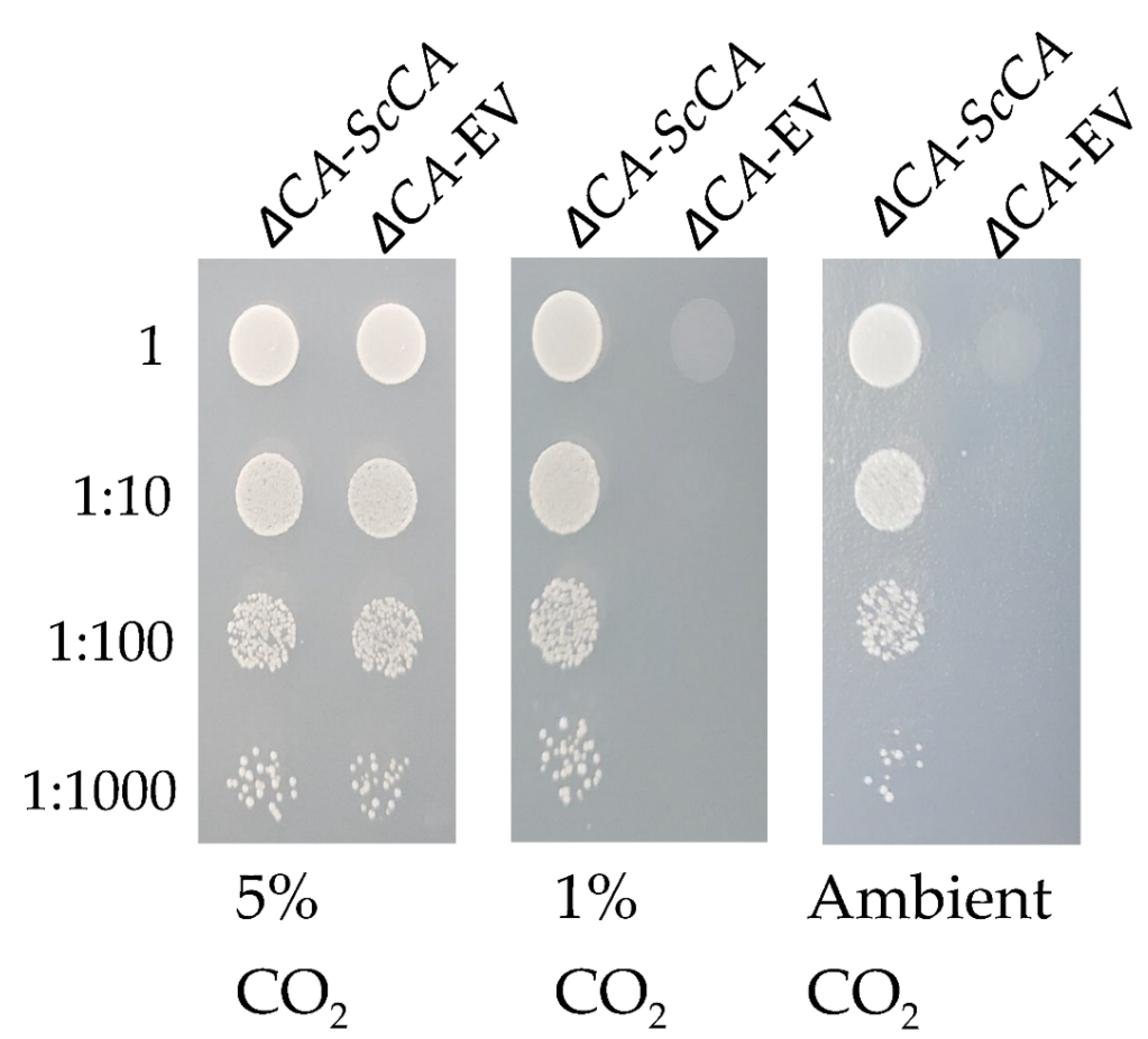
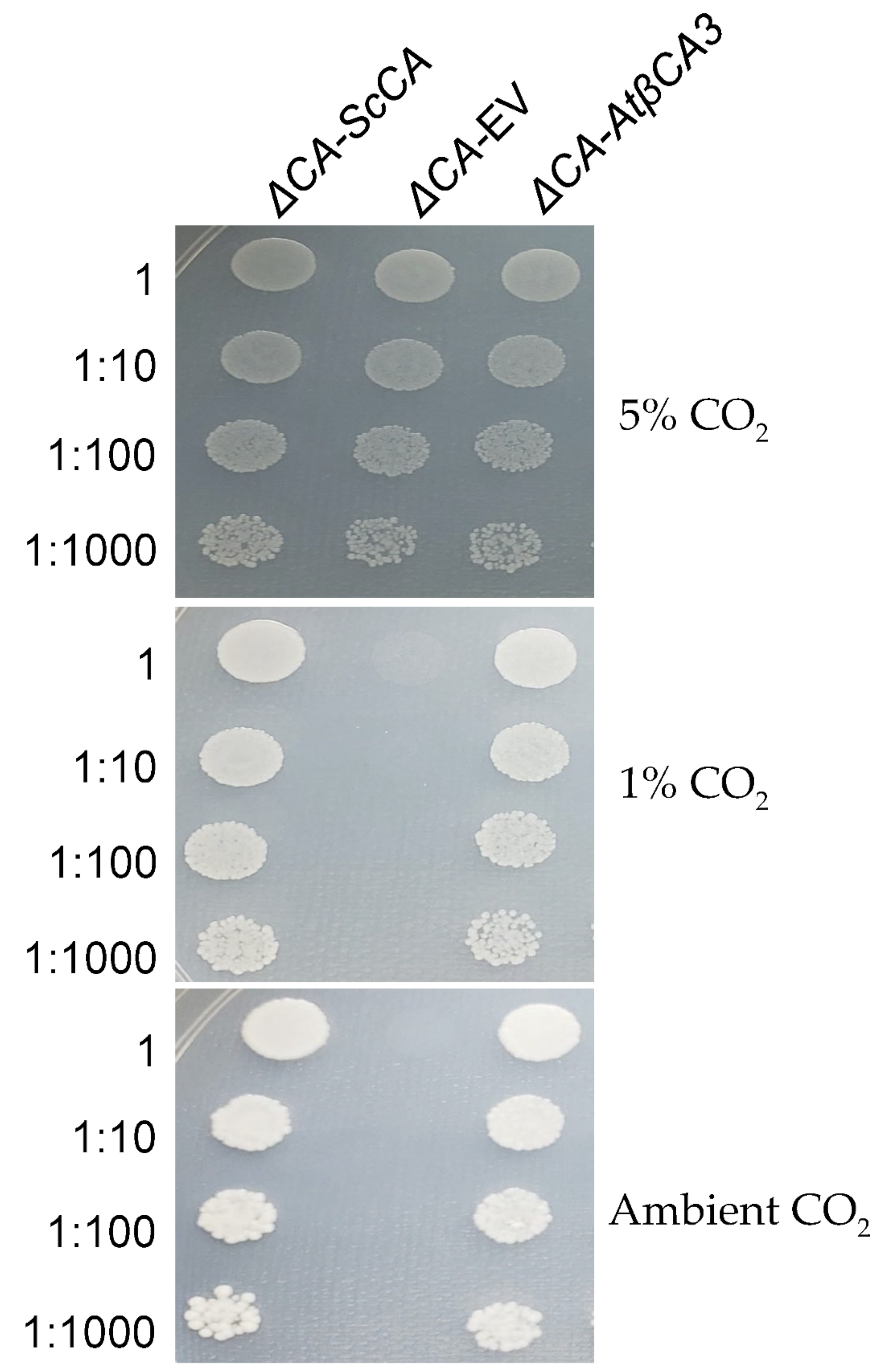
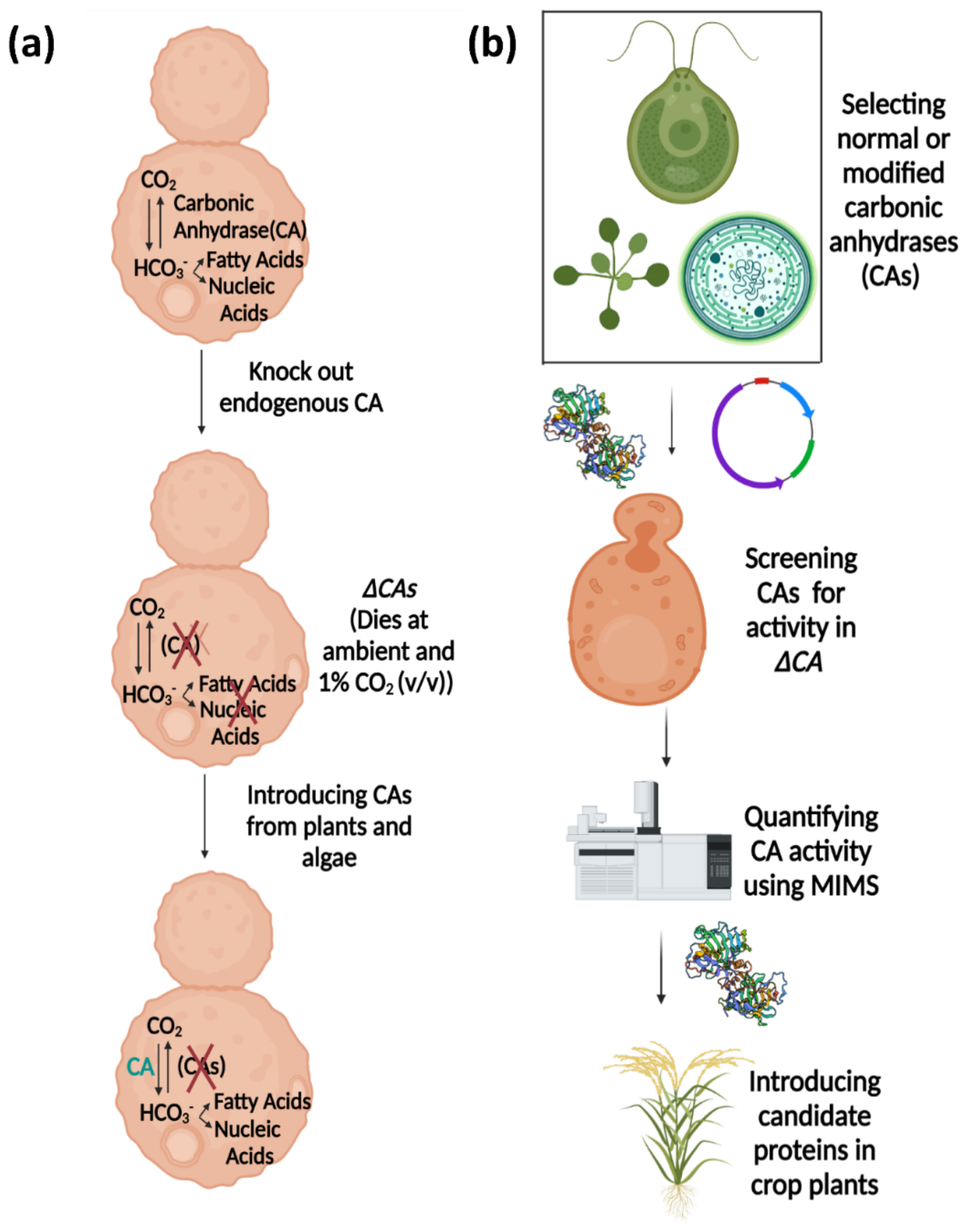
2.1. The S. cerevisiae Strain ΔCA Has a High-CO2-Dependent Growth Phenotype

The CO2 level requirement of ΔCA was characterized by conducting growth assays on solid media supplemented with different levels of CO2: 5% (v/v) CO2 in air, 1% (v/v) CO2 in air, and ambient CO2 (~0.04% (v/v) CO2 in the air) (Figure 1). For these experiments, ΔCA-EV refers to the ΔCA strain transformed with an empty vector (EV) containing a selectable gene. For the positive control, ΔCA was transformed with a vector containing the S. cerevisiae NCE103 gene so that it expresses the native CA (ΔCA-ScCA). Unlike the positive control, ΔCA only grows at 5% CO2 and dies at 1% and ambient CO2. These results agree with an early report by Aguilera et al. [18]. To investigate the biochemical deficiency underlying the high-CO2-dependent phenotype of ΔCA, we incorporated radiolabeled 14C-acetic acid into ΔCA-EV, ΔCA-ScCA, and ΔCA-hCA-YCO (ΔCA complemented with a S. cerevisiae codon-optimized (YCO) version of hCA) for one hour in ambient-CO2 conditions and assayed 14C incorporation into lipids using a silicone oil filtering centrifugation assay. The incorporation of radiolabeled 14C in neutral lipids was higher in cells reconstituted with hCA compared to ΔCA-EV after one hour. Furthermore, ΔCA-hCA-YCO had twice the incorporation of 14C in neutral lipids compared to ΔCA after one hour (Figure 2).

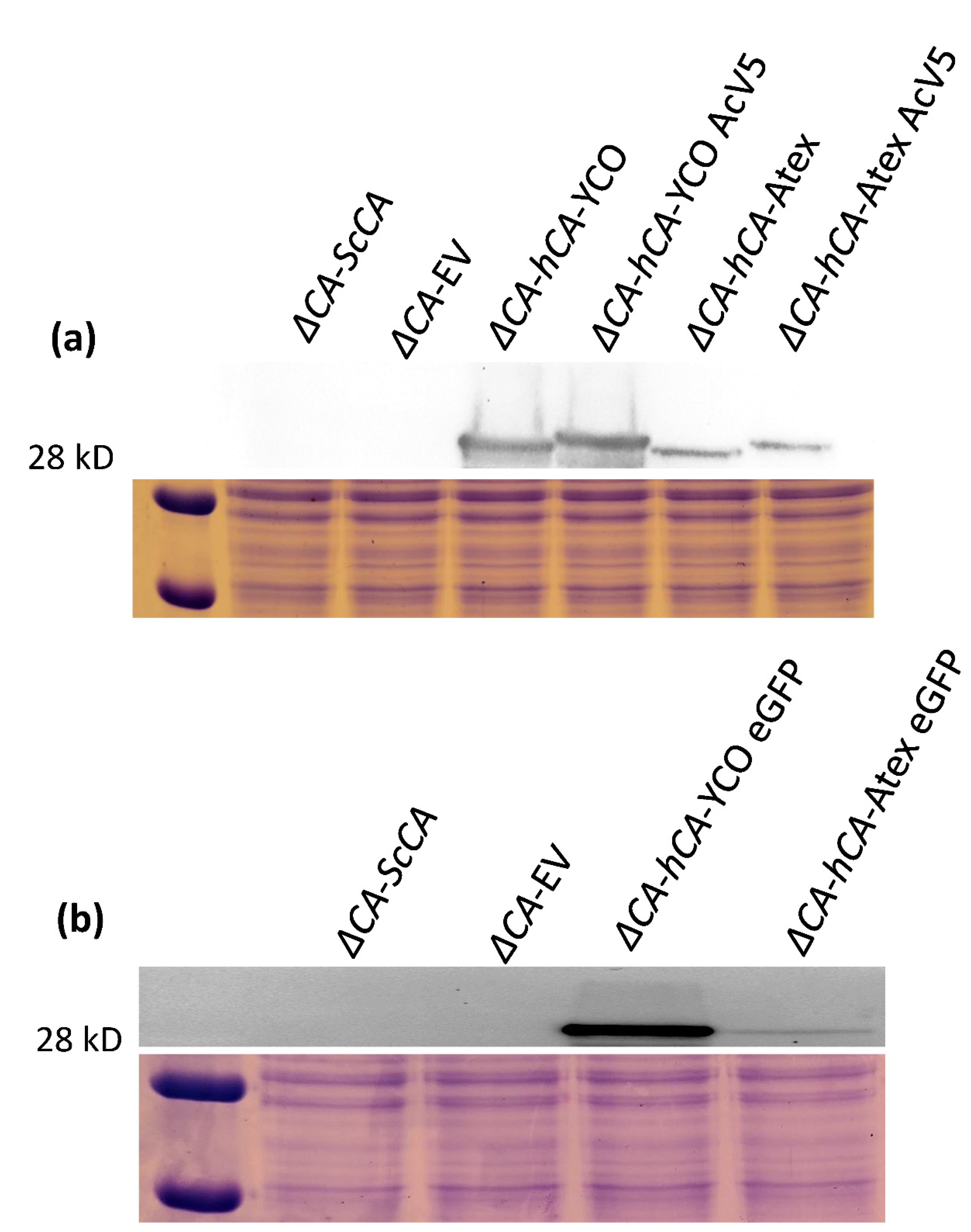
2.2. ΔCA Can Be Used as a Heterologous Complementation System to Detect CA Activity of Normal and Modified CAs

The CO2 growth requirement of ΔCA was used to characterize the CA activity of normal and modified CAs. hCA complemented the ΔCA phenotype in ambient CO2 and 1% CO2 (Figure 3 and Figure 4) [29]. In the liquid growth assay, we observed that ΔCA-hCA-YCO grew faster than ΔCA-EV in ambient CO2. However, in both conditions, ΔCA-ScCA growth was faster compared to ΔCA-EV (Figure 3a,b). In the growth assay on solid media, ΔCA-hCA-YCO grew at a rate similar to ΔCA-ScCA in ambient CO2 and 1% CO2 (Figure 4). Next, we transformed ΔCA with YCO hCA and Arabidopsis codon-optimized (Atex) hCA and checked their effect on CA activity. Additionally, we added the tags AcV5 and eGFP to ΔCA-hCA-YCO and ΔCA-hCA-Atex to see if they affected the growth of the S. cerevisiae. The growth assays on solid media show that the modified hCA variants complemented ΔCA in ambient CO2 and 1% CO2 (Figure 5). We also compared the expression of hCA in the complemented lines by analyzing the protein’s abundance via Western blots. In the strains complemented with the YCO genes (ΔCA-hCA-YCO, ΔCA-hCA-YCO AcV5, and ΔCA-hCA-YCO eGFP), hCA expression was higher compared to strains complemented with Atex genes (ΔCA-hCA-Atex, ΔCA-hCA-Atex AcV5, and ΔCA-hCA-Atex eGFP; Figure 6b). The protein expression of hCA was not affected by the addition of the AcV5 and eGFP tags in strains complemented with the genes optimized for S. cerevisiae and Arabidopsis (Figure 6a,b and Figure S2).

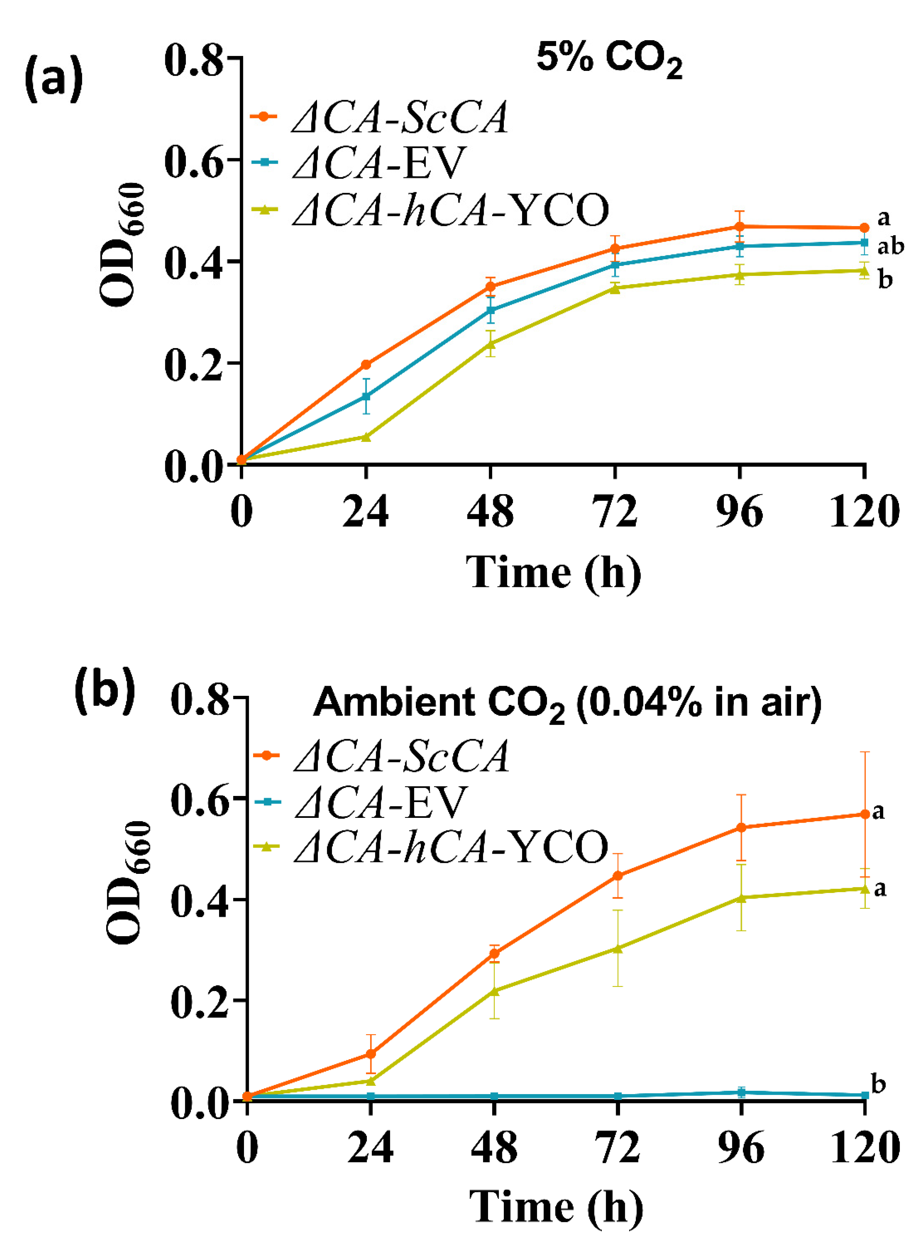
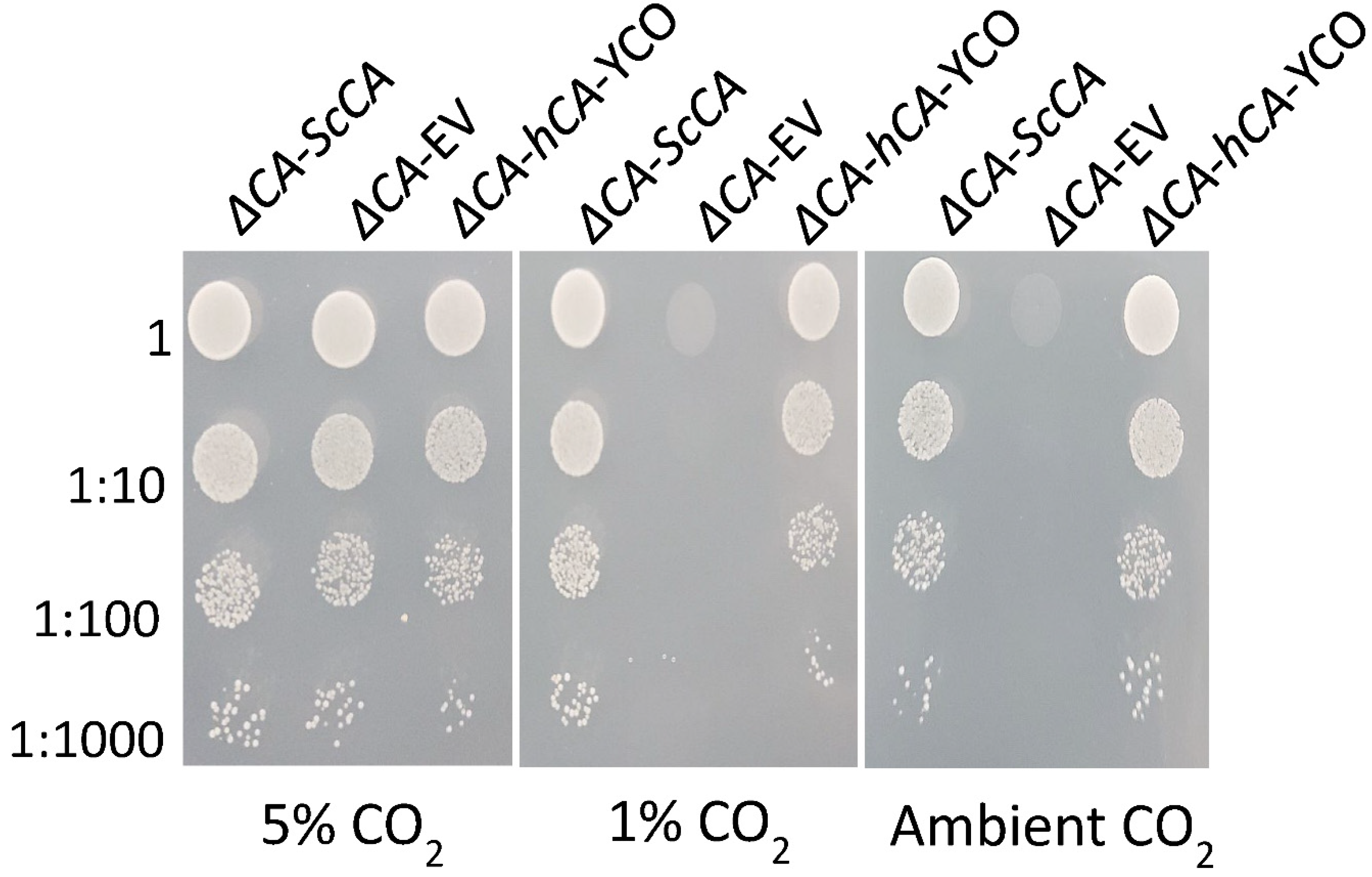
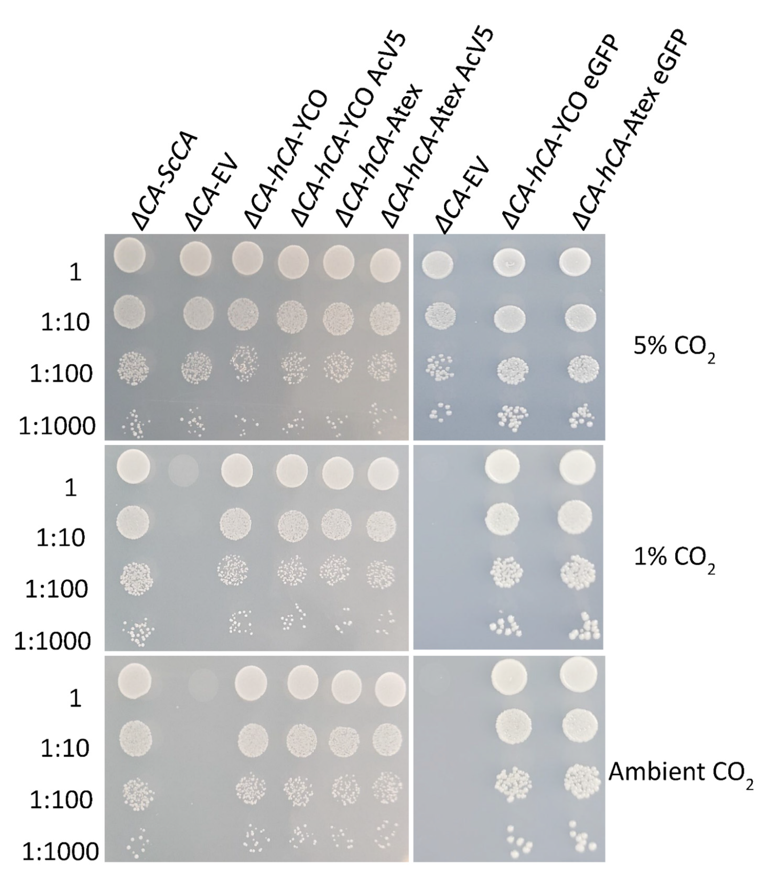
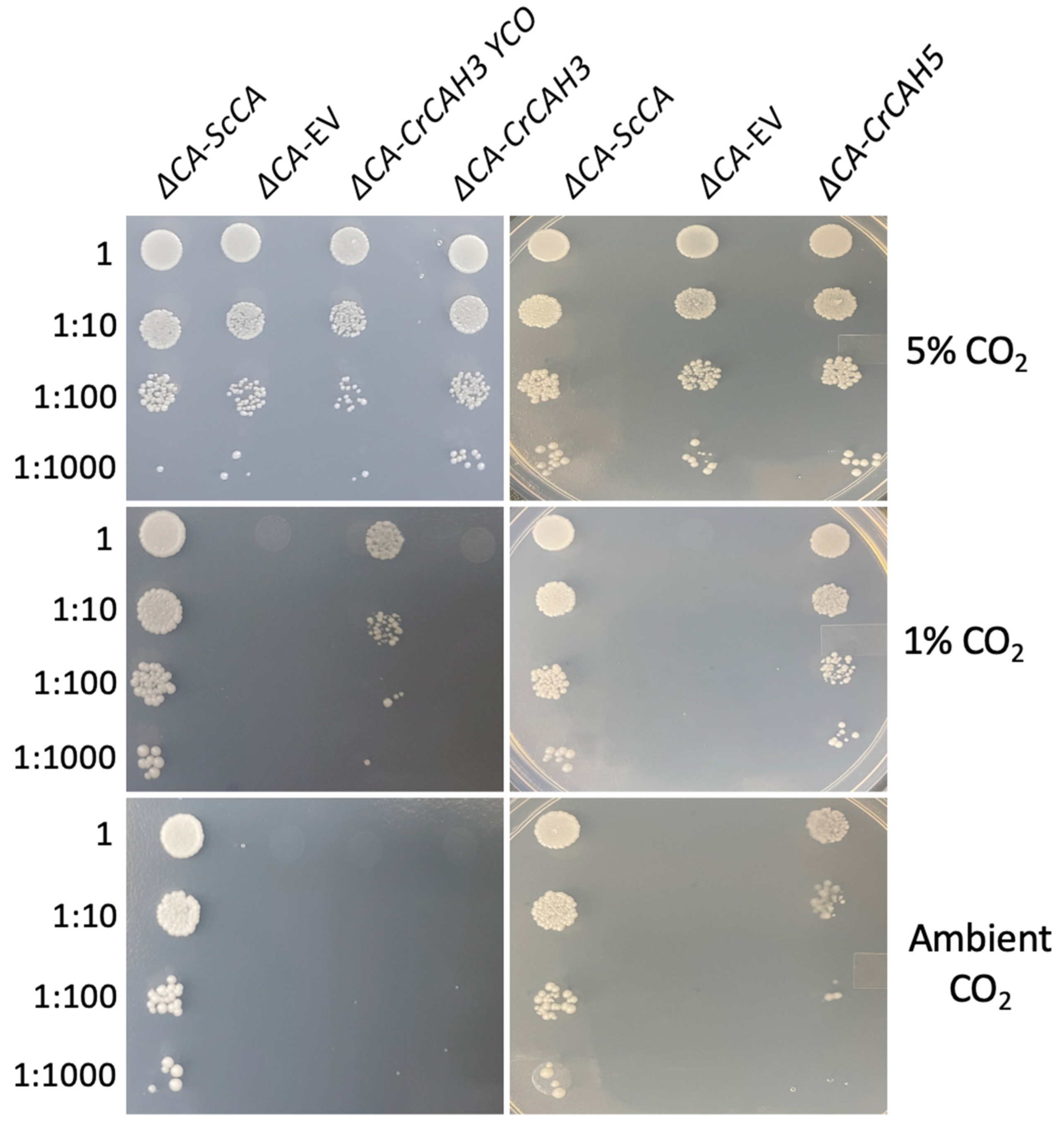
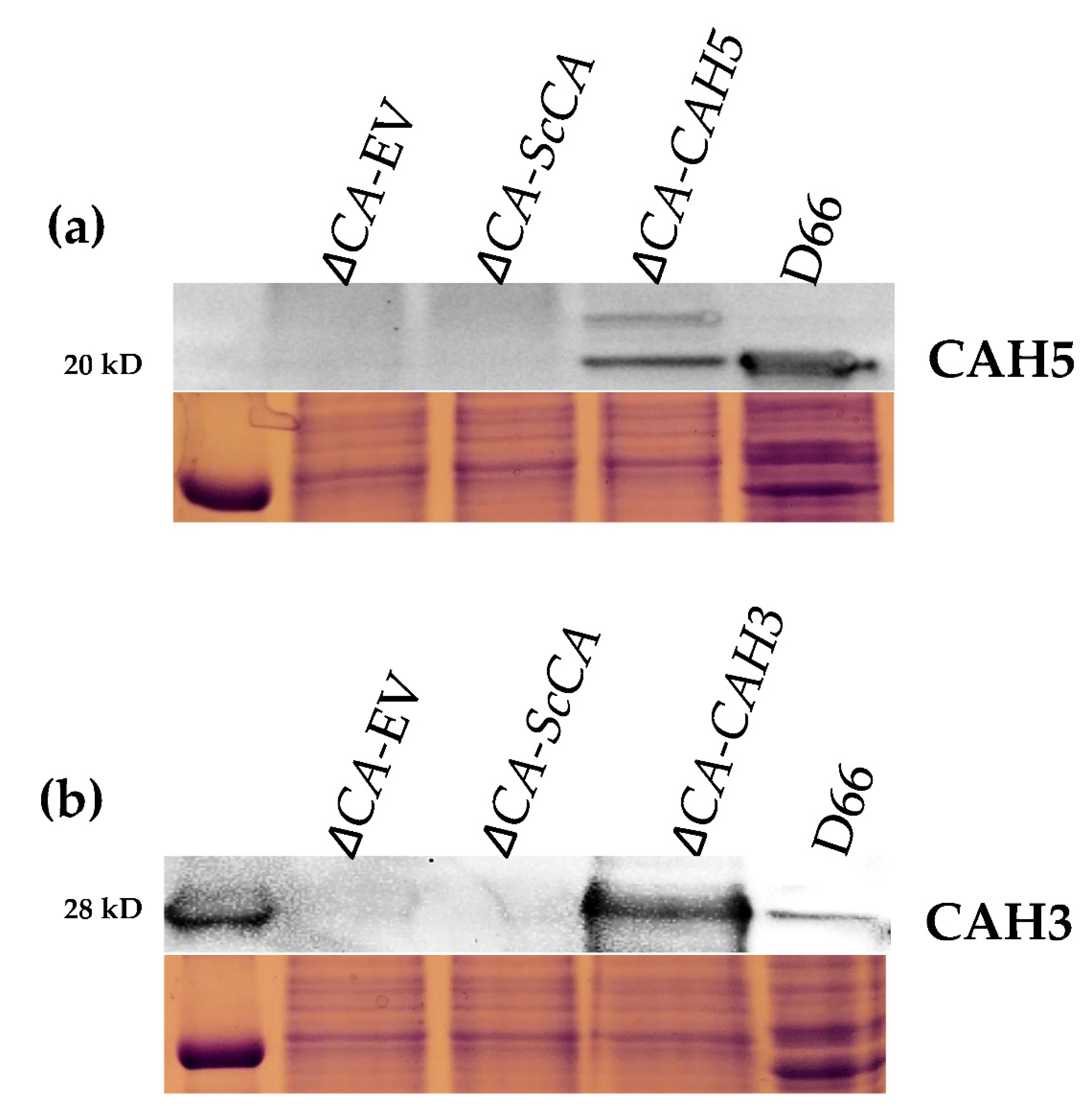
2.3. Plant and Algal Carbonic Anhydrases Show CA Activity in ΔCA-Based Heterologous Complementation System

To test the hypothesis that the ΔCA-based heterologous complementation system can rapidly detect the activity of different algal CA isoforms, we expressed the C. reinhardtii β-carbonic anhydrase CAH5 and α-carbonic anhydrase CAH3 in ΔCA. Expression of CAH5 restored a normal growth phenotype in the ΔCA mutant when cells were grown in ambient CO2 and 1% CO2 (Figure 7). CAH5 protein expression was detected in ΔCA-CrCAH5 and in the positive control D66 (a wild-type C. reinhardtii strain) (Figure 8a). The full-length coding sequence (CDS) was used for the expression of CAH5 in ΔCA. The Western blot shows that S. cerevisiae was able to process the N-terminal mitochondrial sequence of CAH5 (Figure 8a). We observed a full-length polypeptide of 27.8 kD and a cleaved polypeptide of 20.4 kD, which is similar to the size observed in the positive control. This is the first report showing that CAH5 is an active CA in a heterologous system. The expression of CAH3 in ΔCA-CrCAH3-YCO restored growth in 1% CO2, but no growth was observed in ambient CO2 (Figure 7). In the ΔCA-CrCAH3 strain containing the native C. reinhardtii CAH3 gene, growth was not restored in either limiting-CO2 condition. The Western blot shows CAH3 expression in ΔCA-CrCAH3-YCO and in the positive control (Figure 8b).
To test the activity of plant carbonic anhydrases in ΔCA, we transformed the S. cerevisiae mutant with the cytosolic carbonic anhydrase βCA3 from Arabidopsis. Normal growth was observed in ΔCA-AtβCA3 at all three CO2 levels (Figure 9). This result suggests that βCA3 is an active CA.

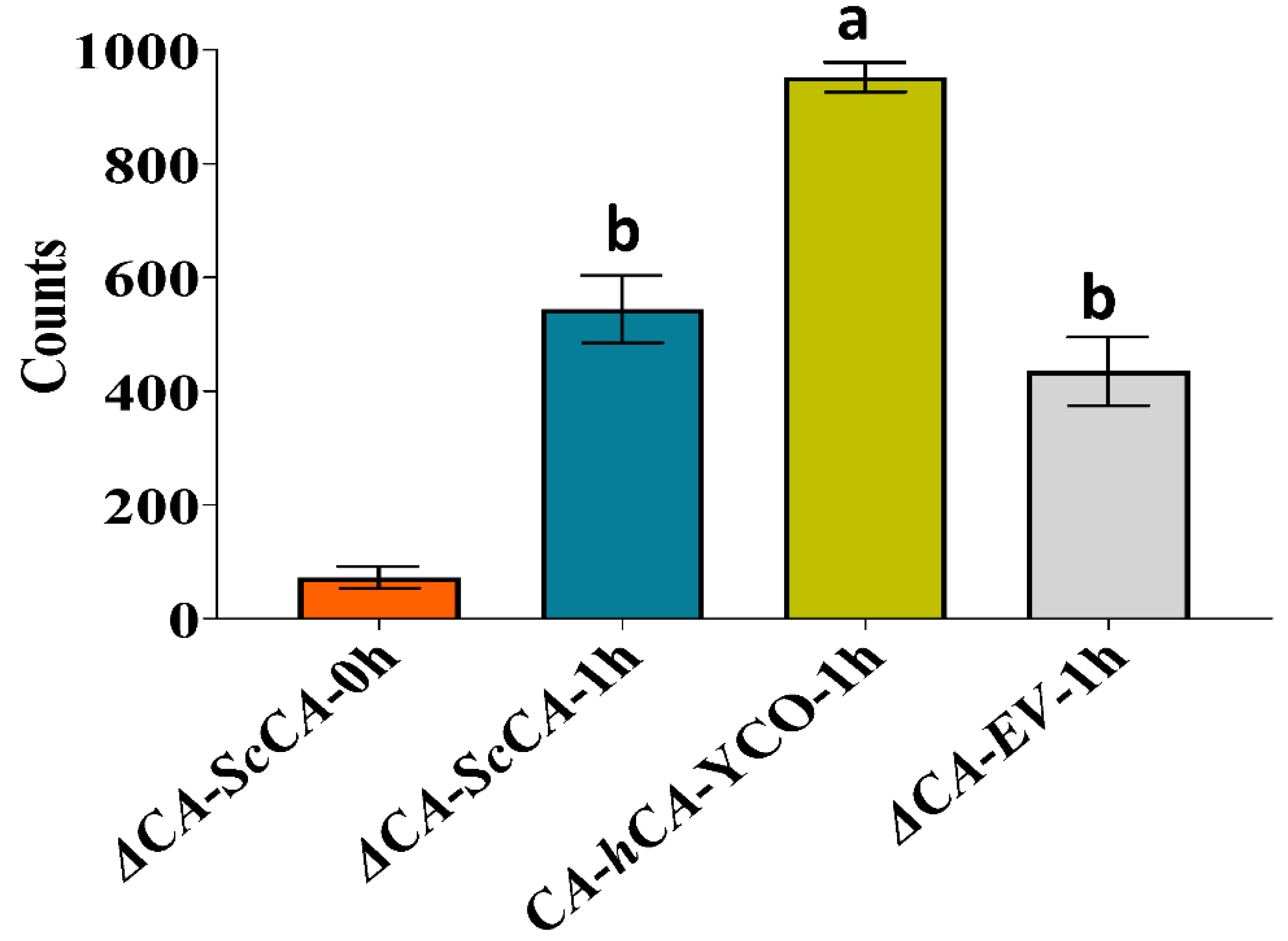
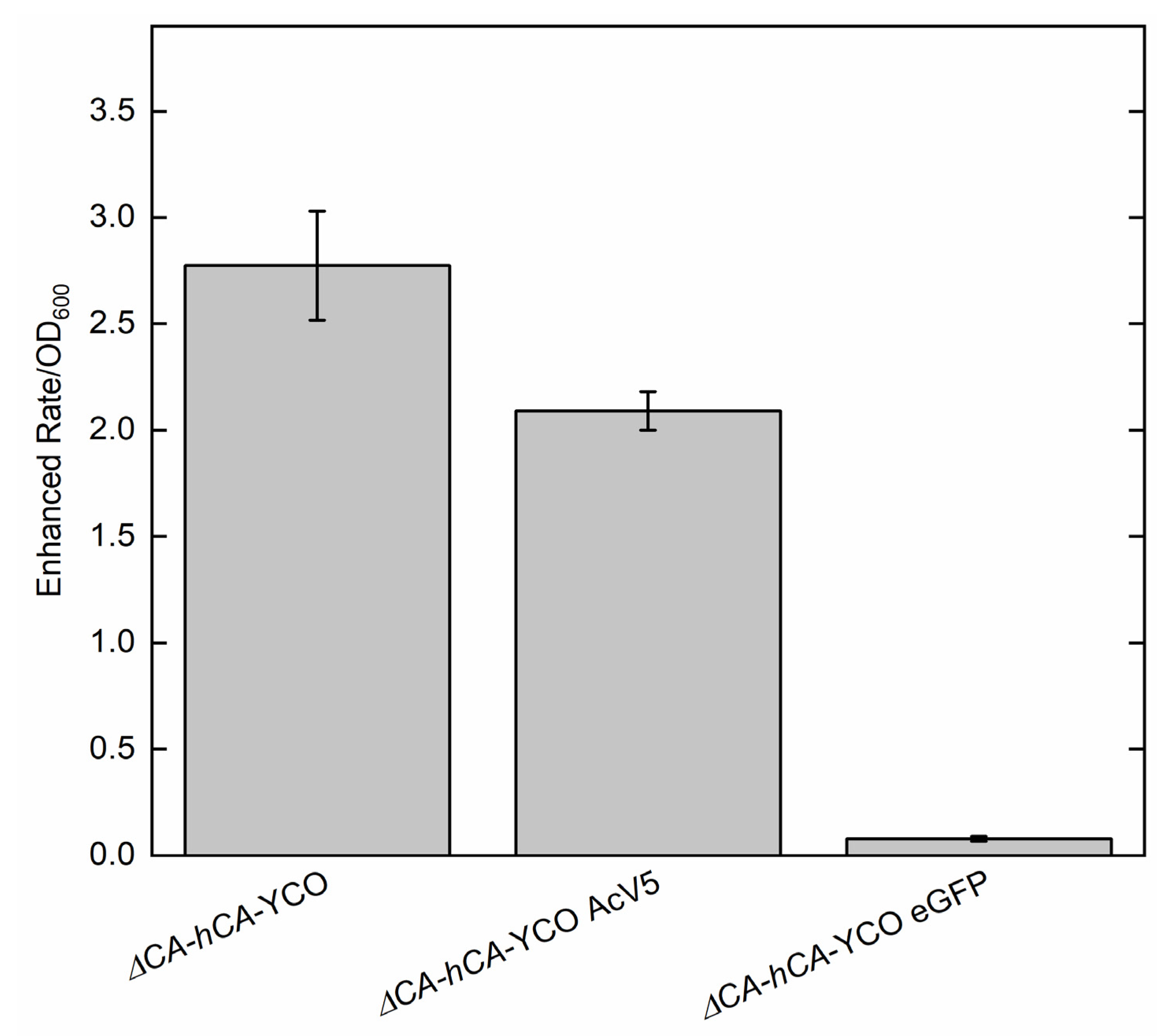
2.4. ΔCA-Based Heterologous Complementation System Can Be Used to Quantify CA Activity Using Wilbur–Anderson Assay and MIMS

To rapidly quantify the activity of normal or modified CAs in the ΔCA system, we used the Wilbur–Anderson assay and MIMS. The Wilbur–Anderson assay was successful in demonstrating the CA activity of ΔCA strains complemented with different variants of hCA (Table 1). CA activity was recorded as 4.7 ± 0.5 WAU mg−1 in ΔCA-hCA-YCO, which was the highest of all tested strains. The addition of AcV5 and eGFP decreased the CA activity to 3.9 ± 0.4 WAU mg−1 and 2.5 ± 0.3 WAU mg−1, respectively. In the strains using Atex genes, CA activity was further reduced. The strain ΔCA-hCA-Atex showed CA activity around 1.9 ± 0.2 WAU mg−1. Similar to the tagged YCO strains, the addition of AcV5 and eGFP decreased CA activity to 0.9 ± 0.1 WAU mg−1 and 1.4 ± 0.2 WAU mg−1, respectively. CA activity was also measured in the ΔCA strains using MIMS (Figure 10). We found that the cell lysate in ΔCA-hCA-YCO exhibited maximum CA activity. The AcV5 and eGFP tags reduced the CA activity significantly, consistent with measurements obtained using the Wilbur–Anderson assay.

3. Discussion

In this report, an S. cerevisiae CA knock-out strain (ΔNCE103 or ΔCA) was utilized as a successful heterologous system for screening active carbonic anhydrases from plants and algae. Additionally, ΔCA has previously been reported as a potential tool for accelerating the discovery of non-sulfonamide-based CAIs (carbonic anhydrase inhibitors) for the treatment of CA-related diseases, such as glaucoma [30].
In this study, ΔCA was generated and found to have a high-CO2-dependent phenotype, meaning it requires high-CO2 conditions to survive (Figure 1). This clear high CO2 growth requirement indicates that ΔCA strains can be used for fast and accurate screening of CA activity or active bicarbonate transport. To confirm this hypothesis, we tested CAs from human red blood cells (hCA), Arabidopsis, and C. reinhardtii in the heterologous S. cerevisiae system. Along with rapidly screening for CA activity, ΔCA can also be used for the estimation of CA enzymatic activity using the Wilbur–Anderson assay and MIMS.
In mammals, CAs are expressed in almost all tissues and are involved in oxygen transport between lungs, red blood cells and tissues; pH regulation; ion exchange in the kidney; and electrical activity in the retina and nervous system [31,32,33]. Autotrophic organisms use CAs in CCMs, where CAs are involved in increasing inorganic carbon for carbon fixation [3]. In contrast, very little is known about the physiological role of CAs in heterotrophic microbes. According to Aguilera et al. [18], the CA-deficient S. cerevisiae mutant’s need for elevated CO2 concentrations originates from three bicarbonate-dependent carboxylation reactions catalyzed by pyruvate decarboxylase, acetyl-CoA carboxylase, and carbamoyl phosphate synthetase. These enzymes are involved in the synthesis of C4 intermediates, fatty acids, arginine, and uracil, respectively [34]. These observations demonstrate that the S. cerevisiae CA is a key biosynthetic enzyme responsible for the viability of S. cerevisiae under aerobic conditions. Since ΔCA was complemented at air levels of CO2 by the addition of hCA (Figure 3 and Figure 4), we investigated whether fatty acid biosynthesis is bicarbonate-dependent at air levels of CO2 (Figure 2). S. cerevisiae uses acetyl-CoA as a building block to synthesize neutral lipids (NL) such as triglycerides (TGs) and sterol-esters (SEs). Acetyl-CoA is first converted into malonyl-CoA by acetyl-CoA carboxylase, using HCO3 as a substrate. S. cerevisiae cells deficient in acetyl-CoA carboxylase are not able to make long chain saturated fatty acids for de novo growth [35]. To test our hypothesis, we introduced 14C-acetic acid to ΔCA-ScCA, ΔCA-EV, and ΔCA-hCA grown on air levels of CO2 for one hour. Acetic acid is rapidly converted into acetyl-CoA by acetyl-CoA synthetase (ACS2), which makes 14C-acetic acid, a suitable radiolabeling substrate in S. cerevisiae. We observed that radiolabeled 14C is incorporated into the chloroform–methanol fraction containing NLs. The incorporation of radiolabeled 14C into NLs occurs at a higher count in ΔCA-hCA than in ΔCA-ScCA and ΔCA-EV (Figure 2). This result confirmed that CAs produce HCO3 for the NL biosynthesis pathway. However, there are other bicarbonate-requiring pathways that are also involved in limiting the growth of ΔCA in air. Hence, if we introduce CAs or HCO3 transporters from plants and algae, they can help to increase the HCO3 pool required for different biological processes in the cell. This makes the ΔCA heterologous complementation system useful for identifying new bicarbonate transporters or CAs as suitable candidates to improve photosynthetic efficiency in C3 crop plants.
This report shows that hCA displayed sufficient CA activity to rescue ΔCA grown on air levels of CO2 (Figure 3 and Figure 4). The results extend the work of Sangkaew et al. [30] who used this ΔCA system to screen CA inhibitors. To test modified CAs, S. cerevisiae codon-optimized (YCO) and Arabidopsis codon-optimized (Atex) hCA genes with added eGFP and AcV5 tags were used to complement ΔCA (Figure 5. The hCA protein was detected in all the hCA variants, but the amount of protein was highest in S. cerevisiae codon-optimized hCA (Figure 6a,b). The different variants of hCA complement ΔCA even though the protein content differs in the strains. This relates to the concept that only a low amount of CA activity is needed to maintain vital biological functions in S. cerevisiae since CA is such a fast enzyme. This concept is also supported by plant studies showing that the majority of CA activity within the plant needs to be removed in order to observe a growth phenotype [10]. Although all the hCA variants rescued ΔCA, CA activity was highest in the strain using S. cerevisiae codon-optimized hCA (Table 1 and Figure 10). The low CA activity in Atex strains might also be because of low protein expression (Figure 6a,b). In terms of the effect of added tags, the MIMS and Wilbur–Anderson assay data showed that the addition of longer tags resulted in a larger reduction in CA activity. The hCA tagged with eGFP has lower CA activity compared to the hCA tagged with AcV5 (Table 1 and Figure 10). These results suggest that the ΔCA system can be used to rapidly test the suitability of carbonic anhydrases before introducing them into C3 plants. Tags such as eGFP and AcV5 are widely used to determine the subcellular location of CAs, but there is a possibility that these tags can affect the functionality of the proteins. Thus, the ΔCA-based heterologous complementation system provides a rapid pipeline for the systematic assessment of normal and modified CAs before introducing them into C3 plants. Mathematical models predict that installing a CCM into C3 plants could improve leaf CO2 uptake by up to 60% [36,37]. Hence, using ΔCA in conjugation with the Wilbur–Anderson assay and MIMS can shorten the process of selecting suitable CCM components from cyanobacteria and algae to transform into terrestrial C3 plants.
To check the CA activity from an algal system, we expressed the β-carbonic anhydrase, CAH5, and the α-carbonic anhydrase, CAH3, from C. reinhardtii in ΔCA. CAH5 is present in the mitochondrial matrix and is required to maintain optimal rates of photoautotrophic growth on ambient levels of CO2 [25]. CAH5 restores the growth of ΔCA at air levels of CO2 and 1% CO2, but the rescued phenotype is weak compared to ΔCA-ScCA (Figure 7). This might indicate that CAH5 is a low-activity CA. Mitochondrial CA in C. reinhardtii is encoded by two genes (CAH4 and CAH5). This genetic redundancy could help the cell increase the amount of CA in the mitochondria since the enzyme has low activity, but it seems wasteful to produce large amounts of a protein with low activity rather than making a protein with high activity. The α carbonic anhydrase CAH3 was also tested in ΔCA. It is located in the thylakoid lumen in C. reinhardtii and has also been identified as an important component to maintain the CCM at low CO2 levels [38,39]. It was previously reported that CrCAH3 is different from other α carbonic anhydrases given that it has an optimum CA activity at lower pH values than CAs of the same type, which normally operate at pH 7.0 and higher [6,40]. S. cerevisiae codon-optimized CAH3 restored the normal growth phenotype in ΔCA at 1% CO2 but not at air levels, indicating that CAH3 activity might have been reduced because the S. cerevisiae cytoplasmic pH is around 7 (Figure 7). The Western blot clearly shows that CrCAH3 is produced in ΔCA (Figure 8b). The chloroplast transit peptide was removed from CrCAH3. The low activity could also be attributed to our use of a truncated version of CAH3 in ΔCA, although the protein length we used was reported to be the mature protein size [6,26]. Another possibility is that CAH3 requires post-translational modifications. Blanco-Rivero, et al. [41] reported that kinase activity is needed to activate CAH3 inside the lumen. The complementation of CrCAH5 and CrCAH3 (both YCO) in ΔCA suggests that the ΔCA-based heterologous complementation system can detect active CA enzymes from algal systems.
In general, α-carbonic anhydrases are structurally simpler than β-carbonic anhydrases and often have high specific activity. Humans only have α-carbonic anhydrases, while plants and algae have a wide variety of carbonic anhydrase families from α, β, γ, and θ classes. These CAs are localized to different intercellular and intracellular locations. Hence, the ΔCA heterologous complementation system could also be used as a tool to differentiate activity between CA families, which could help researchers select better CA candidates to integrate into C3 crop plants.
To check if ΔCA is rescued by a plant’s CA, we used β carbonic anhydrase βCA3, which localizes in the cytosol of Arabidopsis. βCA3 rescued ΔCA on 1% CO2 and air levels of CO2, showing that it is an active CA enzyme (Figure 9).
After transforming crop plants with a protein, the main challenge occurs upon trying to determine if the protein is functional once it is correctly localized. The addition of a fluorescent tag reveals if the protein is in the correct location, but it cannot report if the protein is in the correct orientation in the membrane or if it is functional. In addition, when GFP is added to a protein, the protein is modified because an entire second protein has been attached. This modification could potentially alter the CAs’ activity. The heterologous ΔCA-based system is an important tool for rapidly checking the activity of normal or modified CAs before integrating them into C3 crop plants to improve photosynthetic efficiency (Figure 11a,b). The ΔCA system can also check the activity of proteins retargeted to different compartments in plants.

4. Materials and Methods

4.1. S. cerevisiae Strain and Growth Conditions

Saccharomyces cerevisiae strain DDY2 (S. cerevisiae W303-1a diploid variant) was used as the starting stock for the generation of a CA knock-out (ΔNCE103). Yeast minimal media (YM) supplemented with 6.7 g/L yeast nitrogen base, 20% (w/v) dextrose, and an amino acid mix was used to grow liquid cultures of the S. cerevisiae cells. YM plates were made by adding 1.5% (w/v) agar to liquid YM. The ΔCA generated in this study was grown in 5% (v/v) CO2 in air at 30 °C, unless otherwise stated. Liquid cultures were grown on a rotary shaker at 30 °C in 5% (v/v) CO2 in air and ambient CO2 (0.04% (v/v) CO2 in air). S. cerevisiae cells were grown on YM plates in three different CO2 conditions: 5% (v/v) CO2 in the air, 1% (v/v) CO2 in the air, and ambient CO2 (0.04% (v/v) CO2 in the air) at 30 °C. Where applicable, amino acid mixes were added in the following order for the strains generated in this report: amino acid mix made without tryptophan (ΔCA), amino acid mix made without histidine and tryptophan (ΔCA-EV, ΔCA-ScCA, ΔCA-hCA-YCO, ΔCA-hCA-YCO AcV5, ΔCA-hCA-Atex, ΔCA-hCA-Atex AcV5, ΔCA-CrCAH5, ΔCA-CrCAH3-YCO, ΔCA-CrCAH3, and ΔCA-AtβCA3) and amino acid mix made without uracil and tryptophan (ΔCA-hCA-YCO eGFP, ΔCA-hCA-Atex eGFP).

4.2. Generation of S. cerevisiae Carbonic Anhydrase Knock-Out

The construction of the NCE103 deletion in the diploid strain DDY2 was carried out by PCR-targeting with a TRP1 disruption cassette flanked by short homology regions of the NCE103 gene [42]. The disruption cassette was obtained by amplifying the TRP1 cassette from the plasmid pRS304 ([43] and Table S1). pRS304 was used as a template in a PCR reaction to amplify TRP1 with NCE103 flanking sequences using oligonucleotides DDO-1976 and -1977. This DNA was concentrated by ethanol precipitation and transformed into S. cerevisiae strain DDY2. The transformation mix was plated onto minimal media lacking tryptophan to screen for ΔNCE103 mutants. The successful haploid knock-outs were confirmed by PCR using primers described in Table S1. Correctly targeted strains were sporulated to haploid, and Trp+ isolates were re-confirmed by PCR as ΔNCE103 mutants.

4.3. Genetic Constructs and Vectors

To express the mature peptide versions of human CAII (HCAII; Genbank ID AK312978) and CrCAH3-YCO (referred to as yeast codon-optimized (YCO)) in ΔNCE103, the hCA and CrCAH3 gene was synthesized by GenScript in pENTR and cloned into destination vectors MGO515 (-HIS) and MGO528 (-URA) using Gateway cloning. The hCA gene was codon optimized for S. cerevisiae (referred to as yeast codon-optimized (YCO)) and Arabidopsis (referred to as Arabidopsis codon-optimized (Atex)). A C-terminal AcV5 tag and eGFP tag were added to the hCA gene (Figure S1). hCA (with or without Acv5 tag) coding sequences were commercially synthesized (Genscript) as gateway-enabled entry vectors (i.e., included flanking attL sites). A second set of hCAII genes without the stop codon were also made, for use in GFP C-terminal fusion constructs. The hCAII coding sequences (CDS) were cloned into plasmids from the Advanced Gateway® adapted pRS series of yeast expression plasmids [44] using Gateway LR Clonase II enzyme mix (Invitrogen™)—essentially swapping out the ccdB bacterial lethality cassette for the given hCAII CDS between the attR1/R2 sites (Figure S1). The yeast expression construct library was obtained through Addgene (https://www.addgene.org/) (kit #1000000011). pAG423GPD-ccdB (internally designated MG0515; HIS) was used for full-length CDS clones. pAG426GPD-ccdB-eGFP (internally designated MG0528; URA) was used for GFP fusions (i.e., hCAII CDS without stop codon). All E. coli cloning steps used One Shot™ OmniMAX™ (Peachtree Corners, GA, USA) 2 T1R Chemically Competent E. coli cells (Invitrogen). All final plasmids were sanger sequenced to confirm accuracy of the clones using Wizard® Plus SV Minipreps DNA Purification Systems (Promega, Madison, WI, USA), BigDye® sequencing chemistry (Thermofisher Scientific, Waltham, MA, USA), and ZR DNA Sequencing Clean-Up Kit (Zymo Research, Irvine, CA, USA). Internal catalogue designations for final yeast hCAII expression constructs were: MG0515.54: GPD-hCAII (YCO)-stop; MG0515.55: GPD-hCAII (YCO)-AcV5-stop; MG0515.56: GPD-hCAII (Atex)-stop; MG0515.57: GPD-hCAII (Atex)-AcV5-stop; MG0528.20: GPD-hCAII (YCO)-nostop-eGFP; MG0528.21: GPD-hCAII (Atex)-nostop-eGFP.
The genes CrCAH5, CrCAH3, ScCA, and AtβCA3 were amplified by PCR from C. reinhardtii (D66) and A. thaliana (Col-0). The genes were cloned into the expression vector pDD506 using ClaI/XhoI sites for constitutive expression under control of the ADH1 promoter (Figure S1). All DNA constructs were verified by DNA sequencing.

4.4. S. cerevisiae CA Knock-Out Transformation

The plasmid MG0515 containing hCA-YCO, hCA-YCO AcV5, hCA-Atex, hCA-Atex AcV5, and CrCAH3-YCO, the plasmid MGO528 containing hCA-YCO eGFP, and hCA-Atex eGFP, and the plasmid pDD506 containing CrCAH5, CrCAH3, and AtβCA3 were transformed in E. coli TOP10 cells (One Shot™ TOP10 Chemically Competent E. coli). The plasmids were extracted from the transformed E. coli cultures using a GeneJET Plasmid Miniprep Kit (Thermo Scientific™) according to manufacturer’s instructions. The plasmids were transformed in the ΔNCE103 mutant using a S. cerevisiae transformation protocol as described by Gietz and Schiestl [45]. The positive colonies were screened by colony PCR using primers complementary to the genes (Table S1).

4.5. High-CO2-Dependence Growth Assay

S. cerevisiae cell cultures were initiated from −80 °C glycerol stocks. The liquid cultures were grown to log phase in liquid YM in 5% CO2 at 30 °C. The cultures were reinoculated for the growth assay and grown in 5% CO2 and ambient CO2. The optical density at 600 nm (OD600) of the cultures was adjusted to an initial OD600 of 0.01. Relative growth rates were measured in liquid YM by monitoring the cultures’ OD600 using a spectrometer (Thermo Fisher Scientific, Waltham, MA, USA). For the measurement of relative growth rates on solidified YM, the OD600 of the cultures was standardized to 0.01, and 10 µL of serial dilutions were spotted onto YM plates. The plates were incubated at 30 °C in 5%, 1%, and ambient CO2 for 72 h. The plates were photographed after 72 h.

4.6. Immunological Detection of Expressed Proteins in S. cerevisiae

Membrane-enriched protein fractions of S. cerevisiae were prepared according to Zhang et al. [46]. Proteins from the S. cerevisiae cell lysate were mixed with 2× Laemmli sample buffer and β-mercaptoethanol and heated to 95 °C for ~7 min prior to resolution by SDS-PAGE on 12% (v/v) polyacrylamide gels (Mini-PROTEAN TGX, Bio-Rad Laboratories, Hercules, CA, USA). The expression of tagged proteins was detected immuno-chemically after transferring the proteins to a PVDF membrane. The membrane was blocked in TTBS (TBS with 0.1% (v/v) Tween) with 1% (w/v) bovine serum albumin at 4 °C overnight. The membrane was treated with monoclonal antibodies for 1 h at room temperature with the following dilutions: 1:1000 for hCA (Sigma-Aldrich, Darmstadt, Germany), 1:20,000 for CrCAH5 (Agrisera, Vännäs, Sweden) and 1:10,000 for CrCAH3 (Agrisera). The anti-CrCAH3 antibody is commercially available at Agrisera and the anti-CrCAH5 antibody was custom made by Agrisera. The membrane was treated with secondary anti-rabbit antibody (HRP conjugated from Bio-Rad) for 1 h at room temperature with the following dilutions: 1:500 for hCA (Sigma-Aldrich, Darmstadt, Germany) and 1:4000 for CrCAH5 and CrCAH3. Antibody binding was visualized by fluorescence detection with Thermo Scientific™ Pierce™ ECL Western Blotting Substrate on a Chemi-Doc XRS (Bio-Rad, Hercules, CA, USA).

4.7. Analysis of Neutral Lipid Synthesis Using Radiolabel 14C-Acetic Acid

14C-acetic acid was incorporated in S. cerevisiae using the protocol described by Rogers and Henne [22], except that 50 μL of radiolabeling media was added to 1 mL of cell suspension (final 14C-acetic acid concentration = 1.25 μCi/mL). Here, a 1 h radiolabeling pulse incubation was used to label neutral lipid species in ΔCA, ΔCA-ScCA and ΔCA-hCA-YCO.

4.8. Carbonic Anhydrase Activity Assay

CA activity was measured by the Wilbur–Anderson assay according to Mitra et al. [47]. The isotope exchange membrane-inlet mass spectrometry (MIMS) technique was used to measure CA activity as described by Price and Badger [48]. Briefly, 20 μL of S. cerevisiae cell lysate was added to a temperature controlled, 2 mL reaction cuvette connected to the inlet of a Finnegan DELTA-V Isotope Ratio Mass Spectrometer (Thermo Fisher Scientific). The 2 mL CA assay consisted of 100 mM HEPES-KOH (pH 7.4), 5 mM dithiothreitol, and 13C18O2. The enhanced rate of 18O exchange between 13C18O2 and H216O was calculated as a ratio between the increase in 18O loss from 13C18O2 in the presence of CA compared to the uncatalyzed rate. Three technical replicates were run at 25 °C for each CA enzyme tested [10].

Supplementary Materials

The following supporting information can be downloaded at: https://www.mdpi.com/article/10.3390/plants11141882/s1; Figure S1: Plasmids pDD506(HIS3), MGO515(HIS3), and MGO528(URA3) were used in this study for the overexpression of proteins in S. cerevisiae, Figure S2: Relative intensity of the modified hCA bands for the immunoblot experiment shown in Figure 6a,b; Table S1: Primers used in this study for cloning.

Author Contributions

A.K.R. and J.V.M. designed the research; A.K.R., M.G., R.J.D., R.W.K. and J.V.M. performed the research. All authors along with A.B.C. and D.D. provided critical feedback and helped shape the research, analysis, and manuscript. All authors have read and agreed to the published version of the manuscript.

Funding

This work was supported by the Realizing Improved Photosynthetic Efficiency (RIPE) initiative awarded to JVM by the University of Illinois, Champaign, IL, USA. RIPE is made possible through support from the Bill & Melinda Gates Foundation, FFAR, and FCDO, grant no. OPP1172157. This study was also supported in part by the Division of Chemical Sciences, Geosciences, and Biosciences, Office of Basic Energy Sciences, Department of Energy (grant no. DE-SC0001685) to A.B.C.

Institutional Review Board Statement

Not applicable.

Informed Consent Statement

Not applicable.

Data Availability Statement

All of the new data is contained within this article or the supplementary material.

Acknowledgments

The authors express their gratitude to Lillian LaPlace, Isaiah C. Pabuayon and Timothy Chen, Louisiana State University, Baton Rouge, LA, USA, for their valuable suggestions in improving the manuscript. The authors are also thankful to BioRender.com for help in creating Figure 11a,b.

Conflicts of Interest

The authors declare no conflict of interest.

References

  1. Supuran, C.T. Structure and function of carbonic anhydrases. Biochem. J. 2016, 473, 2023–2032. [Google Scholar] [CrossRef] [PubMed]
  2. DiMario, R.J.; Machingura, M.C.; Waldrop, G.L.; Moroney, J.V. The many types of carbonic anhydrases in photosynthetic organisms. Plant Sci. 2018, 268, 11–17. [Google Scholar] [CrossRef] [PubMed]
  3. Sültemeyer, D.; Schmidt, C.; Fock, H.P. Carbonic anhydrases in higher plants and aquatic microorganisms. Plant Physiol. 1993, 88, 179–190. [Google Scholar] [CrossRef]
  4. Moroney, J.V.; Ma, Y.; Frey, W.D.; Fusilier, K.A.; Pham, T.T.; Simms, T.A.; DiMario, R.J.; Yang, J.; Mukherjee, B. The carbonic anhydrase isoforms of Chlamydomonas reinhardtii: Intracellular location, expression, and physiological roles. Photosynth. Res. 2011, 109, 133–149. [Google Scholar] [CrossRef] [PubMed]
  5. Badger, M. The roles of carbonic anhydrases in photosynthetic CO2 concentrating mechanisms. Photosynth. Res. 2003, 77, 83. [Google Scholar] [CrossRef]
  6. Karlsson, J.; Clarke, A.K.; Chen, Z.Y.; Hugghins, S.Y.; Park, Y.I.; Husic, H.D.; Moroney, J.V.; Samuelsson, G. A novel alpha-type carbonic anhydrase associated with the thylakoid membrane in Chlamydomonas reinhardtii is required for growth at ambient CO2. EMBO J. 1998, 17, 1208–1216. [Google Scholar] [CrossRef] [Green Version]
  7. Espie, G.S.; Kimber, M.S. Carboxysomes: Cyanobacterial RubisCO comes in small packages. Photosynth. Res. 2011, 109, 7–20. [Google Scholar] [CrossRef]
  8. Price, G.D.; Coleman, J.R.; Badger, M.R. Association of carbonic anhydrase activity with carboxysomes isolated from the cyanobacterium Synechococcus PCC7942. Plant Physiol. 1992, 100, 784–793. [Google Scholar] [CrossRef] [Green Version]
  9. Hatch, M.D.; Burnell, J.N. Carbonic Anhydrase Activity in Leaves and Its Role in the First Step of C4 Photosynthesis. Plant Physiol. 1990, 93, 825–828. [Google Scholar] [CrossRef] [Green Version]
  10. DiMario, R.J.; Giuliani, R.; Ubierna, N.; Slack, A.D.; Cousins, A.B.; Studer, A.J. Lack of leaf carbonic anhydrase activity eliminates the C4 carbon-concentrating mechanism requiring direct diffusion of CO2 into bundle sheath cells. Plant Cell Environ. 2022, 45, 1382–1397. [Google Scholar] [CrossRef]
  11. DiMario, R.J.; Quebedeaux, J.C.; Longstreth, D.J.; Dassanayake, M.; Hartman, M.M.; Moroney, J.V. The Cytoplasmic Carbonic Anhydrases βCA2 and βCA4 Are Required for Optimal Plant Growth at Low CO2. Plant Physiol. 2016, 171, 280–293. [Google Scholar] [CrossRef] [PubMed] [Green Version]
  12. Medina-Puche, L.; Castelló, M.J.; Canet, J.V.; Lamilla, J.; Colombo, M.L.; Tornero, P. β-carbonic anhydrases play a role in salicylic acid perception in Arabidopsis. PLoS ONE 2017, 12, e0181820. [Google Scholar] [CrossRef] [PubMed] [Green Version]
  13. Hines, K.M.; Chaudhari, V.; Edgeworth, K.N.; Owens, T.G.; Hanson, M.R. Absence of carbonic anhydrase in chloroplasts affects C3 plant development but not photosynthesis. Proc. Natl. Acad. Sci. USA 2021, 118, e2107425118. [Google Scholar] [CrossRef]
  14. Atkinson, N.; Mao, Y.; Chan, K.X.; McCormick, A.J. Condensation of Rubisco into a proto-pyrenoid in higher plant chloroplasts. Nat. Commun. 2020, 11, 6303. [Google Scholar] [CrossRef] [PubMed]
  15. Hennacy, J.H.; Jonikas, M.C. Prospects for engineering biophysical CO2 concentrating mechanisms into land plants to enhance yields. Annu. Rev. Plant Biol. 2020, 71, 461–485. [Google Scholar] [CrossRef] [Green Version]
  16. Long, B.M.; Hee, W.Y.; Sharwood, R.E.; Rae, B.D.; Kaines, S.; Lim, Y.-L.; Nguyen, N.D.; Massey, B.; Bala, S.; von Caemmerer, S.; et al. Carboxysome encapsulation of the CO2-fixing enzyme Rubisco in tobacco chloroplasts. Nat. Commun. 2018, 9, 3570. [Google Scholar] [CrossRef] [Green Version]
  17. Amoroso, G.; Morell-Avrahov, L.; Müller, D.; Klug, K.; Sültemeyer, D. The gene NCE103 (YNL036w) from Saccharomyces cerevisiae encodes a functional carbonic anhydrase and its transcription is regulated by the concentration of inorganic carbon in the medium. Mol. Microbiol. 2005, 56, 549–558. [Google Scholar] [CrossRef]
  18. Aguilera, J.; Van Dijken, J.P.; De Winde, J.H.; Pronk, J.T. Carbonic anhydrase (Nce103p): An essential biosynthetic enzyme for growth of Saccharomyces cerevisiae at atmospheric carbon dioxide pressure. Biochem. J. 2005, 391, 311–316. [Google Scholar] [CrossRef] [Green Version]
  19. Whitney, P.A.; Cooper, T.G. Urea Carboxylase and Allophanate Hydrolase: Two components of adenosine triphosphate:urea amido-lyase in Saccharomyces cerevisisae. J. Biol. Chem. 1972, 247, 1349–1353. [Google Scholar] [CrossRef]
  20. Mishina, M.; Rogguenkamp, R.; Schweizer, E. Yeast mutants defective in acetyl-coenzyme A carboxylase and biotin: Apocarboxylase ligase. Eur. J. Biochem. 1980, 111, 79–87. [Google Scholar] [CrossRef]
  21. Magnuson, K.; Jackowski, S.; Rock, C.O.; Cronan, J.E. Regulation of fatty acid biosynthesis in Escherichia coli. Microbiol. Rev. 1993, 57, 522–542. [Google Scholar] [CrossRef] [PubMed]
  22. Rogers, S.; Henne, W.M. Analysis of Neutral Lipid Synthesis in Saccharomyces cerevisiae by Metabolic Labeling and Thin Layer Chromatography. J. Vis. Exp. 2021, 168, e62201. [Google Scholar] [CrossRef] [PubMed]
  23. Eriksson, M.; Karlsson, J.; Ramazanov, Z.; Gardeström, P.; Samuelsson, G. Discovery of an algal mitochondrial carbonic anhydrase: Molecular cloning and characterization of a low-CO2-induced polypeptide in Chlamydomonas reinhardtii. Proc. Natl. Acad. Sci. USA 1996, 93, 12031–12034. [Google Scholar] [CrossRef] [PubMed] [Green Version]
  24. Fang, W.; Si, Y.; Douglass, S.; Casero, D.; Merchant, S.S.; Pellegrini, M.; Ladunga, I.; Liu, P.; Spalding, M.H. Transcriptome-wide changes in Chlamydomonas reinhardtii gene expression regulated by carbon dioxide and the CO2-concentrating mechanism regulator CIA5/CCM1. Plant Cell 2012, 24, 1876–1893. [Google Scholar] [CrossRef] [PubMed] [Green Version]
  25. Rai, A.K.; Chen, T.; Moroney, J.V. Mitochondrial carbonic anhydrases are needed for optimal photosynthesis at low CO2 levels in Chlamydomonas. Plant Physiol. 2021, 187, 1387–1398. [Google Scholar] [CrossRef] [PubMed]
  26. Hanson, D.T.; Franklin, L.A.; Samuelsson, G.; Badger, M.R. The Chlamydomonas reinhardtii cia3 mutant lacking a thylakoid lumen-localized carbonic anhydrase is limited by CO2 supply to Rubisco and not photosystem II function in vivo. Plant Physiol. 2003, 132, 2267–2275. [Google Scholar] [CrossRef]
  27. Park, Y.-I.; Karlsson, J.; Rojdestvenski, I.; Pronina, N.; Klimov, V.; Öquist, G.; Samuelsson, G. Role of a novel photosystem II-associated carbonic anhydrase in photosynthetic carbon assimilation in Chlamydomonas reinhardtii. FEBS Lett. 1999, 444, 102–105. [Google Scholar] [CrossRef] [Green Version]
  28. Wilbur, K.M.; Anderson, N.G. Electrometric and colorimetric determination of carbonic anhydrase. J. Biol. Chem. 1948, 176, 147–154. [Google Scholar] [CrossRef]
  29. Clark, D.; Rowlett, R.S.; Coleman, J.R.; Klessig, D.F. Complementation of the yeast deletion mutant DeltaNCE103 by members of the beta class of carbonic anhydrases is dependent on carbonic anhydrase activity rather than on antioxidant activity. Biochem. J. 2004, 379, 609–615. [Google Scholar] [CrossRef]
  30. Sangkaew, A.; Krungkrai, J.; Yompakdee, C. Development of a high throughput yeast-based screening assay for human carbonic anhydrase isozyme II inhibitors. AMB Express 2018, 8, 124. [Google Scholar] [CrossRef]
  31. Dodgson, S.J.; Forster, R.E.; Schwed, D.A.; Storey, B.T. Contribution of matrix carbonic anhydrase to citrulline synthesis in isolated guinea pig liver mitochondria. J. Biol. Chem. 1983, 258, 7696–7701. [Google Scholar] [CrossRef]
  32. Hazen, S.A.; Waheed, A.; Sly, W.S.; Lanoue, K.F.; Lynch, C.J. Differentiation-dependent expression of CA V and the role of carbonic anhydrase isozymes in pyruvate carboxylation in adipocytes. FASEB J. 1996, 10, 481–490. [Google Scholar] [CrossRef]
  33. Nocentini, A.; Donald. W., A.; Supuran, C.T. Human carbonic anhydrases: Tissue distribution, physiological role, and druggability. In Carbonic Anhydrases; Academic Press: Cambridge, MA, USA, 2019; pp. 151–185. [Google Scholar]
  34. Lacroute, F.; Piérard, A.; Grenson, M.; Wiame, J.M. The biosynthesis of carbamoyl phosphate in Saccharomyces cerevisiae. Microbiology 1965, 40, 127–142. [Google Scholar]
  35. Roggenkamp, R.; Numa, S.; Schweizer, E. Fatty acid-requiring mutant of Saccharomyces cerevisiae defective in acetyl-CoA carboxylase. Proc. Natl. Acad. Sci. USA 1980, 77, 1814–1817. [Google Scholar] [CrossRef] [PubMed] [Green Version]
  36. Long, S.P.; Marshall-Colon, A.; Zhu, X.G. Meeting the global food demand of the future by engineering crop photosynthesis and yield potential. Cell 2015, 161, 56–66. [Google Scholar] [CrossRef] [PubMed] [Green Version]
  37. McGrath, J.M.; Long, S.P. Can the cyanobacterial carbon-concentrating mechanism increase photosynthesis in crop species? A Theoretical Analysis. Plant Physiol. 2014, 164, 2247–2261. [Google Scholar] [CrossRef] [Green Version]
  38. Moroney, J.V.; Tolbert, N.E.; Sears, B.B. Complementation analysis of the inorganic carbon concentrating mechanism of Chlamydomonas reinhardtii. Mol. Gen. Genet. MGG 1986, 204, 199–203. [Google Scholar] [CrossRef]
  39. Spalding, M.H.; Spreitzer, R.J.; Ogren, W.L. Reduced inorganic carbon transport in a CO2-requiring mutant of Chlamydomonas reinhardii. Plant Physiol. 1983, 73, 273–276. [Google Scholar] [CrossRef] [Green Version]
  40. Benlloch, R.; Shevela, D.; Hainzl, T.; Grundström, C.; Shutova, T.; Messinger, J.; Samuelsson, G.; Sauer-Eriksson, A.E. Crystal structure and functional characterization of photosystem II-associated carbonic anhydrase CAH3 in Chlamydomonas reinhardtii. Plant Physiol. 2015, 167, 950–962. [Google Scholar] [CrossRef] [Green Version]
  41. Blanco-Rivero, A.; Shutova, T.; Román, M.J.; Villarejo, A.; Martinez, F. Phosphorylation controls the localization and activation of the lumenal carbonic anhydrase in Chlamydomonas reinhardtii. PLoS ONE 2012, 7, e49063. [Google Scholar] [CrossRef] [Green Version]
  42. Gueldener, U.; Heinisch, J.; Koehler, G.J.; Voss, D.; Hegemann, J.H. A second set of loxP marker cassettes for Cre-mediated multiple gene knockouts in budding yeast. Nucleic Acids Res. 2011, 30, e23. [Google Scholar] [CrossRef] [PubMed] [Green Version]
  43. Sikorski, R.S.; Hieter, P. A system of shuttle vectors and yeast host strains designed for efficient manipulation of DNA in Saccharomyces cerevisiae. Genetics 1989, 122, 19–27. [Google Scholar] [CrossRef] [PubMed]
  44. Alberti, S.; Gitler, A.D.; Lindquist, S. A suite of Gateway® cloning vectors for high-throughput genetic analysis in Saccharomyces cerevisiae. Yeast 2007, 24, 913–919. [Google Scholar] [CrossRef] [PubMed] [Green Version]
  45. Gietz, R.D.; Schiestl, R.H. Frozen competent yeast cells that can be transformed with high efficiency using the LiAc/SS carrier DNA/PEG method. Nat. Protoc. 2007, 2, 1–4. [Google Scholar] [CrossRef] [PubMed]
  46. Zhang, T.; Lei, J.; Yang, H.; Xu, K.; Wang, R.; Zhang, Z. An improved method for whole protein extraction from yeast Saccharomyces cerevisiae. Yeast 2011, 28, 795–798. [Google Scholar] [CrossRef]
  47. Mitra, M.; Lato, S.M.; Ynalvez, R.A.; Xiao, Y.; Moroney, J.V. Identification of a new chloroplast carbonic anhydrase in Chlamydomonas reinhardtii. Plant Physiol. 2004, 135, 173–182. [Google Scholar] [CrossRef] [Green Version]
  48. Price, G.D.; Badger, M.R. Expression of human carbonic anhydrase in the canobacterium Synechococcus PCC7942 creates a high CO2-requiring phenotype: Evidence for a central role for carboxysomes in the CO2 concentrating mechanism. Plant Physiol. 1989, 91, 505–513. [Google Scholar] [CrossRef] [Green Version]
Figure 1. S. cerevisiae CA knock-out (ΔCA) cannot grow in limiting-CO2 conditions. ΔCA-EV cells and S. cerevisiae CA knock-out out cells complemented with ScCA (ΔCA-ScCA) were plated in 10 µL spots on YM (-his,-trp) plates and incubated at 30 °C in 5%, 1%, and ambient (0.04%) CO2 for 3 days. The cells were standardized to an initial OD600 of 0.1 and serially diluted before plating.
Plants 11 01882 g001
Figure 2. ΔCA complemented with yeast codon-optimized (YCO) human CA (hCA) incorporates 14C-acetic acid into neutral lipids at a faster rate than ΔCA-EV and ΔCA-ScCA. ΔCA-ScCA, ΔCA-hCA-YCO, and ΔCA-EV cells were grown in liquid YM (-his,-trp) supplemented with 14C-acetic acid for one hour in air levels of CO2. ΔCA-ScCA cells supplemented with 14C-acetic acid for zero hours is shown as a negative control. 14C incorporation was measured using a silicone oil filtering centrifugation assay. Bars represent means, and error bars represent standard errors (n = 3). Statistical significance among different groups was computed with ANOVA and Tukey’s post hoc HSD test (p < 0.05), and different statistical groups are represented by the letters a and b above bars.
Plants 11 01882 g002
Figure 3. ΔCA-hCA-YCO can grow in limiting-CO2 conditions in liquid media, similar to ΔCA-ScCA. ΔCA-ScCA, ΔCA-EV, and ΔCA-hCA-YCO cells were grown in liquid YM (-his,-trp) and incubated at 30 °C in (a) 5% and (b) ambient CO2 for 120 h. The cultures were standardized to an initial OD600 of 0.01 in 50 mL. Points in the graph represent means, and error bars represent standard errors (n = 3). Statistical significance for the last time point (120 h) was computed with ANOVA and Tukey’s post hoc HSD test (p < 0.05), and different statistical groups are represented by letters.
Plants 11 01882 g003
Figure 4. ΔCA-hCA-YCO can grow in limiting-CO2 conditions on solid media, similar to ΔCA-ScCA. ΔCA-ScCA, ΔCA-EV, and ΔCA-hCA-YCO cells were grown to logarithmic phase then plated in 10 µL spots on YM (-his,-trp) plates and incubated at 30 °C in 5%, 1%, and ambient (0.04%) CO2 for 3 days. The cells were standardized to an initial OD600 of 0.1 and serially diluted before plating.
Plants 11 01882 g004
Figure 5. ΔCA complemented with modified hCAs can grow in limiting-CO2 conditions. Modified ΔCA-hCA constructs still grow in limiting-CO2 conditions regardless of codon optimization or added tags. ΔCA-ScCA, ΔCA-EV, ΔCA-hCA-YCO, ΔCA-hCA-YCO AcV5, ΔCA-hCA-YCO eGFP, ΔCA-hCA-Atex, ΔCA-hCA-Atex AcV5, and ΔCA-hCA-Atex eGFP cells were grown to logarithmic phase then plated in 10 µL spots on YM (-his,-trp) plates and incubated at 30 °C in 5%, 1%, and ambient (0.04%) CO2 for 3 days. The cells were standardized to an initial OD600 of 0.1 and serially diluted before plating.
Plants 11 01882 g005
Figure 6. Immunological detection of modified hCAs in ΔCA. (a) Immunoblot showing hCA expression in ΔCA-ScCA, ΔCA-EV, ΔCA-hCA-YCO, ΔCA-hCA-YCO AcV5, ΔCA-hCA-Atex, and ΔCA-hCA-Atex AcV5. (b) Immunoblot showing hCA expression in ΔCA-ScCA, ΔCA-EV, ΔCA-hCA-YCO eGFP, and ΔCA-hCA-Atex eGFP. Cells were grown in liquid YM (-his,-trp) and 5% CO2 for 72 h prior to extracting protein. Below the immunoblots are SDS-Page gels loaded with the same protein samples and stained with Coomassie Blue.
Plants 11 01882 g006
Figure 7. ΔCA complemented with CrCAH3 and CrCAH5 can grow in limiting-CO2 conditions. ΔCA complemented with CrCAH3 (YCO) can grow in 1% CO2, while ΔCA complemented with CrCAH5 can grow in 1% CO2 and ambient CO2. ΔCA-ScCA, ΔCA-EV, ΔCA-CrCAH3-YCO, ΔCA-CrCAH3, and ΔCA-CrCAH5 cells were grown to logarithmic phase then plated in 10 µL spots on YM (-his,-trp) plates and incubated at 30 °C in 5% CO2, 1% CO2, and ambient CO2 (0.04%) for 3 days. The cells were standardized to an initial OD600 of 0.1 and serially diluted before plating.
Plants 11 01882 g007
Figure 8. Immunological detection of CrCAH5 and CrCAH3 in ΔCA. (a) Immunoblot showing CrCAH5 expression in ΔCA-EV, ΔCA-ScCA, ΔCA-CrCAH5, and wild-type C. reinhardtii cells (D66). (b) Immunoblot showing CrCAH3 expression in ΔCA-EV, ΔCA-ScCA, ΔCA-CrCAH3, and wild-type C. reinhardtii cells (D66). S. cerevisiae cells were grown in liquid YM (-his,-trp) and 5% CO2 for 72 h prior to extracting protein. C. reinhardtii cells were grown in MIN media and low CO2 conditions for 12 h prior to extracting protein. Below the immunoblots are SDS-Page gels loaded with the same protein samples and stained with Coomassie Blue.
Plants 11 01882 g008
Figure 9. ΔCA complemented with AtβCA3 can grow in limiting-CO2 conditions. ΔCA-ScCA, ΔCA-EV, and ΔCA-AtβCA3 cells were grown to logarithmic phase then plated in 10 µL spots on YM (-his,-trp) plates and incubated at 30 °C in 5% CO2, 1% CO2, and ambient CO2 (0.04%) for 3 days. The cells were standardized to an initial OD600 of 0.01 and serially diluted before plating.
Plants 11 01882 g009
Figure 10. MIMS can be used to measure CA activity in ΔCA strains. MIMS assays were performed at 25 °C and pH 7.4 on protein extracts from ΔCA-hCA-YCO, ΔCA-hCA-YCO AcV5, and ΔCA-hCA-YCO eGFP. All strains were grown in ambient CO2 for 48 h. The enhanced rate of 18O exchange between 13C18O2 and H216O was calculated as the ratio between the catalyzed rate of 18O loss from 13C18O2 and the uncatalyzed rate in the absence of CA. Columns and error bars represent the mean ± standard deviation of three technical replicates for each S. cerevisiae strain.
Plants 11 01882 g010
Figure 11. Model proposing the use of ΔCA-based heterologous complementation systems to detect CA activity. In (a) the importance of carbonic anhydrase (CA) for the growth of S. cerevisiae at ambient CO2 is shown and (b) discusses the pipeline to use ΔCA as a useful tool for rapidly detecting CA activity before introducing normal or modified CAs in crop plants.
Plants 11 01882 g011
Table 1. The Wilbur–Anderson assay can be used to measure CA activity in the ΔCA mutant. The Wilbur–Anderson CA assay was performed on protein extracts from ΔCA strains grown in 5% CO2 for 72 h. One WAU = (t0 − t)/t where t0 is the time for the uncatalyzed reaction and t is the time for the enzyme-catalyzed reaction. The negative control (ΔCA-EV) has an activity of 0.9 ± 1 WAU mg−1.
Biochemical TraitΔCA-hCA-YCOΔCA-hCA-YCO AcV5ΔCA-hCA-AtexΔCA-hCA-Atex AcV5ΔCA-hCA-YCO eGFPΔCA-hCA-Atex eGFPΔCA-EVΔCA-ScCA
Specific activity4.7 ± 0.5 WAU mg−13.9 ± 0.4 WAU mg−11.9 ± 0.2 WAU mg−10.9 ± 0.1 WAU mg−12.5 ± 0.3 WAU mg−11.4 ± 0.2 WAU mg−10.9 ± 0.1 WAU mg−11.2 ± 0.1 WAU mg−1
Publisher’s Note: MDPI stays neutral with regard to jurisdictional claims in published maps and institutional affiliations.

