Article

Isolation, Identification, and Biocontrol Potential of Root Fungal Endophytes Associated with Solanaceous Plants against Potato Late Blight (Phytophthora infestans)

Department of Phytopathology, Institute of Phytomedicine, Faculty of Agricultural Sciences, University of Hohenheim, Otto-Sander-Str. 5, D-70599 Stuttgart, Germany
*
Author to whom correspondence should be addressed.
Plants 2022, 11(12), 1605; https://doi.org/10.3390/plants11121605
Submission received: 1 June 2022 / Revised: 10 June 2022 / Accepted: 14 June 2022 / Published: 18 June 2022
(This article belongs to the Special Issue Advances in Alternative Measures in Plant Protection)

Abstract

:
Late blight of potato caused by Phytophthora infestans is one of the most damaging diseases affecting potato production worldwide. We screened 357 root fungal endophytes isolated from four solanaceous plant species obtained from Kenya regarding their in vitro antagonistic activity against the potato late blight pathogen and evaluated their performance in planta. Preliminary in vitro tests revealed that 46 of these isolates showed potential activity against the pathogen. Based on their ITS-sequences, 37 out of 46 endophytes were identified to species level, three isolates were connected to higher taxa (phylum or genus), while two remained unidentified. Confrontation assays, as well as assays for volatile or diffusible organic compounds, resulted in the selection of three endophytes (KB1S1-4, KA2S1-42, and KB2S2-15) with a pronounced inhibitory activity against P. infestans. All three isolates produce volatile organic compounds that inhibit mycelial growth of P. infestans by up to 48.9%. The addition of 5% extracts obtained from KB2S2-15 or KA2S1-42 to P. infestans sporangia entirely suppressed their germination. A slightly lower inhibition (69%) was achieved using extract from KB1S1-4. Moreover, late blight symptoms and the mycelial growth of P. infestans were completely suppressed when leaflets were pre-treated with a 5% extract from these endophytes. This might suggest the implementation of such biocontrol candidates or their fungicidal compounds in late blight control strategies.

1. Introduction

Since its earliest epidemic outbreak in the 1840s, late blight of potato incited by Phytophthora infestans (Mont.) de Bary has been the most severe biotic constraint threatening potato production worldwide. Under favorable environmental conditions and host susceptibility, the heterothallic fungal-like oomycete can massively produce aerially dispersible sporangia that can either directly germinate or release large numbers of water motile zoospores able to completely destroy not only potato, but also tomato crops within a few days [1,2,3]. The global economic impact associated with late blight on potato in terms of losses incurred due to damage and costs of disease control is conservatively estimated at more than 6 billion USD annually, with losses being heavier in developing rather than developed countries [1,4]. In Sub-Saharan Africa, losses attributed to late blight range from 30–75% [5]. However, when the disease is initiated early in the cropping season losses might increase up to 100% [6].
Over several decades, fighting late blight disease has largely been based on the use of chemicals [7,8,9,10]. Recently, ecologists have succeeded in raising public concern and awareness about the impacts of agrochemicals on the environment [11]. From this perspective, increased public and scientific desire has been elevated to develop alternative environmental-friendly control strategies that might be less harmful to both the consumer and the ecosystem [12,13,14,15]. Introducing beneficial microorganisms as biological control agents (BCAs) might represent a sustainable and reliable solution to replace chemical fungicides in late blight management. Nonpathogenic endophytic microorganisms have been shown to have potential as BCAs in many agricultural systems [16,17]. Endophytic microorganisms may exert their biocontrol activities by producing antimicrobial compounds that suppress plant pathogens or by inducing defense reactions within the plants [18,19]. Moreover, endophytes can also bestow other beneficial properties to their hosts (e.g., nitrogen fixation and/or phytohormone production), which may lead to a reduced use of agrochemicals and maintenance of biodiversity in plant-associated communities [20]. Several attempts regarding the activities of BCAs against the potato late blight pathogen have been reported [21,22]. In 1997, Ng and Webster [23] demonstrated that treating potato foliage with crude extracts of Xenorhabdus bovienii, led to a development of late blight symptoms in only 4% of the pathogen-treated plants compared to the untreated control [23]. Daayf et al. [24] established that Bacillus subtilis and Rahnella aquatilis restricted the growth of P. infestans in vitro, reporting inhibitions up to 81%. They also showed that Serratia plymuthica could inhibit the growth of the pathogen by >75% on detached potato leaves. In a potted plant experiment, a commercial preparation of B. subtilis applied as a foliar spray immediately after P. infestans inoculation was effective against late blight, suppressing the disease to below 40% [25]. Loliam et al. [26] demonstrated that Streptomyces rubrolavendulae could increase the survival of tomato and chili seedlings from 51.42% to 88.57% and 34.10% to 76.71%, respectively, in P. infestans-infested soils. Fungal endophytes isolated from Espeletia spp., including Trichoderma asperellum, Aureobasidium pullulans, Nigrospora oryzae, Chaetomium globosum, and Penicillium commune completely inhibited growth of P. infestans in vitro, while Paecilomyces sinensis and Pestalotiopsis disseminate recorded inhibitions of >60% [27]. Moreover, Gupta [28] also showed that lesion areas caused by P. infestans on detached potato leaves were significantly reduced after treatment with spore suspensions of Trichoderma viride, Penicillium viridcatum, Myrothecium verrucaria, or Trichoderma harzianum compared to the untreated control. Under controlled conditions, Trichoderma atroviride was observed to reduce late blight disease severity by 27% relative to the untreated control in potato plants [29].
Solanaceous plants providing diverse niches for endophytic associations have been shown to harbor a diversity of endophytic fungal species in their leaves, stems, and roots that enhance growth and suppress plant pathogens [14,30,31]. Kim et al. [12] showed that fermentation broths of F. oxysporum EF119, isolated from roots of red pepper, controlled late blight by >90% compared to the control in intact tomato seedlings grown under controlled conditions. Andrade-Linares et al. [14] observed that tomato plants colonized by dark septate endophytes isolated from tomato roots recorded enhanced shoot biomass during early stages of vegetative growth. Recently, de Vries et al. [32] have shown that a root endophyte, Phoma eupatorii, could suppress mycelial growth of a broad spectrum of P. infestans isolates in vitro and also protect tomato plants through the production of anti-oomycete compounds in planta.
Despite several attempts to characterize endophytes successfully controlling late blight disease on potatoes, their effects often did not deliver consistent disease suppression comparable to their chemical counterparts [2,3,4,6]. Due to this lack of consistency of biocontrol activity, research efforts in terms of discovering new bioactive endophytes preferably with multiple modes of action should be accelerated. Hence, the objectives of the present study were (i) to isolate root endophytic fungi associated with four solanaceous plants obtained from diverse regions in Kenya, (ii) to characterize these endophytes according to their phylogeny, (iii) to evaluate their inhibitory effects against P. infestans in co-culture, (iv) to explore the modes of action of the most successful fungal endophytes by studying the suppressive effects of their diffusible and volatile metabolites and, (v) to validate the efficacy of these endophytes in combating P. infestans in planta.

2. Results

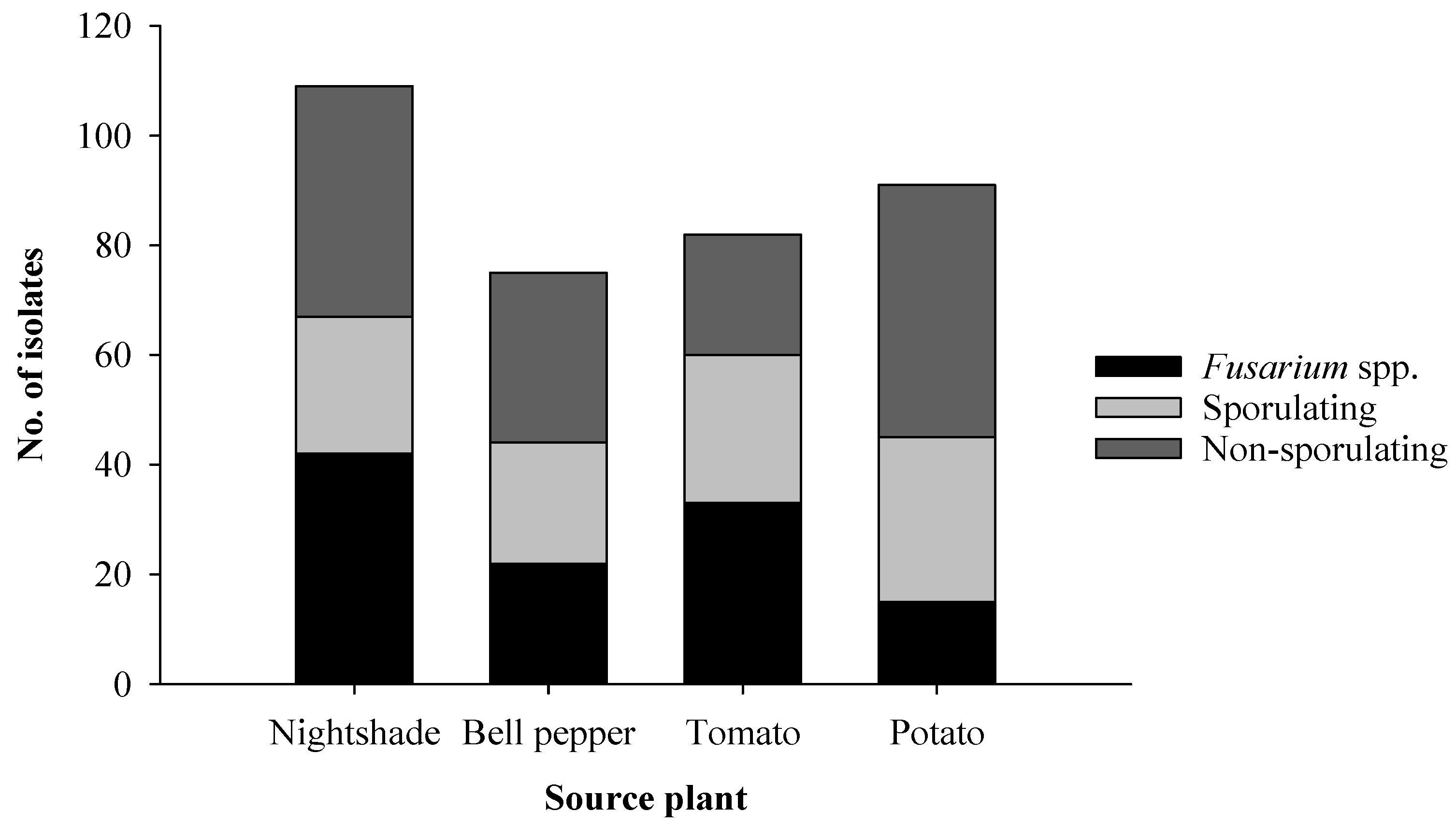
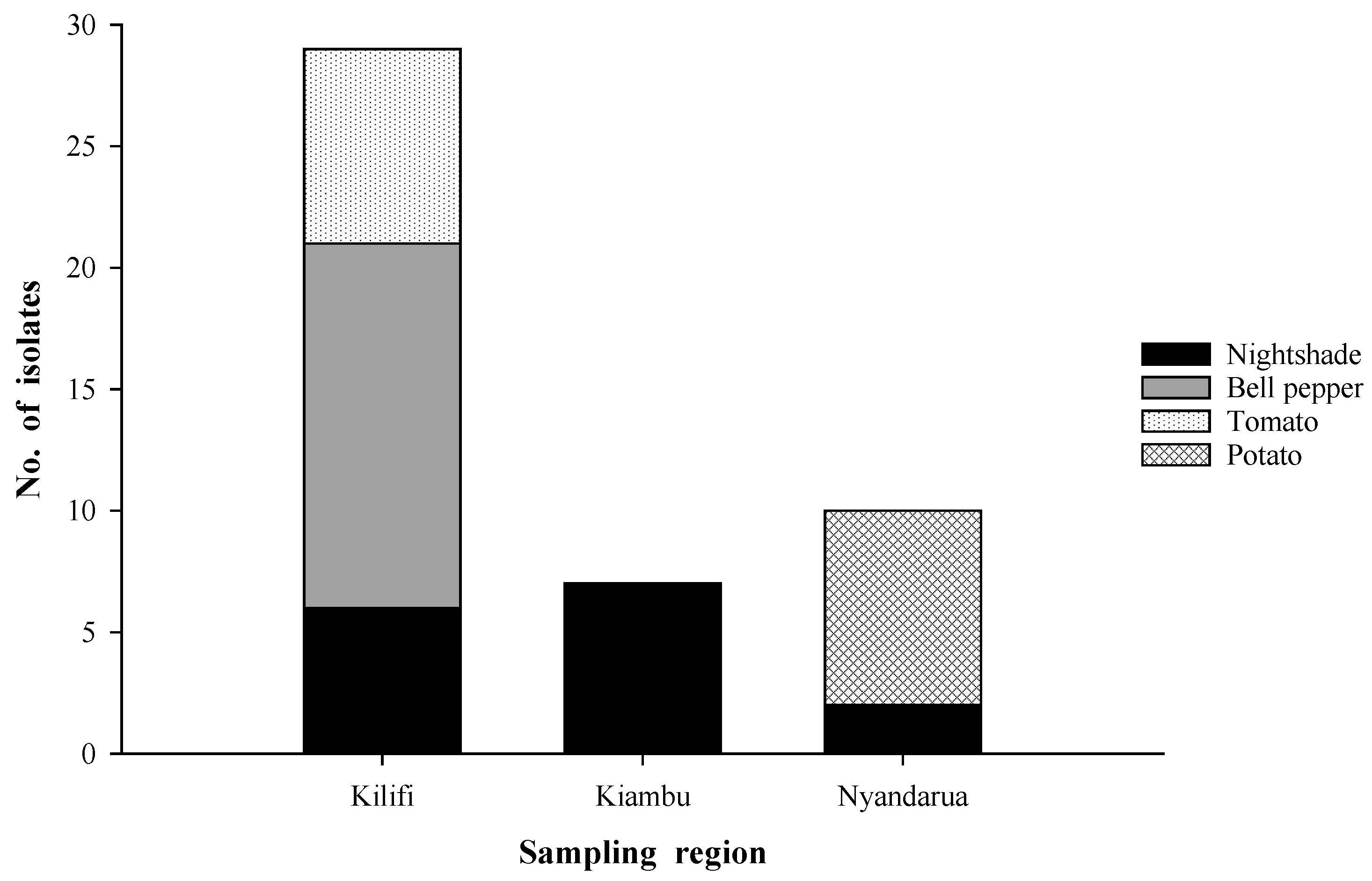
2.1. Isolation of Endophytic Fungi from Roots of Solanaceous Plants

A total of 357 isolates of fungal endophytes were obtained from roots of potato, tomato, bell pepper, and nightshade from Nyandarua, Kiambu and Kilifi regions in Kenya. The highest number of endophytes were isolated from nightshade (30.5%), followed by potato (25.5%), tomato (23%), and bell pepper (21%) (Figure 1). Among all endophytes, 112 (31.4%) were identified as Fusarium spp., 104 (29.1%) readily sporulated on PDA, while 141, accounting for 39.5%, formed no spores on this medium.

2.2. Screening of Endophytes for Anti-Oomycete Activity

The results obtained from the primary high throughput screening assay showed that 64 of the total isolates (n = 357) had potential activity against P. infestans. However, four of these proved fastidious and ceased to grow in culture while 14 were morphologically identified as Fusarium spp. and excluded from subsequent analyses. Among the remaining 46 potentially active isolates, 63% were obtained from Kilifi (Figure 2), with those isolated from bell pepper in this region accounting for 32.6% of the total number of potential antagonists. Other isolates from Kilifi showing potential activity against P. infestans were obtained from tomato (17.4%) and nightshade (13%). All antagonistic isolates from Kiambu were from nightshade, representing 15.2% of the selected potential antagonists while those from Nyandarua were isolated from potato and nightshade accounting for 17.4% and 4.3%, respectively. Generally, the number of antagonists isolated per plant species was 15 isolates each for bell pepper and nightshade and eight isolates each for tomato and potato. Interestingly, only nightshade plants were accompanied by potential antagonists from all three regions.

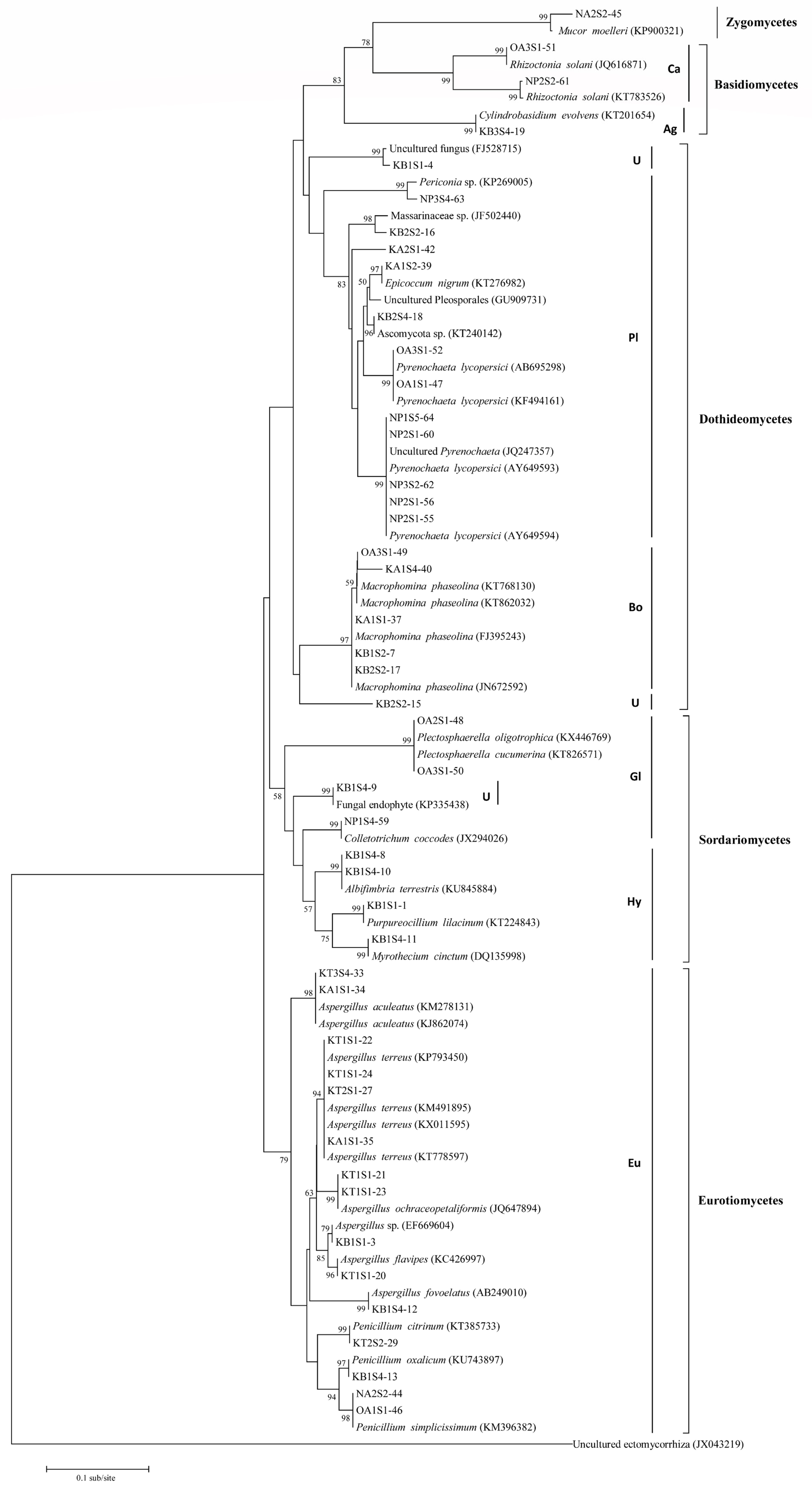
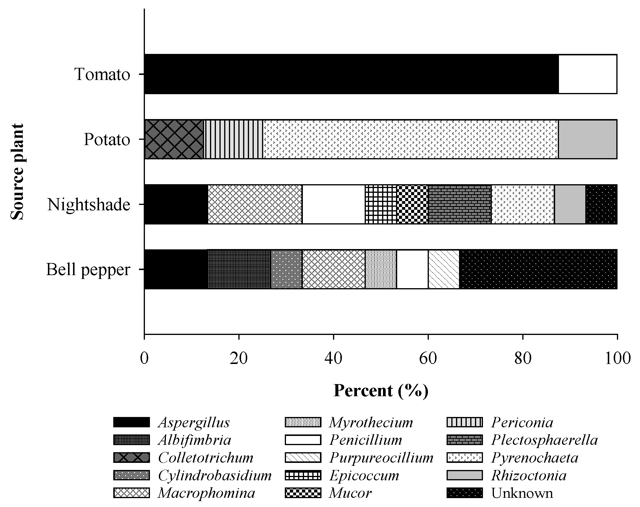
2.3. Characterization of the Fungal Endophytes

Based on their ITS sequences, 46 endophytic fungal isolates screened in the dual culture were characterized. The isolates were considered conspecific to species on the NCBI database when their ITS sequences (ITS1-5.8S-ITS4) matched those of the reference with an identity of ≥99% [33]. Based on this criterion, 37 of the 46 sequences were identified to the species level, three isolates were affiliated to higher taxa (phylum or order), while two matched unidentified fungi (Table 1).
The similarities of the remaining four endophytes (NP3S4-63, KA2S1-42, KB2S2-16, and KB2S2-15) did not meet the threshold and showed associations to higher taxa (genus, family, and order). The 37 isolates belonged to 18 species within the 13 genera including: Albifimbria, Aspergillus, Myrothecium, Cylindobasidium, Epicoccum, Macrophomina, Penicillium, Plectosphaerella, Purpureocillium, Pyrenochaeta, Rhizoctonia, Mucor, and Colletotrichum.
The 46 endophytic isolates and their closest BLAST entries form eight distinctive clades, which agrees nicely with their taxonomic identity (Figure 3). Three of the clades represent the two fungal phyla Zygomycota and Basidiomycota, which contain one and three fungal species, respectively. The Zygomycete Mucor moelleri was found to belong to the order Mucorales, while the Basidiomycetes could be placed in two orders, Cantharellales (Rhizoctonia solani) and Agaricales (Cylindrobasidium evolvens), according to Crous et al. [34]. More than 90% of the endophytic isolates were Ascomycetes distributed into three classes, namely Dothideomycetes, Sordariomycetes, and Eurotiomycetes, which fell into four clades (Figure 3). Dothideomycetes and Eurotiomycetes form the bulk of the identified Ascomycetes representing 45.2% and 35.7%, respectively. All Eurotiomycetes identified belong to the order Eurotiales, while the identified Dothiodeomycetes comprise the orders Pleosporales and Botryosphaeriales [34]. Two isolates (KB1S1-4 and KB2S2-15) cluster with the Dothideomycetes, however, they could not be placed in any of the two orders with absolute certainty. Identified Sordariomycetes form the minority of identified Ascomycetes (19%) and are grouped into two orders, Hypocreales and Glomerellales (Figure 3). Unknown isolate KB1S4-9 clustered with Sordariomycetes and was inferred to belong to the Glomerellales as it grouped with species within this order [34]. The closest BLAST match for isolate KB2S2-15 formed the eighth clade (Figure 3).
A deeper look at the distribution of fungal taxa with potential activity against P. infestans in relation to the source host plant showed that tomato accommodated two genera, namely Aspergillus (87.5%) and Penicillium (12.5%), while four genera, with Pyrenochaeta being the most abundant (62.5%), were found in potato (Figure 4).
On the other hand, nightshade and bell pepper harbored more diverse fungi giving rise to eight and seven known genera, respectively. Both host plants also harbored unknown species, with the proportion being greater in bell pepper (33.3%) than in nightshade (6.7%).

2.4. In Vitro Activity of Endophytic Fungi against Mycelial Growth of P. infestans

In confrontation assays, mycelial growth retardation of P. infestans in the presence of one of the 46 fungal endophytic isolates varied significantly (Table 2). T. harzianum along with two endophytes (KB1S2-7 and KA1S1-34) suppressed mycelial growth of the pathogen by 84.5%, 78.2%, and 76.5%, respectively. The other endophytes, however, were either only moderate (KB1S4-10 and KT1S1-21), or slight (KT2S2-29 and KB2S2-15) growth inhibitors. Macroscopic observations of the interaction zones showed that some endophytes completely overgrew the pathogen. Members of these endophytes including T. harzianum, Mucor moelleri, and Macrophomina phaseolina recorded the highest inhibition (70.4–84.5%) (Table 2). Albifimbria terrestris and Penicillium simplicissimum partially overgrew pathogen colonies giving moderate growth inhibition (51.8–56.3%). In other cases, the growth of both endophyte and pathogen stopped once their colonies came into contact. Mycelial growth inhibition in this group was dependent on the growth rate of the endophytes. Fungal endophytes within the genera Aspergillus, Albifimbria, Macrophomina, and Cylindrobasidium showed this type of interaction. Interestingly, other endophytes within the genera Aspergillus, Penicillium, Purpureocillium, and Pyrenochaeta created inhibition zones with the pathogen. These endophytes showed slight to moderate inhibition percentages (13.3–46.9%) and the inhibition areas created between endophyte and pathogen varied markedly (Table 2). Three unidentified isolates (KB1S1-4, KA2S1-42, and KB2S2-15) formed significantly (α = 0.05) larger inhibition zones measuring 18.8, 18.3, and 14.8 mm, respectively.

2.5. Potential of Endophytes Secreting Volatile Organic Compounds Active against P. infestans

Based on the results obtained from dual culture experiments, six endophytic fungal isolates (NA2S2-45, KB2S2-17, KB1S1-4, KA2S1-42, KB2S2-15, and KB2S4-8) were selected and subjected to further characterization regarding their ability to produce volatile organic compounds (VOCs) with anti-oomycete activity. All endophytes were found to produce VOCs with varying activities against mycelial growth of P. infestans (Figure 5).
M. moelleri (NA2S2-45), M. phaseolina (KB2S2-17), and an unidentified Pleosporales species (KA2S1-42) retarded the growth of the pathogen to a similar extent, recording inhibition percentages of 56.7%, 50.8%, and 48.9%, respectively. Lower suppression levels were observed in the case of Alibifimbria terrestris KB1S4-8 (18.4%) and two unidentified fungi, KB1S1-4 (26.8%) and KB2S2-15 (16.7%).

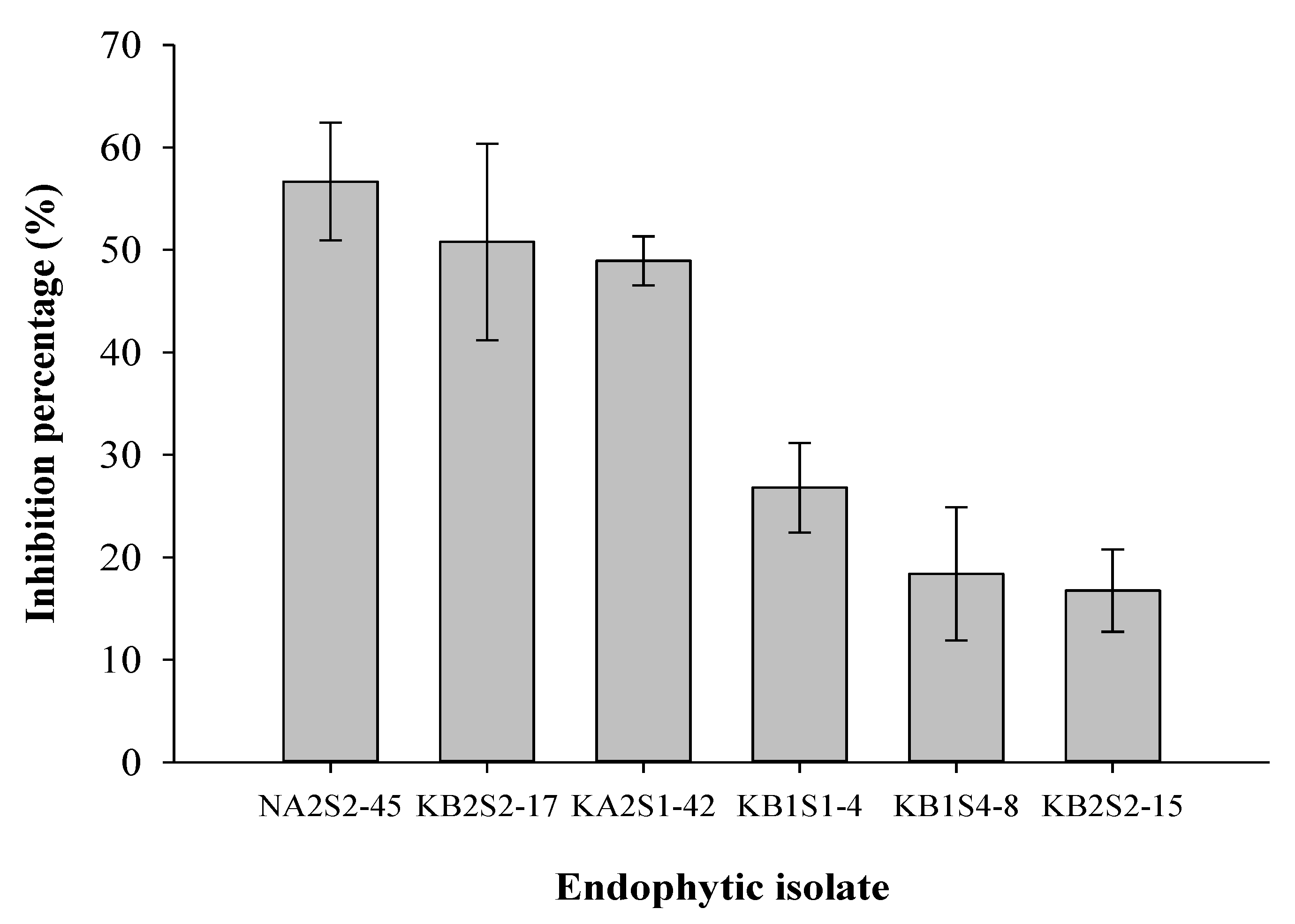
2.6. Effect of Crude Extracts from Selected Endophytes on Sporangial Germination of P. infestans

To assess the potential of crude extracts (CEs) obtained from selected fungal isolates against P. infestans, sporangia were allowed to germinate in the presence of 5% of individual CEs and germination was evaluated after 16 h. Sporangia germination in 5% acetone (solvent control) recorded ≥90% and differed only insignificantly from that of the water control. Sporangia reacted differentially to the individual CEs applied (Table 3).
While the crude extract (5%) from NA2S2-45 significantly retarded sporangia germination (30%), no suppressive effect on germ tube growth was detected. A similar dosage of the crude extract from KB1S1-4 suppressed sporangial germination by approximately 70% and diminished germ tube elongation by >80% compared to the solvent control. The amendment of P. infestans sporangia with CEs obtained from two endophytic fungi (KB2S2-15 and KA2S1-42) entirely suppressed their germination (Table 3). As a result, CEs from KB2S2-15, KA2S1-42, and KB1S1-4 showed suppressive effects against sporangia germination and germ tube development and hence were selected for further in vivo investigations.

2.7. In Vivo Activity of the Crude Extracts against P. infestans

To validate the in vitro results, the effect of CEs on leaf blight development on detached potato leaves was investigated. CEs were incorporated into sporangial suspension to give a dosage of (5%, w:v). This mixture was inoculated on the abaxial surface of detached leaflets on either side of the midrib. The results obtained from two independent experiments revealed that detached leaflets treated with either acetone or CEs in the absence of the pathogen did not show any phytotoxic damage (Figure 6b,d–f).
Under a high relative humidity, necrotic lesions covered with white fluffy hyphae and sporangia of P. infestans developed on the inoculated leaflets treated only with water (control) within 7 d (Figure 6A). Similarly, no decrease in lesion development or mycelial colonization was detected on inoculated leaflets treated with 5% acetone compared with the water control treatment (Figure 6B). In contrast, late blight symptoms and mycelial growth were completely suppressed when the systemic broad-spectrum fungicide Infinito® was applied (Figure 6C). Interestingly, the application of 5% CEs of the endophytes KB1S1-4, KA2S1-42, and KB2S2-15 yielded neither visible lesions nor hyphal growth of P. infestans on the inoculated leaflets (Figure 6D–F).

3. Discussion

The screening and identification of microorganisms with antagonistic properties against soilborne pathogens are indispensable first steps in the search for potential biocontrol agents [27,33]. In the current study, root endophytic fungi were isolated from four solanaceous plant species obtained from three regions in Kenya with diverse climatic conditions and soil properties (Table 1). This might increase the heterogeneity and number of fungal species obtained, and hence the possibility of identifying endophytes with unusual adaptation strategies and potential bioactivity against P. infestans [27]. The sampling regions were a few to several hundred kilometers apart (1000 km maximum distance), differed in elevation (2532 m maximum difference), climate, and physio-chemical soil properties (Table S1).
In addition to soil and climatic factors, host plant species also influence the composition of root microbiomes, with roots being shown to harbor more endophytic biodiversity than the rest of the plant organs [35,36]. These factors may have contributed to the realization of 357 endophytic fungal isolates from Kilifi, Nyandarua, and Kiambu from the roots of four solanaceous plant species sampled, namely S. tuberosum, L. esculentum, S. nigrum, and C. annuum. However, only a limited proportion of approximately 13% of the isolates showed potential activity against P. infestans. Interestingly, 63% of the endophytes were obtained from Kilifi (Figure 2), a non-potato growing region, implying that spatial separation could be a predominant factor limiting pathogen–antagonist interactions. Similar findings of the occurrence of unique endophytic fungal species from Kilifi were reported by Bogner et al. [37]. These authors attributed their findings to the hot and humid climate experienced in the coastal region.
The type of host plant may also have been an influential factor in the availability of antagonists. Bell pepper and nightshade each gave rise to the largest number of antagonistic endophytes (Figure 2), which was consistent with the finding of Kim et al. [12], that red pepper roots harbored endophytic fungi with potent activity against P. infestans. The endophytes from nightshade and bell pepper were significantly more diverse than those from other host plants (Figure 4). In these two plant species, unidentified endophytes were also captured, all isolated from Kilifi. The diversity of antagonists was lower in potato and least in tomato (Figure 4).
Although challenges in the use of sequence information on the NCBI database for molecular identification of fungi have been cited, it remains a useful tool in the identification of species, especially for non-sporulating endophytes [27,38]. In the current study, there was considerable agreement between the phylogenetic relationships of the endophytes and their corresponding closest BLAST matches, as demonstrated by the high bootstrap support at the terminal nodes (Table 1 and Figure 3). The exceptions were isolates KB2S2-15 and KA2S1-42, for which BLAST matches with only little similarity were found that did not cluster in the phylogeny. These endophytes were non-sporulating on different nutrient media, hampering morphological characterization. Similarly, isolates KB1S1-4, KB1S4-9, KB2S2-16, and KB2S4-18 were also non-sporulating and their identity remained concealed, as their closest BLAST matches were unknown fungal species. The isolates KB1S1-3, NP2S1-60, and NP3S4-63 could not be identified to the species level, but molecular and morphological traits established their genera as Aspergillus, Pyrenochaeta, and Periconia, respectively. On a broader perspective, most of the antagonistic endophytes characterized in this study were Ascomycetes, while the occurrence of Basidiomycetes and Zygomycetes was limited to one or a few isolates. The Ascomycota is the largest and most diverse fungal phylum, and its members are usually the dominant endophytes in plants in relation to other fungal groups [39,40,41,42].
By observing the interactions of 46 potential fungal endophytes in dual culture with P. infestans, some antagonists rapidly grew over the pathogen colony resulting in the highest inhibition percentages. Apart from M. moelleri, this group comprises mainly of members of possible potato pathogens. While the pathogenicity of these endophytes has not been yet verified, M. moelleri has been recently indicated as a potential antagonist against different tomato pathogens including Athelia rolfsii and Colletotrichum gloeosporiodes [43]. T. harzianum T16 also showed a similar interaction with P. infestans. Previous reports of the activity of Trichoderma spp. against P. infestans and other pathogens have demonstrated their capacity for complete growth over pathogen colonies in dual culture, mycoparasitism, and antibiosis through the production of active compounds such as 6-pentyl-α-pyrone, viridiofungin A, harzianolide, and harzianic acid [44,45,46,47]. In exclusion of putative potato pathogens, Aspergillus spp., Penicillium spp., Albifimbria terrestris, and Cylindrobasidium evolvens were among the antagonistic fungal endophytes identified. Members of Aspergillus and Penicillium have been shown to have endophytic lifestyles in plants as well as exhibiting bioactivity against plant pathogens (e.g., Botrytis cinerea, Trichothecium roseum, Sclerotinia sclerotiorum, Fusarium oxysporum, and Rhizoctonia solani) with A. niger, A. terreus, and P. citrinum being some of the well-studied antagonists [33,39,42,48,49]. In addition, a protein derived from Aspergillus giganteus was shown to have activity against P. infestans in vitro [50]. The Basidiomycete C. evolvens, as a saprophyte capable of infecting wounded stem and root tissue, is associated with decay in woody plants [51,52]. In this study, C. evolvens was isolated as an endophyte from the roots of C. annuum and showed a moderate antagonistic activity against P. infestans in dual culture (Table 2). Likewise, A. terrestris was isolated from the roots of C. annuum and is morphologically similar to A. verrucaria, whose basionym was Myrothecium verrucaria, a known plant pathogen that has been formulated into bioherbicides or nematicides [53,54,55]. Interestingly, some endophytes exerted their antagonism by antibiosis as indicated by the inhibition zones. In a comparable study, CEs of Aureobasidium pullulans isolated from Espeletia spp., a native Andean plant, showed in vitro activity against P. infestans [27].
In addition, endophytic fungi are also capable of producing VOCs. VOCs from M. moelleri, M. phaseolina, and an unknown endophyte KA2S1-42 showed a higher mycelial growth inhibition of P. infestans than those released by KB1S1-4, A. terrestris, and KB2S2-15 (Figure 5). Previous reports have shown that Mucor spp. are able to produce ethanol while M. phaseolina has been associated with the production of several fatty acid methyl esters with an inhibitory activity against Sclerotinia sclerotiorum [42,56]. On the other hand, A. verrucaria excreted bioactive compounds, including antibacterial cyclopeptides that showed herbicidal activities as well [57,58]. Moreover, the A. verrucaria isolate SYE-1 exhibited a wide antifungal activity against B. cinerea, Lasiodiplodia theobromae, and Elsinoë ampelina on grapevine [59]. Myrothecium inundatum, a close relative of A. terrestris, has been implicated in the production of a variety of hydrocarbon VOCs, or their derivatives, potent against Pythium ultimum and S. sclerotiorum [60].
VOCs have been indicated to have a greater coverage of soil and organic substrata than diffusible organic compounds (DOCs), a characteristic attributed to their gaseous state [61]. In the current study, three unknown antagonists KA2S1-42, KA1S1-4, and KB2S2-15 showed the capability of producing VOCs as well as DOCs that affect P. infestans. In addition, M. moelleri showed a high inhibition of P. infestans mycelium both in dual culture and through the production of VOCs. These findings led to the selection of the antagonistic endophytes KA1S1-4, KB2S2-15, KA2S1-42, and M. moelleri for further testing while putative pathogens were left out of subsequent studies.
Consistent with dual culture findings, CEs from the antagonistic endophytes KB1S1-4, KB2S2-15, and KA2S1-42, showed a suppressive activity against sporangia germination and the germ tube growth of P. infestans. While CEs from KB1S1-4 at a concentration of 5% exhibited considerable suppressive effects against sporangial germination and germ tube elongation of the pathogen, CEs from KB2S2-15 and KA2S1-42 entirely blocked sporangia germination (Table 3). In a similar set up, Linkies et al. [62] found that upon exposure to ethyl-acetate extracts from Chaetomium cochliodes and C. elatum at a dosage of 30%, sporangia germination of P. infestans was considerably inhibited. However, this dosage is much higher (six-fold) than that used in our study.
On the other hand, although CEs from M. moelleri (NA2S2-45) showed a relatively weak activity against sporangia germination, it did not inhibit germ tube development, suggesting that the anti-Phytophthora metabolites produced by this fungus are rather confined to the gaseous phase, which could not be extracted from the culture filtrates in concentrations sufficient to affect the germination of P. infestans sporangia. Several reports agree with the ability of endophytic, rhizospheric, or phyllospheric fungi in producing diffusible compounds with anti-oomycete activity [12,27,45,63,64]. Tellenbach et al. [65] reported that a strain of Phialocephala europaea was able to produce metabolites containing sclerin and sclerotinin with activity against Phytophthora citricola. Our earlier studies showed that strain T23 of T. harzianum (recently re-identified as T. asperellum) secreted viridiofungin A with a strong inhibitory effect against sporangia germination of P. infestans [45]. In the current study, although the biologically active metabolites produced by KB1S1-4, KB2S2-15, and KA2S1-42 have not been identified, it was evident that these endophytes produce potent compounds with a strong activity against P. infestans.
In the detached leaflet assay, the application of CEs from the endophytes KB1S1-4, KB2S2-15, and KA2S1-42 entirely protected inoculated leaflets and suppressed the hyphal growth of P. infestans (Figure 6). Likewise, Bae et al. [66] found that CEs from Trichoderma atroviride showed inhibitory activities against Phytophthora sojae, P. capsici, and P. melonis and induced defense reactions in the detached leaves of pepper and tomato plants. Furthermore, Kim et al. [12] reported a substantial in vivo activity of Fusarium oxysporum strain EF119 against tomato late blight. Similar results were also reported by Chandrakala et al. [64], where culture filtrates from T. virens and T. viride proved to be effective against sporangia germination of P. infestans and impeded the establishment of late blight on potato.
Overall, we report preliminary evidence for targeting the late blight pathogen by particular fungal endophytes both in vitro and in vivo. Based on their promising in vitro performance against the late blight pathogen, three endophytes (KA1S1-4, KB2S2-15, and KA2S1-42) were selected from a total of 357 isolates. These endophytic isolates proved to be very active in protecting potato leaflets from P. infestans. This biological activity is associated with DOCs and VOCs with anti-oomycete properties. However, the purification and elucidation of the chemical structures of these bioactive molecules have not been accomplished in this study. Therefore, it is still a rich field for future investigations, particularly in terms of clarifying modes of action of the metabolites involved to determine whether they target single or multiple pathways in P. infestans. This will shed light on the possibility of combining them to improve their efficacy and hence both the reliability and durability of the biocontrol.

4. Materials and Methods

4.1. Sampling Regions and Collection of Plant Materials

Root samples from four solanaceous plant species including potato (Solanum tuberosum L.), tomato (Lycopersicon esculentum L. Mill.), bell pepper (Capsicum annuum L.), and African nightshade (Solanum nigrum L.), were collected from three sampling regions in Kenya (Nyandarua, Kilifi, and Kiambu) with variable soil types. From each region, root systems of twelve apparently healthy plants were obtained along with representative soil samples. Immediately after collection, intact roots were washed thoroughly under running tap water to remove soil and adhering debris, air dried, and stored at 4 °C until further processing. In order to determine their physio-chemical properties, soil samples were analyzed at the Soil Science laboratories of Jomo Kenyatta University of Agriculture and Technology (Juja, Kenya) (Table S1). pH and electrical conductivity (EC) were determined in a soil: water mixture (2:5, w:v), while total soil organic carbon was measured using the Walkley–Black rapid titration method [67].

4.2. Isolation of Fungal Root Endophytes and P. infestans

Root segments of 3–4 cm from primary and secondary roots of each plant were surface sterilized by immersion in 70% alcohol for 1 min and immediately transferred into 2.5% sodium hypochlorite (Carl Roth, Karlsruhe, Germany) for 5 min. Root segments were rinsed three times with sterile distilled water for 5 min and blotted between sterile paper towels to remove excess moisture. To ascertain the success of surface sterilization, sterilized root segments were briefly imprinted on PDA (Figure S1). Subsequently, the ends of the root segments were trimmed off and further divided into three fragments of approx. 1 cm length, which were placed on PDA amended with 0.05 g/L chloramphenicol (Merck KGaA, Darmstadt, Germany). Finally, plates were sealed with Parafilm and incubated at 22 ± 1 °C in the dark. Only endophytic fungi emerging from successfully surface sterilized roots (Figure S2) were subcultured onto fresh PDA. Isolates were purified through either single-spore isolation or hyphal tip transfer for sporulating and non-sporulating colonies, respectively. Pathogens (e.g., Fusarium spp.) were excluded from further tests.
P. infestans was isolated from single lesions of potato leaves obtained from a late blight susceptible variety (Duke of York) growing under field conditions in Hohenheim, Stuttgart, Germany. To induce fresh sporulation, blighted leaves were placed in a humid chamber at 20 °C for 48 h. Colonized leaf segments were placed between two surface sterilized tuber slices (5 mm thickness) and incubated for 7 d at 20 °C in the dark (Figure S3). Mycelia emerging from the upper side of a slice were transferred to unclarified V8-based agar medium containing 200 mL V8 juice, 2 g CaCO3, 0.05 g β-sitosterol, 0.05 g ampicillin, 0.05 g vancomycin, 0.01 g pentachloronitrobenzene (PCNB), and 15 g agar in 800 mL deionized water. A pure culture of the pathogen generated from the tip of a single hypha was maintained on corn meal agar (CMA, 17 g/L; Sigma-Aldrich, Munich, Germany) and reactivated after every 3–4 transfers on CMA by inoculation on sterile potato leaflets.

4.3. Primary Screening of Endophytes for Anti-Oomycete Activity against P. infestans

A high throughput confrontation assay was established to screen 357 potential antagonistic endophytes against P. infestans. Owing to the slow growth of P. infestans, 5 mm Ø agar plugs from the border of an actively growing colony were inoculated on the center of 20% V8 agar plates and incubated at 20 °C for 72 h in darkness. Subsequently, agar plugs (5 mm Ø) from four different endophytic fungi were placed equidistant, 5 mm from the edge of the Petri plates pre-inoculated with the pathogen (Figure S4). Control treatments were set up in a similar way in the absence of endophytes. Isolates observed to retard the growth of P. infestans compared to the control were selected for further testing and purified to generate axenic cultures.

4.4. DNA Extraction, PCR Conditions, and Sequencing

Genomic DNA was extracted from mycelia of root endophytic fungi selected from the screening experiment using the method described by Liu et al. [68]. PCR was conducted in a 40 μL reaction mixture using ITS1 (5′-TCCGTAGGTGAACCTGCGG-3′) and ITS4 (5′-TCCTCCGCTTATTGATATGC-3′) primers [69] to amplify the internal transcribed spacer regions (ITS1-5.8S-ITS2). A single PCR reaction was comprised of 8 µL of Phusion® HF buffer (5×), 0.8 µL of dNTPs (10 mM), 1 µL of each of the forward and reverse primers (10 µM), 0.4 µL of Phusion® polymerase (2 U/µL), and 27.8 µL of ultra-pure water. The PCR program started with an initial denaturation step at 98 °C for 30 s, followed by 35 cycles at 98 °C for 10 s, 54 °C for 20 s, and 72 °C for 35 s and a final extension step at 72 °C for 10 min. Success of amplification was ascertained on 1% agarose gels visualized with 0.05% ethidium bromide with the aid of a gel documentation system (Quantum 1100 PEQLAB, VWR, Darmstadt, Germany). Amplified PCR products were purified using Innu PREP PCR-pure kit (Analytik Jena AG, Jena, Germany) and their concentration adjusted to meet requirements for Sanger sequencing by Source Bioscience Company (Berlin, Germany). Sequences from single reads with the forward primer (ITS1) were trimmed and edited with GENtle v 2.0 and compared to those deposited at the NCBI database. Endophytic fungi whose sequences showed a similarity of >99% to database entries were considered identical to the reference fungi [33], and their taxonomic classification carried out using the MYCOBANK Database [34]. Evolutionary history was deduced using the Neighbour-Joining method as described by Saitou and Nei [70] with a bootstrap test of 1000 replicates to determine the percentage when replicate trees of related taxa clustered together [71]. The maximum composite likelihood method [72] was used to calculate the evolutionary distances, used to infer the phylogenetic tree. Finally, sequence data were submitted to GenBank.

4.5. Establishment the Anti-Oomycete Activity of the Endophytes

To validate the inhibitory effect of screened fungal endophytes against P. infestans, a dual culture assay was performed. The assay was similar to that described under 4.3 except that the pathogen and each unique endophytic isolate were inoculated at 7 cm distance. P. infestans was also inoculated on 20% V8 agar plates 72 h prior the antagonistic isolates. Three replicates were prepared for each isolate and plates inoculated only with the pathogen served as control. Trichoderma harzianum (T16) shown to have antagonistic properties against several plant pathogens [45,46] was used as a positive control, while pathogen colonies grown in the absence of the endophytes served as negative control. The experiment was laid out in a randomized complete block design. The initial (72 h post inoculation; hpi) and the final (12 d post inoculation; dpi) colony diameter of the pathogen were measured. The percentage of mycelial growth inhibition was calculated using the formula ascribed by Edgington et al. [73]. In addition, the inhibition zones created between the pathogen and the tested endophytic fungal colonies were recorded.

4.6. Impact of Volatile Organic Compounds Extracted from the Endophytes on Mycelial Growth of P. infestans

The ability of selected endophytic fungi to produce volatile organic compounds (VOCs) was assessed following the modified procedure of El-Hasan et al. [74]. Briefly, agar plugs (5 mm Ø) bearing P. infestans mycelium were placed on the center of V8 agar plates and incubated for 72 h. Subsequently, the lid was replaced with an upside-down agar plate inoculated with an agar plug colonized by an endophyte. Both Petri dishes were separated with a sterile cellophane sheet and held together with Parafilm. Positive and negative controls consisted of T. harzianum and sterile PDA plugs, respectively. Mycelial growth inhibition (in %) was calculated as described above.

4.7. Suppressive Activity of Crude Extracts of Root Endophytic Fungi against Sporangial Germination of P. infestans

Putative endophytic fungi that formed inhibition zones in dual culture and/or produced active VOCs were tested for their ability to produce diffusible organic compounds with anti-oomycete activity. To this end, 1 L of 20% V8 broth was inoculated with ten agar plugs (5 mm Ø) of an actively growing endophyte culture. The cultures were incubated on an orbital shaker at 125 rpm and 20 °C in the dark for 12 d. Control treatments, where the endophytes were absent, were set up. Crude extracts (CEs) were prepared from culture filtrates according to El-Hasan et al. [45]. The resulting CE residues were re-dissolved in 2 mL acetone.
Sporangia of P. infestans were produced on detached potato leaflets. For this purpose, an agar plug (3 × 3 mm) was placed on the abaxial side of a detached leaflet surface sterilized with 2.5% sodium hypochlorite. A drop of sterile deionized water was introduced on the interface and the inoculated leaflets were placed in a humid chamber made by inverting the Petri dish containing water agar over lids containing sterile moist filter paper. Cultures were incubated at 20/18 °C (light/dark) for 7 d in a growth chamber with a 16 h light cycle. Sporangia were harvested by briefly vortexing infected leaflets in a sterile centrifuge tube containing V8 liquid medium. Mycelia and leaf fragments were trapped using double layers of sterile muslin cloth and the resulting sporangial suspension (5 × 10⁴ sporangia/mL) was used in subsequent experiments.
To determine the effect of CEs on sporangia germination, 25 µL of each extract were combined with an equal volume of water in a sterile 1.5 mL microcentrifuge tube. The tubes were aseptically left open for three hours to allow solvent evaporation. Subsequently, 475 µL of the sporangia suspension were added to the CE solution. Cultures were incubated at 20 °C in the dark to allow sporangia germination, the experiment was terminated after 16 h by the addition of 100 µL lactophenol blue. Acetone was used as solvent control. The percentage of germinated sporangia and the length of germ tubes were determined using a light microscope (Axioskop 2, Carl Zeiss Microscopy GmbH, Göttingen, Germany). Images were taken with an AxioCam MRm digital camera (Carl Zeiss) and measurements determined with the corresponding AxioVision software (SE64 Release 4.8.3 SP1; Carl Zeiss).

4.8. Activity of the Crude Extracts against P. infestans on Detached Potato Leaflets

For the in vivo bioassay, leaflets were harvested from the fourth to sixth fully expanded leaves obtained from potato plants (var. Duke of York) grown under greenhouse conditions. CEs that showed activity against germination of P. infestans sporangia were tested for their activity in vivo against the pathogen on detached leaflets. Acetone was evaporated from the crude extracts as described above, and each extract reconstituted to give a concentration of 5% in a sporangial suspension. Two droplets of 50 µL each were inoculated on the abaxial surface of detached leaflets on either side of the midrib. Control treatments were set up in a similar way and comprised water, 5% acetone, and a conventional fungicide (Infinito®; Bayer CropScience, Langenfeld, Germany). Leaflets were placed individually in a humid chamber and incubated for 7 d at 20/18 °C (light/dark) with a 16 h photoperiod. All treatments were set up in four replicates. The assay was conducted twice.

4.9. Data Analysis

If not mentioned elsewhere, experiments were repeated at least twice in triplicate in a completely randomized design. All statistical analyses were performed using SAS software (SAS Institute Inc.). The MIXED and GLIMMIX procedures were applied to determine whether there were treatment effects in the experiments conducted within this study. Data were assessed and transformed when necessary to ensure they met model assumptions. Pairwise comparisons among treatments were carried out using Tukey test (α = 0.05).

Supplementary Materials

The following supporting information can be downloaded at: https://www.mdpi.com/article/10.3390/plants11121605/s1, Table S1. Characteristics of sampling regions; Figure S1. Isolation of fungal root endophytes. (a) Endophytic fungi emerging from cultured roots, (b) Clean short press plate, (c) Contaminated short press plate; Figure S2. Isolation of fungal root endophytes from solanaceous plants, (A) Endophytic fungi arising from cultured roots, (B) Pure fungal colonies; Figure S3. Isolation of P. infestans (a) P. infestans mycelia growing on the surface of an inoculated potato tuber slice (b) Pure colony of P. infestans on 20% V8 agar (c) Mycelia of P. infestans on potato leaflets inoculated with the pathogen; Figure S4. Dual antagonistic assay of fungal endophytes and P. infestans. (a) Co-culture of P. infestans (center colony) with four root endophytes to screen for activity against the pathogen’s mycelia (b) Setup of dual culture assay on the introduction of an endophyte 72 h after P. infestans (left colony) inoculation (c) Schematic representation of the measurement (R2) used in calculating percentage of inhibition.

Author Contributions

Conceptualization, A.E.-H., G.N. and R.T.V.; methodology, A.E.-H. and G.N.; validation, G.N. and A.E.-H.; formal analysis, A.E.-H., G.N. and T.I.L.; investigation, G.N.; resources, A.E.-H. and G.N.; data curation, G.N. and A.E.-H.; writing—original draft preparation, A.E.-H. and G.N.; writing—review and editing, A.E.-H., T.I.L. and R.T.V.; visualization, G.N. and A.E.-H.; supervision, A.E.-H. and R.T.V.; project administration, R.T.V.; funding acquisition, G.N. and R.T.V. All authors have read and agreed to the published version of the manuscript.

Funding

This research was funded by the German Academic Exchange Service (DAAD), grant number A/12/97759, and Food Security Center (FSC), University of Hohenheim, no grant number.

Institutional Review Board Statement

Not applicable.

Informed Consent Statement

Not applicable.

Data Availability Statement

The data presented in this study are all contained within the figures and tables.

Acknowledgments

The kind assistance of Heike Popovitsch, Bianka Maiwald and Sybille Berger of the Institute of Phytomedicine, University of Hohenheim is greatly acknowledged.

Conflicts of Interest

The authors declare no conflict of interest.

References

  1. Haverkort, A.J.; Struik, P.C.; Visser, R.G.F.; Jacobsen, E. Applied biotechnology to combat late blight in potato caused by Phytophthora Infestans. Potato Res. 2009, 52, 249–264. [Google Scholar] [CrossRef]
  2. Kromann, P.; Miethbauer, T.; Ortiz, O.; Forbes, G.A. Review of potato biotic constraints and experiences with integrated pest management interventions. In Integrated Pest Management; Pimental, D., Peshin, R., Eds.; Springer: Dordrecht, The Netherlands, 2014; pp. 245–268. [Google Scholar]
  3. Ivanov, A.A.; Ukladov, E.O.; Golubeva, T.S. Phytophthora infestans: An Overview of Methods and Attempts to Combat Late Blight. J. Fungi 2021, 7, 1071. [Google Scholar] [CrossRef] [PubMed]
  4. Fry, W.E.; Birch, P.R.J.; Judelson, H.S.; Grünwald, N.J.; Danies, G.; Everts, K.L.; Gevens, A.J.; Gugino, B.K.; Johnson, D.A.; Johnson, S.B.; et al. Five reasons to consider Phytophthora infestans a reemerging pathogen. Phytopathology 2015, 105, 966–981. [Google Scholar] [CrossRef] [PubMed] [Green Version]
  5. Olanya, O.M.; Adipala, E.; Hakiza, J.J.; Kedera, J.C.; Ojiambo, P.; Mukalazi, J.M.; Forbes, G.; Nelson, R. Epidemiology and population dynamics of Phytophthora infestans in sub Saharan Africa. Afr. Crop Sci. J. 2001, 9, 185–194. [Google Scholar] [CrossRef]
  6. Chepsergon, J.; Motaung, T.E.; Bellieny-Rabelo, D.; Moleleki, L.N. Organize, Don’t Agonize: Strategic Success of Phytophthora Species. Microorganisms 2020, 8, 917. [Google Scholar] [CrossRef]
  7. Leach, S.S. Effects of copper and copper fungicide soil residues on Phytophthora infestans. Am. Potato J. 1966, 43, 431–438. [Google Scholar] [CrossRef]
  8. Bruck, R.I.; Fry, W.E.; Apple, A.E. Effect of metalaxyl, an acylalanine fungicide, on developmental stages of Phytophthora infestans. Phytopathology 1980, 70, 597–601. [Google Scholar] [CrossRef]
  9. Wale, S.; Platt, H.W.; Cattlin, N.D. Introduction. In Diseases, Pests and Disorders of Potatoes: A Colour Handbook; Wale, S.H., Platt, W., Cattlin, N.D., Eds.; Manson Publishing: London, UK, 2008; pp. 6–8. [Google Scholar]
  10. Cohen, Y.; Rubin, A.E.; Galperin, M. Effective control of two genotypes of Phytophthora infestans in the field by three oxathiapiprolin fungicidal mixtures. PLoS ONE 2021, 16, e0258280. [Google Scholar] [CrossRef]
  11. Rani, L.; Thapa, K.; Kanojia, N.; Sharma, N.; Singh, S.; Grewal, A.; Srivastav, A.L.; Kaushal, J. An extensive review on the consequences of chemical pesticides on human health and environment. J. Clean. Prod. 2021, 283, 124657. [Google Scholar] [CrossRef]
  12. Kim, H.-Y.; Choi, G.J.; Lee, H.B.; Lee, S.-W.; Lim, H.K.; Jang, K.S.; Son, S.W.; Lee, S.O.; Cho, K.Y.; Sung, N.D.; et al. Some fungal endophytes from vegetable crops and their anti-oomycete activities against tomato late blight. Lett. Appl. Microbiol. 2007, 44, 332–337. [Google Scholar] [CrossRef]
  13. Portz, D.; Koch, E.; Slusarenko, A.J. Effects of garlic (Allium sativum) juice containing allicin on Phytophthora infestans and downy mildew of cucumber caused by Pseudoperonospora cubensis. Eur. J. Plant Pathol. 2008, 122, 197–206. [Google Scholar] [CrossRef]
  14. Andrade-Linares, D.R.; Grosch, R.; Restrepo, S.; Krumbein, A.; Franken, P. Effects of dark septate endophytes on tomato plant performance. Mycorrhiza 2011, 21, 413–422. [Google Scholar] [CrossRef] [PubMed]
  15. Hashemi, M.; Tabet, D.; Sandroni, M.; Benavent-Celma, C.; Seematti, J.; Andersen, C.; Grenville-Briggs, L. The hunt for sustainable biocontrol of oomycete plant pathogens, a case study of Phytophthora infestans. Fungal Biol. Rev. 2022, 40, 53–69. [Google Scholar] [CrossRef]
  16. Miller, S.L. Functional diversity in fungi. Can. J. Bot. 1995, 73, 50–57. [Google Scholar] [CrossRef]
  17. Berg, G.; Krechel, A.; Lottmann, J.; Faltin, F.; Ulrich, A.; Hallmann, J.; Grosch, R. Endophytes: A new source for multi-target Biological Control Agents? IOBC/WPRS Bull. 2004, 27, 161–165. [Google Scholar]
  18. van Loon, L.C.; Bakker, P.A.H.M.; Pieterse, C.M.J. Systemic resistance induced by rhizospere bacteria. Annu. Rev. Phytopathol. 1998, 36, 453–483. [Google Scholar] [CrossRef] [Green Version]
  19. Kloepper, J.W.; Ryu, C.-M. Bacterial endophytes as elicitors of induced systemic resistance. In Microbial Root Endophytes, SOIL Biology; Schulz, B., Boyle, C., Sieber, T.N., Eds.; Springer Science + Business Media Verlag: Berlin, Germany, 2006; Volume 9, pp. 33–52. [Google Scholar]
  20. Feng, Y.; Shen, D.; Song, W. Rice endophyte Pantoea agglomerans YS19 promotes host plant growth and affects allocations of host photosynthates. J. Appl. Microbiol. 2006, 100, 938–945. [Google Scholar] [CrossRef]
  21. Mizubuti, E.S.G.; Júnior, L.V.; Gregory, F.A. Management of late blight with alternative products. Pest. Technol. 2007, 1, 106–116. [Google Scholar]
  22. De Vrieze, M.; Germanier, F.; Vuille, N.; Weisskopf, L. Combining Different Potato-Associated Pseudomonas Strains for Improved Biocontrol of Phytophthora infestans. Front. Microbiol. 2018, 9, 2573. [Google Scholar] [CrossRef]
  23. Ng, K.K.; Webster, J.M. Oomycete activity of Xenorhabdus bovienii. Can. J. Plant Pathol. 1997, 2, 125–132. [Google Scholar] [CrossRef]
  24. Daayf, F.; Adam, L.; Fernando, W.G.D. Comparative screening of bacteria for biological control of potato late blight (strain US-8), using in-vitro, detached-leaves, and whole-plant testing systems. Can. J. Plant Pathol. 2003, 25, 276–284. [Google Scholar] [CrossRef]
  25. Stephan, D.; Schmitt, A.; Carvalho, S.M.; Seddon, B.; Koch, E. Evaluation of biocontrol preparations and plant extracts for the control of Phytophthora infestans on potato leaves. Eur. J. Plant Pathol. 2005, 112, 235–246. [Google Scholar] [CrossRef]
  26. Loliam, B.; Morinaga, T.; Chaiyanan, S. Biocontrol of Phytophthora infestans, fungal pathogen of seedling damping off disease in economic plant nursery. Psyche 2012, 2012, 324317. [Google Scholar] [CrossRef]
  27. Miles, L.A.; Lopera, C.A.; González, S.; Cepero de García, M.C.; Franco, A.E.; Restrepo, S. Exploring the biocontrol potential of fungal endophytes from an Andean Colombian Paramo ecosystem. BioControl 2012, 57, 697–710. [Google Scholar] [CrossRef]
  28. Gupta, J. Efficacy of biocontrol agents against Phytophthora infestans on potato. Int. J. Eng. Sci. 2016, 6, 2249–2252. [Google Scholar]
  29. Al-Mughrabi, K.I. Biological control of Phytophthora infestans of potatoes using Trichoderma atroviride. Pest. Technol. 2008, 2, 104–108. [Google Scholar]
  30. Andrade-Linares, D.R.; Grosch, R.; Franken, P.; Rexer, K.-H.; Kost, G.; Restrepo, S.; de Garcia, M.C.C.; Maximova, E. Colonization of roots of cultivated Solanum lycopersicum by dark septate and other ascomycetous endophytes. Mycologia 2011, 103, 710–721. [Google Scholar] [CrossRef]
  31. Kannan, K.P.; Muthumary, J. Comparative analysis of endophytic mycobiota in different tissues of medicinal plants. Afr. J. Microbiol. Res. 2012, 6, 4219–4225. [Google Scholar]
  32. de Vries, S.; von Dahlen, J.K.; Schnake, A.; Ginschel, S.; Schulz, B.; Rose, L.E. Broad-spectrum inhibition of Phytophthora infestans by root endophytes. FEMS Microbiol. Ecol. 2018, 94, fiy037. [Google Scholar] [CrossRef] [Green Version]
  33. Kusari, P.; Kusari, S.; Spiteller, M.; Kayser, O. Endophytic fungi harbored in Cannabis sativa L.: Diversity and potential as biocontrol agents against host plant-specific phytopathogens. Fungal Divers. 2012, 60, 137–151. [Google Scholar] [CrossRef]
  34. Crous, P.W.; Gams, W.; Stalpers, J.A.; Robert, V.; Stegehuis, G. MycoBank: An online initiative to launch mycology into the 21st century. Stud. Mycol. 2004, 50, 19–22. [Google Scholar]
  35. Lv, Y.; Zhang, F.; Chen, J.; Cui, J.; Xing, Y.; Li, X.; Guo, S. Diversity and antimicrobial activity of endophytic fungi associated with the alpine plant Saussurea involucrata. Biol. Pharm. Bull. 2010, 33, 1300–1306. [Google Scholar] [CrossRef] [PubMed] [Green Version]
  36. Bokati, D.; Herrera, J.; Poudel, R. Soil influences colonization of root-associated fungal endophyte communities of maize, wheat and their progenitors. J. Mycol. 2016, 2016, 8062073. [Google Scholar] [CrossRef]
  37. Bogner, C.W.; Kariuki, G.M.; Elashry, A.; Sichtermann, G.; Buch, A.; Mishra, B.; Thines, M.; Grundler, F.M.W.; Schouten, A. Fungal root endophytes of tomato from Kenya and their nematode biocontrol potential. Mycol. Prog. 2016, 15, 30. [Google Scholar] [CrossRef]
  38. Arnold, A.E.; Lutzoni, F. Diversity and host range of foliar fungal endophytes: Are tropical leaves biodiversity hotspots? Ecology 2007, 88, 541–549. [Google Scholar] [CrossRef]
  39. Vitorino, L.C.; Silva, F.G.; Soares, M.A.; Souchie, E.L.; Lima, W.C. The isolation and characterization of endophytic microorganisms from Hyptis marrubioides Epling roots. Afr. J. Biotechnol. 2012, 11, 12766–12772. [Google Scholar]
  40. Karst, J.; Piculell, B.; Brigham, C.; Booth, M.; Hoeksema, J.D. Fungal communities in soils along a vegetative ecotone. Mycologia 2013, 105, 61–70. [Google Scholar] [CrossRef]
  41. Lau, M.K.; Arnold, A.E.; Johnson, N.C. Factors influencing communities of foliar fungal endophytes in riparian woody plants. Fungal Ecol. 2013, 6, 365–378. [Google Scholar] [CrossRef]
  42. Chowdhary, K.; Kaushik, N. Fungal endophyte diversity and bioactivity in the Indian medicinal plant Ocimum sanctum. PLoS ONE 2015, 10, e0141444. [Google Scholar] [CrossRef]
  43. Nartey, L.; Pu, Q.; Zhu, W.; Zhang, S.; Li, J.; Yao, Y.; Hu, X. Antagonistic and plant growth promotion effects of Mucor moelleri, a potential biocontrol agent. Microbiol. Res. 2022, 255, 126922. [Google Scholar] [CrossRef]
  44. El-Hasan, A.; Walker, F.; Schöne, J.; Buchenauer, H. Antagonistic effect of 6-pentyl-alpha-pyrone produced by Trichoderma harzianum toward Fusarium moniliforme. J. Plant Dis. Prot. 2007, 114, 62–68. [Google Scholar] [CrossRef]
  45. El-Hasan, A.; Walker, F.; Schöne, J.; Buchenauer, H. Detection of viridiofungin A and other antifungal metabolites excreted by Trichoderma harzianum active against different plant pathogens. Eur. J. Plant Pathol. 2009, 124, 457–470. [Google Scholar] [CrossRef]
  46. El-Hasan, A.; Walker, F.; Klaiber, I.; Schöne, J.; Pfannstiel, J.; Voegele, R.T. New Approaches to Manage Asian Soybean Rust (Phakopsora pachyrhizi) Using Trichoderma spp. or Their Antifungal Secondary Metabolites. Metabolites 2022, 12, 507. [Google Scholar] [CrossRef]
  47. Matroudi, S.; Zamani, M.R.; Motallebi, M. Antagonistic effects of three species of Trichoderma sp. on Sclerotinia sclerotiorum, the causal agent of canola stem rot. Egypt. J. Biol. 2009, 11, 37–44. [Google Scholar]
  48. Tripathi, A.; Sharma, N.; Tripathi, N. Biological control of plant diseases: An overview and the Trichoderma system as biocontrol agents. In Management of Fungal Plant Pathogens; Arya, A., Perelló, A.E., Eds.; CAB International: Oxfordshire, UK, 2010; pp. 110–121. [Google Scholar]
  49. Massimo, N.C.; Devan, M.M.N.; Arendt, K.R.; Wilch, M.H.; Riddle, J.M.; Furr, S.H.; Steen, C.; U’Ren, J.M.; Sandberg, D.C.; Arnold, E. Fungal endophytes in above-ground tissues of desert plants: Infrequent in culture, but highly diverse and distinctive symbionts. Microb. Ecol. 2015, 70, 61–76. [Google Scholar] [CrossRef]
  50. Vila, L.; Lacadena, V.; Fontanet, P.; del Pozo, M.A.; Segundo, B.S. A protein from the mold Aspergillus giganteus is a potent inhibitor of fungal plant pathogens. Mol. Plant-Microbe Interact. 2001, 14, 1327–1331. [Google Scholar] [CrossRef] [Green Version]
  51. Vasiliauskas, R.; Stenlid, J. Fungi inhabiting stems of Picea abies in a managed stand in Lithuania. For. Ecol. Manag. 1998, 109, 119–126. [Google Scholar] [CrossRef]
  52. Vasiliauskas, R.; Stenlid, J. Population structure and genetic variation in Cylindrobasidium evolvens. Mycol. Res. 1998, 102, 1453–1458. [Google Scholar] [CrossRef]
  53. Warrior, P.; Rehberger, L.A.; Beach, M.; Grau, P.A.; Kirfman, G.W.; Conley, J.M. Commercial development and introduction of DiTeraTM, a new nematicide. Pest. Manag. Sci. 1999, 59, 376–379. [Google Scholar] [CrossRef]
  54. Hoagland, R.E.; Boyette, D.C.; Abbas, H.K. Myrothecium verrucaria isolates and formulations as bioherbicide agents for kudzu. Biocontrol Sci. Technol. 2007, 17, 721–731. [Google Scholar] [CrossRef]
  55. Lombard, L.; Houbraken, J.; Decock, C.; Samson, R.A.; Meijer, M.; Réblová, M.; Groenewald, J.Z.; Crous, P.W. Generic hyper-diversity in Stachybotriaceae. Persoonia 2016, 36, 156–246. [Google Scholar] [CrossRef] [PubMed] [Green Version]
  56. Kurakov, A.V.; Lavrent, R.B.; Nechitailo, T.Y.; Golyshin, P.N.; Zvyagintsev, D.G. Diversity of facultatively anaerobic microscopic mycelial fungi in soils. Microbiology 2008, 77, 90–98. [Google Scholar] [CrossRef]
  57. Zou, X.; Niu, S.; Ren, J.; Li, E.; Liu, X.; Che, Y. Verrucamides A–D, antibacterial cyclopeptides from Myrothecium verrucaria. J. Nat. Prod. 2011, 74, 1111–1116. [Google Scholar] [CrossRef] [PubMed]
  58. Walker, H.L.; Tilley, A.M. Evaluation of an isolate of Myrothecium verrucaria from sicklepod (Senna obtusifolia) as a potential mycoherbicide agent. Biol. Control 1997, 10, 104–112. [Google Scholar] [CrossRef]
  59. Li, Z.; Chang, P.; Gao, L.; Wang, X. The endophytic fungus Albifimbria verrucaria from wild grape as an antagonist of Botrytis cinerea and other grape pathogens. Phytopathology 2019, 110, 843–850. [Google Scholar] [CrossRef]
  60. Banerjee, D.; Strobel, G.A.; Booth, E.; Geary, B.; Sears, J.; Spakowicz, D.; Busse, S. An endophytic Myrothecium inundatum producing volatile organic compounds. Mycosphere 2010, 1, 229–240. [Google Scholar]
  61. Boddy, L. Interactions between fungi and other microbes. In The Fungi, 3rd ed.; Watkinson, S.C., Boddy, L., Money, N.P., Eds.; Academic Press: London, UK, 2016; pp. 337–360. [Google Scholar]
  62. Linkies, A.; Jacob, S.; Zink, P.; Maschemer, M.; Maier, W.; Koch, E. Characterization of cultural traits and fungicidal activity of strains belonging to the fungal genus Chaetomium. J. Appl. Microbiol. 2021, 131, 375–391. [Google Scholar] [CrossRef]
  63. Bae, S.J.; Mohanta, T.K.; Chung, J.Y.; Ryu, M.; Park, G.; Shim, S.; Hong, S.-B.; Seo, H.; Bae, D.-W.; Bae, I.; et al. Trichoderma metabolites as biological control agents against Phytophthora pathogens. Biol. Control 2016, 92, 128–138. [Google Scholar] [CrossRef]
  64. Chandrakala, A.; Chadrashekar, S.C.; Jyothi, G.; Ravikumar, B.M. Effect of cell-free culture filtrates of bio-control agents on the spore germination and infection by Phytophthora infestans causing late blight of potato. Glob. J. Biol. Agric. Health Sci. 2012, 1, 40–45. [Google Scholar]
  65. Tellenbach, C.; Sumarah, M.W.; Grünig, C.R.; Miller, J.D. Inhibition of Phytophthora species by secondary metabolites produced by the dark septate endophyte Phialocephala europaea. Fungal Ecol. 2013, 6, 12–18. [Google Scholar] [CrossRef]
  66. Bae, H.; Roberts, D.P.; Lim, H.-S.; Strem, M.D.; Park, S.-C.; Ryu, C.-M.; Melnick, R.L.; Bailey, B.A. Endophytic Trichoderma isolates from tropical environments delay disease onset and induce resistance against Phytophthora capsici in hot pepper using multiple mechanisms. Mol. Plant-Microbe Interact. 2011, 24, 336–351. [Google Scholar] [CrossRef] [PubMed] [Green Version]
  67. Schulte, E.E.; Hoskins, B. Recommended soil organic matter tests. In Recommended Soil Testing Procedures for the Northeastern United States, The Northeast Coordinating Committee for Soil Testing (NEC 1812), Northeastern Regional Publication Cooperative Bulletin No. 493, 3rd ed.; Agricultural Experiment Station, Univ. of Delaware: Newark, NY, USA, 2011; pp. 63–74. [Google Scholar]
  68. Liu, D.; Coloe, S.; Baird, R.; Pedersen, J. Rapid mini-preparation of fungal DNA for PCR. J. Clin. Microbiol. 2000, 38, 471. [Google Scholar] [CrossRef] [PubMed]
  69. White, T.J.; Bruns, T.; Lee, S.; Taylor, J. PCR protocols: A guide to methods and applications. In Amplification and Direct Sequencing of Fungal Ribosomal DNA Genes for Phylogenetics; Innis, M.A., Gelfand, D.H., Sninsky, J.J., White, T.J., Eds.; Academic Press Inc.: San Diego, CA, USA, 1990; pp. 315–322. [Google Scholar]
  70. Saitou, N.; Nei, M. The neighbor-joining method: A new method for reconstructing phylogenetic trees. Mol. Biol. Evol. 1987, 4, 406–425. [Google Scholar]
  71. Felsenstein, J. Confidence limits on phylogenies: An approach using the bootstrap. Evolution 1985, 39, 783–791. [Google Scholar] [CrossRef]
  72. Tamura, K.; Nei, M.; Kumar, S. Prospects for inferring very large phylogenies by using the neighbor-joining method. Proc. Natl. Acad. Sci. USA 2004, 101, 11030–11035. [Google Scholar] [CrossRef] [Green Version]
  73. Edgington, L.V.; Khew, K.L.; Barron, G.L. Fungitoxic spectrum of benzimidazole compounds. Phytopathology 1971, 61, 42–44. [Google Scholar] [CrossRef]
  74. El-Hasan, A.; Schöne, J.; Höglinger, B.; Walker, F.; Voegele, R.T. Assessment of the antifungal activity of selected biocontrol agents and their secondary metabolites against Fusarium graminearum. Eur. J. Plant Pathol. 2018, 150, 91–103. [Google Scholar] [CrossRef]
Figure 1. Distribution of the total fungal root endophytes found in four solanaceous host plants and classified into three categories: fusarial, sporulating, and non-sporulating endophytes.
Plants 11 01605 g001
Figure 2. Distribution of root fungal endophytes with potential activity against P. infestans in relation to host plant and sampling region.
Plants 11 01605 g002
Figure 3. Phylogram of 46 rDNA ITS sequences of root endophytic fungi and their closest BLAST matches based on neighbor-joining analysis. Bootstrap values of >50% are shown at branching points. Ca: Cantharellales; Ag: Agaricales; U: Unknown; Pl: Pleosporales; Bo: Botryosphaeriales; Gl: Glomerellales; Hy: Hypocreales; and Eu: Eurotiales.
Plants 11 01605 g003
Figure 4. Comparative distribution of taxa with antagonistic activity against P. infestans from four solanaceous plant species.
Plants 11 01605 g004
Figure 5. Suppressive effects (%) of the VOCs secreted by the endophytic fungi against mycelial growth of P. infestans. Error bars represent the standard error of means (n = 8).
Plants 11 01605 g005
Figure 6. Detached potato leaflets treated or not with crude extracts of the endophytes and inoculated (+Pi, uppercase latters) or not (−Pi, lowercase letters) with sporangial suspension of P. infestans. (A,a): water control, (B,b): solvent control, (C,c): Infinito®, (D,d): KB1S1-4, (E,e): KB2S1-15, and (F,f): KA2S1-42.
Plants 11 01605 g006
Table 1. Molecular identity of endophytic fungi from four solanaceous plants.
IsolatesAccession NumbersHost PlantBest BLAST Match (Accession Number)Identity (%)
KB1S2-7MG214581Capsicum annumMacrophomina phaseolina (KT862032)99
NA2S2-45MG214587Solanum nigrumMucor moelleri (KP900321)99
KA1S1-37MG214583S. nigrumMacrophomina phaseolina (FJ395243)100
OA3S1-49MG214585S. nigrumMacrophomina phaseolina (KT768130)100
KA1S4-40MG214584S. nigrumMacrophomina phaseolina (KT768130)100
OA3S1-51MG214604S. nigrumRhizoctonia solani (JQ616871)100
NP2S2-61MG214605S. tuberosumRhizoctonia solani (KT783526)99
KA1S1-34MG214568S. nigrumAspergillus aculeatus (KJ862074)100
KB1S1-3MG214573C. annumAspergillus sp. (EF669604)99
KT1S1-20MG214569Lycopersicon esculentumAspergillus flavipes (KC426997)100
OA1S1-46MG214592S. nigrumPenicillium simplicissimum (KM396382)99
KB1S4-8MG214560C. annumAlbifimbria terrestris (KU845884)99
KB2S2-17MG214582C. annumMacrophomina phaseolina (JN672592)100
KB2S4-18MG214565C. annumAscomycota sp. (KT240142)100
KB1S4-10MG214561C. annumAlbifimbria terrestris (KU845884)99
KT1S1-21MG214571L. esculentumAspergillus ochraceopetaliformis (JQ647894)100
KT1S1-24MG214575L. esculentumAspergillus terreus (KP793450)99
KT2S1-27MG214576L. esculentumAspergillus terreus (KX011595)100
KB1S4-13MG214590C. annumPenicillium oxalicum (KU743897)100
KB3S4-19MG214579C. annumCylindrobasidium evolvens (KT201654)100
KT3S4-33MG214567L. esculentumAspergillus aculeatus (KM278131)100
KT1S1-22MG214574L. esculentumAspergillus terreus (KT778597)100
OA3S1-52MG214598S. nigrumPyrenochaeta lycopersici (AB695298)99
KB2S2-16MG214586C. annumMassarinaceae sp. (JF502440)97
KB1S4-9MG214563C. annumFungal endophyte isolate (KP335438)99
NP1S4-59MG214578S. tuberosumColletotrichum coccodes (JX294026)99
NA2S2-44MG214591S. nigrumPenicillium simplicissimum (KM396382)99
KA1S2-39MG214580S. nigrumEpicoccum nigrum (KT276982)100
OA2S1-48MG214595S. nigrumPlectosphaerella oligotrophica (KX446769)99
KB1S4-11MG214588C. annumMyrothecium cinctum (DQ135998)100
OA3S1-50MG214594S. nigrumPlectosphaerella cucumerina (KT826571)100
KB1S4-12MG214570C. annumAspergillus foveolatus (AB249010)100
KT1S1-23MG214572L. esculentumAspergillus ochraceopetaliformis (JQ647894)100
KA1S1-35MG214577S. nigrumAspergillus terreus (KM491895)99
NP1S5-64MG214602S. tuberosumPyrenochaeta lycopersici (AY649593)99
NP2S1-55MG214599S. tuberosumPyrenochaeta lycopersici (AY649594)100
NP2S1-60MG214603S. tuberosumUncultured Pyrenochaeta (JQ247357)100
KB1S1-1MG214596C. annumPurpureocillium lilacinum (KT224843)100
NP3S4-63MG214593S. tuberosumPericonia sp. (KP269005)97
KA2S1-42MG214566S. nigrumUncultured Pleosporales (GU909731)94
NP2S1-56MG214600S. tuberosumPyrenochaeta lycopersici (AY649594)100
NP3S2-62MG214601S. tuberosumPyrenochaeta lycopersici (AY649594)100
OA1S1-47MG214597S. nigrumPyrenochaeta lycopersici (KF494161)100
KB1S1-4MG214562C. annumUncultured fungus (FJ528715)99
KT2S2-29MG214589L. esculentumPenicillium citrinum (KT385733)100
KB2S2-15MG214564C. annumUncultured ectomycorrhiza (JX043219)91
Table 2. Mycelial growth inhibition of P. infestans and inhibition zones caused by endophytic fungi (arranged in descending order in terms of their mycelial growth inhibition) in dual culture.
IsolateHighest BLAST AffinitiesMycelial Growth Inhibition (%) *Inhibition Zone (mm) *
T16Trichoderma harzianum84.5 a-
KB1S2-7Macrophomina phaseolina78.8 ab-
KA1S1-34Aspergillus aculeatus76.7 ab-
NA2S2-45Mucor moelleri73.8 ab-
KB2S2-17Macrophomina phaseolina72.0 ab-
KA1S1-37Macrophomina phaseolina70.4 ab-
OA3S1-49Macrophomina phaseolina70.2 ab-
KA1S4-40Macrophomina phaseolina69.2 ab-
OA3S1-51Rhizoctonia solani69.2 ab-
NP2S2-61Rhizoctonia solani68.3 bc-
KB1S1-3Aspergillus sp.68.2 bc-
OA1S1-46Penicillium simplicissimum56.3 dc-
KB2S4-18Ascomycota sp.54.1 de-
KT1S1-23Aspergillus ochraceopetaliformis53.5 de9.3 cd
KA1S1-35Aspergillus terreus52.0 d–f7.0 c–e
KB1S4-10Albifimbria terrestris51.8 d–f-
KT1S1-21Aspergillus ochraceopetaliformis49.5 d–g-
KT1S1-24Aspergillus terreus47.0 d–h-
NP1S4-59Colletotrichum coccodes46.9 d–h-
KT2S1-27Aspergillus terreus46.5 d–h-
NA2S2-44Penicillium simplicissimum45.0 d–i-
NP1S5-64Pyrenochaeta lycopersici43.9 d–j10.0 cd
KB1S4-13Penicillium oxalicum43.5 d–j-
NP2S1-55Pyrenochaeta lycopersici42.9 e–k7.6 c–e
KB1S4-8Albifimbria terrestris41.5 e–k-
NP2S1-60Uncultured Pyrenochaeta39.4 f–l17.0 a
KB1S1-1Purpureocillium lilacinum39.0 f–l6.5 de
KA1S2-39Epicoccum nigrum39.0 f–l-
NP3S4-63Periconia sp.38.4 g–m11.0 bc
KB3S4-19Cylindrobasidium evolvens37.3 g–m-
KA2S1-42Uncultured Pleosporales37.0 g–n18.3 a
NP2S1-56Pyrenochaeta lycopersici36.5 g–n11.3 bc
KT3S4-33Aspergillus aculeatus36.4 g–o-
NP3S2-62Pyrenochaeta lycopersici34.5 h–o7.3 c–e
KT1S1-20Aspergillus flavipes32.2 i–p-
OA1S1-47Pyrenochaeta lycopersici31.4 j–q8.0 c–e
KT1S1-22Aspergillus terreus31.1 j–q-
OA2S1-48Plectosphaerella oligotrophica30.5 k–q-
KB1S4-11Myrothecium cinctum28.1 l–r-
OA3S1-50Plectosphaerella cucumerina26.1 m–r-
OA3S1-52Pyrenochaeta lycopersici24.7 n–r-
KB1S1-4Uncultured fungus24.2 o–r18.8 a
KB1S4-12Aspergillus foveolatus22.3 p–s-
KB2S2-16Massarinaceae sp.20.1 q–s-
KB1S4-9Fungal endophyte isolate20.0 q–s-
KT2S2-29Penicillium citrinum18.9 rs4.8 e
KB2S2-15Uncultured ectomycorrhiza13.3 s14.8 ab
* Means with the same letter are not significantly different at α = 0.05.
Table 3. Effect of crude extracts from the endophytes on sporangial germination and germ tube elongation of P. infestans.
CEs SourceInhibition (%) 1
Sporangial Germination 2Germ tube Growth 3
KB2S2-15100.00 a100.00 a
KA2S1-42100.00 a100.00 a
KB1S1-469.38 b82.73 b
NA2S2-4530.04 c−1.92 e
Acetone (solvent) control0.00 d0.00 e
(1) Inhibition percentages calculated relative to the solvent control. Means with the same letter within each column are not significantly different at α = 0.05, (p < 0.0001), n (2) = 1600, n (3) = 160.
Publisher’s Note: MDPI stays neutral with regard to jurisdictional claims in published maps and institutional affiliations.

