
A Skin Cancer Classification Method Based on Discrete Wavelet Down-Sampling Feature Reconstruction

Abstract
:1. Introduction
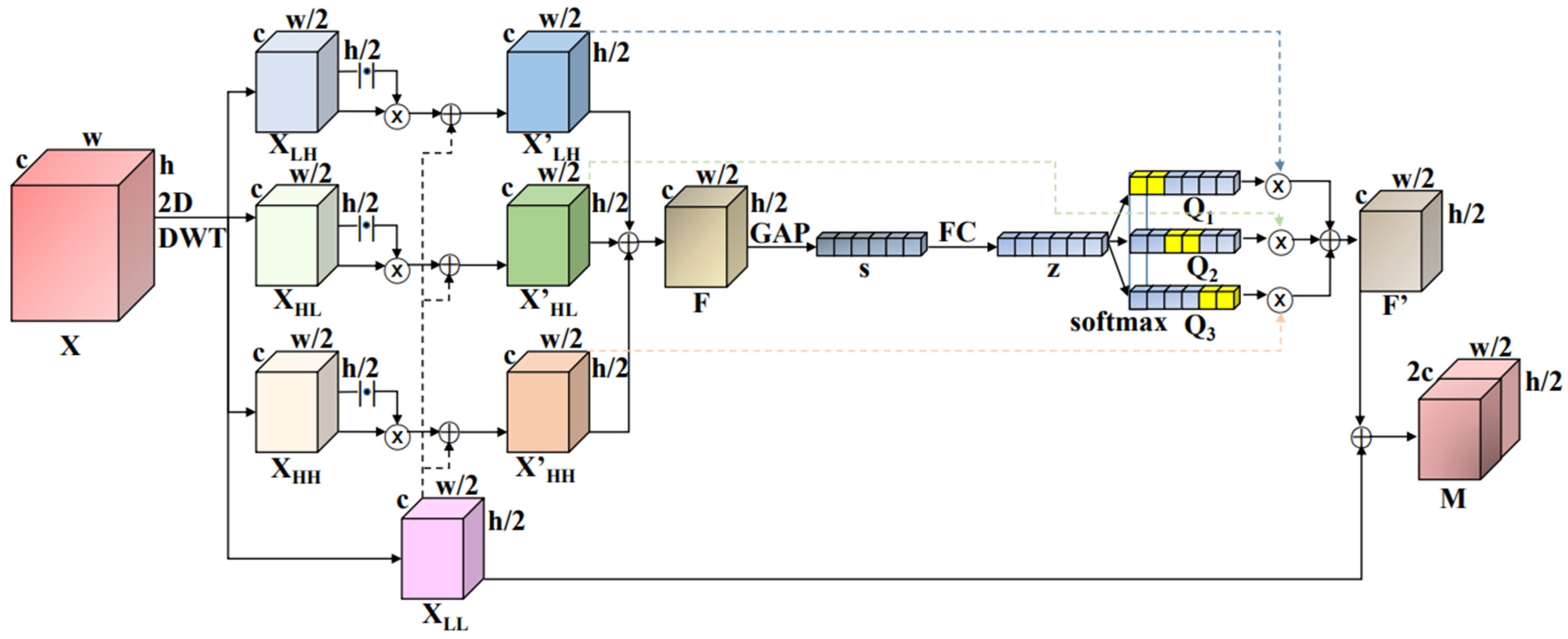
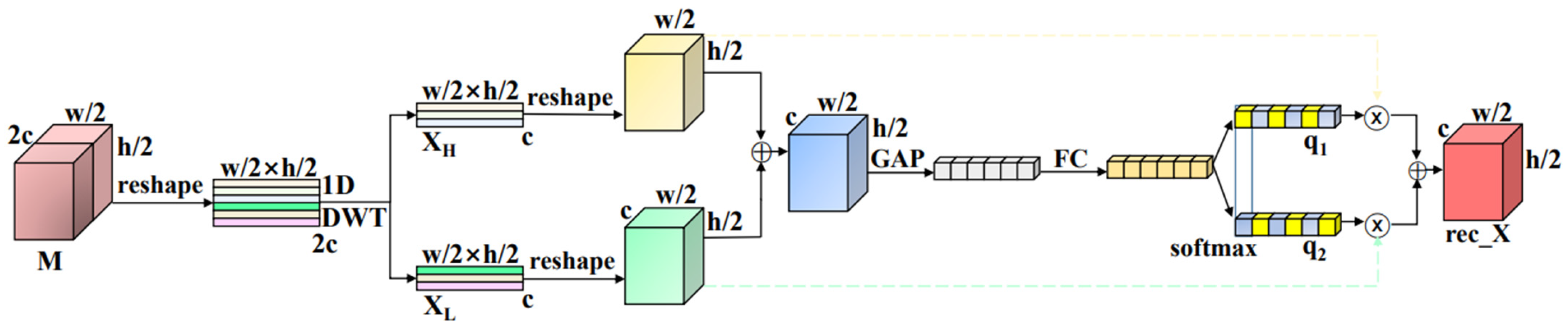
- In the current study, we developed a wavelet down-sampling feature reconstruction method to address the problem of traditional down-sampling feature information loss, introducing a multichannel attention mechanism to effectively combine low-frequency components with high-frequency components, fully utilizing channel information to reveal finer details.
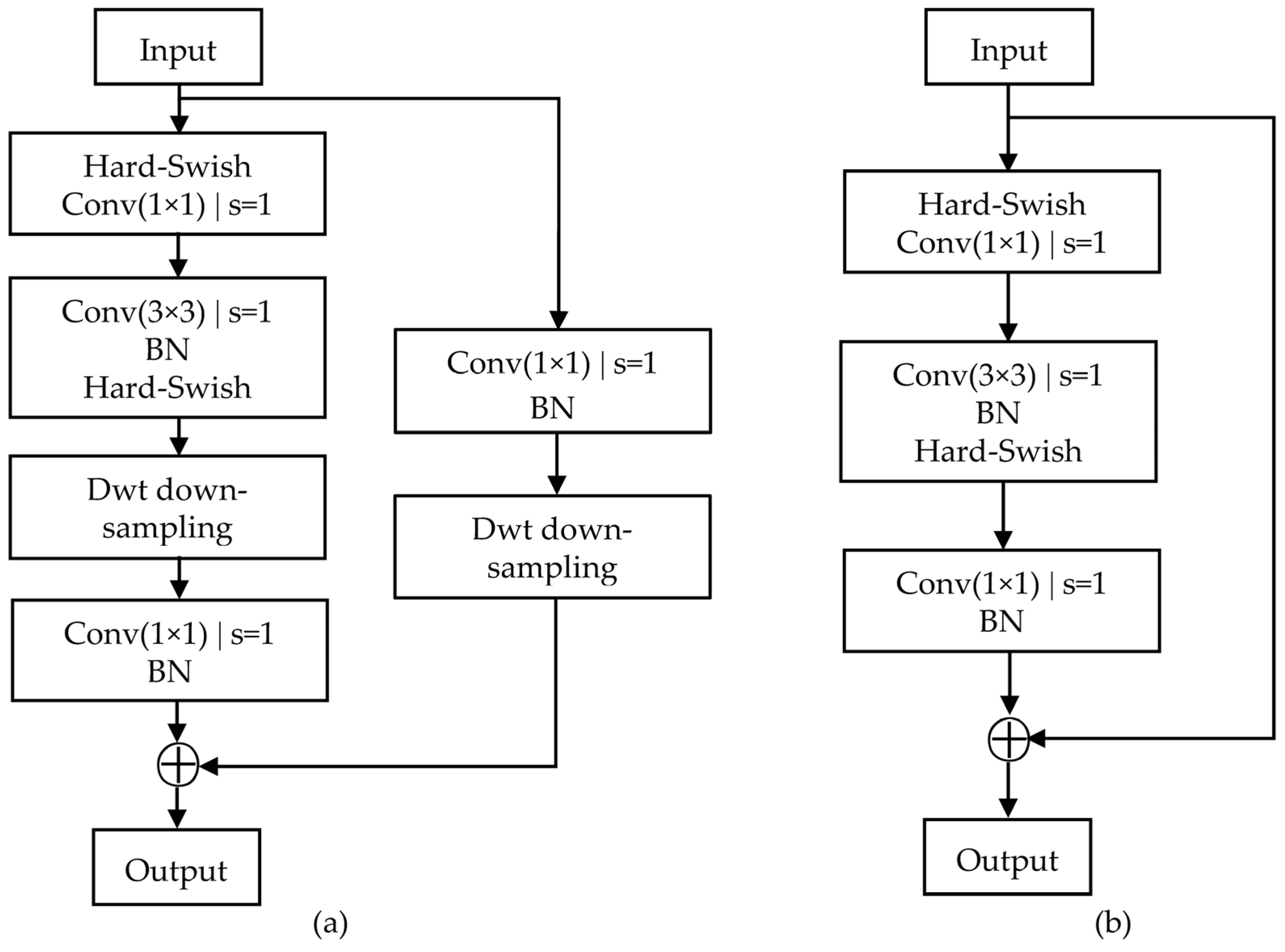
- This paper developed a wavelet down-sampling feature reconstruction method-based convolutional neural network as a feature extractor to classify melanoma skin cancer using skin pathological mirror.
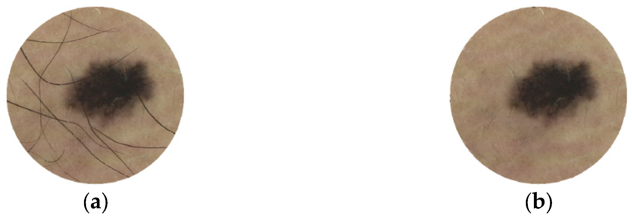
- A data augmentation and hair removal algorithm is used to pre-process the skin pathological mirror dataset HAM1000, and the pre-processing method is tested on the pre-processing HAM10000 dataset using transfer learning. The experimental results show that the proposed method has high accuracy for melanoma skin cancer classification.
2. Related Work
3. Wavelet Down-Sampling Reconstruction
4. Skin Cancer Classification Model
5. Experiments and Analysis of Results
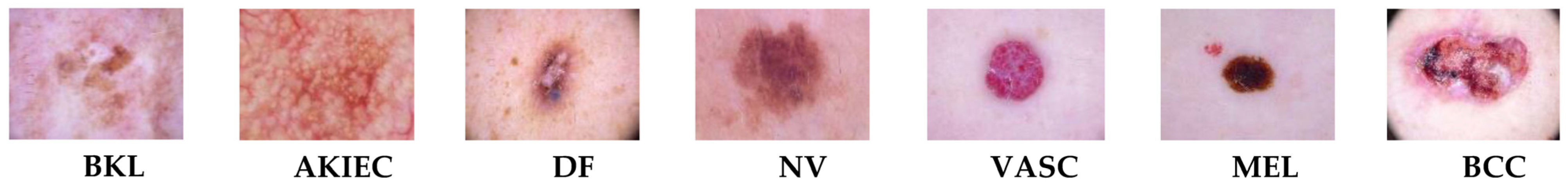
5.1. Dataset and Pre-Processing
| Algorithm 1: hair removal algorithm |
| Input: skin mirror image (img) Output: restoration skin mirror image (img1)
|
5.2. Experimental Environment and Evaluation Indicators
5.3. Comparative Experiments
6. Conclusions
Author Contributions
Funding
Data Availability Statement
Conflicts of Interest
References
- Siegel, R.L.; Miller, K.D.; Jemal, A. Cancer statistics, 2019. CA A Cancer J. Clin. 2019, 69, 7–34. [Google Scholar] [CrossRef] [PubMed]
- Koh, H.K. Melanoma screening: Focusing the public health journey. Arch. Dermatol. 2007, 143, 101–103. [Google Scholar] [CrossRef] [PubMed]
- Zunair, H.; Hamza, A.B. Melanoma detection using adversarial training and deep transfer learning. Phys. Med. Biol. 2020, 65, 135005. [Google Scholar] [CrossRef] [PubMed]
- LeCun, Y.; Bengio, Y.; Hinton, G. Deep learning. Nature 2015, 521, 436–444. [Google Scholar] [CrossRef]
- Qiu, C.H.; Huang, C.F.; Xia, S.R.; Kong, D.X. Application review of artificial intelligence in medical images aided diagnosis. Space Med. Med. Eng. 2021, 34, 407–414. [Google Scholar]
- Chen, Y.D.; Zhang, Q.; Lan, L.; Peng, L.; Yin, J. A Review of Deep Convolutional Neural Networks in Medical Image Segmentation. Chin. J. Health Inform. Manag. 2021, 18, 278–284. [Google Scholar]
- Codella, N.; Cai, J.; Abedini, M.; Garnavi, R.; Halpern, A.; Smith, J.R. Deep learning, sparse coding, and SVM for melanoma recognition in dermoscopy images. In Machine Learning in Medical Imaging: 6th International Workshop, MLMI 2015, Proceedings of the MICCAI 2015, Munich, Germany, 5 October 2015; Held in Conjunction with MICCAI 2015; Springer International Publishing: Cham, Switzerland, 2015; pp. 118–126. [Google Scholar]
- Pomponiu, V.; Nejati, H.; Cheung, N.M. Deepmole: Deep neural networks for skin mole lesion classification. In Proceedings of the 2016 IEEE International Conference on Image Processing (ICIP), Phoenix, AZ, USA, 25–28 September 2016; pp. 2623–2627. [Google Scholar]
- Esteva, A.; Kuprel, B.; Novoa, R.A.; Ko, J.; Swetter, S.M.; Blau, H.M.; Thrun, S. Dermatologist-level classification of skin cancer with deep neural networks. Nature 2017, 542, 115–118. [Google Scholar] [CrossRef]
- Khan, M.A.; Sharif, M.I.; Raza, M.; Anjum, A.; Saba, T.; Shad, S.A. Skin lesion segmentation and classification: A unified framework of deep neural network features fusion and selection. Expert Syst. 2022, 39, e12497. [Google Scholar] [CrossRef]
- Ahmed, S.A.A.; Yanikoğlu, B.; Göksu, Ö.; Aptoula, E. Skin lesion classification with deep CNN ensembles. In Proceedings of the 2020 28th Signal Processing and Communications Applications Conference (SIU), Gaziantep, Turkey, 5–7 October 2020; pp. 1–4. [Google Scholar]
- Abayomi-Alli, O.O.; Damasevicius, R.; Misra, S.; Maskeliunas, R.; Abayomi-Alli, A. Malignant skin melanoma detection using image augmentation by oversamplingin nonlinear lower-dimensional embedding manifold. Turk. J. Electr. Eng. Comput. Sci. 2021, 29, 2600–2614. [Google Scholar] [CrossRef]
- Zeiler, M.D.; Fergus, R. Stochastic pooling for regularization of deep convolutional neural networks. In Proceedings of the 1st International Conference on Learning Representations, ICLR 2013, Scottsdale, AZ, USA, 2–4 May 2013. [Google Scholar]
- He, K.; Zhang, X.; Ren, S.; Sun, J. Deep residual learning for image recognition. In Proceedings of the IEEE Conference on Computer Vision and Pattern Recognition, Las Vegas, NV, USA, 27–30 June 2016; pp. 770–778. [Google Scholar]
- Zhao, G.; Wang, J.; Zhang, Z. Random Shifting for CNN: A Solution to Reduce Information Loss in Down-Sampling Layers. In Proceedings of the International Joint Conference on Artificial Intelligence (IJCAI), Melbourne, Australia, 25 August 2017; pp. 3476–3482. [Google Scholar]
- Jiang, Z.T.; Qin, J.Q.; Zhang, S.Q. Parameterized pooling convolution neural network for image classification. Acta Electron. Sin. 2020, 48, 1729. [Google Scholar]
- Daubechies, I. Ten Lectures on Wavelets. Comput. Phys. 1992, 6, 697. [Google Scholar] [CrossRef]
- Hong, J.; Wang, Z.; Qu, C.; Zhou, Y.; Shan, T.; Zhang, J.; Hou, Y. Investigation on overcharge-caused thermal runaway of lithium-ion batteries in real-world electric vehicles. Appl. Energy 2022, 321, 119229. [Google Scholar] [CrossRef]
- Hong, J.; Zhang, H.; Xu, X. Thermal fault prognosis of lithium-ion batteries in real-world electric vehicles using self-attention mechanism networks. Appl. Therm. Eng. 2023, 226, 120304. [Google Scholar] [CrossRef]
- Bruna, J.; Mallat, S. Invariant scattering convolution networks. IEEE Trans. Pattern Anal. Mach. Intell. 2013, 35, 1872–1886. [Google Scholar] [CrossRef]
- Liu, P.; Zhang, H.; Zhang, K.; Lin, L.; Zuo, W. Multi-level wavelet-CNN for image restoration. In Proceedings of the IEEE Conference on Computer Vision and Pattern Recognition Workshops, Salt Lake City, UT, USA, 18–22 June 2018; pp. 773–782. [Google Scholar]
- Li, Q.; Shen, L.; Guo, S.; Lai, Z. Wavelet integrated CNNs for noise-robust image classification. In Proceedings of the IEEE/CVF Conference on Computer Vision and Pattern Recognition, Seattle, WA, USA, 13–19 June 2020; pp. 7245–7254. [Google Scholar]
- Xu, D. Application of wavelet transform-based image processing techniques. J. Soochow Univ. (Nat. Sci.) 2002, 1, 45–49. [Google Scholar]
- Xu, K.; Qin, M.; Sun, F.; Wang, Y.; Chen, Y.K.; Ren, F. Learning in the frequency domain. In Proceedings of the IEEE/CVF Conference on Computer Vision and Pattern Recognition, Seattle, WA, USA, 13–19 June 2020; pp. 1740–1749. [Google Scholar]
- Li, X.; Wang, W.; Hu, X.; Yang, J. Selective kernel networks. In Proceedings of the IEEE/CVF Conference on Computer Vision and Pattern Recognition, Long Beach, CA, USA, 15–20 June 2019; pp. 510–519. [Google Scholar]
- Dang, L.; Pang, P.; Lee, J. Depth-wise separable convolution neural network with residual connection for hyperspectral image classification. Remote Sens. 2020, 12, 3408. [Google Scholar] [CrossRef]
- Avenash, R.; Viswanath, P. Semantic Segmentation of Satellite Images using a Modified CNN with Hard-Swish Activation Function. In Proceedings of the nternational Joint Conference on Computer Vision, Imaging and Computer Graphics (VISIGRAPP), Prague, Czech Republic, 25–27 February 2019; pp. 413–420. [Google Scholar]
- Bronskill, J.; Gordon, J.; Requeima, J.; Nowozin, S.; Turner, R. Tasknorm: Rethinking batch normalization for meta-learning. In Proceedings of the International Conference on Machine Learning, Virtual, 13–18 July 2020; pp. 1153–1164. [Google Scholar]
- Tschandl, P.; Rosendahl, C.; Kittler, H. The HAM10000 dataset, a large collection of multi-source dermatoscopic images of common pigmented skin lesions. Sci. Data 2018, 5, 180161. [Google Scholar] [CrossRef]
- Xie, F.; Qin, S.; Jiang, Z.; Meng, R. Unsupervised repair of hair-occluded information for skin melanoma image. Chin. J. Sci. Instrum. 2009, 30, 699–705. [Google Scholar]
- Youwen, G.; Benjun, Z.; Xiaofei, H. Research on image recognition of convolution neural network based on data augmentation. Comput. Technol. Dev. 2018, 28, 62–65. [Google Scholar]
- Krizhevsky, A.; Sutskever, I.; Hinton, G.E. Imagenet classification with deep convolutional neural networks. Commun. ACM 2017, 60, 84–90. [Google Scholar] [CrossRef]
- Bansal, M.; Kumar, M.; Sachdeva, M.; Mittal, A. Transfer learning for image classification using VGG19: Caltech-101 image data set. J. Ambient. Intell. Humaniz. Comput. 2021, 14, 3609–3620. [Google Scholar] [CrossRef] [PubMed]
- Sandler, M.; Howard, A.; Zhu, M.; Zhmoginov, A.; Chen, L.C. Mobilenetv2: Inverted residuals and linear bottlenecks. In Proceedings of the IEEE Conference on Computer Vision and Pattern Recognition, Salt Lake City, UT, USA, 18–22 June 2018; pp. 4510–4520. [Google Scholar]
- Huang, G.; Liu, Z.; Van Der Maaten, L.; Weinberger, K.Q. Densely connected convolutional networks. In Proceedings of the IEEE Conference on Computer Vision and Pattern Recognition, Honolulu, HI, USA, 21–26 July 2017; pp. 4700–4708. [Google Scholar]
- Tan, M.; Le, Q. Efficientnet: Rethinking model scaling for convolutional neural networks. In Proceedings of the 36th International Conference on Machine Learning, Long Beach, CA, USA, 10–15 June 2019; Volume 97, pp. 6105–6114. [Google Scholar]
- Gao, M. Soft Attention Improves Skin Cancer Classification Performance. In Interpretability of Machine Intelligence in Medical Image Computing, and Topological Data Analysis and Its Applications for Medical Data: 4th International Workshop, Proceedings of the iMIMIC 2021, and 1st International Workshop, Strasbourg, France, 27 September 2021; TDA4MedicalData 2021, Held in Conjunction with MICCAI 2021; Springer Nature: Berlin, Germany, 2021; Volume 12929. [Google Scholar]
- Gessert, N.; Nielsen, M.; Shaikh, M.; Werner, R.; Schlaefer, A. Skin lesion classification using ensembles of multi-resolution EfficientNets with meta data. MethodsX 2020, 2020, 100864. [Google Scholar] [CrossRef] [PubMed]
- Shen, S.; Xu, M.; Zhang, F.; Shao, P.; Liu, H.; Xu, L.; Zhang, C.; Liu, P.; Yao, P.; Xu, R.X. Erratum to “A Low-Cost High-Performance Data Augmentation for Deep Learning-Based Skin Lesion Classification”. BME Front. 2023, 4, 0011. [Google Scholar] [CrossRef]
- Lan, Z.; Cai, S.; He, X.; Wen, X. FixCaps: An improved capsules network for diagnosis of skin cancer. IEEE Access 2022, 10, 76261–76267. [Google Scholar] [CrossRef]


| Base Model | Input Backbone | Residual Module | Data Augmentation | AC/% | F1/% |
|---|---|---|---|---|---|
| ResNet50 | √ | 90.48 | 90.69 | ||
| √ | √ | 93.45 | 93.67 | ||
| √ | √ | 94.51 | 94.64 | ||
| √ | √ | 93.94 | 94.03 | ||
| √ | √ | √ | 95.28 | 95.39 |
| Model | AC/% | F1/% |
|---|---|---|
| AlexNet | 87.81 | 88.13 |
| VGG19 | 90.34 | 90.61 |
| MobileNet-V2 | 88.23 | 88.86 |
| DenseNet-121 | 92.93 | 93.14 |
| EfficientNet-B0 | 93.62 | 93.78 |
| Proposed model | 95.84 | 95.96 |
| References | AC/% | F1/% | Parameters/MB |
|---|---|---|---|
| ResNet50 + SA [37] | 91.55 | 91.30 | 91.2 |
| CNN ensembles [11] | 93.09 | - | - |
| IRv2-SA [37] | 93.47 | 93.65 | 181.3 |
| Loss balance + ensemble [38] | 92.60 | - | - |
| Low-cost augmentation + CNN [39] | 95.79 | 86.14 | 42.0 |
| FixCaps [40] | 96.49 | - | 1.86 |
| Our method | 95.84 | 95.96 | 60.9 |
| References | AUC | Category | ||||||
|---|---|---|---|---|---|---|---|---|
| AKIEC | BCC | BKL | DF | MEL | NV | VASC | ||
| ResNet50 + SA [37] | 0.980 | 0.981 | 0.996 | 0.964 | 0.971 | 0.973 | 0.979 | 0.999 |
| CNN ensembles [11] | 0.929 | 0.902 | 0.934 | 0.885 | 0.968 | 0.925 | 0.951 | 0.941 |
| IRv2-SA [37] | 0.985 | 0.981 | 0.998 | 0.982 | 0.973 | 0.974 | 0.984 | 1.000 |
| Loss balance + ensemble [38] | 0.941 | 0.919 | 0.947 | 0.908 | 0.980 | 0.931 | 0.960 | 0.942 |
| Low-cost augmentation + CNN [39] | 0.976 | 0.988 | 0.989 | 0.968 | 0.972 | 0.943 | 0.970 | 0.995 |
| FixCaps [40] | - | - | - | - | - | - | - | - |
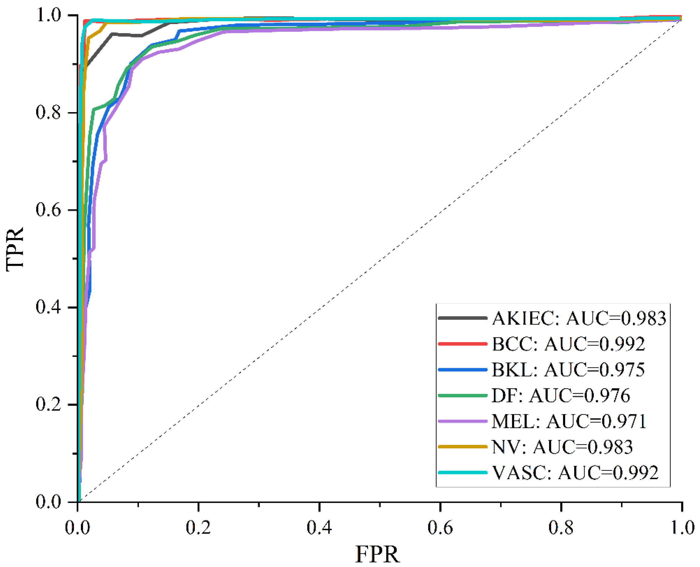
| Our method | 0.982 | 0.983 | 0.992 | 0.975 | 0.976 | 0.971 | 0.983 | 0.992 |
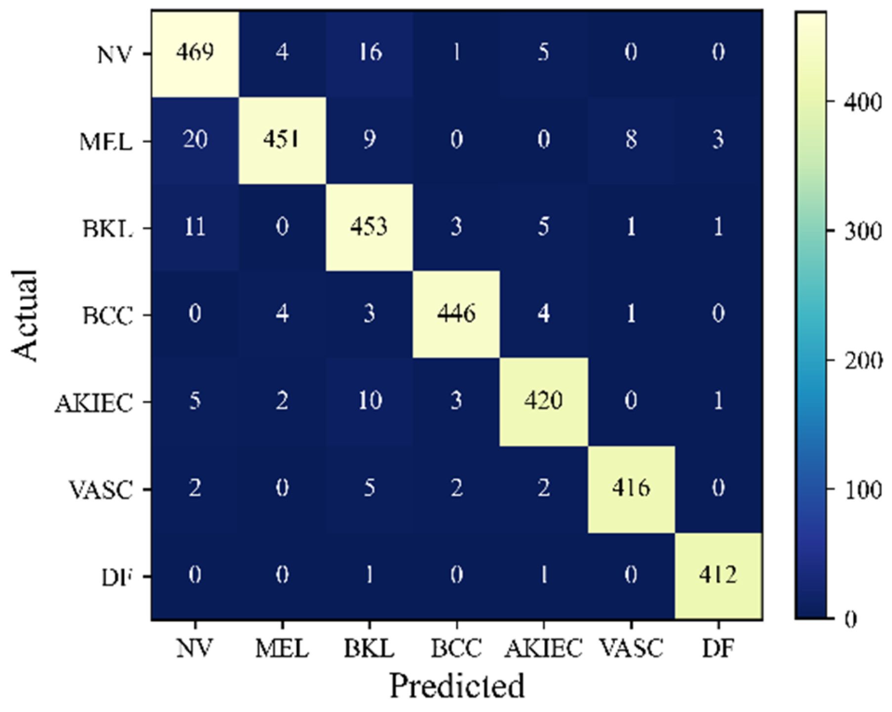
| Category | Precision/% | Recall/% | F1-Score/% |
|---|---|---|---|
| NV | 92.50 | 94.75 | 93.61 |
| MEL | 97.83 | 91.85 | 94.75 |
| BKL | 91.15 | 95.57 | 93.31 |
| BCC | 98.02 | 97.38 | 97.70 |
| AKIEC | 96.11 | 95.24 | 95.67 |
| VASC | 97.65 | 97.42 | 97.54 |
| DF | 98.80 | 99.52 | 99.16 |
Disclaimer/Publisher’s Note: The statements, opinions and data contained in all publications are solely those of the individual author(s) and contributor(s) and not of MDPI and/or the editor(s). MDPI and/or the editor(s) disclaim responsibility for any injury to people or property resulting from any ideas, methods, instructions or products referred to in the content. |
© 2023 by the authors. Licensee MDPI, Basel, Switzerland. This article is an open access article distributed under the terms and conditions of the Creative Commons Attribution (CC BY) license (https://creativecommons.org/licenses/by/4.0/).
Share and Cite
Wu, Q.-e.; Yu, Y.; Zhang, X. A Skin Cancer Classification Method Based on Discrete Wavelet Down-Sampling Feature Reconstruction. Electronics 2023, 12, 2103. https://doi.org/10.3390/electronics12092103
Wu Q-e, Yu Y, Zhang X. A Skin Cancer Classification Method Based on Discrete Wavelet Down-Sampling Feature Reconstruction. Electronics. 2023; 12(9):2103. https://doi.org/10.3390/electronics12092103
Chicago/Turabian StyleWu, Qing-e, Yao Yu, and Xinyang Zhang. 2023. "A Skin Cancer Classification Method Based on Discrete Wavelet Down-Sampling Feature Reconstruction" Electronics 12, no. 9: 2103. https://doi.org/10.3390/electronics12092103