Article

Staphylococcus aureus Small-Colony Variants from Airways of Adult Cystic Fibrosis Patients as Precursors of Adaptive Antibiotic-Resistant Mutations

by
Guillaume Millette
1,†,
David Lalonde Séguin
1,†,
Charles Isabelle
1,
Suzanne Chamberland
1,
Jean-François Lucier
1,
Sébastien Rodrigue
1,
André M. Cantin
2 and
François Malouin
1,*
1
Département de Biologie, Faculté des Sciences, Université de Sherbrooke, Sherbrooke, QC J1K 2R1, Canada
2
Service de Pneumologie, Département de Médecine, Faculté de Médecine et des Sciences de la Santé, Université de Sherbrooke, Sherbrooke, QC J1H 5N4, Canada
*
Author to whom correspondence should be addressed.
These authors contributed equally to this work.
Antibiotics 2023, 12(6), 1069; https://doi.org/10.3390/antibiotics12061069
Submission received: 18 May 2023 / Revised: 14 June 2023 / Accepted: 15 June 2023 / Published: 17 June 2023

Abstract

:
Prototypic Staphylococcus aureus and their small-colony variants (SCVs) are predominant in cystic fibrosis (CF), but the interdependence of these phenotypes is poorly understood. We characterized S. aureus isolates from adult CF patients over several years. Of 18 S. aureus-positive patients (58%), 13 (72%) were positive for SCVs. Characterization included genotyping, SCCmec types, auxotrophy, biofilm production, antibiotic susceptibilities and tolerance, and resistance acquisition rates. Whole-genome sequencing revealed that several patients were colonized with prototypical and SCV-related clones. Some clonal pairs showed acquisition of aminoglycoside resistance that was not explained by aminoglycoside-modifying enzymes, suggesting a mutation-based process. The characteristics of SCVs that could play a role in resistance acquisition were thus investigated further. For instance, SCV isolates produced more biofilm (p < 0.05) and showed a higher survival rate upon exposure to ciprofloxacin and vancomycin compared to their prototypic associated clones. SCVs also developed spontaneous rifampicin resistance mutations at a higher frequency. Accordingly, a laboratory-derived SCV (ΔhemB) acquired resistance to ciprofloxacin and gentamicin faster than its parent counterpart after serial passages in the presence of sub-inhibitory concentrations of antibiotics. These results suggest a role for SCVs in the establishment of persistent antibiotic-resistant clones in adult CF patients.

1. Introduction

Cystic fibrosis (CF) is a common life-shortening autosomal recessive disorder promoting chronic polymicrobial infections and bronchopulmonary inflammation that ultimately cause respiratory insufficiency. Staphylococcus aureus is one the most prevalent pathogens recovered from the lungs of CF patients [1,2]. Recent data show a noticeable increase in the prevalence of S. aureus among CF patients together with an increased life expectancy of adult patients [1,2]. The clinical impact of S. aureus in CF lung infections is not completely understood. Some studies noted a deleterious impact of S. aureus on airway inflammation, especially in children [3,4], whereas others reported no impact or even a better prognosis following S. aureus colonization [5,6,7]. However, it has been reported that S. aureus small-colony variants (SCVs) [8,9,10] and MRSA [11,12,13] worsen prognostics and increase mortality rate. A better understanding of the contribution of S. aureus, especially SCV and MRSA, to the health of adult CF patients is needed.
Subpopulations of SCVs are often found in the airways of CF patients [7,9,14]. In contrast to prototypical S. aureus, SCVs exhibit small, non-pigmented colonies less susceptible to aminoglycoside antibiotics due to their metabolic defect, which can often be restored by supplemental hemin, menadione, or thymidine [15,16]. SCVs express a set of virulence factors that favor host tissue colonization and intracellular persistence [14,17,18,19,20,21,22]. As a result, SCVs are associated with chronic infections, namely in the respiratory tract of patients with CF.
Antibiotic prophylaxis and treatment regimens have been used in an attempt to reduce S. aureus chronic pulmonary infections [23,24]. While both resulted in fewer S. aureus infections, the positive impact on lung function was unclear [23,24]. However, it is clear that the increasing emergence of antibiotic-resistant bacteria in CF patients is a negative side-effect of such practices [25]. S. aureus manages to survive antibiotic treatments via different mechanisms, including biofilm production and the formation of SCVs [14,26]. While it is known that SCVs are inherently tolerant to many different antibiotics [16,27], it remains unclear whether they favor the acquisition of long-term resistance mechanisms that could be detrimental to CF patients in the long run. A recent study has shown that E. coli persister cells, a subpopulation of temporarily antibiotic-tolerant bacterial cells due to their slow growth [28], are able to resume growth after lethal stress, facilitating the development of clinical antibiotic resistance [29]. S. aureus SCVs, which are similar to persisters (i.e., slow growth and tolerance to antibiotics), may also facilitate the acquisition of antibiotic resistance.
The impact of environmental factors on the incidence of S. aureus and SCVs is still not completely understood, but it is thought that exposure to some antibiotics may increase the presence of SCVs and persistent strains of S. aureus. Aminoglycoside therapy is associated with the formation of SCV auxotroph to hemin or menadione [20]. Trimethoprim-sulfamethoxazole treatments are linked to the emergence of thymidine-dependent SCV [30]. Exposure to fluoroquinolones induces the SOS response, which potentially increases the formation of SCVs [31]. Aside from antibiotic therapy, other factors can trigger the SCV phenotype: a pseudomonal quorum-sensing molecule, 4-hydroxy-2-heptylquinoline-N-oxide (HQNO) [32,33,34], the mammalian intracellular milieu [35], reactive aldehydes [36], permanent activation of the bacterial stringent response [37], CO2 auxotrophy [16], and oxidative stress [38]. As a result, new SCV-inducing factors and environmental conditions are uncovered, and such selective pressures could be more commonly found in infected patients than previously thought possible.
Few studies have addressed the status of S. aureus and SCV colonization in CF patients and their contribution to the pathology (for a review, see [14,22]). Firstly, the aim of this work was to characterize S. aureus isolates from the respiratory tract of adult CF patients. We looked for the SCV phenotype, conducted genetic profiling, determined antibiotic susceptibility profiles, and measured biofilm production of S. aureus isolates. In addition, this study aimed to investigate how SCVs may contribute to the development of antibiotic resistance in the airways of CF patients, thus emphasizing the clinical importance of this phenotype.

2. Results

2.1. S. aureus Infections among CF Patients

For at least one visit, 18 (58%) of the 31 patients in this study tested positive for S. aureus. Except for patient 17, from whom only an SCV could be isolated, SCVs were isolated exclusively from patients colonized with prototypical S. aureus. SCVs were found among 13 (72%) of the 18 patients who had been colonized by S. aureus.

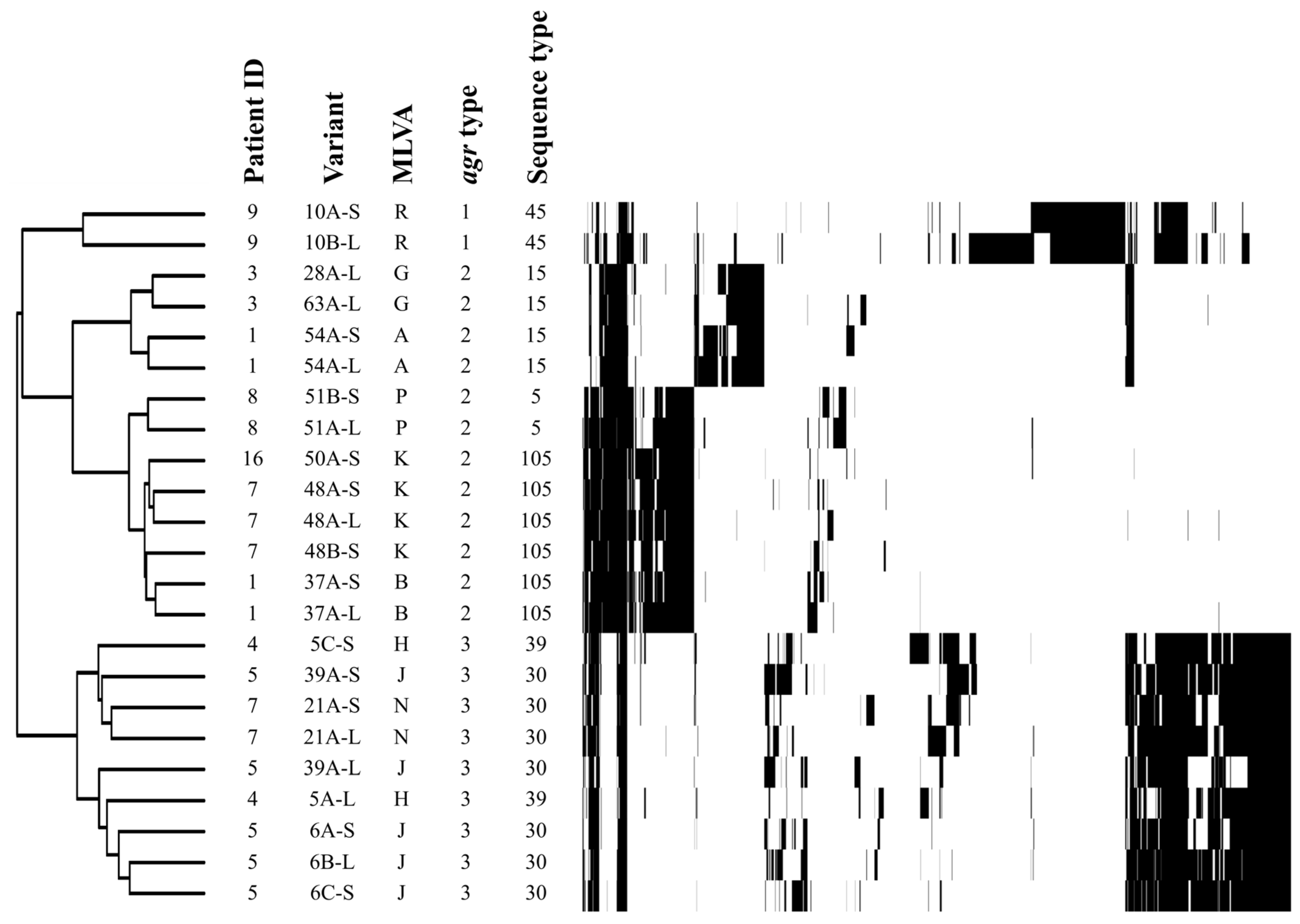
2.2. Genotype Profiles

From a total of 158 S. aureus isolates collected from the 18 patients who tested positive for S. aureus over the course of this study, 77 distinct S. aureus isolates were selected for further analyses. The individuality of these non-redundant 77 isolates was based on source (patient), colony morphology (prototype or SCV), type of auxotrophy if SCV, MLVA and agr typing, antibiotic susceptibility profile, and presence of the mecA gene (Table 1). All isolates were classified into 33 different profiles (A to GG) by MLVA. From these 77 distinct analyzed S. aureus isolates, a total of 26 SCV isolates were found. In some patients, prototypic S. aureus (large colonies; -L suffix in Table 1) and co-isolated SCVs (-S suffix in Table 1) had identical MLVA and agr types, indicative of genetic relatedness. From these distinct 77 analyzed S. aureus isolates, 12 MRSA isolates were found in this study and could be divided into three lineages based on MLVA and agr types (Table 1). All MRSA isolates had the SCCmec type II.
As mentioned above, MLVA and agr types suggested the presence of sequential clonal strains that persisted within the same patient for at least two visits (several months apart). Some of these clonal strains included prototypic–SCV pairs that were submitted to whole-genome sequencing. High-impact single-nucleotide polymorphisms (SNPs) were then used to create a phylogenetic tree to confirm the clonality of the sequential or prototypic–SCV pairs, as seen in Figure 1.

2.3. Auxotrophy of SCVs

Because their growth was enhanced by the presence of hemin and/or menadione, 12/26 (46%) of all SCVs appeared to be defective in electron transport. Three patients were colonized by a type of SCV for which abundant growth could only be restored by adding both menadione and hemin (Table 1). Only one thymidine-dependent SCV was found in a single patient. The majority of the SCVs retrieved from the patients (13/26 or 50%) were auxotroph for an unknown factor, meaning that their growth could not be complemented by either hemin, menadione, or thymidine.

2.4. Antibiotic Susceptibility Profiles

Table 1 shows the susceptibility profiles of the 77 distinct isolates to t10 different antibiotics. In the 18 patients who tested positive for S. aureus, 94% (17/18) were colonized by S. aureus isolates that were intermediate (I) or resistant (R) to at least one antibiotic (Table 1; see supplemental Table S1 for precise MIC values). Resistance to ciprofloxacin and erythromycin was found in 50% (9/18) and 61% (11/18) of these patients, respectively. Resistance to trimethoprim-sulfamethoxazole was found only in two patients (four distinct isolates) and was not exclusively associated with the presence of thymidine-dependent SCVs. Over the course of the study, 33% (6/18) of S. aureus-positive patients were colonized with MRSA (oxacillin MIC ≥ 64 µg/mL). All MRSA isolates were found to also be resistant to ciprofloxacin (MIC ≥ 64 µg/mL) and erythromycin (MIC ≥ 64 µg/mL). No resistance to rifampicin or vancomycin was found.
Since many SCVs were dependent on supplemental hemin or menadione or both for abundant growth, it was expected that the reduced proton motive force (PMF) of SCVs would reduce their susceptibility to aminoglycosides, which rely on an active PMF to cross the cell membrane barrier [39]. Indeed, a total of 23 SCVs out of 26 distinct SCV isolates (88%) showed an intermediate level of susceptibility (MIC of 8 µg/mL) to gentamicin or tobramycin, while 10 out of 26 (38%) were resistant (MIC > 32 µg/mL) to at least 1 aminoglycoside. Interestingly, in many cases, intermediate resistance or resistance to aminoglycosides seemed to gradually increase with time among isolates that were genetically related. Table 2 shows such cases found in eight different patients where the initial clone was susceptible to gentamicin or tobramycin and where other isolates from the same visit or from subsequent visits showed an increased resistance that could not be associated with acquisition of aminoglycoside-modifying enzymes. Most of the time, an SCV was found among those isolates, and this was also true for MRSA strains (Table 2).

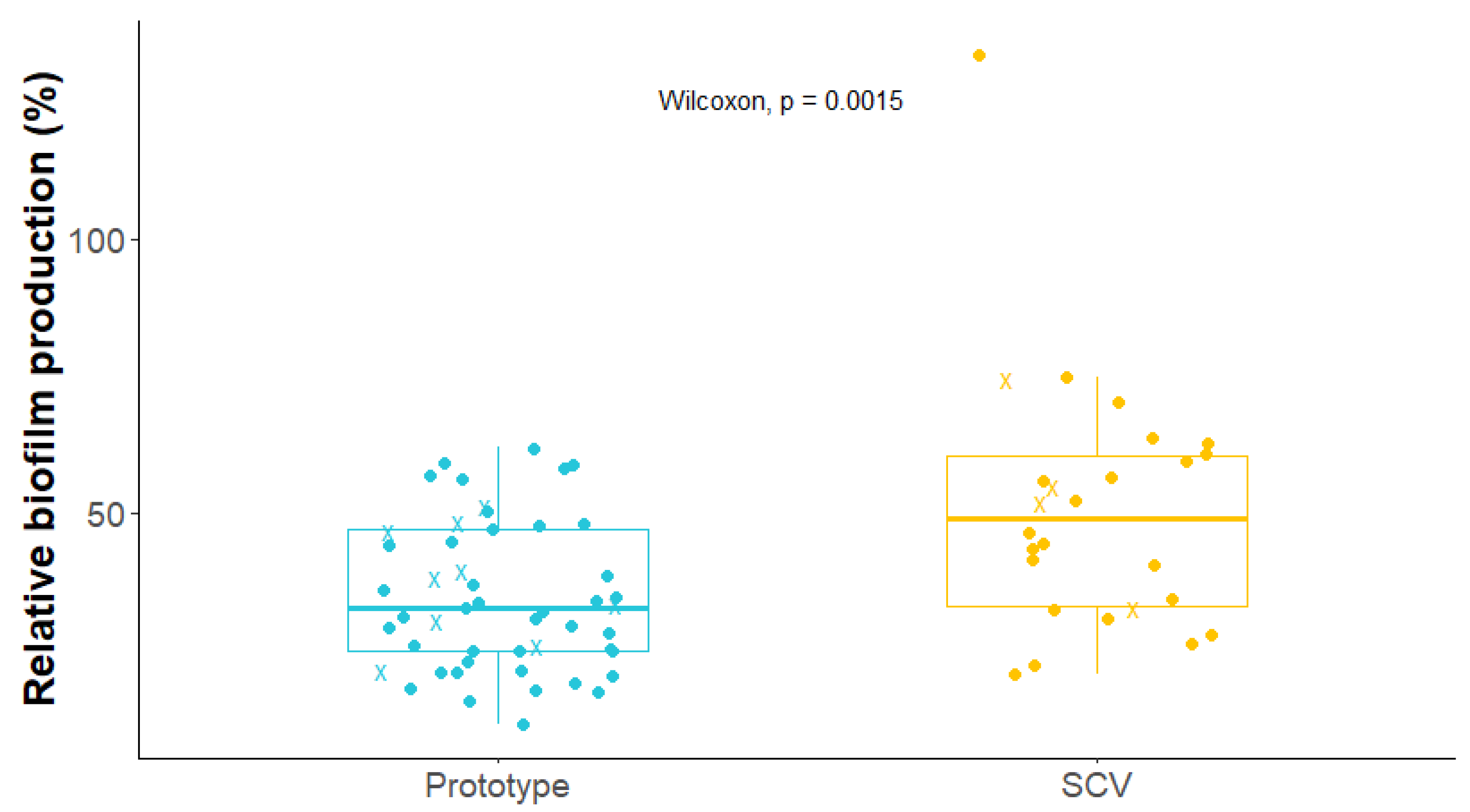
2.5. Biofilm Production by S. aureus Isolates

Because the presence of biofilm often impacts on the susceptibility of bacteria to antibiotics, the relative biofilm production capacity of prototypical and SCV isolates was determined. As shown in Figure 2, SCVs (n = 26) produced significantly more biofilm than prototypical (n = 51) S. aureus isolates (p-value = 0.0015; Wilcoxon test) with a median relative biofilm production that was 1.50-fold higher than that of the prototypical strains.

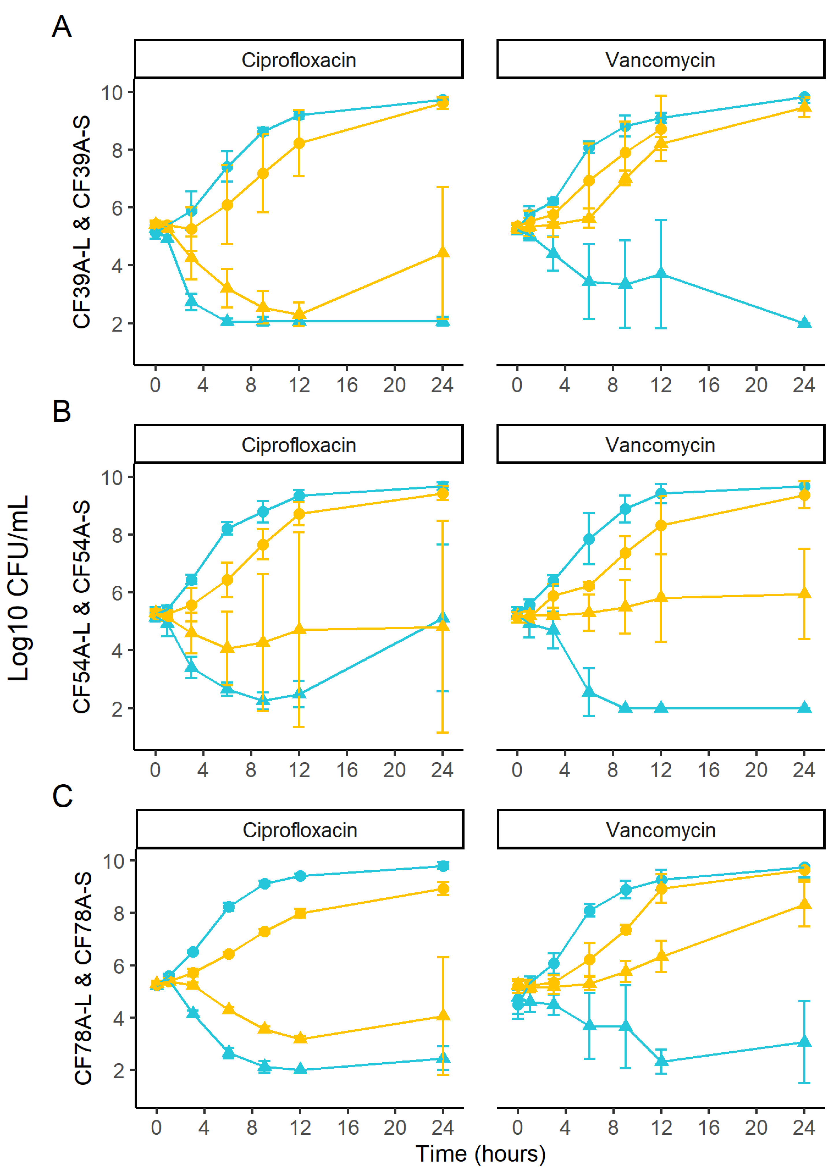
2.6. Kill Kinetics of Related SCV and Prototypic Isolates

We investigated whether antibiotics killed and/or inhibited growth of SCV and prototypical isolates to the same extent. To do so, we selected one related SCV–prototype pair from each of three different patients, resulting in a total of three pairs. The pairs used were CF54A, CF39A, and CF78A (-S and -L) from patients 1, 5, and 10, respectively. The related SCV and prototype isolates had identical MICs for ciprofloxacin and vancomycin, respectively (CF39A-L or -S: 2 and 1 µg/mL; CF54A-L or -S: 0.5 and 1 µg/mL; CF78A-L or -S: 0.25–0.5 and 1 µg/mL; MIC values can be found in Table S1). As expected, prototypes grew faster than SCVs in the control condition without antibiotic (Figure 3). On the other hand, SCVs better survived ciprofloxacin treatment, showing a much slower killing rate than that seen for prototypical isolates (Figure 3, left panels). Moreover, all three SCVs either maintained viability or showed growth in the presence of vancomycin compared to their related prototypical counterparts (Figure 3, right panels).

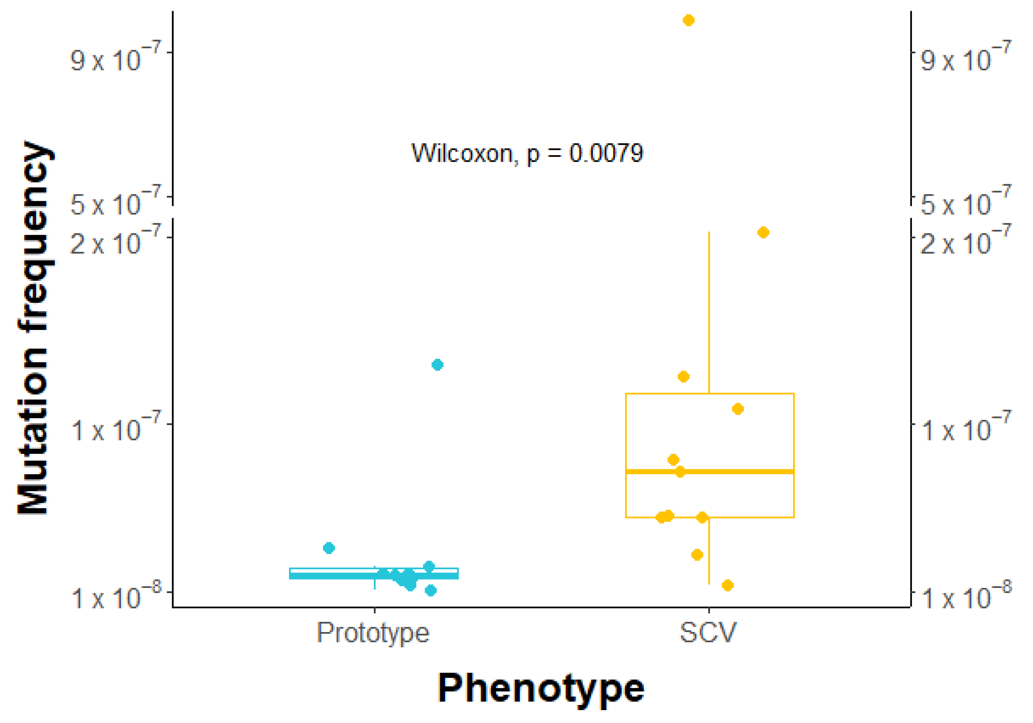
2.7. Higher Frequency of Rifampicin-Induced Resistant Mutants in SCVs

To assess whether SCVs tend to acquire beneficial mutations more easily than prototypic isolates under antibiotic pressure, we exposed 10 prototypes and 11 SCVs to rifampicin at a concentration equivalent to 10 times their MIC. Results showed that SCVs exhibited over three times higher mutant frequency than prototypes (median of 1.94 × 10⁻⁸ vs. 7.44 × 10⁻⁸, as shown in Figure 4), which was found to be statistically significant. These findings suggest that SCVs have a greater tendency to develop mutation-based antibiotic resistance than prototypes.

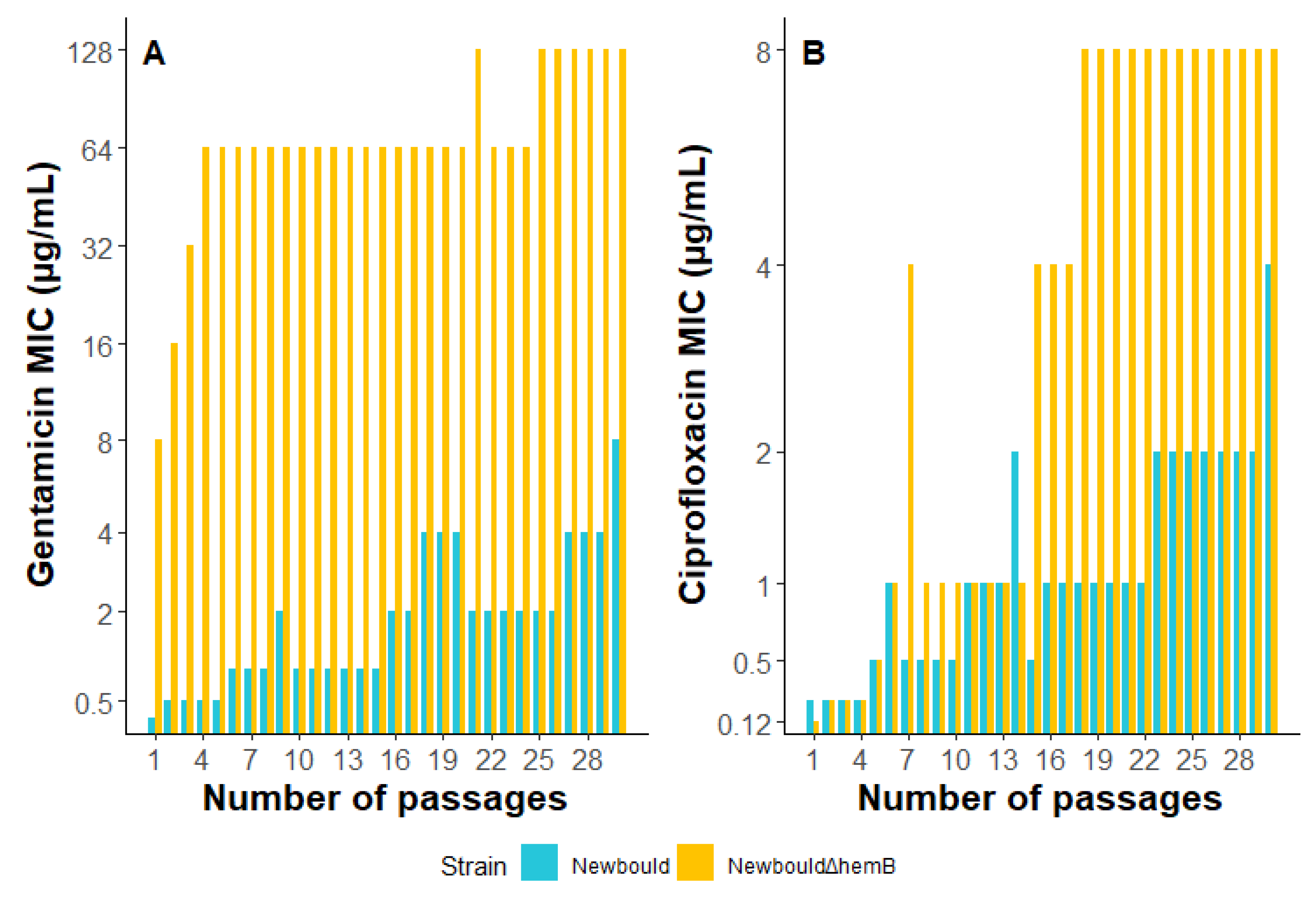
2.8. S. aureus Exposure to Sub-Inhibitory Concentrations of Gentamicin or Ciprofloxacin

S. aureus Newbould and NewbouldΔhemB were sequentially exposed to sub-inhibitory concentrations of gentamicin and ciprofloxacin for a total of 30 passages (Figure 5). A NewbouldΔhemB-resistant population (MIC > 16 µg/mL) emerged after only 2 passages when exposed to gentamicin and even reached an MIC of 128 µg/mL after 30 passages, whereas the bacterial population derived from the prototypical strain Newbould only achieved an intermediate level of resistance (MIC of 8 µg/mL) after all the passages. Furthermore, both strains produced ciprofloxacin-resistant populations (MIC > 4 µg/mL) but Newbould achieved resistance after 30 passages, whereas NewbouldΔhemB only required 15 passages to achieve and maintain resistance. These results suggest that in the presence of antibiotics, SCVs may develop higher levels of resistance, and more rapidly, than prototypical S. aureus.

3. Discussion

S. aureus is one of the most frequently isolated pathogen in the CF respiratory tract [1,7]. Although its prevalence is particularly high in the earlier years of life, it is still common for adults to be infected. This study reports the prevalence and the characteristics of S. aureus in an adult CF patient population. In agreement with previous studies [40,41], genotyping results demonstrated the important diversity of S. aureus isolates that can colonize the airways of CF patients (within and across patients). The co-isolation of S. aureus normal-growth (prototypic) and SCV phenotypes with identical genotype profiles may reflect the phenotype-switching phenomenon previously observed in other studies of CF patients [21,42]. On the other hand, the detection of isolates with similar genotypic profiles among different patients, such as some of the MRSA isolates detected in this study, may reflect nosocomial transmission from non-CF patients or cross-contamination between patients frequenting the same CF clinic. Indeed, CF50A-S (patient 16), CF48A-S, and CF48A-L (patient 7) are all MRSA and were closely related (Figure 1). According to their sequence type, they all belong to the clonal complex 5, which includes hospital-associated MRSA USA100 [43]. While it has frequently been reported for P. aeruginosa [44,45], transmission of S. aureus between patients is not as frequent [46,47]. In our present study, all isolated MRSA showed identical MLVA profile, agr type, and SCCmec type.
In addition to its genetic diversity, S. aureus isolates showed multiple antibiotic resistance profiles (Table 1). Of interest, several aminoglycoside-intermediate or resistant strains of S. aureus for which resistance was not explained by the acquisition of common AMEs (Table 2) were identified in this study. As suggested before, such phenotypes may reflect the adaptability of S. aureus in stressful environments such as the respiratory tract of CF patients [48].
In this study, SCVs were superior biofilm producers compared to prototypical isolates (Figure 2). Biofilms often contain subpopulations of persister cells. It has previously been suggested that metabolically inactive persister cells tolerate high antibiotic exposure, even for concentrations above the determined MIC of the strain [49], and that this favors the emergence of antibiotic resistance [29]. Since SCVs both produce large amounts of biofilm and already share many similarities with persister cells (e.g., slow growth), we investigated whether antibiotics killed SCV and prototypic isolates in the same way (Figure 3). In a time-kill assay comparing prototypic and SCV isolates showing identical antibiotic MICs, SCVs demonstrated greater survival than their genetically related prototypic counterparts in the presence of ciprofloxacin or vancomycin. Among the different features allowing vancomycin resistance, two that could apply to SCVs have previously been described: cell wall thickening and a reduced growth rate [50]. While the SCVs’ reduced growth rate is self-explanatory, sigB overexpression, a typical trait of the SCVs [33,51], is associated with cell wall thickening [52] and has also been described in vancomycin-resistant S. aureus strains [53]. As for the SCVs’ increased tolerance to ciprofloxacin, a possible explanation is that the SCV is less susceptible to the reactive oxygen species (ROS) induced by ciprofloxacin [38]. A longer exposure to ciprofloxacin would then be required to obtain a lethal level of ROS in SCVs.
After testing the kill kinetics, it was assessed whether SCVs had a higher mutation frequency compared to prototypic isolates (Figure 4), which is a characteristic of persister cells [29]. Related SCV and prototypical clinical strains were exposed to rifampicin at concentrations exceeding their MIC, and resistant CFUs were counted and normalized to the initial inoculum. SCVs were found to be over 3 times more likely to develop resistance to rifampicin, and the range of mutation frequencies observed for SCVs was wider than that measured for prototypic isolates. Further studies could explore the cause of resistance in the mutants by whole-genome sequencing and what drives some SCVs to acquire resistance more easily. Overall, this increased mutation rate could represent another factor facilitating antibiotic resistance development over time.
The effect of sub-MIC concentrations of gentamicin and ciprofloxacin on the emergence of resistance in SCVs versus prototypic isolates was also investigated (Figure 5). For both antibiotics, SCVs developed higher resistance levels earlier than prototypic isolates. One possible explanation is the greater tolerance of SCVs to killing by antibiotics, which allows a larger proportion of the population to survive (Figure 3). With this, combined with the higher frequency of mutations shown in Figure 4, SCVs would then be more prone to developing beneficial mutations for resistance in fewer passages (Figure 5). However, the precise cause of resistance was not determined. Whole-genome sequencing of the isolates at different passages could provide valuable information about intermediate-step mutations and ultimate full resistance mutations occurring in such experiments. Furthermore, additional passages in antibiotic-free media could be performed to verify if the resistance of both SCV and prototype is maintained. Further research is needed to better understand the specific mechanisms of resistance to the antibiotics.
New studies investigating the impact of SCVs on patient health should include robust and extensive means of detection of SCVs, considering unknown auxotrophies. Most of the SCVs retrieved in this study were auxotroph for an unknown factor. As stated earlier, the SCV phenotype may be triggered in several forms [8,17,31,32,33,34,35,36,37,38,54,55] and many may remain undiscovered in the clinical laboratory. Notably, while we have tested for the most common auxotrophies (hemin, menadione, and thymidine), we did not test reversion of the SCV phenotype by CO2 or fatty acid complementation, which also have previously been described to be responsible for the SCV phenotype [55,56,57,58]. Among our collection of isolates, we found a menadione-dependent SCV (patient 7, CF21A-S) that could be complemented for normal growth and pigmentation by its co-prototypical strain (CF21A-L, Figure S1). A similar phenomenon was previously observed with thymidine-dependent SCVs [59], but never with SCVs of other auxotrophies. Even if factors providing normal growth to SCVs remain to be determined, inter-strain complementation may importantly contribute to the course of an infection. Indeed, the capacity of SCVs to grow normally in the presence of their prototypical counterparts may increase their fitness in S. aureus-favorable environments and inversely allow a rapid return to the SCV persistent phenotype in harsh environments [42].
The use of MLVA and agr typing (Table 1), and to some extent whole-genome sequencing (Figure 1), allowed the identification of sequential clonal strains and prototypic–SCV pairs persisting in patients. Their presence certainly represents the capacity of S. aureus to colonize adult CF airways successfully for months. Since we found SCVs to withstand antibiotics better and to establish resistance faster than prototypic isolates, we propose that SCVs can facilitate faster acquisition of antibiotic resistance in subsequent sequential isolates. Whole-genome sequence analyses should help to identify mutations inducing the SCV phenotype and/or antibiotic resistance. For example, CF5C-S and CF48B-S, two SCVs whose auxotrophies were not supplemented by hemin, menadione, or thymidine, were mutated in the cysG and menA genes, respectively, which encode enzymes involved in either heme or menaquinone biosynthesis (Table S2). Further analysis is required to determine if and how these mutations are responsible for the SCV phenotype, but they most likely affect the respiratory chain. Furthermore, a preliminary review of the SNPs that were used to demonstrate clonality of related prototypic and SCV pairs (Figure 1) identified some SNPs impacting DNA repair among different SCVs, e.g., dUTP pyrophosphatase, deoxyribose-phosphate aldolase, and recombinase. As previously described, strains defective for DNA repair and maintenance are often hypermutators with an increased mutation rate that favors the emergence of antibiotic resistance [60]. SCVs displaying such SNPs could facilitate acquisition of full antibiotic resistance in either the SCV or prototypical phenotypes since these can be reversible. As an example, SNPs were more closely examined in three sequential isolates showing important changes in their antibiotic susceptibility profiles (Table 2 and Table S3; mutations in Table S3 were confirmed by Sanger sequencing). When isolates CF63A-S and CF63A-L were compared to their sequential clone precursor, CF28A-L, all three isolates showed a frameshift SNP in the ksgA gene. This gene’s functions are to catalyze proper processing of rRNA during ribosome biogenesis and to favor translation fidelity [61]. It also has a DNA glycosylase-apurinic/apyrimidinic site lyase activity, allowing repair of mismatched DNA strands [62]. In previously published articles, it has been shown that ksgA mutations not only conferred modest resistance to kasugamycin, an aminoglycoside, but also favored higher rates of high-level-resistance mutants [63]. These results suggest that the inactivation of ksgA might facilitate the acquisition of mutations for antibiotic resistance, which would be coherent with our observations. Furthermore, CF63A-S and CF63A-L, but not the precursor CF28A-L, share a moderate-impact SNP (amino-acid substitution) in gene lacD (Table S3). Interestingly, a study investigating amicoumacin A resistance in S. aureus identified both ksgA and lacD as mutated genes responsible for an increase in resistance [64]. However, contradictory results were obtained for other aminoglycosides (e.g., kanamycin, gentamicin, and tobramycin), as a ksgA mutant was found to be more susceptible to these [65]. Furthermore, at this time, the role of LacD in S. aureus antibiotic resistance seems farfetched, but the closely related Class I aldolase LacD.1 in Streptococcus pyogenes was proposed to influence the transcription of virulence genes through a direct and specific association with the transcription regulator RopB (Rgg) [66,67,68]. Further experiments should be conducted to evaluate the role of ksgA and lacD in the emergence of antibiotic resistance.
In conclusion, while SCVs have previously been identified as contributing to the persistence of infection in the CF airway [14,21], SCVs can potentially represent yet another challenge since they can acquire mutational antibiotic resistance more easily than prototypical strains (Figure 5). Due to their high biofilm production (Figure 2), ability to tolerate antibiotic action (Figure 3), and increased beneficial mutation rate (Figure 4), SCVs could accumulate useful mutations under selective pressure (e.g., antibiotic therapy) and then revert to a normal-growth and more virulent phenotype with a now reduced susceptibility to antibiotics. This study also identified associated prototypic–SCV MRSA pairs in some patients, which is further cause for concern. Hence, the phenotypic switch from SCVs to prototypic S. aureus and vice versa apparently occurring in patients may represent properties that favor the establishment and persistence of S. aureus in adult CF patients and could also reduce antibiotic susceptibility over time. Since modulators of the CFTR function have been shown to reduce CF lung colonization by MRSA [69], it remains to be seen if such novel therapies could also prevent the emergence of SCVs and break this vicious circle.

4. Materials and Methods

4.1. Reference Strains and Growth Conditions

The S. aureus reference strains used in this study were ATCC 29213, Newbould (ATCC 29740), and its stable SCV derivative NewbouldΔhemB [70]. ATCC 29213 was used for quality control in antibiotic susceptibility tests with clinical isolates, while Newbould and NewbouldΔhemB were used for serial passage on sub-inhibitory concentrations of gentamicin and ciprofloxacin. Unless otherwise stated, TSA and TSB (BD, Mississauga, ON, Canada) were generally used as growth media.

4.2. Patients and Specimens

A total of 31 adult patients (12 males and 19 females) regularly treated at the Cystic Fibrosis outpatient clinic of the Centre Hospitalier Universitaire de Sherbrooke (CHUS, Sherbrooke, QC, Canada) were enrolled in the study from 2008 to 2018. The age of patients ranged from 18 to 41 years old (mean of 25). Specimens were obtained from sputum samples or deep throat swabs when no bronchial secretions were produced. For each patient, specimens from a routine visit or hospitalization (exacerbations) were collected and analyzed. Every patient visit was separated by three to eight months. This study was approved by the ethics review board of the Centre Intégré Universitaire de Santé et de Services Sociaux de l’Estrie (CIUSSS)—CHUS, and prior informed consent was obtained from all subjects recruited in this study (ethics protocol #2008-82, 06-158).

4.3. Microbiology of Clinical Samples

For all specimens, detection of S. aureus was performed using routine clinical laboratory methods. To further confirm the presence of prototypical S. aureus and to facilitate detection of SCVs, a portion of each sample was also treated as follows. For sputum samples, 100 µLof dithiothreitol (100 mg/mL) was added to 100 mg of sputum [71,72]. From this solution, 100 µL was then added to 10 mL of “NaCl broth” that consisted of 7.5 g of tryptone, 1.25 g of neutralized liver digest, 2.5 g of yeast extract, 37.5 g of NaCl, 5 g of (D-) mannitol, 2 g of glucose, and 0.5 g of thymidine in a final volume of 500 mL. For deep throat specimens, swabs were directly used to inoculate 10 mL of NaCl broth. Cultures were aerobically incubated at 35 °C with shaking (225 rpm) for 48 h. Aliquots were then spread on two different media. Mannitol-salt agar (MSA) was used for detection of prototypical S. aureus, while brain-heart infusion agar (BHIA) supplemented with 10 µg/mL of nalidixic acid, 10 µg/mL of polymyxin B, and 2 µg/mL of gentamicin (BHIA-SCV) was used to suppress Pseudomonas sp. and to support the growth of SCVs [71,73,74,75].
From each sample, a maximum of 8 prototypical S. aureus and SCV colonies were isolated from MSA or BHIA-SCV. Very small and non-pigmented colonies were considered to be SCVs. All prototypical colonies and SCVs were confirmed to be S. aureus by PCR detection of the femA gene [76] and of the 668 pb product amplified from the nuc gene by using the following primer sequences: nuc FWD 5′-GGCATCTAGAGCTAAGTCGTGGCATATGTATGGC-3′ and nuc REV 5′-CCGCACTAGTCCTTGACCTGAATCAGCGTTG-3′. The mecA gene coding for the low-affinity penicillin-binding protein responsible for methicillin resistance in S. aureus [77] was also identified by PCR. All isolates were kept frozen at −80 °C. The S. aureus clinical isolates obtained are described in Table 1.

4.4. Genotyping and Selection of Representative Isolates

Multiple-locus variable-number of tandem repeat analysis (MLVA) was use to address the genetic profiles of confirmed S. aureus colonies [78]. The MLVA is based on five loci: sdr, clfA, clfB, ssp, and spa. Band profiles were both visually evaluated and analyzed by using Quantity One software (version 4.6.3 BioRad, Munich, Germany). Any two MLVA patterns differing by one or more bands were considered distinct types. Accessory gene regulator (agr) typing was carried out by group-specific multiplex PCR amplification, as described previously [79]. For each patient, a selection of representative isolates was made based on MLVA and agr profiles, the presence or absence of mecA (and SCCmec types—see below), the normal or SCV phenotype, and the various auxotrophies for SCVs. Clones were defined as strains with the same MLVA and agr profile, while sequential clonal strains were defined as clones isolated from distinct visits of the same patients.

4.5. Genome Sequencing and Annotation

To confirm the clonality of clinical isolates previously determined by MLVA typing, 23 sequential clonal strains taken from lineages that acquired significant antibiotic resistance and included SCV isolates were further analyzed using Illumina sequencing. DNA was first extracted using the GenElute bacterial genomic DNA kit (Sigma–Aldrich, Oakville, ON, Canada). Purifications were made on double solid phase reversible immobilization (SPRI) with carboxyl-coated magnetic beads with an adjusted ratio of 0.7, to obtain fragments longer than 300 bp. Using the QIAseq FX DNA library kit (Qiagen, Toronto, ON, Canada), adapters were ligated, then the libraries were amplified by PCR and validated using a 2100 Bioanalyzer (Agilent Technologies, Mississauga, ON, Canada). The Quant-iT PicoGreen dsDNA assay kit (Invitrogen, Burlington, ON, Canada) was used to quantify gDNA before pooling libraries. Library pool sequencing was performed on an Illumina HiSeq 2000 sequencing system at the Centre d’innovation Genome Quebec platform to generate 125 bp paired-ends reads. Sequenced data quality was verified using the FastQC control tool v0.11.4. Briefly, sequencing reads where trimmed using trimmomatic v0.32 with the following parameters: ILLUMINACLIP:new_illumina_adaptors.fa:2:30:15 TRAILING:30 MINLEN:50. Afterwards, reads were aligned with bwa mem v0.7.10 using reference genome from NCBI Staphylococcus aureus subsp. aureus str. Newman NC_009641.1 and sorted by coordinates using picard v1.123. GATK v3.3.0 RealignerTargetCreator and IndelRealigner steps were run on aligned reads. Picard v1.123 MarkDuplicates is executed to locate and tag duplicate reads in a BAM along with metrics file. Next, the VCF file was generated using GATK v3.3.0 HaplotypeCaller module on previously generated bam using these parameters: --emitRefConfidence GVCF --variant_index_type LINEAR --variant_index_parameter 128000 -dt none -nct 1. Finally, the VCF file was annotated using the snpEff v3.6 using database Staphylococcus_aureus_subsp_aureus_str_newman and the following parameters: -noStats -ud 0.
Finally, a phylogenetic tree was created by comparing the SNP difference between the sequenced strains. All VCF files from every strain were imported to a custom Sqlite database and queried for high-impact snps. These high-impact snps were then clustered using R 4.1.2 with hclust, using the complete method where distance was computed using the dist function and the manhattan method. Cran package gplots was used to generate heatmap. Sequences of the 23 sequential clonal strains were submitted on PubMLST (https://pubmlst.org/saureus/ accessed on 15 May 2023) to determine their sequence type, using 7 house-keeping genes (arcC, aroE, glpF, gmk, pta, tpi, and yqi).

4.6. Minimal Inhibitory Concentrations (MICs)

Antibiotic susceptibility profiles were evaluated by a broth microdilution method as recommended by the Clinical and Laboratory Standards Institute [80] with some modifications. Trimethoprim-sulfamethoxazole (T-S at a ratio of 1:19) was evaluated using Cation Adjusted Mueller-Hinton Broth (CAMHB). Tobramycin (TOB), gentamicin (GEN), ciprofloxacin (CIP), erythromycin (ERY), tetracycline (TET), rifampicin (RIF), and vancomycin (VAN) were tested using BHI to allow substantial growth of all S. aureus isolates including SCVs. Resistance for clindamycin (CLI) was tested for all isolates of the first visit (Table 1, V1) and erythromycin-resistant isolates were further tested for inducible resistance to CLI by the ERY-CLI D-test [80]. For thymidine-dependent SCVs, which showed reversion in BHI, MICs for GEN, TOB, and T-S were performed using CAMHB. SCVs with unknown auxotrophy were tested using both BHI and CAMHB. Oxacillin (OXA) was tested in CAMHB supplemented with 2% NaCl. All tested antibiotics were purchased from Sigma-Aldrich (Oakville, ON, Canada).

4.7. Staphylococcal Cassette Chromosome mec (SCCmec) Typing

SCCmec types were detected using a multiplex PCR assay described previously [81] on all oxacillin-resistant isolates (≥4 µg/mL; referred as MRSA in the text).

4.8. Aminoglycoside Resistance Determinants

The aminoglycoside-resistant isolates in this study were screened by PCR for the presence of genes encoding aminoglycoside-modifying enzymes (AMEs) as described in Mitchell et al. [82].

4.9. Auxotrophy of SCVs

Auxotrophy of SCVs for hemin, thymidine, and menadione was determined using an agar diffusion method. Directly taken from the frozen culture stock and following overnight growth on TSA, 3 to 4 SCV colonies were pooled and evenly spread onto new TSA plates. Wells were punched into the TSA and supplements were added as follows: hemin or menadione each at 10 µg/well; a mixture of both hemin (10 µg) and menadione (10 µg) in the same well; and thymidine at 100 µg/well. Auxotrophy was detected by abundant growth of normal-sized colonies around wells containing specific supplement or combination of supplements after 18 h of incubation at 35 °C. The assay was carried out in duplicate.

4.10. Biofilm Production

The extent of biofilm production was measured by spectrophotometry after growth in 96-well microtiter plates using crystal violet staining, as previously described [17]. The results were collected from at least three independent experiments in which each strain’s biofilm formation was measured in four replicates. The mean biofilm production of each individual strain was expressed relatively to the mean biofilm production of the calibrator strain S. epidermidis ATCC 35984 (100%) measured at the same time in each 96-well plate and all experiments.

4.11. Kill Kinetics

Antibiotic efficacy against SCVs and prototypical isolates was studied in time-kill experiments, as described in Boulanger et al. [83]. Pairs of genetically related SCVs and prototypical isolates co-isolated in patients were selected for this in vitro study. Bacteria were inoculated at 105–106 CFU/mL. BHI was used for SCVs and CAMHB was used for prototypical isolates; these conditions were chosen so that their growth density could be comparable after 24 h. The cultures were incubated for 24 h at 35 °C with shaking (225 rpm). Samples were taken prior to the addition of antibiotics (T = 0) and at several time points during the 24 h incubation period. Samples were serially diluted and plated on TSA to determine viable bacterial counts. TSA plates were incubated 24 h at 35 °C for prototypical isolates, while for SCVs the incubation was prolonged to 48 h at 35 °C to allow detection of growth. Ciprofloxacin and vancomycin were used at the MIC of each pair of strains. Data were collected from at least three independent assays.

4.12. Selection of Rifampicin-Resistant Mutants in Prototypic and SCV Backgrounds

Pairs of genetically related SCVs and prototypical isolates co-isolated in patients were selected for this experiment. The selected SCVs (-S) and prototypic (-L) isolates were: Patient 1, CF37A-S, CF54A-S, CF37A-L, CF54A-L; Patient 3, CF63A-S, CF28A-L, CF63A-L; Patient 5, CF6A-S, CF6C-S, CF39A-S, CF6B-L, CF39A-L; Patient 7, CF21A-S, CF48A-S, CF48B-S, CF21A-L, CF48A-L; Patient 8, CF51B-S, CF51A-L; Patient 16, CF50A-S, CF50A-L. Bacteria were suspended in PBS, and approximately 3 × 10⁸ CFU were plated in triplicates on TSA plates containing 0.3 µg/mL of rifampicin (i.e., ~10–30 × MIC). Plates were incubated 24 h at 35 °C for prototypical isolates, while for SCVs the incubation was prolonged to 48 h at 35 °C to allow detection of growth. CFUs were normalized with the exact initial inoculum on plates. Data were collected from three independent assays.

4.13. Selection of Resistance Using Sub-Inhibitory Concentrations of Antibiotics

Sequential passages into broth containing sub-inhibitory concentrations of gentamicin or ciprofloxacin were used to compare the rate of emergence of resistant mutants from the prototypical S. aureus strain Newbould and its SCV derivative NewbouldΔhemB. In 96-well plates, gentamicin and ciprofloxacin were serially diluted 2-fold in TSB, yielding concentrations ranging from 128 to 0.25 µg/mL and 8 to 0.015 µg/mL, respectively. A bacterial suspension of 105 to 106 CFU/mL was then inoculated into the wells. Following a 24 h or 48 h incubation at 35 °C for Newbould and NewbouldΔhemB, respectively, MICs were determined, and cultures growing one dilution below the MIC breakpoint were used to inoculate a new series of plates containing antibiotics. A total of 30 passages were completed for each strain and MICs were determined at every passage.

Supplementary Materials

The following supporting information can be downloaded at: https://www.mdpi.com/article/10.3390/antibiotics12061069/s1, Table S1: Clonal diversity and antibiotic minimal inhibitory concentrations (MICs) for 77 S. aureus isolates from 18 adult CF patients. Table S2: Possible SCV-inducting high-impact SNP mutations found in isolates CF5C-S and CF48B-S of unknown auxotrophy. Table S3: High- and moderate-impact SNP mutations found in CF28A-L, CF63A-L and CF63A-S isolates showing changes in antibiotic susceptibility over time. Figure S1: Auxotrophy complementation of a SCV by its co-isolated prototypical isolate.

Author Contributions

Conceptualization, F.M., A.M.C.; methodology, G.M., D.L.S. and F.M.; software J.-F.L.; validation, G.M., D.L.S. and F.M.; formal analysis, D.L.S., G.M., C.I. and F.M.; investigation, D.L.S., G.M. and C.I.; resources, F.M., S.R. and A.M.C.; data curation, D.L.S., G.M., S.C. and J.-F.L.; writing—original draft preparation, G.M., D.L.S. and S.C.; writing—review and editing, F.M., S.C. and G.M.; visualization, G.M., D.L.S., S.C. and F.M.; supervision, F.M., A.M.C. and S.R.; project administration, F.M.; funding acquisition, F.M. and A.M.C. All authors have read and agreed to the published version of the manuscript.

Funding

This research was funded by Cystic Fibrosis Canada, grant number 3010. The authors also acknowledge support from the Natural Sciences and Engineering Research Council of Canada (grant number 2020-04811 to F.M.). G.M. was a recipient of an Alexandre-Graham-Bell doctoral research studentship from NSERC and a studentship from the Fonds de Recherche du Québec-Nature et Technology (FRQ-NT) during this study.

Institutional Review Board Statement

The study was conducted in accordance with the Declaration of Helsinki and approved by the Ethics Committee of Centre Intégré Universitaire de Santé et de Services Sociaux de l’Estrie (CIUSSS)-CHUS (protocol #2008-82, 06-158, 24 July 2007, and renewed each year).

Informed Consent Statement

Informed consent was obtained from all subjects involved in the study.

Data Availability Statement

Genome sequences are available on NCBI (SRA data: PRJNA973120 Temporary Submission ID: SUB13343251) and MICs are available in Table S1.

Acknowledgments

We thank all CF patients for their willingness and enthusiasm in participating in this research project. We also thank the personnel from the CF clinic and from the clinical microbiology laboratory of the CHUS for the initial handling of clinical specimens. Finally, we thank Calcul Québec, Compute Canada, and the Centre de Calcul Scientifique of the Université de Sherbrooke for access and technical support in using the Mammouthmp2 supercomputer.

Conflicts of Interest

The authors declare no conflict of interest.

References

  1. The Canadian Cystic Fibrosis Registry Annual Report. In Cystic Fibrosis Canada [Internet]. 2023, pp. 1–54. Available online: www.cysticfibrosis.ca (accessed on 1 May 2023).
  2. 2021 Annual Data Report—Cystic Fibrosis Foundation Patient Registry. 2022. Available online: https://www.cff.org/medical-professionals/patient-registry (accessed on 1 May 2023).
  3. Armstrong, D.S.; Hook, S.M.; Jamsen, K.M.; Nixon, G.M.; Carzino, R.; Carlin, J.B.; Robertson, C.F.; Grimwood, K. Lower airway inflammation in infants with cystic fibrosis detected by newborn screening. Pediatr. Pulmonol. 2005, 40, 500–510. [Google Scholar] [CrossRef]
  4. Sagel, S.D.; Gibson, R.L.; Emerson, J.; McNamara, S.; Burns, J.L.; Wagener, J.S.; Ramsey, B.W. Inhaled Tobramycin in Young Children Study Group; Cystic Fibrosis Foundation Therapeutics Development Network. Impact of Pseudomonas and Staphylococcus infection on inflammation and clinical status in young children with cystic fibrosis. J. Pediatr. 2009, 154, 183–188. [Google Scholar] [CrossRef] [Green Version]
  5. Cogen, J.D.; Hall, M.; Faino, A.V.; Ambroggio, L.; Blaschke, A.J.; Brogan, T.V.; Cotter, J.M.; Gibson, R.L.; Grijalva, C.G.; Hersh, A.L.; et al. Antibiotics and outcomes of CF pulmonary exacerbations in children infected with MRSA and Pseudomonas aeruginosa. J. Cyst. Fibros. 2022, 22, 313–319. [Google Scholar] [CrossRef] [PubMed]
  6. Ahlgren, H.G.; Benedetti, A.; Landry, J.S.; Bernier, J.; Matouk, E.; Radzioch, D.; Lands, L.C.; Rousseau, S.; Nguyen, D. Clinical outcomes associated with Staphylococcus aureus and Pseudomonas aeruginosa airway infections in adult cystic fibrosis patients. BMC Pulm. Med. 2015, 15, 67. [Google Scholar] [CrossRef] [PubMed] [Green Version]
  7. Schwerdt, M.; Neumann, C.; Schwartbeck, B.; Kampmeier, S.; Herzog, S.; Görlich, D.; Dübbers, A.; Große-Onnebrink, J.; Kessler, C.; Küster, P.; et al. Staphylococcus aureus in the airways of cystic fibrosis patients—A retrospective long-term study. Int. J. Med. Microbiol. 2018, 308, 631–639. [Google Scholar] [CrossRef]
  8. Wolter, D.J.; Emerson, J.C.; McNamara, S.; Buccat, A.M.; Qin, X.; Cochrane, E.; Houston, L.S.; Rogers, G.B.; Marsh, P.; Prehar, K.; et al. Staphylococcus aureus small-colony variants are independently associated with worse lung disease in children with cystic fibrosis. Clin. Infect. Dis. 2013, 57, 384–391. [Google Scholar] [CrossRef]
  9. Junge, S.; Görlich, D.; den Reijer, M.; Wiedemann, B.; Tümmler, B.; Ellemunter, H.; Dübbers, A.; Küster, P.; Ballmann, M.; Koerner-Rettberg, C.; et al. Factors Associated with Worse Lung Function in Cystic Fibrosis Patients with Persistent Staphylococcus aureus. Omri A, editor. PLoS ONE 2016, 11, e0166220. [Google Scholar] [CrossRef] [Green Version]
  10. Wolter, D.J.; Onchiri, F.M.; Emerson, J.; Precit, M.R.; Lee, M.; McNamara, S.; Nay, L.; Blackledge, M.; Uluer, A.; Orenstein, D.M.; et al. Prevalence and clinical associations of Staphylococcus aureus small-colony variant respiratory infection in children with cystic fibrosis (SCVSA): A multicentre, observational study. Lancet Respir. Med. 2019, 7, 1027–1038. [Google Scholar] [CrossRef] [PubMed]
  11. Harik, N.S.; Com, G.; Tang, X.; Melguizo Castro, M.; Stemper, M.E.; Carroll, J.L. Clinical characteristics and epidemiology of methicillin-resistant Staphylococcus aureus (MRSA) in children with cystic fibrosis from a center with a high MRSA prevalence. Am. J. Infect. Control. 2016, 44, 409–415. [Google Scholar] [CrossRef]
  12. Dasenbrook, E.C.; Checkley, W.; Merlo, C.A.; Konstan, M.W.; Lechtzin, N.; Boyle, M.P. Association between respiratory tract methicillin-resistant Staphylococcus aureus and survival in cystic fibrosis. JAMA 2010, 303, 2386–2392. [Google Scholar] [CrossRef] [Green Version]
  13. Kahl, B.C. Impact of Staphylococcus aureus on the pathogenesis of chronic cystic fibrosis lung disease. Int. J. Med. Microbiol. 2010, 300, 514–519. [Google Scholar] [CrossRef] [PubMed]
  14. Kahl, B.C.; Becker, K.; Löffler, B. Clinical Significance and Pathogenesis of Staphylococcal Small Colony Variants in Persistent Infections. Clin. Microbiol. Rev. 2016, 29, 401–427. [Google Scholar] [CrossRef] [Green Version]
  15. Miller, M.H.; Edberg, S.C.; Mandel, L.J.; Behar, C.F.; Steigbigel, N.H. Gentamicin uptake in wild-type and aminoglycoside-resistant small-colony mutants of Staphylococcus aureus. Antimicrob. Agents Chemother. 1980, 18, 722–729. Available online: http://www.ncbi.nlm.nih.gov/pubmed/7447428 (accessed on 1 May 2023). [CrossRef] [Green Version]
  16. Proctor, R.A.; Von Eiff, C.; Kahl, B.C.; Becker, K.; McNamara, P.; Herrmann, M.; Peters, G. Small colony variants: A pathogenic form of bacteria that facilitates persistent and recurrent infections. Nat. Rev. Microbiol. 2006, 4, 295–305. [Google Scholar] [CrossRef] [PubMed]
  17. Mitchell, G.; Brouillette, E.; Séguin, D.L.; Asselin, A.-E.; Jacob, C.L.; Malouin, F. A role for sigma factor B in the emergence of Staphylococcus aureus small-colony variants and elevated biofilm production resulting from an exposure to aminoglycosides. Microb. Pathog. 2010, 48, 18–27. [Google Scholar] [CrossRef] [PubMed]
  18. Mitchell, G.; Lamontagne, C.-A.; Brouillette, E.; Grondin, G.; Talbot, B.G.; Grandbois, M.; Malouin, F. Staphylococcus aureus SigB activity promotes a strong fibronectin-bacterium interaction which may sustain host tissue colonization by small-colony variants isolated from cystic fibrosis patients. Mol. Microbiol. 2008, 70, 1540–1555. [Google Scholar] [CrossRef]
  19. Moisan, H.; Brouillette, E.; Jacob, C.L.; Langlois-Bégin, P.; Michaud, S.; Malouin, F. Transcription of virulence factors in Staphylococcus aureus small-colony variants isolated from cystic fibrosis patients is influenced by SigB. J. Bacteriol. 2006, 188, 64–76. [Google Scholar] [CrossRef] [Green Version]
  20. Balwit, J.M.; van Langevelde, P.; Vann, J.M.; Proctor, R.A. Gentamicin-resistant menadione and hemin auxotrophic Staphylococcus aureus persist within cultured endothelial cells. J. Infect. Dis. 1994, 170, 1033–1037. Available online: http://www.ncbi.nlm.nih.gov/pubmed/7930701 (accessed on 1 May 2023). [CrossRef]
  21. Tuchscherr, L.; Löffler, B.; Proctor, R.A. Persistence of Staphylococcus aureus: Multiple Metabolic Pathways Impact the Expression of Virulence Factors in Small-Colony Variants (SCVs). Front. Microbiol. 2020, 11, 1028. [Google Scholar] [CrossRef]
  22. Proctor, R. Respiration and Small Colony Variants of Staphylococcus aureus. Microbiol. Spectr. 2019, 7, 1–15. [Google Scholar] [CrossRef] [PubMed]
  23. Rosenfeld, M.; Rayner, O.; Smyth, A.R. Prophylactic anti-staphylococcal antibiotics for cystic fibrosis. Cochrane Database Syst. Rev. 2020, 2020, CD001912. [Google Scholar] [CrossRef]
  24. Lo, D.K.H.; Muhlebach, M.S.; Smyth, A.R. Interventions for the eradication of meticillin-resistant Staphylococcus aureus (MRSA) in people with cystic fibrosis. Cochrane Database Syst. Rev. 2018, 2018, CD009650. [Google Scholar] [CrossRef] [PubMed]
  25. Bell, S.C.; Mall, M.A.; Gutierrez, H.; Macek, M.; Madge, S.; Davies, J.C.; Burgel, P.-R.; Tullis, E.; Castaños, C.; Castellani, C.; et al. The Lancet Respiratory Medicine Commission on the Future of Care of Cystic Fibrosis. Lancet Respir. Med. 2020, 8, 65. [Google Scholar] [CrossRef] [PubMed] [Green Version]
  26. Otto, M. Staphylococcal Biofilms. Microbiol. Spectr. 2018, 6, 4. [Google Scholar] [CrossRef] [PubMed]
  27. Gläser, R.; Becker, K.; Von Eiff, C.; Meyer-Hoffert, U.; Harder, J. Decreased Susceptibility of Staphylococcus aureus Small-Colony Variants toward Human Antimicrobial Peptides. J. Invest. Dermatol. 2014, 134, 2347–2350. [Google Scholar] [CrossRef] [PubMed] [Green Version]
  28. Fisher, R.A.; Gollan, B.; Helaine, S. Persistent bacterial infections and persister cells. Nat. Rev. Microbiol. 2017, 15, 453–464. [Google Scholar] [CrossRef]
  29. Windels, E.M.; Michiels, J.E.; Fauvart, M.; Wenseleers, T.; Van den Bergh, B.; Michiels, J. Bacterial persistence promotes the evolution of antibiotic resistance by increasing survival and mutation rates. ISME J. 2019, 13, 1239–1251. [Google Scholar] [CrossRef]
  30. Kriegeskorte, A.; Lorè, N.I.; Bragonzi, A.; Riva, C.; Kelkenberg, M.; Becker, K.; Proctor, R.A.; Peters, G.; Kahl, B.C. Thymidine-Dependent Staphylococcus aureus Small-Colony Variants Are Induced by Trimethoprim-Sulfamethoxazole (SXT) and Have Increased Fitness during SXT Challenge. Antimicrob. Agents Chemother. 2015, 59, 7265–7272. [Google Scholar] [CrossRef] [Green Version]
  31. Vestergaard, M.; Paulander, W.; Ingmer, H. Activation of the SOS response increases the frequency of small colony variants. BMC Res. Notes. 2015, 8, 749. [Google Scholar] [CrossRef] [Green Version]
  32. Hoffman, L.R.; Deziel, E.; D’Argenio, D.A.; Lepine, F.; Emerson, J.; McNamara, S.; Gibson, R.L.; Ramsey, B.W.; Miller, S.I. Selection for Staphylococcus aureus small-colony variants due to growth in the presence of Pseudomonas aeruginosa. Proc. Natl. Acad. Sci. USA 2006, 103, 19890–19895. [Google Scholar] [CrossRef] [Green Version]
  33. Mitchell, G.; Séguin, D.L.; Asselin, A.-E.; Déziel, E.; Cantin, A.M.; Frost, E.H.; Michaud, S.; Malouin, F. Staphylococcus aureus sigma B-dependent emergence of small-colony variants and biofilm production following exposure to Pseudomonas aeruginosa 4-hydroxy-2-heptylquinoline-N-oxide. BMC Microbiol. 2010, 10, 33. [Google Scholar] [CrossRef] [Green Version]
  34. Lightbown, J.W.; Jackson, F.L. Inhibition of cytochrome systems of heart muscle and certain bacteria by the antagonists of dihydrostreptomycin: 2-alkyl-4-hydroxyquinoline N-oxides. Biochem. J. 1956, 63, 130–137. [Google Scholar] [CrossRef] [Green Version]
  35. Vesga, O.; Groeschel, M.C.; Otten, M.F.; Brar, D.W.; Vann, J.M.; Proctor, R.A. Staphylococcus aureus small colony variants are induced by the endothelial cell intracellular milieu. J. Infect. Dis. 1996, 173, 739–742. [Google Scholar] [CrossRef] [Green Version]
  36. Bui, L.M.G.; Turnidge, J.D.; Kidd, S.P. The induction of Staphylococcus aureus biofilm formation or Small Colony Variants is a strain-specific response to host-generated chemical stresses. Microbes Infect. 2015, 17, 77–82. [Google Scholar] [CrossRef]
  37. Gao, W.; Chua, K.; Davies, J.K.; Newton, H.J.; Seemann, T.; Harrison, P.; Holmes, N.E.; Rhee, H.-W.; Hong, J.-I.; Hartland, E.; et al. Two novel point mutations in clinical Staphylococcus aureus reduce linezolid susceptibility and switch on the stringent response to promote persistent infection. PLoS Pathog. 2010, 6, e1000944. [Google Scholar] [CrossRef] [PubMed] [Green Version]
  38. Painter, K.L.; Strange, E.; Parkhill, J.; Bamford, K.B.; Armstrong-James, D.; Edwards, A.M. Staphylococcus aureus adapts to oxidative stress by producing H2O2-resistant small-colony variants via the SOS response. Infect. Immun. 2015, 83, 1830–1844. [Google Scholar] [CrossRef] [PubMed] [Green Version]
  39. Taber, H.W.; Mueller, J.P.; Miller, P.F.; Arrow, A.S. Bacterial uptake of aminoglycoside antibiotics. Microbiol. Rev. 1987, 51, 439–457. [Google Scholar] [CrossRef] [PubMed]
  40. Wieneke, M.K.; Dach, F.; Neumann, C.; Görlich, D.; Kaese, L.; Thißen, T.; Dübbers, A.; Kessler, C.; Große-Onnebrink, J.; Küster, P.; et al. Association of Diverse Staphylococcus aureus Populations with Pseudomonas aeruginosa Coinfection and Inflammation in Cystic Fibrosis Airway Infection. mSphere. 2021, 6, e0035821. [Google Scholar] [CrossRef]
  41. Bernardy, E.E.; Petit, R.A.; Raghuram, V.; Alexander, A.M.; Read, T.D.; Goldberg, J.B. Genotypic and phenotypic diversity of Staphylococcus aureus isolates from cystic fibrosis patient lung infections and their interactions with Pseudomonas aeruginosa. MBio 2020, 11, 1–18. [Google Scholar] [CrossRef]
  42. Kittinger, C.; Toplitsch, D.; Folli, B.; Landgraf, L.M.; Zarfel, G. Phenotypic Stability of Staphylococcus aureus Small Colony Variants (SCV) Isolates from Cystic Fibrosis (CF) Patients. Int. J. Environ. Res. Public. Health 2019, 16, 1940. [Google Scholar] [CrossRef] [Green Version]
  43. Grundstad, M.L.; Parlet, C.P.; Kwiecinski, J.M.; Kavanaugh, J.S.; Crosby, H.A.; Cho, Y.-S.; Heilmann, K.; Diekema, D.; Horswill, A.R. Quorum Sensing, Virulence, and Antibiotic Resistance of USA100 Methicillin-Resistant Staphylococcus aureus Isolates. mSphere 2019, 4, e00553-19. [Google Scholar] [CrossRef] [Green Version]
  44. Parkins, M.D.; Somayaji, R.; Waters, V.J. Epidemiology, Biology, and Impact of Clonal Pseudomonas aeruginosa Infections in Cystic Fibrosis. Clinical microbiology reviews. Clin. Microbiol. Rev. 2018, 31, e00019-18. [Google Scholar] [CrossRef] [PubMed] [Green Version]
  45. Thornton, C.S.; Parkins, M.D. Microbial Epidemiology of the Cystic Fibrosis Airways: Past, Present, and Future. In Seminars in Respiratory and Critical Care Medicine; Thieme Medical Publishers, Inc.: New York, NY, USA, 2023. [Google Scholar]
  46. Biggs, S.L.; Jennison, A.V.; Bergh, H.; Graham, R.; Nimmo, G.; Whiley, D. Limited evidence of patient-to-patient transmission of Staphylococcus aureus strains between children with cystic fibrosis, Queensland, Australia. PLoS ONE 2022, 17, e0275256. [Google Scholar] [CrossRef] [PubMed]
  47. Westphal, C.; Görlich, D.; Kampmeier, S.; Herzog, S.; Braun, N.; Hitschke, C.; Mellmann, A.; Peters, G.; Kahl, B.C.; Staphylococcal CF Study Group; et al. Antibiotic Treatment and Age Are Associated with Staphylococcus aureus Carriage Profiles during Persistence in the Airways of Cystic Fibrosis Patients. Front. Microbiol. 2020, 11, 230. [Google Scholar] [CrossRef] [PubMed]
  48. Rumpf, C.; Lange, J.; Schwartbeck, B.; Kahl, B.C. Staphylococcus aureus and cystic fibrosis—A close relationship. What can we learn from sequencing studies? Pathogens 2021, 10, 1177. [Google Scholar] [CrossRef]
  49. Harms, A.; Maisonneuve, E.; Gerdes, K. Mechanisms of bacterial persistence during stress and antibiotic exposure. Science 2016, 354, aaf4268. [Google Scholar] [CrossRef] [PubMed]
  50. Howden, B.P.; Davies, J.K.; Johnson, P.D.R.; Stinear, T.P.; Grayson, M.L. Reduced vancomycin susceptibility in Staphylococcus aureus, including vancomycin-intermediate and heterogeneous vancomycin-intermediate strains: Resistance mechanisms, laboratory detection, and clinical implications. Clin. Microbiol. Rev. 2010, 23, 99–139. [Google Scholar] [CrossRef] [PubMed] [Green Version]
  51. Mitchell, G.; Fugère, A.; Gaudreau, K.P.; Brouillette, E.; Frost, E.H.; Cantin, A.M.; Malouin, F. SigB Is a Dominant Regulator of Virulence in Staphylococcus aureus Small-Colony Variants. PLoS ONE 2013, 8, 1–14. [Google Scholar] [CrossRef] [Green Version]
  52. Morikawa, K.; Maruyama, A.; Inose, Y.; Higashide, M.; Hayashi, H.; Ohta, T. Overexpression of Sigma Factor B, Urges Staphylococcus aureus to Thicken the Cell Wall and to Resist β-Lactams. Biochem. Biophys. Res. Commun. 2001, 288, 385–389. [Google Scholar] [CrossRef]
  53. Chen, H.Y.; Chen, C.C.; Fang, C.S.; Hsieh, Y.T.; Lin, M.H.; Shu, J.C. Vancomycin activates σ B in vancomycin-resistant Staphylococcus aureus resulting in the enhancement of cytotoxicity. PLoS ONE 2011, 6, e24472. [Google Scholar] [CrossRef] [Green Version]
  54. von Eiff, C.; Peters, G.; Becker, K. The small colony variant (SCV) concept—The role of staphylococcal SCVs in persistent infections. Injury. 2006, 37 (Suppl. 2), S26–S33. [Google Scholar] [CrossRef]
  55. Schleimer, N.; Kaspar, U.; Drescher, M.; Seggewiß, J.; von Eiff, C.; Proctor, R.A.; Peters, G.; Kriegeskorte, A.; Becker, K. The energy-coupling factor transporter module EcfAA’T, a novel candidate for the genetic basis of fatty acid-auxotrophic small-colony variants of Staphylococcus aureus. Front. Microbiol. 2018, 9, 1863. [Google Scholar] [CrossRef] [Green Version]
  56. Bhattacharyya, S.; Jaiswal, N.K.; Kumar, M.; Ansari, M.A.A.; Sarfraz, A. Breast abscess in a case of duct atresia caused by CO2-auxotrophic small colony variants of Staphylococcus aureus: Case report and review of literature. J. Nat. Sci. Biol. Med. 2015, 6, 475–476. [Google Scholar] [CrossRef] [PubMed] [Green Version]
  57. Gómez-González, C.; Acosta, J.; Villa, J.; Barrado, L.; Sanz, F.; Orellana, M.A.; Otero, J.R.; Chaves, F. Clinical and molecular characteristics of infections with CO2-dependent small-colony variants of Staphylococcus aureus. J. Clin. Microbiol. 2010, 48, 2878–2884. [Google Scholar] [CrossRef] [PubMed] [Green Version]
  58. Bazaid, A.S.; Forbes, S.; Humphreys, G.J.; Ledder, R.G.; O’Cualain, R.; McBain, A.J. Fatty Acid Supplementation Reverses the Small Colony Variant Phenotype in Triclosan-Adapted Staphylococcus aureus: Genetic, Proteomic and Phenotypic Analyses. Sci Rep. 2018, 8, 3876. [Google Scholar] [CrossRef] [Green Version]
  59. Zander, J.; Besier, S.; Saum, S.H.; Dehghani, F.; Loitsch, S.; Brade, V.; Wichelhaus, T.A. Influence of dTMP on the phenotypic appearance and intracellular persistence of Staphylococcus aureus. Infect. Immun. 2008, 76, 1333–1339. [Google Scholar] [CrossRef] [Green Version]
  60. Oliver, A. Mutators in cystic fibrosis chronic lung infection: Prevalence, mechanisms, and consequences for antimicrobial therapy. Int. J. Med. Microbiol. 2010, 300, 563–572. [Google Scholar] [CrossRef]
  61. Boehringer, D.; O’Farrell, H.C.; Rife, J.P.; Ban, N. Structural insights into methyltransferase KsgA function in 30S ribosomal subunit biogenesis. J. Biol. Chem. 2012, 287, 10453–10459. [Google Scholar] [CrossRef] [Green Version]
  62. Zhang-Akiyama, Q.-M.; Morinaga, H.; Kikuchi, M.; Yonekura, S.-I.; Sugiyama, H.; Yamamoto, K.; Yonei, S. KsgA, a 16S rRNA adenine methyltransferase, has a novel DNA glycosylase/AP lyase activity to prevent mutations in Escherichia coli. Nucleic Acids Res. 2009, 37, 2116–2125. [Google Scholar] [CrossRef] [Green Version]
  63. Ochi, K.; Kim, J.-Y.; Tanaka, Y.; Wang, G.; Masuda, K.; Nanamiya, H.; Okamoto, S.; Tokuyama, S.; Adachi, Y.; Kawamura, F. Inactivation of KsgA, a 16S rRNA methyltransferase, causes vigorous emergence of mutants with high-level kasugamycin resistance. Antimicrob. Agents Chemother. 2009, 53, 193–201. [Google Scholar] [CrossRef] [PubMed] [Green Version]
  64. Lama, A.; Pané-Farré, J.; Chon, T.; Wiersma, A.M.; Sit, C.S.; Vederas, J.C.; Hecker, M.; Nakano, M.M. Response of methicillin-resistant Staphylococcus aureus to amicoumacin a. PLoS ONE 2012, 7, e34037. [Google Scholar] [CrossRef] [PubMed] [Green Version]
  65. Zou, J.; Zhang, W.; Zhang, H.; Zhang, X.D.; Peng, B.; Zheng, J. Studies on aminoglycoside susceptibility identify a novel function of KsgA to secure translational fidelity during antibiotic stress. Antimicrob. Agents Chemother. 2018, 62, 1–13. [Google Scholar] [CrossRef] [PubMed] [Green Version]
  66. Loughman, J.A.; Caparon, M.G. A novel adaptation of aldolase regulates virulence in Streptococcus pyogenes. EMBO J. 2006, 25, 5414–5422. [Google Scholar] [CrossRef] [Green Version]
  67. Loughman, J.A.; Caparon, M.G. Comparative functional analysis of the lac operons in Streptococcus pyogenes. Mol. Microbiol. 2007, 64, 269–280. [Google Scholar] [CrossRef]
  68. Lee, S.J.; Kim, H.S.; Kim, D.J.; Yoon, H.-J.; Kim, K.H.; Yoon, J.Y.; Suh, S.W. Crystal structures of LacD from Staphylococcus aureus and LacD.1 from Streptococcus pyogenes: Insights into substrate specificity and virulence gene regulation. FEBS Lett. 2011, 585, 307–312. [Google Scholar] [CrossRef] [Green Version]
  69. Sheikh, S.; Britt, R.D.; Ryan-Wenger, N.A.; Khan, A.Q.; Lewis, B.W.; Gushue, C.; Ozuna, H.; Jaganathan, D.; McCoy, K.; Kopp, B.T. Impact of elexacaftor-tezacaftor-ivacaftor on bacterial colonization and inflammatory responses in cystic fibrosis. Pediatr. Pulmonol. 2023, 58, 825–833. [Google Scholar] [CrossRef]
  70. Brouillette, E.; Martinez, A.; Boyll, B.J.; Allen, N.E.; Malouin, F. Persistence of a Staphylococcus aureus small-colony variant under antibiotic pressure in vivo. FEMS Immunol. Med. Microbiol. 2004, 41, 35–41. [Google Scholar] [CrossRef] [PubMed] [Green Version]
  71. Gilligan, P.H. Microbiology of airway disease in patients with cystic fibrosis. Clin. Microbiol. Rev. 1991, 4, 35–51. [Google Scholar] [CrossRef] [PubMed]
  72. Hammerschlag, M.R.; Harding, L.; Macone, A.; Smith, A.L.; Goldmann, D.A. Bacteriology of sputum in cystic fibrosis: Evaluation of dithiothreitol as a mucolytic agent. J. Clin. Microbiol. 1980, 11, 552–557. Available online: http://www.ncbi.nlm.nih.gov/pubmed/6776135 (accessed on 1 May 2023). [CrossRef] [Green Version]
  73. Gibson, R.L.; Burns, J.L.; Ramsey, B.W. Pathophysiology and Management of Pulmonary Infections in Cystic Fibrosis. Am. J. Respir. Crit. Care Med. 2003, 168, 918–951. [Google Scholar] [CrossRef] [PubMed]
  74. Estevez, E.G. Bacteriologic plate media: Review of mechanisms of action. Lab. Med. 1984, 15, 258–262. [Google Scholar] [CrossRef]
  75. Murray, C. Manual of Clinical Microbiology, 7th ed.; American Society for Microbiology: Washington, DC, USA, 1999. [Google Scholar]
  76. Hwang, S.Y.; Kim, S.H.; Jang, E.J.; Kwon, N.H.; Park, Y.K.; Koo, H.C.; Jung, W.K.; Kim, J.M.; Park, Y.H. Novel multiplex PCR for the detection of the Staphylococcus aureus superantigen and its application to raw meat isolates in Korea. Int. J. Food Microbiol. 2007, 117, 99–105. [Google Scholar] [CrossRef] [PubMed]
  77. Murakami, K.; Minamide, W.; Wada, K.; Nakamura, E.; Teraoka, H.; Watanabe, S. Identification of methicillin-resistant strains of staphylococci by polymerase chain reaction. J. Clin. Microbiol. 1991, 29, 2240–2244. Available online: http://www.ncbi.nlm.nih.gov/pubmed/1939577 (accessed on 1 May 2023). [CrossRef] [PubMed] [Green Version]
  78. Sabat, A.; Krzyszton-Russjan, J.; Strzalka, W.; Filipek, R.; Kosowska, K.; Hryniewicz, W.; Travis, J.; Potempa, J. New Method for Typing Staphylococcus aureus Strains: Multiple-Locus Variable-Number Tandem Repeat Analysis of Polymorphism and Genetic Relationships of Clinical Isolates. J. Clin. Microbiol. 2003, 41, 1801–1804. [Google Scholar] [CrossRef] [Green Version]
  79. Gilot, P.; Lina, G.; Cochard, T.; Poutrel, B. Analysis of the genetic variability of genes encoding the RNA III-activating components Agr and TRAP in a population of Staphylococcus aureus strains isolated from cows with mastitis. J. Clin. Microbiol. 2002, 40, 4060–4067. [Google Scholar] [CrossRef] [Green Version]
  80. CLSI Clinical and Laboratory Standards Institute. Performance Standards for Antimicrobial Susceptibility Testing; Twenty-Second Informational Supplement; CLSI document M100-S22; Clinical and Laboratory Standards Institute: Wayne, PA, USA, 2018. [Google Scholar]
  81. Zhang, K.; McClure, J.-A.; Elsayed, S.; Louie, T.; Conly, J.M. Novel multiplex PCR assay for characterization and concomitant subtyping of staphylococcal cassette chromosome mec types I to V in methicillin-resistant Staphylococcus aureus. J. Clin. Microbiol. 2005, 43, 5026–5033. [Google Scholar] [CrossRef] [Green Version]
  82. Mitchell, G.; Lafrance, M.; Boulanger, S.; Séguin, D.L.; Guay, I.; Gattuso, M.; Marsault, É.; Bouarab, K.; Malouin, F. Tomatidine acts in synergy with aminoglycoside antibiotics against multiresistant Staphylococcus aureus and prevents virulence gene expression. J. Antimicrob. Chemother. 2012, 67, 559–568. [Google Scholar] [CrossRef] [Green Version]
  83. Boulanger, S.; Mitchell, G.; Bouarab, K.; Marsault, É.; Cantin, A.; Frost, E.H.; Déziel, E.; Malouin, F. Bactericidal effect of tomatidine-tobramycin combination against methicillin-resistant Staphylococcus aureus and Pseudomonas aeruginosa is enhanced by interspecific small-molecule interactions. Antimicrob. Agents Chemother. 2015, 59, 7458–7464. [Google Scholar] [CrossRef] [Green Version]
Figure 1. Phylogenetic tree of SCVs based upon SNP comparison. The phylogeny of the SCVs was assessed upon comparison of the presence/absence of high-impact SNPs (annotated by SNPeff) within each sequenced strain. A heatmap illustrating the dispersion of SNPs was also generated. Each vertical bar represents the presence of an SNP.
Antibiotics 12 01069 g001
Figure 2. Biofilm production of prototypical and SCV S. aureus isolates collected from CF patients. The mean biofilm production for each isolate is indicated on the graph. Blue circles represent prototypic isolates and yellow circles represent SCVs. X symbols represent MRSA isolates. The horizontal bars represent the median relative biofilm production for each group, with the boxes showing the interquartile range and the whiskers indicating the minimum and maximum values (excluding outliers). The mean biofilm production (i.e., each dot) was calculated from at least three independent experiments in which each isolate’s biofilm formation was measured in four replicates. SCVs produced significantly more biofilm than prototypical S. aureus isolates (p-value = 0.0015; Wilcoxon test).
Antibiotics 12 01069 g002
Figure 3. Ciprofloxacin and vancomycin kill kinetics in clinical prototype–SCV pairs. Blue symbols represent prototypic isolates (-L) and yellow symbols represent SCV (-S) isolates. Circles report the control condition (no antibiotic) and triangles report the kill kinetics with the indicated antibiotic. Standard deviations are also indicated. (A) represents CF39A-L and CF39A-S viable counts (CFU/mL); (B) represents CF54A-L and CF54A-S; (C) represents CF78A-L and CF78A-S. The detection limit was 2 log10 CFU/mL. Each condition was tested in triplicates and each symbol represents the mean.
Antibiotics 12 01069 g003
Figure 4. Comparison of rifampicin-induced mutation frequencies between prototypic isolates and SCVs. Ten prototypic isolates and eleven related SCVs were included in this analysis. Mutation frequency was determined by normalizing the number of resistant CFUs growing on the rifampicin plate by the initial inoculum. Blue circles represent prototypes and yellow circles are SCVs. The horizontal bars represent the mutation frequency median for each group, with the boxes showing the interquartile range, and the whiskers indicating the minimum and maximum values (excluding outliers). Results demonstrate that the mutation frequency leading to rifampicin resistance for SCVs is significantly higher than that measured for prototypic isolates (p-value = 0.0079; Wilcoxon test).
Antibiotics 12 01069 g004
Figure 5. Selection of resistance using sequential passages on sub-inhibitory concentrations of antibiotics. S. aureus Newbould (blue bars) and the SCV NewbouldΔhemB (yellow bars) were sequentially exposed to sub-inhibitory concentrations of gentamicin (A) or ciprofloxacin (B) for a total of 30 passages. The bars represent MIC values (the highest value is >128 and >8 µg/mL, respectively, for gentamicin and ciprofloxacin).
Antibiotics 12 01069 g005
Table 1. Clonal diversity, antibiotic susceptibilities, and phenotypic characteristics of 77 S. aureus isolates from 18 adult CF patients.
Patient IDVisitFirst IsolateVariantMLVAagrmecAAntibiotic Susceptibility aAux b
OXATOBGENT-SCIPERYCLITETVANRIF
1V1CF1A-L A2 SSSSSSSSSS
V1 CF1C-SA2 SIISSSSSSSH
V1 CF1D-SA2 ntISntSSRSSSU
V2 CF37B-SA2 SIISRRntSSSU
V3 CF54A-L *A2 SSSSSSntSSS
V4 CF54A-S *A2 SRRSSSntSSSU
V2CF37A-L * B2+RSSSRRntSSS
V2 CF37A-S *B2+RSSSRRntSSSU
2V1CF2A-L C1 SSSSSSSSSS
V3 CF62B-LC1 SSSSSSntSSS
V1CF2C-L D3 SSSSSSSSSS
V1 CF2B-SD3 SISSSSSSSSH
V3 CF62A-LD3 SSSSSSntSSS
V2CF34A-L E1 SSSSSSntSSS
V2CF34B-L F3 SSSSSSntSSS
3V1CF4B-L A2 SSSSSSSSSS
V1 CF4B-SA2 SIISSSSSSSM
V2 CF28B-LA2 SSSSSSntSSS
V2CF28A-L * G2 SSSSSRntSSS
V3 CF63A-SG2 SRSSSRntSSSU
V3 CF63A-L *G2 SRISRRntSSS
4V1CF5A-L * H3 SSSSSSSSSS
V1 CF5C-S *H3 ntIISSSSSSSU
V1CF5E-S I3 SIISSSSSSSH+M
5V1CF6B-L * J3 SRRSSSSSSS
V1 CF6A-S *J3 SRRRSSSSSST
V1 CF6C-S *J3 SRRSSSSSSSH+M
V2 CF39A-L *J3 SRRRSSntSSS
V2 CF39A-S *J3 SRRRSIntSSSU
6V1CF7A-L K2+RRSSRRRSSS
V1 CF7D-LK2 SSSSRRRSSS
V2 CF27A-LK2+RRSSRRntSSS
V3CF49A-L L3 SSSSSSntSSS
V3CF49A-S M3 SISSSSntSSSH
V3 CF49B-SM3 SISSSSntSSSU
7V1CF8A-L N3 SSSSSSSSSS
V2 CF21A-L *N3 SSSSSSntSSS
V2 CF21A-S *N3 SSSSSSntSSSM
V1CF8C-L O3 SSSSSSSSSS
V1 CF21D-LO3 SSSSSSntSSS
V1CF8D-L P3 SSSSSSSSSS
V3CF48A-L * K2+RRSSRRntSSS
V3 CF48A-S *K2+RRSSRRntSSSU
V3 CF48B-S *K2 SRISRRntSISU
8V1CF9A-L K2+RRSSRRR cSSS
V1 CF9F-LK2 SRRSRRISSS
V2CF51A-L * P2 SSSSRRntSSS
V2 CF51B-S *P2 SIISRRntSISU
V3 CF75B-LP2 SSSSRRntSSS
V3CF75A-L Q1 SSSSRSntSSS
9V1CF10B-L * R1 SSSSSRRSSS
V1 CF10A-S *R1 SRRSSRRSSSH + M
10V1CF18A-L S2 SSSSSRR cSSS
V2 CF33A-LS2 SSSSIRntSSS
V3 CF78A-LS2 SISSSSntSSS
V3 CF78A-SS2 SISSSSntSSSM
V3 CF78B-SS2 SISSSSntSSSH
V1CF18E-L T1 SSSSSSSSSS
11V1CF19A-L T1 SSSSRSSSSS
V1CF19B-L U1 SSSSRSSSSS
V1CF19A-S V1 SRRSSSSSSSU
V2CF41A-L W1 SSSSRRntSSS
V2 CF41A-SW1 SISSRSntSSSU
V3CF58A-L X1 SRRRSSntSSS
V3CF58B-L Y1 SISSRSntSSS
12V1CF22A-L Z1 SSSSSSSSSS
13V1CF29A-L AA1 SIISSSRSSS
14V1CF35A-L K2+RSSSRRR cSSS
V2 CF77A-LK2+RSSSRRntSSS
15V1CF43A-L BB3 SSSSRRRSSS
V1CF43B-L CC3 SSSSRRRSSS
16V1CF50A-L K2+RRSSRRSSSS
CF50A-S *K2+RRISRRSSSSH
V2CF111A-L DD2+RRSSRRntSSS
17V2CF86A-S EE2 SSSSSSntRSSH
18V1CF81A-L FF3 SSSSSSSSSS
V2CF91A-L GG1 SSSSSSntSIS
a Ciprofloxacin (CIP), clindamycin (CLI), erythromycin (ERY), gentamicin (GEN), oxacillin (OXA), rifampicin (RIF), tetracycline (TET), tobramycin (TOB), trimethoprim-sulfamethoxazole (T-S), and vancomycin (VAN). Not tested (nt), susceptible (S), intermediate (I), and resistant (R). b Aux, auxotrophy for the normal-growth phenotype: hemin (H), menadione (M), thymidine (T), unknown (U). c These isolates are presumed to be resistant based on the detection of inducible clindamycin resistance. * These isolates were used for whole-genome sequencing.
Table 2. Aminoglycoside susceptibility profiles of genetically related strains for which the presence of aminoglycoside-modifying enzymes (AMEs) does not explain the increase in MIC.
StrainsPatientVisitMLVAmecAMIC (µg/mL) aAMEs b
GENTOBAACANT4ANT9
CF1A-L1V1A 0.25–0.50.5–1---
CF54A-L1V3A 2–42–4---
CF54A-S1V4A >32>32---
CF2C-L2V1D 22---
CF2B-S2V1D 44–8---
CF4B-L3V1A 11---
CF4B-S3V1A 4–84–8---
CF28A-L3V2G 0.50.5---
CF63A-S3V3G 4>32---
CF63A-L3V3G 4–8>32---
CF5A-L4V1H 0.250.5---
CF5C-S4V1H 84–8---
CF7A-L6V1K+0.5>32++-
CF27A-L6V2K+2–4>32++-
CF48A-L7V3K+1>32--+
CF48A-S7V3K+4>32--+
CF51A-L8V2P 2–42–4---
CF51B-S8V2P 8–164–8---
CF10B-L9V1R 0.5–10.5–1---
CF10A-S9V1R >32>32---
a Gentamicin (GEN), tobramycin (TOB). b Aminoglycoside-modifying enzymes (AMEs): aac(6′)-aph(2′′) [AAC], ant(4′)-1 [ANT4], ant(9)-1a [ANT9].
Disclaimer/Publisher’s Note: The statements, opinions and data contained in all publications are solely those of the individual author(s) and contributor(s) and not of MDPI and/or the editor(s). MDPI and/or the editor(s) disclaim responsibility for any injury to people or property resulting from any ideas, methods, instructions or products referred to in the content.

