Newly Developed Electrochemiluminescence Based on Bipolar Electrochemistry for Multiplex Biosensing Applications: A Consolidated Review


Abstract
:1. Introduction
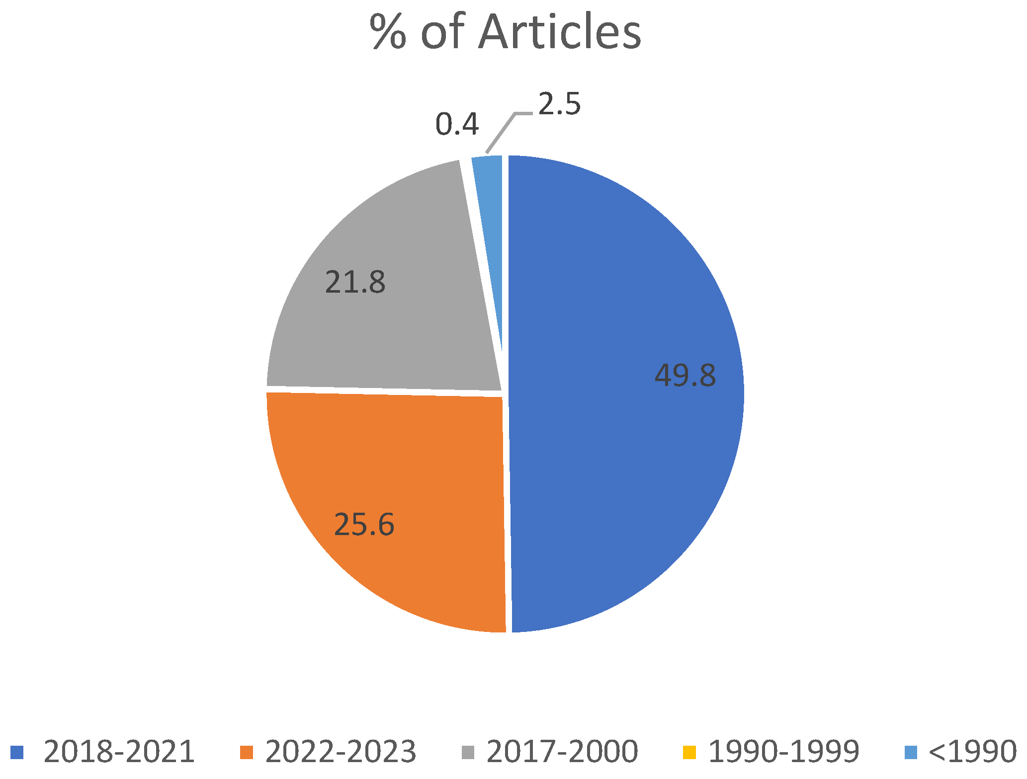
2. Methodology
3. Focus Questions
- (a)
- What are the strengths and limitations of closed bipolar electrochemistry (c-BPE) versus open bipolar electrochemistry (o-BPE) in biosensing applications?
- (b)
- Which configuration of BPE is commonly employed in biosensing for improved multiplex assaying? For what reasons is it predominantly used?
- (c)
- What are the available synthesis and functionalization strategies for bipolar electrodes, and how might they impact their suitability for biosensing applications in ECL-BPE, particularly in multiplex assays?
- (d)
- Over the past half a decade of research, what advancements have been achieved in the development of novel ECL luminophores, and how have these advancements affected the ECL technique?
- (e)
- During the past 5 years, what novel applications of ECL-BPE have emerged for multiplex assaying?
- (f)
- With the focus on research conducted between 2018–2023, what are some of the challenges associated with using ECL-BPE for multiplex assays, and how have they been addressed, if at all?
4. Principles of Bipolar Electrochemistry
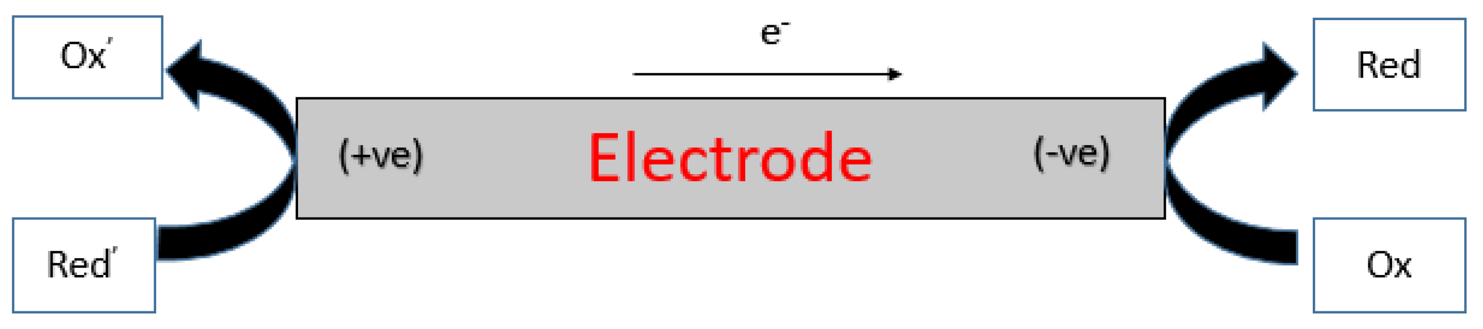
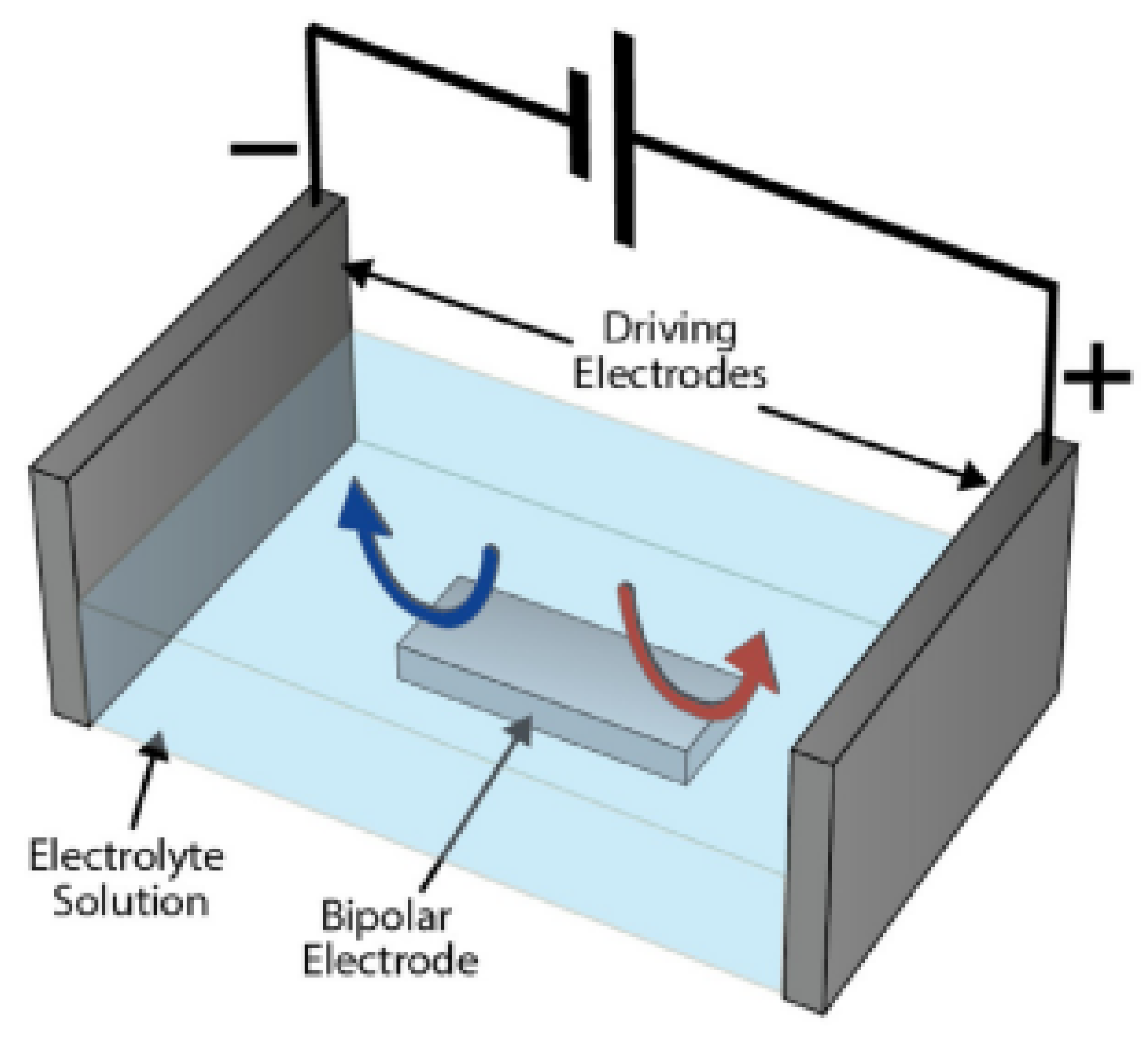
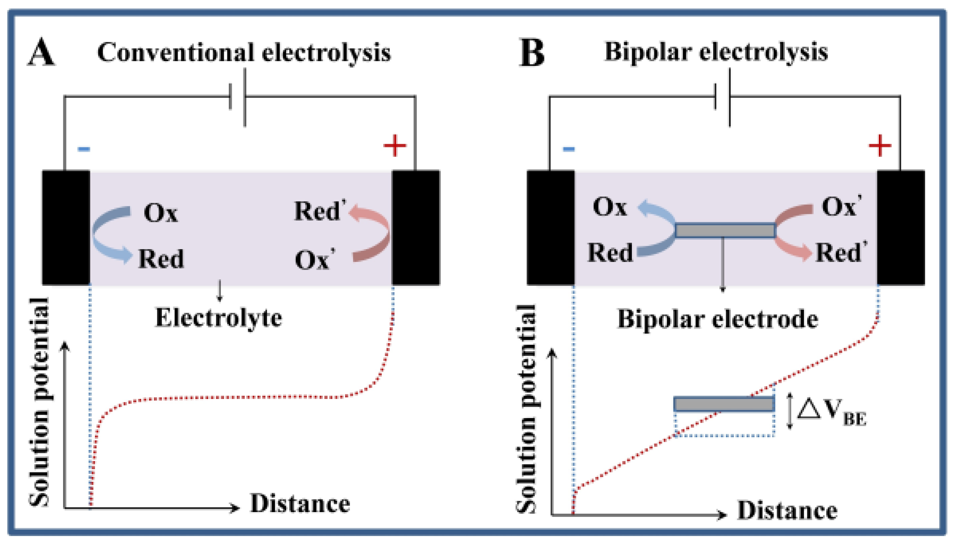
4.1. An Overview of Bipolar Electrochemistry
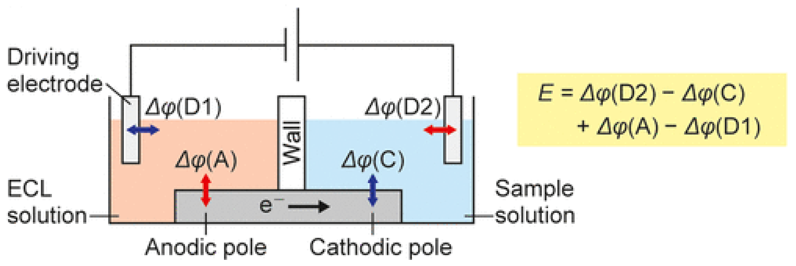
4.2. Fundamentals of Bipolar Electrodes
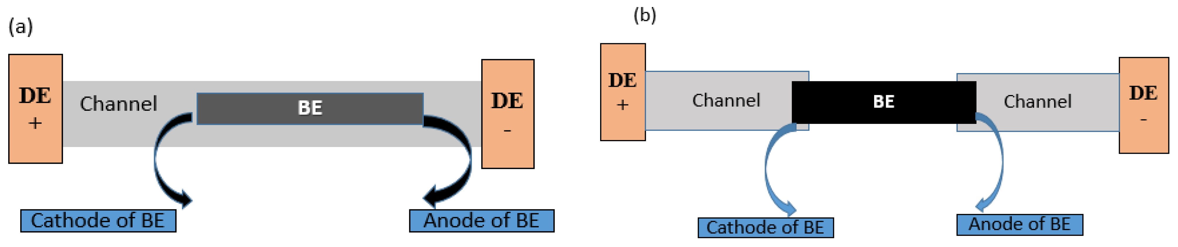
4.3. Configurations of Bipolar Electrochemistry
4.4. Closed vs. Open Bipolar Electrodes: A Comprehensive Review of Their Limitations and Advantages
4.5. Bipolar Electrode Miniaturization: Uncovering the Opportunities and Challenges
4.6. Synthesis and Functionalization Strategies of BEs for Biosensing
5. Fundamentals of Electrochemiluminescence: A Summary
5.1. Recent Advances in ECL Luminophores

| ECL Luminophore | Luminophore Class | Main Co-Reactant | Notable Observations | Ref |
|---|---|---|---|---|
| La3+-BTC MOFs | Inorganic-organic hybrid | - | Prepared as the highly active reactor. | [125] |
| Tr-HOFs | Inorganic metallic/organometallic complexes | - | LoD: 0.28 nM; label-free ECL biosensor; highly improved ECL efficiency that that of Ru(bpy)32+ | [154] |
| Pc gadolinium complex | Metal complex | * In2O3/ZnIn2S4 | Co-reactant possesses unique hollow structure-related advantages. | [156] |
| Zn-MOFs | Inorganic–organic hybrid | - | A co-reactant-free ratiometric ECL biosensor | [157] |
| CDPs and oxidized CDPs | Organic NMs | - | Synthesized by pyrolysis; o-CDPs ECL performances better than CDPs; Ultrasensitive and high selective benign sensor. | [158] |
| Mn doped ZnAgInS/ZnS nanocrystal | NMs | * Snowflake-like MoS2@Cu2S composite | Ultrasensitive, wide linear range, high selectivity, and good stability. | [159] |
| Ru@Zr12-BPDC nanoplate | Organometallic complexes | TPrA | Ultrasensitive detection; LoD of 0.14 fg/mL. | [160] |
| InP/ZnSMPA-MSA | NMs | Co-reactant-free ECL | PSA detection; LoD 0.3 pg·mL−1; protocol simplifies ECL assay procedure and provides an alternative to both annihilation and co-reactant routes. | [161] |
| NH2-Ru@SiO2 NPs | NMs | Nitrogen doped graphene quantum dots | LoD: 1 fg mL−1; self-enhanced luminophore; emitter and co-reactant simultaneously existed in the same NPs | [162] |
| Ru@SiO2-Au nanocomposite | NMs | TPA | Very sensitive H2O2 detection; AuPd NPs used to enhance ECL signal; also served as co-reaction accelerator; nanocomposite quenched by the ferrocene. | [163] |
| Au@SiO2 @ Ru(bpy)32+ doped silica | Organometallic complexes | TPA | Highly sensitive response to glutathione. | [164] |
| Ce3+-Doped TbPO4 | NMs | K2S2O8 | Mucin1 detection; LoD: 0.5 fg·mL−1 study may pave way for further research on novel direct ECL emission of lanthanides | [165] |
| Lu/MoS2 QDs@ZIF-8 | NMs | - | Ultrasensitive detection of microRNA-21; efficiency increased more than luminol–H2O2 system | [166] |
| CD-COF/combined with a CRISPR/Cas12a | NMs | * S2O82−/Bu4N+ | 2.21 fM; introduction of CRISPR/Cas12a system improved sensitivity of the biosensor. | [167] |
| Zr-MOF modified bulk boron carbon oxynitride | Organometallic complexes | S2O82− | 0.2 fg mL−1; high-efficient and low-cost ECL; ultrasensitive detection of breast cancer | [168] |
5.2. Recent Advances in Co-Reactants
| ECL System | LoD and Notable Observations | Ref |
|---|---|---|
| Luminol immobilized on graphite as a paste electrode | 0.7 × 10⁻⁸ μM; two nearly neutral samples spiked with 3% and 6% H2O2. | [176] |
| Luminol nitrogen doped GQDs | 1 × 10⁻⁸ M; detection in water samples. | [185] |
| Luminol-H2O2; Flow injection analysis system with ECL detection | 3.0 × 10⁻⁹ mol/L LoD equal to the level of CL; human serum samples. | [186] |
| Luminol based on synchronous dual sensitization of ECL by using TiNTs and platinum black. | 66 pM (H2O2), 22 nM (resveratrol), and 30 nM (dopamine); human serum samples. | [187] |
| Luminol-based ECL system and fluorine-doped tin oxide making use of electrode modified with Au nanoparticles | 8 nM; Living cell samples. | [188] |
| Luminol-capped AuNP-modified electrode | 1 × 10⁻⁷ mol L−1 | [189] |
5.3. Recent Advances in Electrodes
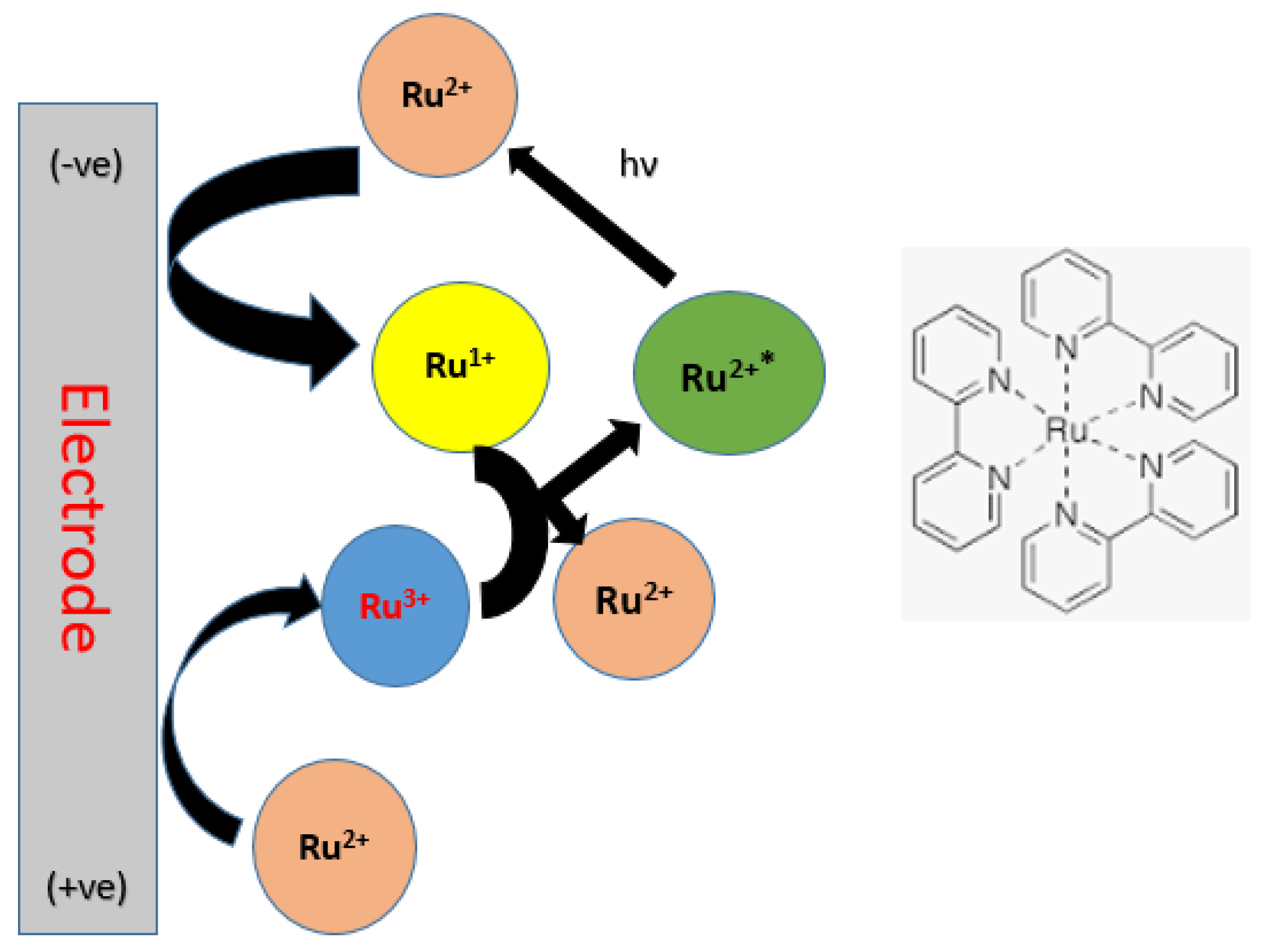
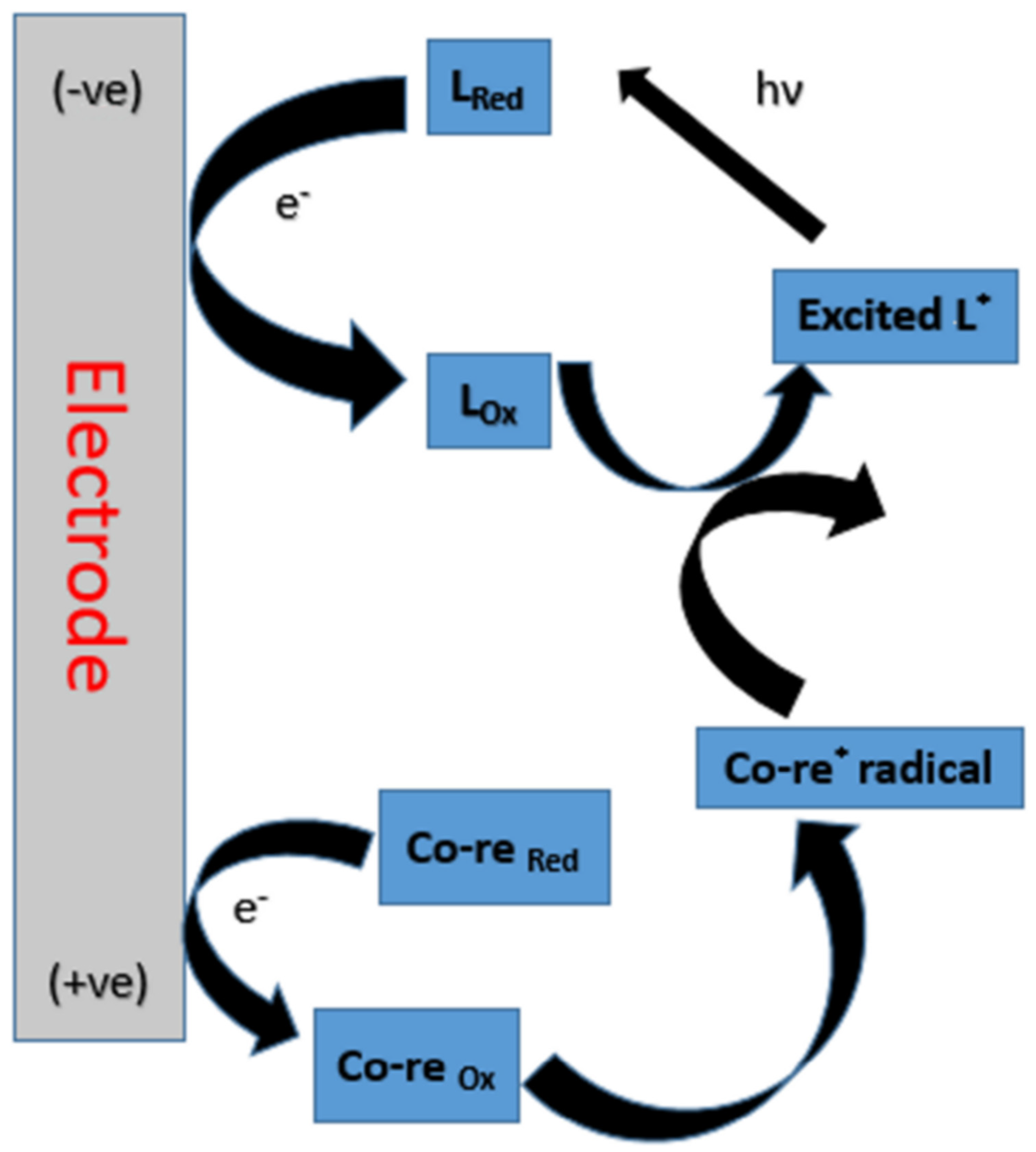
5.4. The Chemistry of ECL Luminophores and Their Mechanism
6. A Critical Review of Recent, Novel Applications of ECL-BPE for Biosensing
| Sample Matrix/Analyte | ECL System | LoD, Nature of BE and Other Notable Remarks | Ref |
|---|---|---|---|
| Blood/carcinoembryonic antigen and H2O2 | Ru(bpy)32+/(NH4)2C2O4/H2O2 | 5.0 pg mL−1 for carcinoembryonic antigen; Pt-graphite paper BE fabricated by depositing Pt NPs. | [41] |
| Blood/ATP, PSA, AFP, and thrombin | Ru(bpy)32+/tripropylamine | Visual detection; Au-ITO hybrid BE; ECL platform enabled sensitive detection and good reproducibility. | [66] |
| Blood/H2O2, vitamin B12, and vitamin C | Luminol/H2O2 | 0.303 μM, 0.109 nM, and 0.96 μM, respectively; laser-induced graphene BE fabricated with polyimide sheet. | [57] |
| Blood/S. typhimurium | ([Ir(ppy)3] and [Ru(bpy)3]2+) | 10 CFU/mL; ITO BE fabricated with immunomagnetic beads. | [67] |
| Blood/multi-assaying of cholesterol, glucose, and lactate | PPC−PBA/Fc/Enzyme interface | 79 μM for cholesterol, 59 μM for glucose, and 86 μM for lactate; powered ITO electrodes. | [77] |
| Blood/cholesterol | Luminol/H2O2 | 0.12 mM; Potential applications in biomedical, food management and in POCT. | [229] |
| Blood/PSA | CdTe QDs and luminol | 0.5 ng/mL (S/N = 3); Au NRs nanocomposite BE. | [230] |
| Blood/aflatoxin M1 | Luminol-functionalized Ag NPs-decorated graphene oxide | 0.01 ng mL−1; gold anodic BE coated with magnetic Fe3O4. | [231] |
| Blood/miRNA-155 and miRNA-126 | CdTe QDs-H2 and Au@g-C3N4 NSs-DNA1/S2O82− | 5.7 and 4.2 fM, respectively; paper-based sensing platform (BE) prepared by wax-printing technology, screen-printing method, and in situ AuNPs. | [76] |
| Plasma/RASSF1A-methylated DNA and SLC5A8 methylated DNA | Luminol loaded into Fe3O4@UiO-66 | Visual detection; HER on Ru NPs electrodeposited on nitrogen-doped graphene-coated Cu foam and electrooxidation of hydrazine on a polycatechol-modified reduced graphene oxide/pencil graphite electrode used as the BE cathodic and anodic reactions, respectively. | [232] |
| Serum/glucose, lactate, and choline. | luminol-H2O2 | 7.57 μM, 8.25 μM and 43.19 μM, respectively: ITO BEs modified to adapt different enzymes; fabricated with: GOD/MWCNTs/CS (LOD/MWCNTs/CS or COD/MWCNTs/C. | [233] |
| Cancer cells/adenosine | Ru(bpy)32+/TPA | 10−15 M; ITO BE modified with complementary single-stranded DNA. | [234] |
| Intracellular/H2O2 and MCF-7 cancer cells | Luminol/H2O2 | 40 cells/mL; AuPd NPs modified BE; wax printing used to fabricate reaction center, and carbon ink-based BE and driving electrodes screen-printed into paper. | [235] |
| H2O2 | Luminol/H2O2 | 0.26 μM; ITO conductive glass as BE. | [236] |
| Blood/cytokeratin 19 fragments | Luminol/H2O2 and O2 | 1.89 pg mL−1; ITO BE; in situ generation of H2O2 and O2 H2O2 and O2 enhanced luminol ECL intensity. | [237] |
7. Conclusions and Prospects
Author Contributions
Funding
Institutional Review Board Statement
Informed Consent Statement
Data Availability Statement
Conflicts of Interest
References
- Wei, H.; Xu, Y.; Xu, W.; Zhou, Q.; Chen, Q.; Yang, M.; Feng, F.; Liu, Y.; Zhu, X.; Yu, M.; et al. Serum Exosomal miR-223 Serves as a Potential Diagnostic and Prognostic Biomarker for Dementia. Neuroscience 2018, 379, 167–176. [Google Scholar] [CrossRef] [PubMed]
- Singh, S.; Kumar, V.; Dhanjal, D.S.; Datta, S.; Prasad, R.; Singh, J. Biological Biosensors for Monitoring and Diagnosis. In Microbial Biotechnology: Basic Research and Applications; Springer: Singapore, 2020; pp. 317–335. [Google Scholar] [CrossRef]
- Su, X.; Sutarlie, L.; Loh, X.J. Sensors, Biosensors, and Analytical Technologies for Aquaculture Water Quality. Research 2020, 2020, 272705. [Google Scholar] [CrossRef] [PubMed] [Green Version]
- Dimitratos, S.D.; Hommel, A.S.; Konrad, K.D.; Simpson, L.M.; Wu-Woods, J.J.; Woods, D.F. Biosensors to monitor water quality utilizing insect odorant-binding proteins as detector elements. Biosensors 2019, 9, 62. [Google Scholar] [CrossRef] [PubMed] [Green Version]
- Villalonga, A.; Sánchez, A.; Mayol, B.; Reviejo, J.; Villalonga, R. Electrochemical biosensors for food bioprocess monitoring. Curr. Opin. Food Sci. 2022, 43, 18–26. [Google Scholar] [CrossRef]
- Kocheril, P.A.; Lenz, K.D.; Mascareñas, D.D.L.; Morales-Garcia, J.E.; Anderson, A.S.; Mukundan, H. Portable Waveguide-Based Optical Biosensor. Biosensors 2022, 12, 195. [Google Scholar] [CrossRef]
- Wasilewski, T.; Neubauer, D.; Kamysz, W.; Gębicki, J. Recent progress in the development of peptide-based gas biosensors for environmental monitoring. Case Stud. Chem. Environ. Eng. 2022, 5, 100197. [Google Scholar] [CrossRef]
- Nnachi, R.C.; Sui, N.; Ke, B.; Luo, Z.; Bhalla, N.; He, D.; Yang, Z. Biosensors for rapid detection of bacterial pathogens in water, food and environment. Environ. Int. 2022, 166, 107357. [Google Scholar] [CrossRef]
- Ye, F.; Zhao, Y.; El-Sayed, R.; Muhammed, M.; Hassan, M. Advances in nanotechnology for cancer biomarkers. Nano Today 2018, 18, 103–123. [Google Scholar] [CrossRef]
- Woo, J.; Kim, J.; Kim, J. Indium tin oxide bipolar electrodes modified with Pt nanoparticles encapsulated inside dendrimers as sensitive electrochemiluminescence platforms. J. Electroanal. Chem. 2022, 906, 115998. [Google Scholar] [CrossRef]
- Liu, G.; Wang, Z.; Lei, C.; Wang, F. An electrochemiluminescence imaging sensor for the analysis of lactate in foods via a single gold microsphere. J. Food Compos. Anal. 2023, 119, 105270. [Google Scholar] [CrossRef]
- Li, X.; Qin, X.; Tian, Z.; Wang, K.; Xia, X.; Wu, Y.; Liu, S. Gold Nanowires Array-Based Closed Bipolar Nanoelectrode System for Electrochemiluminescence Detection of α-Fetoprotein on Cell Surface. Anal. Chem. 2022, 94, 7350–7357. [Google Scholar] [CrossRef]
- Su, Y.; Lai, W.; Liang, Y.; Zhang, C. Novel cloth-based closed bipolar solid-state electrochemiluminescence (CBP-SS-ECL) aptasensor for detecting carcinoembryonic antigen. Anal. Chim. Acta 2022, 1206, 339789. [Google Scholar] [CrossRef]
- Zhu, L.; Lv, X.; Yu, H.; Tan, X.; Rong, Y.; Feng, W.; Zhang, L.; Yu, J.; Zhang, Y. Paper-Based Bipolar Electrode Electrochemiluminescence Platform Combined with Pencil-Drawing Trace for the Detection of M.SssI Methyltransferase. Anal. Chem. 2022, 94, 8327–8334. [Google Scholar] [CrossRef]
- Bhaiyya, M.; Pattnaik, P.K.; Goel, S. Multiplexed and simultaneous biosensing in a 3D-printed portable six-well smartphone operated electrochemiluminescence standalone point-of-care platform. Microchim. Acta 2022, 189, 79. [Google Scholar] [CrossRef]
- Khoshfetrat, S.M.; Dorraji, P.S.; Fotouhi, L.; Hosseini, M.; Khatami, F.; Moazami, H.R.; Omidfar, K. Enhanced electrochemiluminescence biosensing of gene-specific methylation in thyroid cancer patients’ plasma based integrated graphitic carbon nitride-encapsulated metal-organic framework nanozyme optimized by central composite design. Sens. Actuators B Chem. 2022, 364, 131895. [Google Scholar] [CrossRef]
- Fazlali, F.; Hashemi, P.; Khoshfetrat, S.M.; Halabian, R.; Baradaran, B.; Johari-Ahar, M.; Karami, P.; Hajian, A.; Bagheri, H. Electrochemiluminescent biosensor for ultrasensitive detection of lymphoma at the early stage using CD20 markers as B cell-specific antigens. Bioelectrochemistry 2021, 138, 107730. [Google Scholar] [CrossRef]
- Qin, D.; Meng, S.; Wu, Y.; Mo, G.; Deng, B. Aggregation-induced electrochemiluminescence resonance energy transfer with dual quenchers for the sensitive detection of prostate-specific antigen. Sens. Actuators B Chem. 2022, 367, 132176. [Google Scholar] [CrossRef]
- Yang, X.-Y.; Bai, Y.-Y.; Huangfu, Y.-Y.; Guo, W.-J.; Yang, Y.-J.; Pang, D.-W.; Zhang, Z.-L. Ultrasensitive Electrochemiluminescence Biosensor Based on Closed Bipolar Electrode for Alkaline Phosphatase Detection in Single Liver Cancer Cell. Anal. Chem. 2021, 93, 1757–1763. [Google Scholar] [CrossRef]
- Cai, L.; Zhang, J.; Teng, L.; Wang, H.; Fang, G.; Wang, S. Ultrasensitive molecularly imprinted electrochemiluminescence sensor based on highly-conductive rGO-COOH synergically amplify TCPP luminophor signal in aqueous phase system for “switches-controlled” detection of tryptamine. Sens. Actuators B Chem. 2022, 366, 132004. [Google Scholar] [CrossRef]
- Chen, B.; Tao, Q.; OuYang, S.; Wang, M.; Liu, Y.; Xiong, X.; Liu, S. Biocathodes reducing oxygen in BPE-ECL system for rapid screening of E. coli O157:H7. Biosens. Bioelectron. 2023, 221, 114940. [Google Scholar] [CrossRef]
- Song, X.; Zhao, L.; Luo, C.; Ren, X.; Wang, X.; Yang, L.; Wei, Q. Bioactivity-protective electrochemiluminescence sensor using CeO2/Co4N heterostructures as highly effective coreaction accelerators for ultrasensitive immunodetection. Sens. Actuators B Chem. 2022, 355, 131158. [Google Scholar] [CrossRef]
- Teixeira, W.; Pallás-Tamarit, Y.; Juste-Dolz, A.; Sena-Torralba, A.; Gozalbo-Rovira, R.; Rodríguez-Díaz, J.; Navarro, D.; Carrascosa, J.; Gimenez-Romero, D.; Maquieira, Á.; et al. An all-in-one point-of-care testing device for multiplexed detection of respiratory infections. Biosens. Bioelectron. 2022, 213, 114454. [Google Scholar] [CrossRef] [PubMed]
- Qin, X.; Gao, J.; Jin, H.-J.; Li, Z.-Q.; Xia, X.-H. Closed Bipolar Electrode Array for Optical Reporting Reaction-Coupled Electrochemical Sensing and Imaging. Chem. A Eur. J. 2023, 29, e202202687. [Google Scholar]
- Cauteruccio, S.; Pelliccioli, V.; Grecchi, S.; Cirilli, R.; Licandro, E.; Arnaboldi, S. Bipolar Electrochemical Analysis of Chirality in Complex Media through Miniaturized Stereoselective Light-Emitting Systems. Chemosensors 2023, 11, 131. [Google Scholar] [CrossRef]
- Wang, Y.; Jin, R.; Sojic, N.; Jiang, D.; Chen, H.Y. Intracellular Wireless Analysis of Single Cells by Bipolar Electrochemiluminescence Confined in a Nanopipette. Angew. Chem. 2020, 132, 10502–10506. [Google Scholar] [CrossRef]
- Karimian, N.; Hashemi, P.; Afkhami, A.; Bagheri, H. The principles of bipolar electrochemistry and its electroanalysis applications. Curr. Opin. Electrochem. 2019, 17, 30–37. [Google Scholar] [CrossRef]
- Hu, S.; Gao, J. Materials and Physics of Light-Emitting Electrochemical Cells (LECs), 2nd ed.; Elsevier Ltd.: Amsterdam, The Netherlands, 2018; pp. 727–757. [Google Scholar]
- Wang, Y.L.; Cao, J.T.; Liu, Y.M. Bipolar Electrochemistr—A Powerful Tool for Micro/Nano-Electrochemistry. ChemistryOpen 2022, 11, e202200163. [Google Scholar] [CrossRef]
- Bouffier, L.; Zigah, D.; Sojic, N.; Kuhn, A.; Bouffier, L.; Zigah, D.; Sojic, N.; Kuhn, A.; Bouffier, L.; Zigah, D.; et al. Bipolar (bio) electroanalysis. Hal Open Sci. 2021, 14, 65–86. [Google Scholar] [CrossRef]
- Baflnes, D.W.; Khrushchov, N.G. Fluidized Bed Electrodes-Fundamental Measurements and Implications. Dokl. Akad. Nauk TJSSII 1968, 2, 50. [Google Scholar]
- Zhang, X.; Zhai, Q.; Xing, H.; Li, J.; Wang, E. Bipolar Electrodes with 100% Current Efficiency for Sensors. ACS Sens. 2017, 2, 320–326. [Google Scholar] [CrossRef]
- Crooks, R.M. Principles of Bipolar Electrochemistry. ChemElectroChem 2016, 3, 357–359. [Google Scholar] [CrossRef]
- Loget, G.; Zigah, D.; Bouffier, L.; Sojic, N.; Kuhn, A. Bipolar Electrochemistry: Science to Motion and Beyond. Acc. Chem. Res. 2013, 46, 2513–2523. [Google Scholar] [CrossRef]
- Backhurst, J.R.; Coulson, J.M.; Goodridge, F.; Plimley, R.E.; Fleischmann, M. A Preliminary Investigation of Fluidized Bed Electrodes. J. Electrochem. Soc. 1969, 116, 1600. [Google Scholar] [CrossRef]
- Loget, G.; Kuhn, A. Shaping and exploring the micro- and nanoworld using bipolar electrochemistry. Anal. Bioanal. Chem. 2011, 400, 1691–1704. [Google Scholar] [CrossRef]
- Mavré, F.; Anand, R.K.; Laws, D.R.; Chow, K.F.; Chang, B.Y.; Crooks, J.A.; Crooks, R.M. Bipolar electrodes: A useful tool for concentration, separation, and detection of analytes in microelectrochemical systems. Anal. Chem. 2010, 82, 8766–8774. [Google Scholar] [CrossRef] [Green Version]
- Perdue, R.K.; Laws, D.R.; Hlushkou, D.; Tallarek, U.; Crooks, R.M. Bipolar electrode focusing: The effect of current and electric field on concentration enrichment. Anal. Chem. 2009, 81, 10149–10155. [Google Scholar] [CrossRef]
- Laws, D.R.; Hlushkou, D.; Perdue, R.K.; Tallarek, U.; Crooks, R.M. Bipolar electrode focusing: Simultaneous concentration enrichment and separation in a microfluidic channel containing a bipolar electrode. Anal. Chem. 2009, 81, 8923–8929. [Google Scholar] [CrossRef]
- Qi, Z.; You, S.; Liu, R.; Chuah, C.J. Performance and mechanistic study on electrocoagulation process for municipal wastewater treatment based on horizontal bipolar electrodes. Front. Environ. Sci. Eng. 2020, 14, 40. [Google Scholar] [CrossRef]
- Zhang, X.; Ding, S.N. Graphite paper-based bipolar electrode electrochemiluminescence sensing platform. Biosens. Bioelectron. 2017, 94, 47–55. [Google Scholar] [CrossRef]
- Arora, A.; Eijkel, J.C.T.; Morf, W.E.; Manz, A. A wireless electrochemiluminescence detector applied to direct and indirect detection for electrophoresis on a microfabricated glass device. Anal. Chem. 2001, 73, 3282–3288. [Google Scholar] [CrossRef]
- Qi, H.; Zhang, C. Electrogenerated chemiluminescence biosensing. Anal. Chem. 2020, 92, 524–534. [Google Scholar] [CrossRef] [PubMed] [Green Version]
- Knezevic, S.; Bouffier, L.; Liu, B.; Jiang, D.; Sojic, N. Electrochemiluminescence microscopy: From single objects to living cells. Curr. Opin. Electrochem. 2022, 35, 101096. [Google Scholar] [CrossRef]
- Spehar-Délèze, A.M.; Gransee, R.; Martinez-Montequin, S.; Bejarano-Nosas, D.; Dulay, S.; Julich, S.; Tomaso, H.; O’Sullivan, C.K. Electrochemiluminescence dna sensor array for multiplex detection of biowarfare agents. Anal. Bioanal. Chem. 2015, 407, 6657–6667. [Google Scholar] [CrossRef] [PubMed]
- Zhang, H.-R.; Wang, Y.-Z.; Zhao, W.; Xu, J.-J.; Chen, H.-Y. Visual Color-Switch Electrochemiluminescence Biosensing of Cancer Cell Based on Multichannel Bipolar Electrode Chip. Anal. Chem. 2016, 88, 2884–2890. [Google Scholar] [CrossRef] [PubMed]
- Fosdick, S.E.; Knust, K.N.; Scida, K.; Crooks, R.M. Bipolar electrochemistry. Angew. Chem. Int. Ed. 2013, 52, 10438–10456. [Google Scholar] [CrossRef] [PubMed]
- Li, M.; Liu, S.; Jiang, Y.; Wang, W. Visualizing the Zero-Potential Line of Bipolar Electrodes with Arbitrary Geometry. Anal. Chem. 2018, 90, 6390–6396. [Google Scholar] [CrossRef]
- Mele, C.; Lionetto, F.; Bozzini, B. An Erosion-Corrosion Investigation of Coated Steel for Applications in the Oil and Gas Field, Based on Bipolar Electrochemistry. Coatings 2020, 10, 92. [Google Scholar] [CrossRef] [Green Version]
- Rahn, K.L.; Anand, R.K. Recent Advancements in Bipolar Electrochemical Methods of Analysis. Anal. Chem. 2021, 93, 103–123. [Google Scholar] [CrossRef]
- Gamero-Quijano, A.; Molina-Osorio, A.F.; Peljo, P.; Scanlon, M.D. Closed bipolar electrochemistry in a four-electrode configuration. Phys. Chem. Chem. Phys. 2019, 21, 9627–9640. [Google Scholar] [CrossRef]
- Wu, M.S.; Qian, G.S.; Xu, J.J.; Chen, H.Y. Sensitive electrochemiluminescence detection of c-Myc mRNA in breast cancer cells on a wireless bipolar electrode. Anal. Chem. 2012, 84, 5407–5414. [Google Scholar] [CrossRef]
- Goodridge, F. Some recent developments of monopolar and bipolar fluidized bed electrodes. Electrochim. Acta 1977, 22, 929–933. [Google Scholar] [CrossRef]
- Mavré, F.; Chow, K.F.; Sheridan, E.; Chang, B.Y.; Crooks, J.A.; Crooks, R.M. A theoretical and experimental framework for understanding electrogenerated chemiluminescence (ECL) emission at bipolar electrodes. Anal. Chem. 2009, 81, 6218–6225. [Google Scholar] [CrossRef]
- Bouffier, L.; Manojlovic, D.; Kuhn, A.; Sojic, N. Advances in bipolar electrochemiluminescence for the detection of biorelevant molecular targets. Curr. Opin. Electrochem. 2019, 16, 28–34. [Google Scholar] [CrossRef]
- Zhang, J.D.; Zhao, W.W.; Xu, J.J.; Chen, H.Y. Electrochemical behaviors in closed bipolar system with three-electrode driving mode. J. Electroanal. Chem. 2016, 781, 56–61. [Google Scholar] [CrossRef]
- Bhaiyya, M.; Pattnaik, P.K.; Goel, S. Simultaneous detection of Vitamin B12 and Vitamin C from real samples using miniaturized laser-induced graphene based electrochemiluminescence device with closed bipolar electrode. Sens. Actuators A Phys. 2021, 331, 112831. [Google Scholar] [CrossRef]
- Liu, R.; Zhang, C.; Liu, M. Open bipolar electrode-electrochemiluminescence imaging sensing using paper-based microfluidics. Sens. Actuators B Chem. 2015, 216, 255–262. [Google Scholar] [CrossRef]
- Ma, C.; Pei, S.; You, S. Closed bipolar electrode for decoupled electrochemical water decontamination and hydrogen recovery. Electrochem. Commun. 2019, 109, 106611. [Google Scholar] [CrossRef]
- Ongaro, M.; Gambirasi, A.; Ugo, P. Closed Bipolar Electrochemistry for the Low-Potential Asymmetrical Functionalization of Micro-and Nanowires. ChemElectroChem 2016, 3, 450–456. [Google Scholar] [CrossRef]
- Wu, S.; Zhou, Z.; Xu, L.; Su, B.; Fang, Q. Integrating bipolar electrochemistry and electrochemiluminescence imaging with microdroplets for chemical analysis. Biosens. Bioelectron. 2014, 53, 148–153. [Google Scholar] [CrossRef]
- Che, Z.Y.; Wang, X.Y.; Ma, X.; Ding, S.N. Bipolar electrochemiluminescence sensors: From signal amplification strategies to sensing formats. Coord. Chem. Rev. 2021, 446, 214116. [Google Scholar] [CrossRef]
- Koefoed, L.; Pedersen, S.U.; Daasbjerg, K. Bipolar electrochemistry—A wireless approach for electrode reactions. Curr. Opin. Electrochem. 2017, 2, 13–17. [Google Scholar] [CrossRef]
- Eßmann, V.; Zhao, F.; Hartmann, V.; Nowaczyk, M.M.; Schuhmann, W.; Conzuelo, F. In Operando Investigation of Electrical Coupling of Photosystem 1 and Photosystem 2 by Means of Bipolar Electrochemistry. Anal. Chem. 2017, 89, 7160–7165. [Google Scholar] [CrossRef] [PubMed]
- Xu, W.; Fu, K.; Bohn, P.W. Electrochromic sensor for multiplex detection of metabolites enabled by closed bipolar electrode coupling. ACS Sens. 2017, 2, 1020–1026. [Google Scholar] [CrossRef] [PubMed]
- Wu, M.S.; Liu, Z.; Shi, H.W.; Chen, H.Y.; Xu, J.J. Visual electrochemiluminescence detection of cancer biomarkers on a closed bipolar electrode array chip. Anal. Chem. 2015, 87, 530–537. [Google Scholar] [CrossRef]
- Luo, Y.; Lv, F.; Wang, M.; Lu, L.; Liu, Y.; Xiong, X. A multicolor electrochemiluminescence device based on closed bipolar electrode for rapid visual screening of Salmonella typhimurium. Sens. Actuators B Chem. 2021, 349, 130761. [Google Scholar] [CrossRef]
- Guerrette, J.P.; Percival, S.J.; Zhang, B. Fluorescence coupling for direct imaging of electrocatalytic heterogeneity. J. Am. Chem. Soc. 2013, 135, 855–861. [Google Scholar] [CrossRef]
- Dhopeshwarkar, R.; Hlushkou, D.; Nguyen, M.; Tallarek, U.; Crooks, R.M. Electrokinetics in microfluidic channels containing a floating electrode. J. Am. Chem. Soc. 2008, 130, 10480–10481. [Google Scholar] [CrossRef]
- Curulli, A. Electrochemical biosensors in food safety: Challenges and perspectives. Molecules 2021, 26, 2940. [Google Scholar] [CrossRef]
- Jansod, S.; Bakker, E. Tunable Optical Sensing with PVC-Membrane-Based Ion-Selective Bipolar Electrodes. ACS Sens. 2019, 4, 1008–1016. [Google Scholar] [CrossRef] [Green Version]
- Jaworska, E.; Michalska, A.; Maksymiuk, K. Implementation of a Chloride-selective Electrode Into a Closed Bipolar Electrode System with Fluorimetric Readout. Electroanalysis 2020, 32, 812–819. [Google Scholar] [CrossRef]
- Moghaddam, M.R.; Carrara, S.; Hogan, C.F. Multi-colour bipolar electrochemiluminescence for heavy metal ion detection. Chem. Commun. 2019, 55, 1024–1027. [Google Scholar] [CrossRef]
- Chow, K.F.; Mavré, F.; Crooks, J.A.; Chang, B.Y.; Crooks, R.M. A large-scale, wireless electrochemical bipolar electrode microarray. J. Am. Chem. Soc. 2009, 131, 8364–8365. [Google Scholar] [CrossRef] [Green Version]
- Hsueh, A.-J.; Mutalib, N.A.A.; Shirato, Y.; Suzuki, H. Bipolar Electrode Arrays for Chemical Imaging and Multiplexed Sensing. ACS Omega 2022, 7, 20298–20305. [Google Scholar] [CrossRef]
- Wang, F.; Liu, Y.; Fu, C.; Li, N.; Du, M.; Zhang, L.; Ge, S.; Yu, J. Paper-based bipolar electrode electrochemiluminescence platform for detection of multiple miRNAs. Anal. Chem. 2021, 93, 1702–1708. [Google Scholar] [CrossRef]
- Oh, C.; Park, B.; Sundaresan, V.; Schaefer, J.L.; Bohn, P.W. Closed Bipolar Electrode-Enabled Electrochromic Sensing of Multiple Metabolites in Whole Blood. ACS Sens. 2023, 8, 270–279. [Google Scholar] [CrossRef]
- Liu, Y.; Zhang, N.; Pan, J.-B.; Song, J.; Zhao, W.; Chen, H.-Y.; Xu, J.-J. Bipolar Electrode Array for Multiplexed Detection of Prostate Cancer Biomarkers. Anal. Chem. 2022, 94, 3005–3012. [Google Scholar] [CrossRef]
- Liang, Y.; Xue, K.; Shi, Y.; Zhan, T.; Lai, W.; Zhang, C. Dry Chemistry-Based Bipolar Electrochemiluminescence Immunoassay Device for Point-of-Care Testing of Alzheimer-Associated Neuronal Thread Protein. Anal. Chem. 2023, 95, 3434–3441. [Google Scholar] [CrossRef]
- Khan, J.U.; Ruland, A.; Sayyar, S.; Paull, B.; Chen, J.; Innis, P.C. Wireless bipolar electrode-based textile electrofluidics: Towards novel micro-total-analysis systems. Lab Chip 2021, 21, 3979–3990. [Google Scholar] [CrossRef]
- Puneeth, S.B.; Kulkarni, M.B.; Goel, S. Microfluidic viscometers for biochemical and biomedical applications: A review. Eng. Res. Express 2021, 3, 022003. [Google Scholar] [CrossRef]
- Kulkarni, M.B.; Ayachit, N.H.; Aminabhavi, T.M. Recent Advances in Microfluidics-Based Electrochemical Sensors for Foodborne Pathogen Detection. Biosensors 2023, 13, 246. [Google Scholar] [CrossRef]
- Warakulwit, C.; Nguyen, T.; Majimel, J.; Delville, M.H.; Lapeyre, V.; Garrigue, P.; Ravaine, V.; Limtrakul, J.; Kuhn, A. Dissymmetric carbon nanotubes by bipolar electrochemistry. Nano Lett. 2008, 8, 500–504. [Google Scholar] [CrossRef] [PubMed]
- Huang, X.; Li, B.; Lu, Y.; Liu, Y.; Wang, S.; Sojic, N.; Jiang, D.; Liu, B. Direct Visualization of Nanoconfinement Effect on Nanoreactor via Electrochemiluminescence Microscopy. Angew. Chem. Int. Ed. 2023, 62, e202215078. [Google Scholar] [CrossRef] [PubMed]
- Borchers, J.S.; Campbell, C.R.; Van Scoy, S.B.; Clark, M.J.; Anand, R.K. Redox Cycling at an Array of Interdigitated Bipolar Electrodes for Enhanced Sensitivity in Biosensing. ChemElectroChem 2021, 8, 3482–3491. [Google Scholar] [CrossRef]
- Farajikhah, S.; Cabot, J.M.; Innis, P.C.; Paull, B.; Wallace, G. Life-Saving Threads: Advances in Textile-Based Analytical Devices. ACS Comb. Sci. 2019, 21, 229–240. [Google Scholar] [CrossRef] [PubMed]
- Weng, X.; Kang, Y.; Guo, Q.; Peng, B.; Jiang, H. Recent advances in thread-based microfluidics for diagnostic applications. Biosens. Bioelectron. 2019, 132, 171–185. [Google Scholar] [CrossRef]
- Liang, Y.; Lai, W.; Su, Y.; Zhang, C. A novel cloth-based multiway closed bipolar electrochemiluminescence biosensor for accurate detection of uric acid. Microchem. J. 2022, 179, 107657. [Google Scholar] [CrossRef]
- Motaghi, H.; Ziyaee, S.; Mehrgardi, M.A.; Kajani, A.A.; Bordbar, A.-K. Electrochemiluminescence detection of human breast cancer cells using aptamer modified bipolar electrode mounted into 3D printed microchannel. Biosens. Bioelectron. 2018, 118, 217–223. [Google Scholar] [CrossRef]
- Wang, D.; Liang, Y.; Su, Y.; Shang, Q.; Zhang, C. Sensitivity enhancement of cloth-based closed bipolar electrochemiluminescence glucose sensor via electrode decoration with chitosan/multi-walled carbon nanotubes/graphene quantum dots-gold nanoparticles. Biosens. Bioelectron. 2019, 130, 55–64. [Google Scholar] [CrossRef]
- Jaugstetter, M.; Blanc, N.; Kratz, M.; Tschulik, K. Electrochemistry under confinement. Chem. Soc. Rev. 2022, 51, 2491–2543. [Google Scholar] [CrossRef]
- Wang, Y.; Liu, R.; Ma, Y.; Shen, X.; Wang, D. Electrodeposition of Metal Nanoparticles inside Carbon Nanopipettes for Sensing Applications. Anal. Chem. 2022, 94, 16987–16991. [Google Scholar] [CrossRef]
- Manawi, Y.M.; Ihsanullah; Samara, A.; Al-Ansari, T.; Atieh, M.A. A Review of Carbon Nanomaterials’ Synthesis via the Chemical Vapor Deposition (CVD) Method. Materials 2018, 11, 822. [Google Scholar] [CrossRef] [Green Version]
- Zhou, Z.; Zhou, S.; Cheng, X.; Liu, W.; Wu, R.; Wang, J.; Liu, B.; Zhu, J.; Van der Bruggen, B.; Zhang, Y. Ultrathin polyamide membranes enabled by spin-coating assisted interfacial polymerization for high-flux nanofiltration. Sep. Purif. Technol. 2022, 288, 120648. [Google Scholar] [CrossRef]
- Yuan, F.; Qi, L.; Fereja, T.H.; Snizhko, D.V.; Liu, Z.; Zhang, W.; Xu, G. Regenerable bipolar electrochemiluminescence device using glassy carbon bipolar electrode, stainless steel driving electrode and cold patch. Electrochim. Acta 2018, 262, 182–186. [Google Scholar] [CrossRef]
- Anzar, N.; Hasan, R.; Tyagi, M.; Yadav, N.; Narang, J. Carbon nanotube—A review on Synthesis, Properties and plethora of applications in the field of biomedical science. Sens. Int. 2020, 1, 100003. [Google Scholar] [CrossRef]
- Fritea, L.; Banica, F.; Costea, T.O.; Moldovan, L.; Dobjanschi, L.; Muresan, M.; Cavalu, S. Metal Nanoparticles and Carbon-Based Nanomaterials for Improved Performances of Electrochemical (Bio)Sensors with Biomedical Applications. Materials 2021, 14, 6319. [Google Scholar] [CrossRef]
- Silva, R.M.; da Silva, A.D.; Camargo, J.R.; de Castro, B.S.; Meireles, L.M.; Silva, P.S.; Janegitz, B.C.; Silva, T.A. Carbon Nanomaterials-Based Screen-Printed Electrodes for Sensing Applications. Biosensors 2023, 13, 453. [Google Scholar] [CrossRef]
- Lu, W.X.; Bao, N.; Ding, S.N. A bipolar electrochemiluminescence sensing platform based on pencil core and paper reservoirs. RSC Adv. 2016, 6, 25388–25392. [Google Scholar] [CrossRef]
- Ma, Y.; Qu, X.; Liu, C.; Xu, Q.; Tu, K. Metal-Organic Frameworks and Their Composites Towards Biomedical Applications. Front. Mol. Biosci. 2021, 8, 805228. [Google Scholar] [CrossRef]
- Jayapiriya, U.S.; Rewatkar, P.; Goel, S. Miniaturized polymeric enzymatic biofuel cell with integrated microfluidic device and enhanced laser ablated bioelectrodes. Int. J. Hydrog. Energy 2021, 46, 3183–3192. [Google Scholar]
- Antiochia, R.; Tortolini, C.; Tasca, F.; Gorton, L.; Bollella, P. Chapter 1—Graphene and 2D-Like Nanomaterials: Different Biofunctionalization Pathways for Electrochemical Biosensor Development. In Advanced Nanomaterials; Tiwari, A., Ed.; Elsevier: Amsterdam, The Netherlands, 2018; pp. 1–35. [Google Scholar]
- Aljabali, A.A.A.; Bakshi, H.A.; Hakkim, F.L.; Haggag, Y.A.; Al-Batanyeh, K.M.; Al Zoubi, M.S.; Al-Trad, B.; Nasef, M.M.; Satija, S.; Mehta, M.; et al. Albumin Nano-Encapsulation of Piceatannol Enhances Its Anticancer Potential in Colon Cancer Via Downregulation of Nuclear p65 and HIF-1α. Cancers 2020, 12, 113. [Google Scholar] [CrossRef] [Green Version]
- Shen, X.; Liu, X.; Li, T.; Chen, Y.; Chen, Y.; Wang, P.; Zheng, L.; Yang, H.; Wu, C.; Deng, S.; et al. Recent Advancements in Serum Albumin-Based Nanovehicles Toward Potential Cancer Diagnosis and Therapy. Front. Chem. 2021, 9, 746646. [Google Scholar] [CrossRef] [PubMed]
- Zhou, L.; Wang, K.; Sun, H.; Zhao, S.; Chen, X.; Qian, D.; Mao, H.; Zhao, J. Novel Graphene Biosensor Based on the Functionalization of Multifunctional Nano-bovine Serum Albumin for the Highly Sensitive Detection of Cancer Biomarkers. Nano-Micro Lett. 2019, 11, 20. [Google Scholar] [CrossRef] [PubMed] [Green Version]
- Liang, Z.; Liu, Y.; Zhang, Q.; Guo, Y.; Ma, Q. The high luminescent polydopamine nanosphere-based ECL biosensor with steric effect for MUC1 detection. Chem. Eng. J. 2020, 385, 123825. [Google Scholar] [CrossRef]
- Gao, W.; Muzyka, K.; Ma, X.; Lou, B.; Xu, G. A single-electrode electrochemical system for multiplex electrochemiluminescence analysis based on a resistance induced potential difference. Chem. Sci. 2018, 9, 3911–3916. [Google Scholar] [CrossRef] [PubMed] [Green Version]
- Du, F.; Dong, Z.; Liu, F.; Anjum, S.; Hosseini, M.; Xu, G. Single-electrode electrochemical system based on tris(1,10-phenanthroline)ruthenium modified carbon nanotube/graphene film electrode for visual electrochemiluminescence analysis. Electrochim. Acta 2022, 420, 140431. [Google Scholar] [CrossRef]
- Babamiri, B.; Bahari, D.; Salimi, A. Highly sensitive bioaffinity electrochemiluminescence sensors: Recent advances and future directions. Biosens. Bioelectron. 2019, 142, 111530. [Google Scholar] [CrossRef]
- Zhao, Y.; Bouffier, L.; Xu, G.; Loget, G.; Sojic, N. Electrochemiluminescence with semiconductor (nano)materials. Chem. Sci. 2022, 2022, 2528–2550. [Google Scholar] [CrossRef]
- Miao, W. Electrogenerated chemiluminescence and its biorelated applications. Chem. Rev. 2008, 108, 2506–2553. [Google Scholar] [CrossRef]
- Fang, C.; Li, H.; Yan, J.; Guo, H.; Yifeng, T. Progress of the Electrochemiluminescence Biosensing Strategy for Clinical Diagnosis with Luminol as the Sensing Probe. ChemElectroChem 2017, 4, 1587–1593. [Google Scholar] [CrossRef] [Green Version]
- Shi, Z.; Li, G.; Hu, Y. Progress on the application of electrochemiluminescence biosensor based on nanomaterials. Chin. Chem. Lett. 2019, 30, 1600–1606. [Google Scholar] [CrossRef]
- Hiramoto, K.; Villani, E.; Iwama, T.; Komatsu, K.; Inagi, S.; Inoue, K.Y.; Nashimoto, Y.; Ino, K.; Shiku, H. Recent advances in electrochemiluminescence-based systems for mammalian cell analysis. Micromachines 2020, 11, 530. [Google Scholar] [CrossRef]
- Kitte, S.A.; Bushira, F.A.; Xu, C.; Wang, Y.; Li, H.; Jin, Y. Plasmon-Enhanced Nitrogen Vacancy-Rich Carbon Nitride Electrochemiluminescence Aptasensor for Highly Sensitive Detection of miRNA. Anal. Chem. 2022, 94, 1406–1414. [Google Scholar] [CrossRef]
- Wu, K.; Zheng, Y.; Chen, R.; Zhou, Z.; Liu, S.; Shen, Y.; Zhang, Y. Advances in electrochemiluminescence luminophores based on small organic molecules for biosensing. Biosens. Bioelectron. 2023, 223, 115031. [Google Scholar] [CrossRef]
- Rebeccani, S.; Zanut, A.; Santo, C.I.; Valenti, G.; Paolucci, F. A Guide Inside Electrochemiluminescent Microscopy Mechanisms for Analytical Performance Improvement. Anal. Chem. 2022, 94, 336–348. [Google Scholar] [CrossRef]
- Martínez-Periñán, E.; Gutiérrez-Sánchez, C.; García-Mendiola, T.; Lorenzo, E. Electrochemiluminescence Biosensors Using Screen-Printed Electrodes. Biosensors 2020, 10, 118. [Google Scholar] [CrossRef]
- Meng, C.; Knežević, S.; Du, F.; Guan, Y.; Kanoufi, F.; Sojic, N.; Xu, G. Recent advances in electrochemiluminescence imaging analysis. eScience 2022, 2, 591–605. [Google Scholar] [CrossRef]
- Wang, M.; Liu, J.; Liang, X.; Gao, R.; Zhou, Y.; Nie, X.; Shao, Y.; Guan, Y.; Fu, L.; Zhang, J.; et al. Electrochemiluminescence Based on a Dual Carbon Ultramicroelectrode with Confined Steady-State Annihilation. Anal. Chem. 2021, 93, 4528–4535. [Google Scholar] [CrossRef]
- Li, Y.; Ma, X.; Wang, W.; Yan, S.; Liu, F.; Chu, K.; Xu, G.; Smith, Z.J. Improving the limit of detection in portable luminescent assay readers through smart optical design. J. Biophotonics 2020, 13, e201900241. [Google Scholar] [CrossRef]
- Li, Z.; Qin, W.; Liang, G. A mass-amplifying electrochemiluminescence film (MAEF) for the visual detection of dopamine in aqueous media. Nanoscale 2020, 12, 8828–8835. [Google Scholar] [CrossRef]
- Yu, L.; Zhang, Q.; Kang, Q.; Zhang, B.; Shen, D.; Zou, G. Near-Infrared Electrochemiluminescence Immunoassay with Biocompatible Au Nanoclusters as Tags. Anal. Chem. 2020, 92, 7581–7587. [Google Scholar] [CrossRef]
- Ma, C.; Cao, Y.; Gou, X.; Zhu, J.-J. Recent Progress in Electrochemiluminescence Sensing and Imaging. Anal. Chem. 2020, 92, 431–454. [Google Scholar] [CrossRef] [PubMed]
- Gao, H.; Wei, X.; Li, M.; Wang, L.; Wei, T.; Dai, Z. Co-Quenching Effect between Lanthanum Metal–Organic Frameworks Luminophore and Crystal Violet for Enhanced Electrochemiluminescence Gene Detection. Small 2021, 17, 2103424. [Google Scholar] [CrossRef] [PubMed]
- Chang, M.M.; Saji, T.; Bard, A.J. Electrogenerated Chemiluminescence. 30. Electrochemical Oxidation of Oxalate Ion in the Presence of Luminescers in Acetonitrile Solutions. J. Am. Chem. Soc. 1977, 99, 5399–5403. [Google Scholar] [CrossRef]
- Yang, Y.; Jiang, H.; Li, J.; Zhang, J.; Gao, S.Z.; Lu, M.L.; Zhang, X.Y.; Liang, W.; Zou, X.; Yuan, R.; et al. Highly stable Ru-complex-based metal-covalent organic frameworks as a novel type of electrochemiluminescence emitters for ultrasensitive biosensing. Mater. Horiz. 2023. [Google Scholar] [CrossRef]
- Zhang, J.-D.; Lu, L.; Zhu, X.-F.; Zhang, L.-J.; Yun, S.; Duanmu, C.-S.; He, L. Direct Observation of Oxidation Reaction via Closed Bipolar Electrode-Anodic Electrochemiluminescence Protocol: Structural Property and Sensing Applications. ACS Sens. 2018, 3, 2351–2358. [Google Scholar] [CrossRef]
- Liu, J.-L.; Zhang, J.-Q.; Tang, Z.-L.; Zhuo, Y.; Chai, Y.-Q.; Yuan, R. Near-infrared aggregation-induced enhanced electrochemiluminescence from tetraphenylethylene nanocrystals: A new generation of ECL emitters. Chem. Sci. 2019, 10, 4497–4501. [Google Scholar] [CrossRef] [Green Version]
- Fu, L.; Fu, K.; Gao, X.; Dong, S.; Zhang, B.; Fu, S.; Hsu, H.-Y.; Zou, G. Enhanced Near-Infrared Electrochemiluminescence from Trinary Ag–In–S to Multinary Ag–Ga–In–S Nanocrystals via Doping-in-Growth and Its Immunosensing Applications. Anal. Chem. 2021, 93, 2160–2165. [Google Scholar] [CrossRef]
- Redchuk, T.A.; Kaberniuk, A.A.; Verkhusha, V.V. Near-infrared light–controlled systems for gene transcription regulation, protein targeting and spectral multiplexing. Nat. Protoc. 2018, 13, 1121–1136. [Google Scholar] [CrossRef] [Green Version]
- Gao, X.; Fu, K.; Fu, L.; Wang, H.; Zhang, B.; Zou, G. Red-shifted electrochemiluminescence of CdTe nanocrystals via Co2+-Doping and its spectral sensing application in near-infrared region. Biosens. Bioelectron. 2020, 150, 111880. [Google Scholar] [CrossRef]
- He, Y.; Zhang, F.; Zhang, B.; Zou, G. Dichroic Mirror-Assisted Electrochemiluminescent Assay for Simultaneously Detecting Wild-type and Mutant p53 with Photomultiplier Tubes. Anal. Chem. 2018, 90, 5474–5480. [Google Scholar] [CrossRef]
- Han, Q.; Wang, C.; Liu, P.; Zhang, G.; Song, L.; Fu, Y. Three kinds of porphyrin dots as near-infrared electrochemiluminescence luminophores: Facile synthesis and biosensing. Chem. Eng. J. 2021, 421, 129761. [Google Scholar] [CrossRef]
- Gao, H.; Wen, L.; Wu, Y.; Yan, X.; Li, J.; Li, X.; Fu, Z.; Wu, G. Sensitive and Facile Electrochemiluminescent Immunoassay for Detecting Genetically Modified Rapeseed Based on Novel Carbon Nanoparticles. J. Agric. Food Chem. 2018, 66, 5247–5253. [Google Scholar] [CrossRef]
- Zhang, Q.; Zhang, X.; Ma, Q. Recent Advances in Visual Electrochemiluminescence Analysis. J. Anal. Test. 2020, 4, 92–106. [Google Scholar] [CrossRef]
- Fan, Z.; Yao, B.; Ding, Y.; Xie, M.; Zhao, J.; Zhang, K.; Huang, W. Electrochemiluminescence aptasensor for Siglec-5 detection based on MoS2@Au nanocomposites emitter and exonuclease III-powered DNA walker. Sens. Actuators B Chem. 2021, 334, 129592. [Google Scholar] [CrossRef]
- Sun, Y.; Huang, C.; Sun, X.; Wang, Q.; Zhao, P.; Ge, S.; Yu, J. Electrochemiluminescence biosensor based on molybdenum disulfide-graphene quantum dots nanocomposites and DNA walker signal amplification for DNA detection. Microchim. Acta 2021, 188, 353. [Google Scholar] [CrossRef]
- Zhao, J.; Da, J.; Yang, S.S.; Lei, Y.M.; Chai, Y.Q.; Yuan, R.; Zhuo, Y. Efficient electrochemiluminescence of perylene nanocrystal entrapped in hierarchical porous Au nanoparticle-graphene oxide film for bioanalysis based on one-pot DNA amplification. Electrochim. Acta 2020, 332, 135389. [Google Scholar] [CrossRef]
- Wong, J.M.; Zhang, R.; Xie, P.; Yang, L.; Zhang, M.; Zhou, R.; Wang, R.; Shen, Y.; Yang, B.; Wang, H.B.; et al. Revealing Crystallization-Induced Blue-Shift Emission of a Di-Boron Complex by Enhanced Photoluminescence and Electrochemiluminescence. Angew. Chem. Int. Ed. 2020, 59, 17461–17466. [Google Scholar] [CrossRef]
- Zhang, P.; Xue, Z.; Luo, D.; Yu, W.; Guo, Z.; Wang, T. Dual-Peak electrogenerated chemiluminescence of carbon dots for iron ions detection. Anal. Chem. 2014, 86, 5620–5623. [Google Scholar] [CrossRef]
- Li, M.; Chen, T.; Gooding, J.J.; Liu, J. Review of Carbon and Graphene Quantum Dots for Sensing. ACS Sens. 2019, 4, 1732–1748. [Google Scholar] [CrossRef]
- Wei, Z.; Li, H.; Liu, S.; Wang, W.; Chen, H.; Xiao, L.; Ren, C.; Chen, X. Carbon Dots as Fluorescent/Colorimetric Probes for Real-Time Detection of Hypochlorite and Ascorbic Acid in Cells and Body Fluid. Anal. Chem. 2019, 91, 15477–15483. [Google Scholar] [CrossRef]
- Wang, F.-T.; Wang, L.-N.; Xu, J.; Huang, K.-J.; Wu, X. Synthesis and modification of carbon dots for advanced biosensing application. Analyst 2021, 146, 4418–4435. [Google Scholar] [CrossRef] [PubMed]
- Pirsaheb, M.; Mohammadi, S.; Salimi, A. Current advances of carbon dots based biosensors for tumor marker detection, cancer cells analysis and bioimaging. Trends Anal. Chem. 2019, 115, 83–99. [Google Scholar] [CrossRef]
- Liu, J.; Li, R.; Yang, B. Carbon Dots: A New Type of Carbon-Based Nanomaterial with Wide Applications. ACS Cent. Sci. 2020, 6, 2179–2195. [Google Scholar] [CrossRef] [PubMed]
- Xu, L.; Zhang, W.; Shang, L.; Ma, R.; Jia, L.; Jia, W.; Wang, H.; Niu, L. Perylenetetracarboxylic acid and carbon quantum dots assembled synergistic electrochemiluminescence nanomaterial for ultra-sensitive carcinoembryonic antigen detection. Biosens. Bioelectron. 2018, 103, 6–11. [Google Scholar] [CrossRef] [PubMed]
- Zhou, C.; Chen, Y.; You, X.; Dong, Y.; Chi, Y. An Electrochemiluminescent Biosensor Based on Interactions between a Graphene Quantum Dot−Sulfite Co-reactant System and Hydrogen Peroxide. ChemElectroChem 2017, 4, 1783–1789. [Google Scholar] [CrossRef] [Green Version]
- Zhao, L.; Song, X.; Li, Y.; Jia, H.; Zhang, N.; Wei, Q.; Wu, D.; Ju, H. Europium-based metal-organic framework with acid-base buffer structure as electrochemiluminescence luminophore for hyperstatic trenbolone trace monitoring under wide pH range. Biosens. Bioelectron. 2023, 221, 114925. [Google Scholar] [CrossRef]
- He, S.; Ding, Z. Progress in electrochemistry and electrochemiluminescence of metal clusters. Curr. Opin. Electrochem. 2018, 7, 109–117. [Google Scholar] [CrossRef]
- Zhang, S.; Ding, Y.; Wei, H. Ruthenium polypyridine complexes combined with oligonucleotides for bioanalysis: A review. Molecules 2014, 19, 11933–11987. [Google Scholar] [CrossRef] [Green Version]
- Abdussalam, A.; Xu, G. Recent advances in electrochemiluminescence luminophores. Anal. Bioanal. Chem. 2022, 414, 131–146. [Google Scholar] [CrossRef]
- Jin, Z.; Zhu, X.; Wang, N.; Li, Y.; Ju, H.; Lei, J. Electroactive Metal–Organic Frameworks as Emitters for Self-Enhanced Electrochemiluminescence in Aqueous Medium. Angew. Chem. Int. Ed. 2020, 59, 10446–10450. [Google Scholar] [CrossRef]
- Zhang, N.; Wang, X.-T.; Xiong, Z.; Huang, L.-Y.; Jin, Y.; Wang, A.-J.; Yuan, P.-X.; He, Y.-B.; Feng, J.-J. Hydrogen Bond Organic Frameworks as a Novel Electrochemiluminescence Luminophore: Simple Synthesis and Ultrasensitive Biosensing. Anal. Chem. 2021, 93, 17110–17118. [Google Scholar] [CrossRef]
- Li, C.; Yang, J.; Xu, R.; Wang, H.; Zhang, Y.; Wei, Q. Progress and Prospects of Electrochemiluminescence Biosensors Based on Porous Nanomaterials. Biosensors 2022, 12, 508. [Google Scholar] [CrossRef]
- Duan, X.; Zhang, N.; Huang, J.; Xu, R.; Zhao, Z.; Yang, L.; Liu, H.; Wang, Y.; Guo, Y.; Sun, X.; et al. Electrochemiluminescent system based-on novel semi-sandwich Lanthanide metal complex with 3D rod-like nanoflowers In2O3/ZnIn2S4 as co-reaction accelerator for kanamycin detection. Sens. Actuators B Chem. 2023, 379, 133259. [Google Scholar] [CrossRef]
- Wang, X.; Xiao, S.; Yang, C.; Hu, C.; Wang, X.; Zhen, S.; Huang, C.; Li, Y. Zinc–Metal Organic Frameworks: A Coreactant-free Electrochemiluminescence Luminophore for Ratiometric Detection of miRNA-133a. Anal. Chem. 2021, 93, 14178–14186. [Google Scholar] [CrossRef]
- Zhang, G.; Han, Q.; Song, L.; Liu, P.; Kuang, G.; Fu, Y. Oxidized plant leaf-derived carbon dots as novel electrochemiluminescent luminophores for ultrasensitive microRNA-21 detection. Sens. Actuators B Chem. 2021, 346, 130529. [Google Scholar] [CrossRef]
- Wang, C.; Liu, L.; Liu, X.; Chen, Y.; Wang, X.; Fan, D.; Kuang, X.; Sun, X.; Wei, Q.; Ju, H. Highly-sensitive electrochemiluminescence biosensor for NT-proBNP using MoS2@Cu2S as signal-enhancer and multinary nanocrystals loaded in mesoporous UiO-66-NH2 as novel luminophore. Sens. Actuators B Chem. 2020, 307, 127619. [Google Scholar] [CrossRef]
- Yao, L.-Y.; Yang, F.; Liang, W.-B.; Hu, G.-B.; Yang, Y.; Huang, W.; Yuan, R.; Xiao, D.-R. Ruthenium complex doped metal-organic nanoplate with high electrochemiluminescent intensity and stability for ultrasensitive assay of mucin 1. Sens. Actuators B Chem. 2019, 292, 105–110. [Google Scholar] [CrossRef]
- Fu, L.; Gao, X.; Dong, S.; Jia, J.; Xu, Y.; Wang, D.; Zou, G. Coreactant-Free and Direct Electrochemiluminescence from Dual-Stabilizer-Capped InP/ZnS Nanocrystals: A New Route Involving n-Type Luminophore. Anal. Chem. 2022, 94, 1350–1356. [Google Scholar] [CrossRef]
- Luo, L.; Ma, S.; Li, L.; Liu, X.; Zhang, J.; Li, X.; Liu, D.; You, T. Monitoring zearalenone in corn flour utilizing novel self-enhanced electrochemiluminescence aptasensor based on NGQDs-NH2-Ru@SiO2 luminophore. Food Chem. 2019, 292, 98–105. [Google Scholar] [CrossRef]
- Jian, Y.; Wang, H.; Sun, X.; Zhang, L.; Cui, K.; Ge, S.; Yu, J. Electrochemiluminescence cytosensing platform based on Ru(bpy)32+@silica-Au nanocomposite as luminophore and AuPd nanoparticles as coreaction accelerator for in situ evaluation of intracellular H2O2. Talanta 2019, 199, 485–490. [Google Scholar] [CrossRef]
- Cao, N.; Zeng, P.; Zhao, F.; Zeng, B. Au@SiO2@RuDS nanocomposite based plasmon-enhanced electrochemiluminescence sensor for the highly sensitive detection of glutathione. Talanta 2019, 204, 402–408. [Google Scholar] [CrossRef] [PubMed]
- Wang, C.; Han, Q.; Liu, P.; Zhang, G.; Song, L.; Zou, X.; Fu, Y. Novel Enhanced Lanthanide Electrochemiluminescence Luminophores: Ce3+-Doped TbPO4 Facile Synthesis and Detection for Mucin1. Anal. Chem. 2021, 93, 12289–12295. [Google Scholar] [CrossRef] [PubMed]
- Liu, W.; Su, M.; Chen, A.; Peng, K.; Chai, Y.; Yuan, R. Highly Efficient Electrochemiluminescence Based on Luminol/MoS2 Quantum Dots@Zeolitic Imidazolate Framework-8 as an Emitter for Ultrasensitive Detection of MicroRNA. Anal. Chem. 2022, 94, 9106–9113. [Google Scholar] [CrossRef] [PubMed]
- Ma, R.; Jiang, J.; Ya, Y.; Lin, Y.; Zhou, Y.; Wu, Y.; Tan, X.; Huang, K.J.; Du, F.; Xu, J. A carbon dot-based nanoscale covalent organic framework as a new emitter combined with a CRISPR/Cas12a-mediated electrochemiluminescence biosensor for ultrasensitive detection of bisphenol A. Analyst 2023, 148, 1362–1370. [Google Scholar] [CrossRef]
- Xie, J.; Mu, Z.; Qing, M.; Zhou, J.; Sun, S.; Bai, L. A novel binary luminophore based high-efficient electrochemiluminescence biosensor for ultrasensitive detection of human epidermal growth factor receptor-2. Chem. Eng. J. 2022, 450, 138362. [Google Scholar] [CrossRef]
- Voci, S.; Ismail, A.; Pham, P.; Yu, J.; Maziz, A.; Mesnilgrente, F.; Reynaud, L.; Livache, T.; Mailley, P.; Buhot, A.; et al. Wireless Enhanced Electrochemiluminescence at a Bipolar Microelectrode in a Solid-State Micropore. J. Electrochem. Soc. 2020, 167, 137509. [Google Scholar] [CrossRef]
- Swanick, K.N.; Ladouceur, S.; Zysman-Colman, E.; Ding, Z. Self-enhanced electrochemiluminescence of an iridium(III) complex: Mechanistic insight. Angew. Chem. Int. Ed. 2012, 51, 11079–11082. [Google Scholar] [CrossRef]
- Lai, R.Y.; Bard, A.J. Electrogenerated chemiluminescence. 70. The application of ECL to determine electrode potentials of tri-n-propylamine, its radical cation, and intermediate free radical in MeCN/benzene solutions. J. Phys. Chem. A 2003, 107, 3335–3340. [Google Scholar] [CrossRef]
- Sassolas, A.; Blum, L.J.; Leca-Bouvier, B.D. Electrogeneration of polyluminol and chemiluminescence for new disposable reagentless optical sensors. Anal. Bioanal. Chem. 2008, 390, 865–871. [Google Scholar] [CrossRef]
- Marquette, C.A.; Blum, L.J. Applications of the luminol chemiluminescent reaction in analytical chemistry. Anal. Bioanal. Chem. 2006, 385, 546–554. [Google Scholar] [CrossRef]
- Tsafack, V.C.; Marquette, C.A.; Leca, B.; Blum, L.J. An electrochemiluminescence-based fibre optic biosensor for choline flow injection analysis. Analyst 2000, 125, 151–155. [Google Scholar] [CrossRef]
- Harvey, N. Luminescence during electrolysis. J. Phys. Chem. 1929, 33, 1456–1459. [Google Scholar] [CrossRef]
- Nawaz, R.; Rasheed, T.; Bilal, M.; Majeed, S.; Iqbal, Z.; Iqbal, T.; Ali, F. Luminol immobilized graphite electrode as sensitive electrochemiluminescent sensor for the detection of hydrogen peroxide. Sens. Int. 2020, 1, 100027. [Google Scholar] [CrossRef]
- Liu, X.; Shi, L.; Niu, W.; Li, H.; Xu, G. Environmentally Friendly and Highly Sensitive Ruthenium(II) Tris(2,2′-bipyridyl) Electrochemiluminescent System Using 2-(Dibutylamino)ethanol as Co-Reactant. Angew. Chem. Int. Ed. 2007, 46, 421–424. [Google Scholar] [CrossRef]
- Chen, L.; Wei, J.; Chi, Y.; Zhou, S.-F. Tris(2,2′-bipyridyl)ruthenium(II)-Nanomaterial Co-Reactant Electrochemiluminescence. ChemElectroChem 2019, 6, 3878–3884. [Google Scholar] [CrossRef]
- Kamyabi, M.A.; Moharramnezhad, M. Single-step microwave synthesis of a novel ternary nanocomposite as an efficient luminophore and boron nitride quantum dots as a new coreactant for a cathodic ECL monitoring of chlorpyrifos. Anal. Methods 2022, 14, 750–762. [Google Scholar] [CrossRef]
- Xing, H.; Zhai, Q.; Zhang, X.; Li, J.; Wang, E. Boron Nitride Quantum Dots as Efficient Coreactant for Enhanced Electrochemiluminescence of Ruthenium(II) Tris(2,2′-bipyridyl). Anal. Chem. 2018, 90, 2141–2147. [Google Scholar] [CrossRef]
- Tian, L.; Zhao, P.; Wei, J.; Chi, Y.; Zhou, S.-F.; Chen, L. Graphitic Carbon Nitride Nanosheets as Co-reactants for Tris(2,2′-bipyridine)ruthenium(II) Electrochemiluminescence. ChemElectroChem 2019, 6, 1673–1677. [Google Scholar] [CrossRef]
- Xiao, Y.; Wang, G.; Yi, H.; Chen, S.; Wu, Q.; Zhang, S.; Deng, K.; Zhang, S.; Shi, Z.Q.; Yang, X. Electrogenerated chemiluminescence of a Ru(bpy)32+/arginine system: A specific and sensitive detection of acetaminophen. RSC Adv. 2022, 12, 3157–3164. [Google Scholar] [CrossRef]
- Kitte, S.A.; Bushira, F.A.; Li, H.; Jin, Y. Electrochemiluminescence of Ru(bpy)32+/thioacetamide and its application for the sensitive determination of hepatotoxic thioacetamide. Analyst 2021, 146, 5198–5203. [Google Scholar] [CrossRef]
- Lv, L.; Chen, Q.; Jing, C.; Wang, X. An ultrasensitive ratiometric aptasensor based on the dual-potential electrochemiluminescence of Ru(bpy)32+ in a novel ternary system for detection of Patulin in fruit products. Food Chem. 2023, 415, 135780. [Google Scholar] [CrossRef]
- Tian, K.; Li, D.; Tang, T.; Nie, F.; Zhou, Y.; Du, J.; Zheng, J. A novel electrochemiluminescence resonance energy transfer system of luminol-graphene quantum dot composite and its application in H2O2 detection. Talanta 2018, 185, 446–452. [Google Scholar] [CrossRef] [PubMed]
- Ming, C.; Xiuhua, W.; Yifeng, T. A luminol-based micro-flow-injection electrochemiluminescent system to determine reactive oxygen species. Talanta 2011, 85, 1304–1309. [Google Scholar] [CrossRef] [PubMed]
- Ming, L.; Peng, T.; Tu, Y. Multiple enhancement of luminol electrochemiluminescence using electrodes functionalized with titania nanotubes and platinum black: Ultrasensitive determination of hydrogen peroxide, resveratrol, and dopamine. Microchim. Acta 2016, 183, 305–310. [Google Scholar] [CrossRef]
- Li, M.; Gao, H.; Wang, X.; Wang, Y.; Qi, H.; Zhang, C. A fluorine-doped tin oxide electrode modified with gold nanoparticles for electrochemiluminescent determination of hydrogen peroxide released by living cells. Microchim. Acta 2017, 184, 603–610. [Google Scholar] [CrossRef]
- Cui, H.; Wang, W.; Duan, C.-F.; Dong, Y.-P.; Guo, J.-Z. Synthesis, Characterization, and Electrochemiluminescence of Luminol-Reduced Gold Nanoparticles and Their Application in a Hydrogen Peroxide Sensor. Chem.—A Eur. J. 2007, 13, 6975–6984. [Google Scholar] [CrossRef]
- Song, X.; Zhao, L.; Ren, X.; Feng, T.; Ma, H.; Wu, D.; Li, Y.; Luo, C.; Wei, Q. Highly Efficient PTCA/Co3O4/CuO/S2O82-Ternary Electrochemiluminescence System Combined with a Portable Chip for Bioanalysis. ACS Sens. 2022, 7, 2273–2280. [Google Scholar] [CrossRef]
- Zhu, H.; Jiang, D.; Zhu, J.J. High-resolution imaging of catalytic activity of a single graphene sheet using electrochemiluminescence microscopy. Chem. Sci. 2021, 12, 4794–4799. [Google Scholar] [CrossRef]
- Zhang, H.; Gao, W.; Liu, Y.; Sun, Y.; Jiang, Y.; Zhang, S. Electrochemiluminescence-Microscopy for microRNA Imaging in Single Cancer Cell Combined with Chemotherapy-Photothermal Therapy. Anal. Chem. 2019, 91, 12581–12586. [Google Scholar] [CrossRef]
- Juzgado, A.; Soldà, A.; Ostric, A.; Criado, A.; Valenti, G.; Rapino, S.; Conti, G.; Fracasso, G.; Paolucci, F.; Prato, M. Highly sensitive electrochemiluminescence detection of a prostate cancer biomarker. J. Mater. Chem. B 2017, 5, 6681–6687. [Google Scholar] [CrossRef]
- Cheng, H.; Li, X.; Li, T.; Qin, D.; Tang, T.; Li, Y.; Wang, G. Electrospun Nanofibers with High Specific Surface Area to Prepare Modified Electrodes for Electrochemiluminescence Detection of Azithromycin. J. Nanomater. 2021, 2021, 9961663. [Google Scholar] [CrossRef]
- Wang, F.; Fu, C.; Huang, C.; Li, N.; Wang, Y.; Ge, S.; Yu, J. Paper-based closed Au-Bipolar electrode electrochemiluminescence sensing platform for the detection of miRNA-155. Biosens. Bioelectron. 2020, 150, 111917. [Google Scholar] [CrossRef]
- Li, S.; Zhang, D.; Liu, J.; Cheng, C.; Zhu, L.; Li, C.; Lu, Y.; Low, S.S.; Su, B.; Liu, Q. Electrochemiluminescence on smartphone with silica nanopores membrane modified electrodes for nitroaromatic explosives detection. Biosens. Bioelectron. 2019, 129, 284–291. [Google Scholar] [CrossRef]
- Guo, W.; Ding, H.; Gu, C.; Liu, Y.; Jiang, X.; Su, B.; Shao, Y. Potential-Resolved Multicolor Electrochemiluminescence for Multiplex Immunoassay in a Single Sample. J. Am. Chem. Soc. 2018, 140, 15904–15915. [Google Scholar] [CrossRef]
- Gou, X.; Xing, Z.; Ma, C.; Zhu, J.-J. A Close Look at Mechanism, Application, and Opportunities of Electrochemiluminescence Microscopy. Chem. Biomed. Imaging 2023. [Google Scholar] [CrossRef]
- Yoo, S.M.; Jeon, Y.M.; Heo, S.Y. Electrochemiluminescence Systems for the Detection of Biomarkers: Strategical and Technological Advances. Biosensors 2022, 12, 738. [Google Scholar] [CrossRef]
- Yang, L.; Dong, L.; Hall, D.; Hesari, M.; Olivier, Y.; Colman, E.Z.; Ding, Z. Insights into enhanced electrochemiluminescence of a multiresonance thermally activated delayed fluorescence molecule. Smartmat 2022, 4, e1149. [Google Scholar] [CrossRef]
- Hercules, D.M. Chemiluminescence resulting from electrochemically generated species. Science 1964, 145, 808–809. [Google Scholar] [CrossRef]
- Yue, Y.; Xiang, W.; Yuan, Y.; Dong, Y.; Yue, C. Electrochemiluminescence of Ru(bpy)32+ in 1-ethyl-3-methylimidazolium Tetrafluoroborate Salt and Its Application in the Fabrication of Biosensor for Thrombin. J. Electrochem. Soc. 2021, 168, 116509. [Google Scholar] [CrossRef]
- Zu, Y.; Bard, A.J. Electrogenerated Chemiluminescence. 66. The Role of Direct Coreactant Oxidation in the Ruthenium Tris(2,2′)bipyridyl/Tripropylamine System and the Effect of Halide Ions on the Emission Intensity. Anal. Chem. 2000, 72, 3223–3232. [Google Scholar] [CrossRef]
- Li, F.; Cui, H.; Lin, X.-Q. Determination of adrenaline by using inhibited Ru(bpy)32+ electrochemiluminescence. Anal. Chim. Acta 2002, 471, 187–194. [Google Scholar] [CrossRef]
- Qiu, B.; Jiang, X.; Guo, L.; Lin, Z.; Cai, Z.; Chen, G. A highly sensitive method for detection of protein based on inhibition of Ru(bpy)32+/TPrA electrochemiluminescent system. Electrochim. Acta 2011, 56, 6962–6965. [Google Scholar] [CrossRef]
- Guo, L.; Xue, L.; Qiu, B.; Lin, Z.; Kim, D.; Chen, G. Mechanism study on inhibited Ru(bpy)32+ electrochemiluminescence between coreactants. Phys. Chem. Chem. Phys. 2010, 12, 12826–12832. [Google Scholar] [CrossRef] [PubMed]
- Xue, L.; Guo, L.; Qiu, B.; Lin, Z.; Chen, G. Mechanism for inhibition of Ru(bpy)32+/DBAE electrochemiluminescence system by dopamine. Electrochem. Commun. 2009, 11, 1579–1582. [Google Scholar] [CrossRef]
- Di Marzo, N.; Chisci, E.; Giovannoni, R. The Role of Hydrogen Peroxide in Redox-Dependent Signaling: Homeostatic and Pathological Responses in Mammalian Cells. Cells 2018, 7, 156. [Google Scholar] [CrossRef] [Green Version]
- Karimi, A.; Husain, S.W.; Hosseini, M.; Azar, P.A.; Ganjali, M.R. Rapid and sensitive detection of hydrogen peroxide in milk by Enzyme-free electrochemiluminescence sensor based on a polypyrrole-cerium oxide nanocomposite. Sens. Actuators B Chem. 2018, 271, 90–96. [Google Scholar] [CrossRef]
- Andrés, C.M.; Pérez de la Lastra, J.M.; Juan, C.A.; Plou, F.J.; Pérez-Lebeña, E. Chemistry of Hydrogen Peroxide Formation and Elimination in Mammalian Cells, and Its Role in Various Pathologies. Stresses 2022, 2, 256–274. [Google Scholar] [CrossRef]
- Padmakumari Kurup, C.; Abdullah Lim, S.; Ahmed, M.U. Nanomaterials as signal amplification elements in aptamer-based electrochemiluminescent biosensors. Bioelectrochemistry 2022, 147, 108170. [Google Scholar] [CrossRef]
- Wongkaew, N.; Simsek, M.; Griesche, C.; Baeumner, A.J. Functional Nanomaterials and Nanostructures Enhancing Electrochemical Biosensors and Lab-on-a-Chip Performances: Recent Progress, Applications, and Future Perspective. Chem. Rev. 2019, 119, 120–194. [Google Scholar] [CrossRef]
- Wu, M.-S.; Liu, Z.; Xu, J.-J.; Chen, H.-Y. Highly Specific Electrochemiluminescence Detection of Cancer Cells with a Closed Bipolar Electrode. ChemElectroChem 2016, 3, 429–435. [Google Scholar] [CrossRef]
- Heidari, R.; Rashidiani, J.; Abkar, M.; Taheri, R.A.; Moghaddam, M.M.; Mirhosseini, S.A.; Seidmoradi, R.; Nourani, M.R.; Mahboobi, M.; Keihan, A.H.; et al. CdS nanocrystals/graphene oxide-AuNPs based electrochemiluminescence immunosensor in sensitive quantification of a cancer biomarker: p53. Biosens. Bioelectron. 2019, 126, 7–14. [Google Scholar] [CrossRef]
- Guan, W.; Liu, M.; Zhang, C. Electrochemiluminescence detection in microfluidic cloth-based analytical devices. Biosens. Bioelectron. 2016, 75, 247–253. [Google Scholar] [CrossRef]
- Parkhey, P.; Mohan, S.V. Chapter 6.1—Biosensing Applications of Microbial Fuel Cell: Approach Toward Miniaturization. In Microbial Electrochemical Technology; Mohan, S.V., Varjani, S., Pandey, A., Eds.; Elsevier: Amsterdam, The Netherlands, 2019; pp. 977–997. [Google Scholar]
- Liu, X.; Zhao, S.; Tan, L.; Tan, Y.; Wang, Y.; Ye, Z.; Hou, C.; Xu, Y.; Liu, S.; Wang, G. Frontier and hot topics in electrochemiluminescence sensing technology based on CiteSpace bibliometric analysis. Biosens. Bioelectron. 2022, 201, 113932. [Google Scholar] [CrossRef]
- Huang, R.; He, N.; Li, Z. Recent progresses in DNA nanostructure-based biosensors for detection of tumor markers. Biosens. Bioelectron. 2018, 109, 27–34. [Google Scholar] [CrossRef]
- Zhou, J.; Lv, X.; Jia, J.; Din, Z.U.; Cai, S.; He, J.; Xie, F.; Cai, J. Nanomaterials-Based Electrochemiluminescence Biosensors for Food Analysis: Recent Developments and Future Directions. Biosensors 2022, 12, 1046. [Google Scholar] [CrossRef]
- Clark, L.C.; Lyons, C. Electrode Systems for Continuous Monitoring in Cardiovascular Surgery. Ann. N. Y. Acad. Sci. 1962, 102, 29–45. [Google Scholar] [CrossRef]
- Rizwan, M.; Mohd-Naim, N.F.; Keasberry, N.A.; Ahmed, M.U. A highly sensitive and label-free electrochemiluminescence immunosensor for beta 2-microglobulin. Anal. Methods 2017, 9, 2570–2577. [Google Scholar] [CrossRef]
- Gao, W.; Wang, C.; Muzyka, K.; Kitte, S.A.; Li, J.; Zhang, W.; Xu, G. Artemisinin-Luminol Chemiluminescence for Forensic Bloodstain Detection Using a Smart Phone as a Detector. Anal. Chem. 2017, 89, 6160–6165. [Google Scholar] [CrossRef]
- Wu, M.S.; Xu, B.Y.; Shi, H.W.; Xu, J.J.; Chen, H.Y. Electrochemiluminescence analysis of folate receptors on cell membrane with on-chip bipolar electrode. Lab Chip 2011, 11, 2720–2724. [Google Scholar] [CrossRef]
- Zhang, X.; Chen, C.; Yin, J.; Han, Y.; Li, J.; Wang, E. Portable and visual electrochemical sensor based on the bipolar light emitting diode electrode. Anal. Chem. 2015, 87, 4612–4616. [Google Scholar] [CrossRef]
- Tanak, A.S.; Jagannath, B.; Tamrakar, Y.; Muthukumar, S.; Prasad, S. Non-faradaic electrochemical impedimetric profiling of procalcitonin and C-reactive protein as a dual marker biosensor for early sepsis detection. Anal. Chim. Acta X 2019, 3, 100029. [Google Scholar] [CrossRef] [PubMed]
- Villani, E.; Inagi, S. Mapping the Distribution of Potential Gradient in Bipolar Electrochemical Systems through Luminol Electrochemiluminescence Imaging. Anal. Chem. 2021, 93, 8152–8160. [Google Scholar] [CrossRef] [PubMed]
- Bhaiyya, M.L.; Pattnaik, P.K.; Goel, S. Miniaturized Electrochemiluminescence Platform with Laser-Induced Graphene-Based Single Electrode for Interference-Free Sensing of Dopamine, Xanthine, and Glucose. IEEE Trans. Instrum. Meas. 2021, 70, 1–8. [Google Scholar] [CrossRef]
- Stefano, J.S.; Kalinke, C.; da Rocha, R.G.; Rocha, D.P.; da Silva, V.A.O.P.; Bonacin, J.A.; Angnes, L.; Richter, E.M.; Janegitz, B.C.; Muñoz, R.A.A. Electrochemical (Bio)Sensors Enabled by Fused Deposition Modeling-Based 3D Printing: A Guide to Selecting Designs, Printing Parameters, and Post-Treatment Protocols. Anal. Chem. 2022, 94, 6417–6429. [Google Scholar] [CrossRef]
- Bhaiyya, M.; Pattnaik, P.K.; Goel, S. Smartphone Integrated 3D-Printed Standalone Electrochemiluminescence Platform for Cholesterol Detection. In Proceedings of the 2022 IEEE International Symposium on Medical Measurements and Applications, MeMeA 2022—Conference Proceedings, Messina, Italy, 22–24 June 2022. [Google Scholar]
- Lu, H.J.; Zhao, W.; Xu, J.J.; Chen, H.Y. Visual electrochemiluminescence ratiometry on bipolar electrode for bioanalysis. Biosens. Bioelectron. 2018, 102, 624–630. [Google Scholar] [CrossRef]
- Khoshfetrat, S.M.; Bagheri, H.; Mehrgardi, M.A. Visual electrochemiluminescence biosensing of aflatoxin M1 based on luminol-functionalized, silver nanoparticle-decorated graphene oxide. Biosens. Bioelectron. 2018, 100, 382–388. [Google Scholar] [CrossRef]
- Khoshfetrat, S.M.; Seyed Dorraji, P.; Shayan, M.; Khatami, F.; Omidfar, K. Smartphone-Based Electrochemiluminescence for Visual Simultaneous Detection of RASSF1A and SLC5A8 Tumor Suppressor Gene Methylation in Thyroid Cancer Patient Plasma. Anal. Chem. 2022, 94, 8005–8013. [Google Scholar] [CrossRef]
- Xiao, Y.; Xu, L.; Qi, L.W. Electrochemiluminescence bipolar electrode array for the multiplexed detection of glucose, lactate and choline based on a versatile enzymatic approach. Talanta 2017, 165, 577–583. [Google Scholar] [CrossRef]
- Shi, H.W.; Wu, M.S.; Du, Y.; Xu, J.J.; Chen, H.Y. Electrochemiluminescence aptasensor based on bipolar electrode for detection of adenosine in cancer cells. Biosens. Bioelectron. 2014, 55, 459–463. [Google Scholar] [CrossRef]
- Ge, S.; Zhao, J.; Wang, S.; Lan, F.; Yan, M.; Yu, J. Ultrasensitive electrochemiluminescence assay of tumor cells and evaluation of H2O2 on a paper-based closed-bipolar electrode by in-situ hybridization chain reaction amplification. Biosens. Bioelectron. 2018, 102, 411–417. [Google Scholar] [CrossRef]
- Ma, X.; Qi, L.; Gao, W.; Yuan, F.; Xia, Y.; Lou, B.; Xu, G. A portable wireless single-electrode system for electrochemiluminescent analysis. Electrochim. Acta 2019, 308, 20–24. [Google Scholar] [CrossRef]
- Li, X.; Du, Y.; Wang, H.; Ma, H.; Wu, D.; Ren, X.; Wei, Q.; Xu, J.-J. Self-Supply of H2O2 and O2 by Hydrolyzing CaO2 to Enhance the Electrochemiluminescence of Luminol Based on a Closed Bipolar Electrode. Anal. Chem. 2020, 92, 12693–12699. [Google Scholar] [CrossRef]
- Mwanza, C.; Chen, Z.; Zhang, Q.; Chen, S.; Wang, W.; Deng, H. Simultaneous HPLC-APCI-MS/MS quantification of endogenous cannabinoids and glucocorticoids in hair. J. Chromatogr. B Anal. Technol. Biomed. Life Sci. 2016, 1028, 1–10. [Google Scholar] [CrossRef]
- Shah, I.; Petroczi, A.; Uvacsek, M.; Ránky, M.; Naughton, D.P. Hair-based rapid analyses for multiple drugs in forensics and doping: Application of dynamic multiple reaction monitoring with LC-MS/MS. Chem. Cent. J. 2014, 8, 73. [Google Scholar] [CrossRef] [Green Version]
- Wang, Y.; Kan, X. Sensitive and selective “signal-off” electrochemiluminescence sensing of prostate-specific antigen based on an aptamer and molecularly imprinted polymer. Analyst 2021, 146, 7693–7701. [Google Scholar] [CrossRef]
- Babamiri, B.; Salimi, A.; Hallaj, R. A molecularly imprinted electrochemiluminescence sensor for ultrasensitive HIV-1 gene detection using EuS nanocrystals as luminophore. Biosens. Bioelectron. 2018, 117, 332–339. [Google Scholar] [CrossRef]
- Zhang, Y.; Cui, Y.; Sun, M.; Wang, T.; Liu, T.; Dai, X.; Zou, P.; Zhao, Y.; Wang, X.; Wang, Y.; et al. Deep learning-assisted smartphone-based molecularly imprinted electrochemiluminescence detection sensing platform: Protable device and visual monitoring furosemide. Biosens. Bioelectron. 2022, 209, 114262. [Google Scholar] [CrossRef]
- Wang, D.; Jiang, S.; Liang, Y.; Wang, X.; Zhuang, X.; Tian, C.; Luan, F.; Chen, L. Selective detection of enrofloxacin in biological and environmental samples using a molecularly imprinted electrochemiluminescence sensor based on functionalized copper nanoclusters. Talanta 2022, 236, 122835. [Google Scholar] [CrossRef]
- Liu, T.; He, J.; Lu, Z.; Sun, M.; Wu, M.; Wang, X.; Jiang, Y.; Zou, P.; Rao, H.; Wang, Y. A visual electrochemiluminescence molecularly imprinted sensor with Ag+@UiO-66-NH2 decorated CsPbBr3 perovskite based on smartphone for point-of-care detection of nitrofurazone. Chem. Eng. J. 2022, 429, 132462. [Google Scholar] [CrossRef]
- Zhao, W.; Ma, Y.; Ye, J. Development of a novel sensing platform based on molecularly imprinted polymer and closed bipolar electrochemiluminescence for sensitive detection of dopamine. J. Electroanal. Chem. 2021, 888, 115215. [Google Scholar] [CrossRef]
- Wang, H.; Wang, Y.; Cai, L.; Liu, C.; Zhang, B.; Fang, G.; Wang, S. Polythionine-mediated AgNWs-AuNPs aggregation conductive network: Fabrication of molecularly imprinted electrochemiluminescence sensors for selective capture of kanamycin. J. Hazard. Mater. 2022, 434, 128882. [Google Scholar] [CrossRef] [PubMed]
- Liu, G.; Ling, J.; Xie, H.; Li, J. Ultrasensitive molecularly imprinted electrochemiluminescence sensor based on enzyme-encapsulated liposome-linked signal amplification for trace analysis. Sens. Actuators B Chem. 2022, 355, 131263. [Google Scholar] [CrossRef]


Disclaimer/Publisher’s Note: The statements, opinions and data contained in all publications are solely those of the individual author(s) and contributor(s) and not of MDPI and/or the editor(s). MDPI and/or the editor(s) disclaim responsibility for any injury to people or property resulting from any ideas, methods, instructions or products referred to in the content. |
© 2023 by the authors. Licensee MDPI, Basel, Switzerland. This article is an open access article distributed under the terms and conditions of the Creative Commons Attribution (CC BY) license (https://creativecommons.org/licenses/by/4.0/).
Share and Cite
Mwanza, C.; Ding, S.-N. Newly Developed Electrochemiluminescence Based on Bipolar Electrochemistry for Multiplex Biosensing Applications: A Consolidated Review. Biosensors 2023, 13, 666. https://doi.org/10.3390/bios13060666
Mwanza C, Ding S-N. Newly Developed Electrochemiluminescence Based on Bipolar Electrochemistry for Multiplex Biosensing Applications: A Consolidated Review. Biosensors. 2023; 13(6):666. https://doi.org/10.3390/bios13060666
Chicago/Turabian StyleMwanza, Christopher, and Shou-Nian Ding. 2023. "Newly Developed Electrochemiluminescence Based on Bipolar Electrochemistry for Multiplex Biosensing Applications: A Consolidated Review" Biosensors 13, no. 6: 666. https://doi.org/10.3390/bios13060666